Improving Enzyme Properties Through
Directed Evolution
A thesis submitted for the degree of Doctor of Philosophy of
The Australian National University
Joanne Loren Porter
Research School of Chemistry
February 2015
3 6 . 6 < : , . < 0 + , 3 0 5 , : 4 ( 9 * /; / , ( < : ; 9 ( 3 0 ( 5 5 ( ; 0 6 5 ( 3 < 5 0 = , 9 : 0 ; @
;OL(5<SVNVPZHJVU[LTWVYHY`
YLÅLJ[PVUVMV\YOLYP[HNL
0[JSLHYS`WYLZLU[ZV\YUHTL
V\YZOPLSKHUKV\YTV[[V!
-PYZ[[VSLHYU[OLUH[\YLVM[OPUNZ
;OL(5<SVNVYLTHPUZWYVWLY[`VM[OL<UP]LYZP[`;VWYLZLY]L
[OLH\[OLU[PJP[`VMV\YIYHUKPKLU[P[`[OLYLHYLY\SLZ[OH[
NV]LYUOV^V\YSVNVPZ\ZLK
7YLMLYYLKSVNV
;OLWYLMLYYLKSVNVZOV\SKIL\ZLKVUH^OP[LIHJRNYV\UK
;OPZ]LYZPVUPUJS\KLZISHJR[L_[^P[O[OLJYLZ[PU+LLW.VSKPU
LP[OLY74:VY*4@2
)SHJR
>OLYLJVSV\YPZUV[H]HPSHISL[OLISHJRSVNVJHUIL\ZLKVUH
^OP[LIHJRNYV\UK
9L]LYZL
;OLSVNVJHUIL\ZLK^OP[LYL]LYZLKV\[VMHISHJR
IHJRNYV\UKVYVJJHZPVUHSS`HUL\[YHSKHYRIHJRNYV\UK
3VNVHUKHWWYV]HSZJHUILVI[HPULKMYVTIYHUK'HU\LK\H\
+LLW.VSK
*4@2 9.) 74:4L[HSSPJ 74:)SHJR
*4@2 9.) 74:7YVJLZZ)SHJR7YLMLYYLKSVNV )SHJR]LYZPVU
9L]LYZLK]LYZPVU
Any application of the ANU logo on
a coloured background is subject
to approval by the Marketing Of
fi
ce.
Please send to [email protected]
!
"!
Declaration
To the best of my knowledge, the work presented in this thesis does not contain material that has been previously published, except for publications arising from this work that have been referenced within the text.
The work contained in this thesis, except where due acknowledgement has been made, is original work that I carried out during my PhD candidature and has not been submitted previously in whole or in part for any other degree or diploma in any university or tertiary institution.
!
""!
Acknowledgements
First and foremost I wish to thank my supervisor Prof. David Ollis who has offered guidance, support and friendship in perfect measure throughout the course of my candidature. I am immensely grateful to have had such a knowledgeable and enthusiastic mentor.
A huge thank you to Assoc. Prof. Charles Collyer from the University of Sydney and Dr. Paul Carr for their patience when introducing me to X-ray crystallography and for sharing their experience and vast knowledge in this field. Also to Ms. Tracy Murray for her constant support but most especially for her invaluable guidance in the laboratory during the early part of my candidature.
Thanks to Prof. Thomas Huber, Dr. Bradley Stevenson and Dr. Colin Jackson for providing advice and insight during many helpful discussions. I would also like to thank my colleagues at the Research School of Chemistry and especially fellow members of the Ollis Group for providing encouragement and helping to make this an enjoyable experience.
!
"""!
Abstract
Enzymes are specific and economical biocatalysts and as such are highly desirable synthetic tools. While there are many examples of successful applications of enzymes into industrial or commercial processes, the vast potential of enzyme technology is yet to be reached. Protein engineering, specifically directed evolution, is a powerful tool for generating enzymes tailored for specific industrial tasks. The ease of this process dictates the availability of enzymes and their diversity as biocatalysts. This thesis is concerned with the use of directed evolution to enhance the physical and catalytic properties of the enzyme dienelactone hydrolase. This is a small monomeric enzyme with a simple !/" hydrolase fold which provides a catalytic core shown by nature to be capable of fulfilling a wide variety of functions.
Solubility is an important property dictating the suitability of enzymes as industrial biocatalysts in terms of both cost and functional utility. This thesis describes the adaption and use of the dihydrofolate reductase fusion reporter system to select for more soluble dienelactone hydrolase variants, an enzyme that already possesses a good level of solubility. The selection system was modified to incorporate a pre-culturing period, then used to identify mutations in solvent accessible locations that appeared to offer increased solubility. While the original system was capable of selecting for improvements to the solubility of inherently insoluble proteins, the modified system can be used to select for further improvements. This work provides a solid foundation for the continued and future use of the modified dihydrofolate reductase fusion reporter system.
!
"#!
!
#!
Table of Contents
Declaration ... i
Acknowledgements ... ii
Abstract ... iii
Table of Contents ... v
Abbreviations ... vii
List of Publications ... viii
Chapter 1 – Introduction ... 1
1.1 Enzymes ... 1
1.2 Protein Engineering ... 4
1.3 Esterases ... 7
1.4 The !/" Hydrolase Fold ... 8
1.5 Dienelactone Hydrolase ... 10
1.6 Research Objectives ... 16
1.7 Thesis Outline ... 17
!
Chapter 2 – Experimental Methods ... 192.1 Cell Strains and Expression Vectors ... 19
2.2 Molecular Biology ... 20
2.3 Directed Evolution ... 24
2.4 Protein Analysis by SDS-PAGE ... 25
2.5 Enzyme Expression and Purification ... 25
2.6 X-ray Crystallography ... 27
!
Chapter 3 – Improving Enzyme Solubility ... 303.1 Introduction ... 30
3.2 Experimental Methods ... 32
3.3 Results and Discussion ... 37
3.4 Conclusion ... 48
3.5 Future Work ... 48
!
Chapter 4 – Altering Substrate Specificity ... 504.1 Introduction and Chapter Outline ... 50
4.2 Reproduction of Published Article ... 51
4.3 Additional Comments ... 52
4.4 Site-Saturation Mutagenesis ... 53
!
#"!
Chapter 5 – The Role of Compensatory Surface Mutations in Directed Evolution ... 58
5.1 Introduction ... 58
5.2 Reproduction of Published Article ... 59
Chapter 6 – Improving Enzyme Stability in Organic Solvents ... 60
6.1 Introduction ... 60
6.2 Enzyme Catalysis in Organic Solvents ... 61
6.3 Experimental Methods ... 63
6.4 Results and Discussion ... 67
6.5 Bioinformatic Prediction of Stabilising Mutations ... 78
6.6 Conclusion ... 80
6.7 Future Work ... 81
Chapter 7 – Structural Changes Caused by Crystal Packing ... 82
7.1 Introduction ... 82
7.2 Reproduction of Published Article ... 83
Chapter 8 – Conclusion ... 84
References ... 86
Appendices ... 95
A) Supporting Information for Publication 1 ... 95
B) Supporting Information for Publication 2 ... 96
!
#""!
Abbreviations
ASA Accessible Surface Area CFU Colony Forming Units
DHFR Dihydrofolate Reductase
DLH Dienelactone Hydrolase
DMSO Dimethyl Sulfoxide
DNA Deoxyribonucleic Acid
dNTP Deoxyribonucleotide Triphosphate
EDTA Ethylenediaminetetraacetic Acid
GC-MS Gas Chromatography - Mass Spectrometry
GFP Green Fluorescent Protein
HEPES 4-(2-Hydroxyethyl)-1-Piperazineethanesulfonic Acid
LB Luria Bertani
LC-MS Liquid Chromatography - Mass Spectrometry
MH Mueller Hinton
NMR Nuclear Magnetic Resonance PCR Polymerase Chain Reaction PDB Protein Data Bank
PEG Polyethylene Glycol RBS Ribosome Binding Site RMS Root Mean Square
SAXS Small Angle X-ray Scattering
SDS-PAGE Sodium Dodecyl Sulfate Polyacrylamide Gel Electrophoresis
SDM Site-Directed Mutagenesis SSM Site-Saturation Mutagenesis
StEP Staggered Extension Process
TCA Tricarboxylic Acid Tm Melting Temperature
TMP Trimethoprim
!
#"""!
List of Publications
1. J. L. Porter, P. L. S. Boon, T. P. Murray, T. Huber, C. A. Collyer, D. L. Ollis (2014) Directed
evolution of new and improved enzyme functions using an evolutionary intermediate and multi-directional search. ACS Chem. Biol. doi:10.1021/cb500809f.
2. J. L. Porter, C. A. Collyer, D. L. Ollis (2015) Compensatory stabilizing role of surface mutations
during the directed evolution of dienelactone hydrolase for enhanced activity. Protein J.34, 82-89.
3. J. L. Porter, P. D. Carr, C. A. Collyer, D. L. Ollis (2014) Crystallization of dienelactone hydrolase in
!
$!
Introduction
Proteins are complex macromolecules serving important roles in virtually every cellular function. They are made from unique linear sequences of amino acids that dictate the way in which the protein will fold into a unique three-dimensional structure. The folding process is aided by the formation of local structural elements such as !-helices, "-sheets and turns and often the presence of folding co-factors or metal ions. The specific amino acid sequences provide great diversity in protein structure enabling a vast variety of functions. Proteins are classified into types based on their function. Types include: transport, structural, storage, defence, regulatory and signalling or receptor proteins but perhaps the most numerous are enzymes.1
1.1.
Enzymes
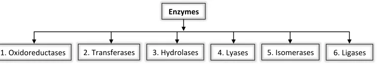
Enzymes are biological catalysts that facilitate the majority of reactions in nature and at any one time are responsible for more chemistry than the combined chemical industry.2 They facilitate many
[image:11.595.119.501.693.758.2]processes fundamental in sustaining life; without enzymes these reactions would not occur on a useful timescale. Enzymes are classified into six classes based on the type of chemical reaction they catalyse (Figure 1.1).1
Figure 1.1 Enzyme classification
!"#$%&'()*'+,-./*/# 0"#1).2/3*)./*/# 4"#56')(7./*/# 8"#96./*/# :"#;/(<*)./*/# ="#9&>./*/#
!
%!
Like all catalysts, enzymes work by lowering the activation energy of a reaction thereby increasing the rate of product formation and enabling reactions to reach their equilibrium state more rapidly. However where non-biological catalysts often require extreme reaction conditions and generate undesired side products, enzymes operate at mild biologically compatible temperatures. More importantly they have the potential to promote a specified and controlled reaction on a target substrate with greatly enhanced reaction rates. Although the catalytic properties of enzymes make them highly desirable synthetic tools, the number in use commercially is somewhat modest.3, 4
1.1.1.
Enzyme Applications
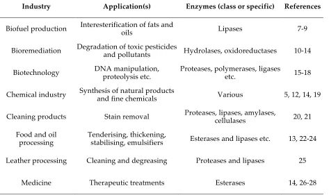
Examples of the utilisation of enzymes in various commercial and industrial processes are given in Table 1.1. The different industrial applications have different requirements on the enzyme. For example, the chemical, medicinal and pharmaceutical industries generally take full advantage of the high substrate and reaction specificity typically associated with enzyme catalysis in order to produce fine chemicals and pharmaceutical compounds of enantiomeric purity.5 However, enzymes
[image:12.595.60.533.486.767.2]such as those utilised in cleaning products, the production of oils and biofuels as well as pulp and textile manufacturing, are required to have broad substrate specificity. In these industries it is advantageous for the enzymes to have this broad specificity since they act upon a range of materials (e.g. various stains, crude oils etc.). In the enzymatic refinement of oils and fats, enzymes are usually added to crude oils to catalyse the transesterification or hydrolysis of triacylglycerols and phospholipids, as such the enzymes need to have broad substrate specificity.6
Table 1.1 Examples of industrial applications of enzymes
Industry Application(s) Enzymes (class or specific) References
Biofuel production Interesterification of fats and oils Lipases 7-9
Bioremediation Degradation of toxic pesticides and pollutants Hydrolases, oxidoreductases 10-14
Biotechnology DNA manipulation, proteolysis etc. Proteases, polymerases, ligases etc. 15-18
Chemical industry Synthesis of natural products and fine chemicals Various 5, 12, 14, 19
Cleaning products Stain removal Proteases, lipases, amylases, cellulases 20, 21
Food and oil
processing Tenderising, thickening, stabilising, emulsifiers Esterases and lipases etc. 13, 22-24
Leather processing Cleaning and degreasing Proteases and lipases 25
!
&!
Pulp and paper
industry Deinking, processing Amylases, cellulases, lipases, xylanases etc. 13
Pharmaceutics Synthesis of pharmaceutical products and intermediates Various 5, 12, 29
Textile manufacturing Scouring, finishing Amylases, cellulases, proteases etc. 13, 30
The most common use of enzymes in everyday life is in household cleaning products and most especially in laundry detergents and washing powders. Enzyme technology was first introduced to this industry in the 1960’s and use is now widespread with most brands on the market containing enzymes.21 Proteases, lipases and amylases can provide a means of breaking down a variety of stains including grease, wine, grass, soil and food stains. In order for enzymes to be suitably utilised in this way, there are certain criteria they need to satisfy. Since the detergents themselves need be inexpensive, perform well and have a long shelf life, the enzymes in them must also be stable, suitably active and cheap to produce meaning they must express well.
In medicine the use of enzyme technology can be sub-categorised into the areas of diagnostics and therapeutics. In diagnostics enzyme concentration can be used as an indicator for disease.31-33 For example, clinical tests for myocardial infarction are based on the detection of
enzymes.34-36 Myocardial infarction is the interruption of blood supply to the heart causing death of
cardiac muscle cells. Detection of enzymes such as creatine kinase and troponin that leak from the damaged tissue into the blood stream can be used for diagnosis. Certain isozymes of creatine kinase and troponin (cardiac I and T) are unique to the heart muscle and their respective concentrations temporarily increase following an infarction.
In therapeutics the most successful applications of enzymes are extracellular, including topical uses for skin diseases and lacerations, removal of toxic substances and treatment of disorders that manifest within the blood circulation. The enzyme L-asparaginase is often used for the treatment of acute lymphoblastic leukemia in combination with other anticancer drugs.28, 37, 38
Certain tumour cells lack the ability to synthesise the essential amino acid L-asparagine and are dependant on external supply. Administration of asparaginase causes rapid depletion of the L-asparagine by hydrolysing it to aspartic acid and ammonia. Depletion of the external supply causes growth suppression and induced apoptosis in tumour cells that lack L-asparaginase synthase.38
Enzymes can be specific and efficient biological catalysts and as such are highly desirable therapeutic agents,26, 39, 40 although unfortunately there are some severe limitations to the use of
!
'!
align purely with the needs of the host organism. For example, if the organism requires small quantities of a particular compound then the enzyme responsible for its production may have poor activity or low levels of expression. Additionally the high level of substrate and reaction specificity associated with enzyme catalysis means finding an appropriate enzyme for an industrial process is not always possible. Techniques in protein engineering have the capability of expanding and improving the current state of enzyme technology as applied to industrial and commercial practices. 41-50
1.2.
Protein Engineering
The properties and characteristics of proteins have been studied for well over a century. However, in more recent times the focus has shifted to improving enzyme properties. The technology and knowledge in the field of genetic engineering has advanced significantly in recent years, such that mutations can be incorporated into target genes with relative ease.51 However, the effect of these
respective mutations is not easy to predict. Consequently the modification of enzymes at a molecular level to improve properties of interest is not a trivial task. Pre-existing knowledge of a candidate enzyme is highly beneficial. This ideally includes:
Sequence and structural information !
Information on close ancestral and evolutionary relations !
An understanding of the active site and reaction mechanism !
The key approaches to protein engineering are rational design and directed evolution. Rational design uses prior knowledge of the candidate protein to predict – manually or computationally – amino acid substitutions that will yield the desired properties. In contrast, directed evolution harnesses the power of natural selection to generate proteins with the desired traits.
1.2.1.
Directed Evolution
The concept of directed or laboratory evolution of proteins is essentially survival of the fittest.52, 53 It
has become a powerful tool not only in engineering enzymes suitable for industrial use but also in highlighting the relationship between protein structure, sequence and function.47-49, 54-56 The overall
!
(!
Figure 1.2 General strategy for the directed evolution of enzymes
The benefit of directed evolution over rational design is that it does not require any forethought to the mutation sites so prior detailed knowledge of the enzyme, although useful, is not essential. Any lack of knowledge is offset by the ‘power in numbers’ approach to library screening. These two quite different approaches are not incompatible and often a hybrid approach will be used, particularly in more recent years. For example, site saturation mutagenesis or randomised mutagenesis but only over a portion of the enzyme rather than the entire enzyme.58
1.2.1.1.
Library Generation
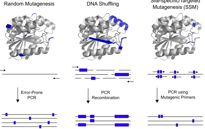
The overall process of introducing mutations and the subsequent selection of desirable traits has been well established. It is the development of the polymerase chain reaction (PCR), a relatively simple laboratory technique that is capable of creating significant genetic diversity that has revolutionised the field.51 Routine methods for library generation include error-prone PCR,
site-saturation mutagenesis and DNA shuffling (Figure 1.3).
Figure 1.3 Common methods of library generation for directed evolution
!"#$%&'()*"+,#,-.- 9:;'/<)33=.#+ /.*,0-1,2.3.245"6+,*,$()*"+,#,-.-'7//(8
>66%60?6%#, ?@!
?@! !,2%&A.#"*.%#
()*"+,#.2'?6.&,6-!
)!
PCR was originally designed to amplify lengths of DNA but has been adapted, in the form of error-prone PCR, to serve as a tool for library generation. DNA polymerases usually have proofreading activity to ensure DNA is replicated with high fidelity. However, as the name suggests, error-prone PCR is intended to introduce random mutations during replication by reducing the fidelity of the DNA polymerase. One approach to reducing the fidelity is to use a polymerase lacking the domain responsible for editing (i.e. Taq polymerase).59, 60 However, the
absence of an editing domain does not give rise to a high error rate and additional agents must be considered. Protocols established to further increase the rate of nucleotide misincorporation include simply varying the nucleotide ratio, increasing the concentration of MgCl2 or the addition of
MnCl2.60
DNA shuffling, otherwise referred to as genetic recombination, involves the fragmentation and subsequent recombination of multiple DNA sequences with the aim of combining beneficial mutations.61 Shuffling is typically performed on a set of naturally occurring homologous sequences
using Deoxyribonuclease I to fragment the genes and self-priming PCR to reassemble the fragments. Site-saturation mutagenesis (SSM) allows the systematic substitution of the wild-type amino acid at a particular position to all twenty possible amino acids.62-64
!
This technique can beconsidered a compromise between rational design and the rigours of directed evolution. In contrast to error-prone PCR, when using SSM, it is highly beneficial to have the relevant prior knowledge of the enzyme in order to target the appropriate residues.
The properties of DNA are not highly sequence dependant so these well established methods of library generation can be applied across many different directed evolution experiments with little alteration. However, the properties of the resulting enzymes can differ dramatically, as such the methods employed to effectively screen these enzyme libraries need to be carefully considered.
1.2.1.2.
Selection or Screening of Libraries
The selection or screening stage is arguably the most difficult step in a directed evolution experiment. A poorly chosen or carelessly executed screen could result in variants with the desired properties remaining unidentified. Recent advances allow for rapid library screening with the compartmentalisation of individual samples or single cells into microdroplets and the use of fluorescence-activated cell sorters (FACS).65-68 However these methods require specialised
equipment and were not used for the work presented in this thesis so will not be discussed further.
!
*!
nutrients essential for cell growth.69 However, the activity of these enzymes can also be utilised as a
marker to evolve improved physical properties for an unrelated target enzyme. For example, systems such as the Dihydrofolate reductase (DHFR) fusion reporter system can be used to improve the solubility or stability of an enzyme.70 This is achieved by fusing its gene to that of an enzyme so
that the fused entity has antibiotic resistance. The DHFR reporter system has been used during the course of this work and is described in greater detail in Chapter 3. Typically selection-based assays are followed up by a secondary screen since the information gained from selection is one-dimensional (i.e. cells that survive selection are known to have the desired property but the degree of improvement is not known).
Table 1.2 Comparison of selection and screening techniques
Technique Library Size Adaptability Dynamic Range Simplicity
Selection 105
!
!
!!!!!
Microtiter plate screen 104
!!!!!
!!!!!
!
Agar plate screen 104 - 105
!!!
!!!
!!!
Microtiter plate screening is more laborious than selection based assays but the information obtained is often more useful. Cells transformed with an enzyme library are grown in microtiter plates – usually single transformants per well. Cells are normally, although not always, lysed and the library is assayed for the desired property. If the reaction involves the formation or degradation of a coloured or fluorescent substance then it can be easily visualised by eye or monitored using a microplate spectrophotometer.71 Alternatively the reaction products can be screened by gas/liquid
chromatography or mass spectrometry. Although microtiter plate screening is versatile and offers great dynamic range, the screenable library size is limited due to practical aspects such as plate preparation and reaction time.
A compromise between the ease of selection and the benefits yielded with a microtiter plate screen is an agar plate screen. These screens offer a robust method of colony assessment, as with selection, while also allowing some measure of the quality of the variants, as with mircotiter plate screening. The critical aspect of agar plate screening is the need for a visual signal to identify colonies expressing an enzyme variant with the desired property. Ideally the catalysis product would be coloured or the reaction would be coupled with a dye and substrate turnover monitored indirectly. The speed that colonies are stained with the indicator colour is used as measure of the catalytic activity. These screening techniques are simpler than microtiter plate screening and larger libraries can be screened but differentiating between catalytic rates is not very precise.
1.3.
Esterases
!
+!
existing weak promiscuous activity can be exploited, so the candidate enzyme should have some level of the desired activity.72, 73 Additionally the candidate enzyme should be relatively stable,
soluble and express well in a recombinant system especially if it is to be used commercially.
Of the six classes of enzymes, hydrolases are the most abundant and are often targets of protein engineering studies since they are the most commonly utilised biocatalysts (Section 1.1.1). These enzymes catalyse the breaking of a single bond through the addition of water. There are a vast variety of hydrolase enzymes and they are further classified into subclasses including carboxylesterases, glycosylases and peptidases. Carboxylesterases include non-specific esterases and lipases, both of which hydrolyse ester bonds. They are distinguished based on substrate specificity; esterases preferentially catalyse the hydrolysis of water-soluble short chain esters while lipases prefer longer chain fatty acids (i.e. lipids).74 Most lipases are either phospholipases (EC
3.1.1.4/5/32) or triacylglycerol lipases otherwise referred to as true lipases (EC 3.1.1.3) that catalyse the conversion of triglycerides to monoglycerides and free fatty acids. True lipases are mostly serine hydrolases belonging to the !/" hydrolase fold superfamily.
1.4.
The
!
/
"
Hydrolase Fold
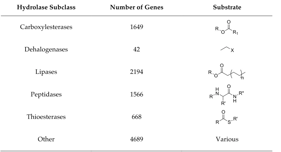
[image:18.595.71.522.480.724.2]The domain folds most frequently found in nature are !/" domains consisting of a ,-sheet surrounded by - helices. The !/" hydrolase fold is a subclass of this fold and is common to a range of enzymes from highly differing phylogenetic origin and catalytic function (Table 1.3).75
Table 1.3 Enzymes of the -/, hydrolase fold family
Hydrolase Subclass Number of Genes Substrate
Carboxylesterases 1649
Dehalogenases 42
Lipases 2194
Peptidases 1566
Thioesterases 668
Other 4689 Various
O R1
O R
X
O O R
n
N H O
R" H
N
R' R
R S
!
.!
In contrast to what the name suggests, the !/" hydrolase fold does not contain purely hydrolytic enzymes, although they were the first discovered and are the most numerous. Enzymes with this characteristic fold can include: proteases, hydrolases, esterases, peroxidases, lipases and dehalogenases. Additionally, there are also a small number of binding proteins that exhibit the -/, hydrolase fold.75-78 The functional diversity of this fold makes it an ideal framework for protein
engineering studies. These enzymes do not necessarily share any significant sequence similarity. They do, however, share remarkably similar tertiary structure and a preserved arrangement of catalytic residues, suggesting evolution from a common ancestor.
1.4.1. Characteristic Features
The canonical !/" hydrolase fold has eight strands forming a central "-sheet, surrounded by six ! -helices. The strands of "-sheet are highly twisted, such that the first and last are orthogonal to each other. Helices connect all but strands one and two, where strand two is anti-parallel to the rest.75
The strand order is 12435678 and topology of the central ,-sheet is +1, +2, -1x, +2x, (+1x)3 (Figure
1.4).79, 80
Figure 1.4 Schematic of the !/" hydrolase canonical fold
The catalytic residues constitute a highly conserved triad, consisting of a nucleophile (serine, cysteine or aspartic acid), an acid and a histidine. The nucleophile is positioned after strand /"#0, the acidic residue is positioned after strand 10#02 and the histidine residue is located after the last strand. Although the nature of the nucleophile differs, it is always the central residue of a sharp
# turn connecting 134526!/"#0 to an !-helix. A highly conserved sequence motif of SR–X–Nu–X–SR– SR (SR = Small Residue, X = Any Residue and Nu = Nucleophile), where the small residue is usually glycine, surrounds the nucleophile and forms the # turn – known as ‘the nucleophile elbow’. This maximises exposure of the nucleophile into the active site cleft and minimises crowding by neighbouring residues. The position of the # turn at the N-terminal end of an !-helix, increases the net reactivity and stabilisation of the nucleophile side chain during catalysis.
! " # $ % & ' (
)
* + ,
-.
/012345678
)9:5;2<=5:2<+>2?:
+012345678 #
!
$7!
1.4.2. Structural Variation and Functional Diversity
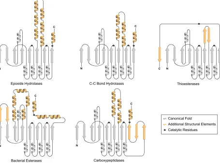
The most significant variation between these enzymes is in the substrate binding region or domain.76, 78 Such that it appears as if nature has taken a highly effective catalytic core and altered
the substrate binding residues to cater for a large variety of substrates (Figure 1.5).
The inherent structural similarity but vast functional diversity exhibited by enzymes of the
!/" hydrolase fold family makes them ideal subjects for protein engineering studies. The framework this fold provides has been adapted by nature enabling it to facilitate many catalytic processes. In relation to the overall structure, dienelactone hydrolase from Pseudomonasknackmussii
[image:20.595.68.523.304.643.2]is one of the simplest of known -/, hydrolase fold enzymes. It has been used to provide the -/, hydrolase fold scaffold that has been used as a basis for the protein engineering and directed evolution experiments presented in this thesis.
Figure 1.5 Additional secondary structural elements exhibited by !/" hydrolase fold enzymes
1.5.
Dienelactone Hydrolase
Dienelactone hydrolase (DLH) is 25.5 kDa monomeric protein that was first identified in
Pseudomonas knackmussii as part of the ,-ketoadipate pathway. This pathway is found in certain
!
!
"
!
! "
!
!
"
!
!
" "#$%&'()*+',$-./(/
0.12(,&.-)"/2(,./(/ 3.,4$%+#(#2&'./(/
56&$(/2(,./(/ !
!
373)0$8')*+',$-./(/ "
3.8$8&1.-)9$-'
!
$$!
strains of bacteria and fungi where it is responsible for the degradation of toxic and stable aromatic compounds. 81, 82
1.5.1.
The
"
-ketoadipate Pathway
Although there are many natural aromatic compounds – i.e. aromatic amino acids, vitamins and melanins – the most abundant is lignin, a natural aromatic polymer.83 Lignin is a constituent of the
plant cell wall. Therefore, it is not surprising that the ,-ketoadipate pathway is found almost exclusively in soil microorganisms, especially bacterial groups associated with plants. Organisms with the capacity to biodegrade these aromatic compounds have the ability to utilise them as a source of energy for growth and replication.
In addition to their natural induction into the environment, large quantities of synthetic aromatic compounds are also introduced through industry. Chlorinated aromatic compounds, such as those found in paints, pesticides and herbicides are the most prevalent of these and are highly toxic to living systems. The ,-ketoadipate pathway consists of three main branches – catechol, protocatechuate and gentisate – and unaltered is unable to degrade haloaromatic compounds. This is likely due to steric constraints or to the electron withdrawing effect of the respective halogen.84
Therefore microorganisms that have evolved pathways for the degradation of these haloaromatics, play an important role in their elimination from the biosphere.
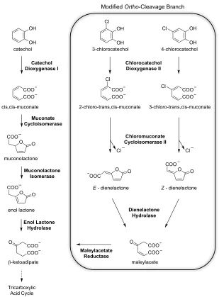
DLH is the third of four enzymes in the modified ortho-cleavage branch that is responsible for the degradation of toxic chloroaromatic compounds (Figure 1.6).85, 86 Organisms that degrade
chloroaromatics using this modified branch, otherwise referred to as the chlorocatechol branch, also co-express the enzymes of the unmodified ,-ketoadipate pathway. The modified branch produces ,-ketoadipate as the final product. This is fed into the unmodified pathway for subsequent thiol cleavage and processing to acetyl-CoA and Succinyl-CoA. These tricarboxylic acid (TCA) cycle intermediates are in turn fed into the TCA cycle to produce metabolic energy. Comparison of the enzymes from the ortho-cleavage and modified ortho-cleavage branches of the ,-ketoadipate pathway present an interesting evolutionary study. While the corresponding enzymes muconate cycloisomerase and chloromuconate cycloisomerase II are almost identical structurally,87-90 that is
not the case for enol lactone hydrolase and dienelactone hydrolase.91 Furthermore a second enzyme
!
$%!
[image:22.595.137.446.54.473.2]!
Figure 1.6 The ortho-cleavage and modified ortho-cleavage catechol branches of the ,-ketoadipate
pathway
DLHs from various chlorobenzoate utilizing bacteria have been classified into three groups. These groups have been assigned based on their specificity toward the Z- (cis-) and E- (trans-) dienelactone substrates. Type I DLHs preferentially catalyse the conversion of the E isomer to maleylacetate and are magnesium dependant. Type II DLHs have the opposite preference from type I in that they hydrolyse only the Z isomer, while type III DLHs – such as the one from P. knackmussii
– can catalyse the hydrolysis of both isomers.
1.5.2.
Structure
The initial structure of type III DLH from P. knackmussii was determined at 2.8 Å resolution then further refined at 1.8 Å from orthorhombic crystals in space group P212121.92, 93 It consists of 236
amino acids forming seven helices and eight strands of "-sheet (Figure 1.7). Seven of the stands are parallel but as is characteristic of !/" hydrolase fold enzymes strand two is anti-parallel to the rest.
Modified Ortho-Cleavage Branch
OH
OH
COO COO catechol
cis,cis-muconate
O O
COO
O O
COO muconolactone
OH
OH 3-chlorocatechol
Cl
OH
OH Cl
4-chlorocatechol
COO COO Cl
3-chloro-trans,cis-muconate COO
COO
2-chloro-trans,cis-muconate Cl
enol lactone
O
O O O
OOC
COO
E - dienelactone Z - dienelactone
COO COO O
maleylacete
Catechol
Dioxygenase I Chlorocatechol Dioxygenase II
Muconate Cycloisomerase
Muconolactone Isomerase
Cl
Dienelactone Hydrolase
COO COO O
Enol Lactone Hydrolase
!-ketoadipate
Chloromuconate Cycloisomerase II
Maleylacetate Reductase
Tricarboxylic Acid Cycle
!
$&!
[image:23.595.130.481.110.716.2]Six of the helices lie on the outer solvent-accessible domains surrounding the "-sheet at the core of the enzyme, the final helix also lies within the inner core.93
Figure 1.7 The !/" hydrolase fold exhibited by DLH shown in (A) three-dimensional form and (B)
in schematic representation with the substrate binding residues in green and residues comprising the oxyanion hole in orange
! " # $ % & ' (
)
*
+ , - .
/
0123456789
:;<=76>7? @>A3
+123456789 B!"$
C!"# ,!'!
@"D" E"D& E(!
F(% G((
-$& H$'
)IJ6K3?B6J3?+A3LJ
0123456789
+123456789
!
"
)IJ6K3?B6J3?+A3LJM$%
B!"$
!
$'!
Unlike other !/" hydrolase fold enzymes (Figure 1.5), DLH has a minimal stretch of peptide responsible for substrate binding. In DLH sections of helix B and loop 14 contain residues involved in substrate binding while in other enzymes, such as acetylcholinesterase, whole domains serve the same purpose.94 Figure 1.7 also shows the residues comprising the oxyanion hole – Q35, E36, I37
and L124. The oxyanion hole creates a hydrophilic region of the active site and is responsible for stabilisation of the enzyme-substrate tetrahedral intermediate during catalysis (discussed later in Section 1.5.3).
The catalytic triad of DLH comprises a nucleophilic cysteine (C123), an aspartic acid (D171) and a histidine (H202).95 The active site pocket extends from a region on the surface of the enzyme
to the central "-pleated sheet. The nucleophile lies at the bottom of this pocket and is the central residue of a sharp # turn that links strand five to helix C. The histidine is in loop 14 between strand eight and helix C, this loop acts as a cover for the active site creating a crevice inaccessible to bulk solvent. The carboxylic acid side chain of D171 is hydrogen bonding distance from H202 forming a charge relay system ensuring de-protonation of the nucleophile for catalysis. Figure 1.8 shows residues of the DLH active site with an inhibitor dienelactam in complex, a complex that mimics the Michaelis complex of the native dienelactone substrates.
Figure 1.8 Active site residues of DLH with the inhibitor dienelactam bound
During crystal structure analysis, it was observed that the side chain of C123 exhibited two distinct conformations. The C123 thiol can rotate about the C- – C, bond, so that it is positioned either into the active site or away from it in a seemingly inactive conformation. The crystal structure showed C123 to be partially oxidised to sulfonic acid, disabling rotation and holding it in the active state.93, 95 It has been suggested that prior to oxidation, the nucleophile exists predominantly in the
inactive conformation, although it is not able to initiate catalysis, it is more energetically favourable. It is thought that upon substrate binding the nucleophile will shift into the active conformation through ionic rearrangements in the active site involving R206 and E36.96, 97
!
$(!
Mutation of the nucleophile to a serine (C123S) produces an enzyme with altered functionality toward its physiological substrates the E- and Z-dienelactones.95 Changing the
nucleophile from cysteine to serine causes the enzyme to become an isomerase catalysing the interconversion of the substrate isomers.98, 99 Both the native and C123S variant have low level
promiscuous hydrolytic activity toward non-physiological ester substrates such as p-nitrophenyl acetate and !-naphthyl acetate. The DLH C123S variant has been used for the work contained within this thesis since unlike cysteine, serine is not prone to oxidation, so use of this variant prevents potential loss of activity during expression and purification stages due to irreversible oxidation. Throughout the subsequent chapters of this thesis the DLH C123S is variant is referred as native.
1.5.3.
Mechanism
The DLH catalysed hydrolysis of its physiological substrates the E- and Z-dienelactones is a variant of the serine protease mechanism.100-102 The DLH catalysed hydrolysis of the non-physiological
substrate p-nitrophenyl acetate does follow this known serine protease mechanism (Scheme 1.1).
Scheme 1.1 DLH C123S catalysed hydrolysis of p-nitrophenyl acetate
The nucleophile attacks the carbonyl carbon of the substrate, leading to formation of the covalent enzyme-substrate tetrahedral intermediate, which is stabilised in the oxyanion hole. Following release of the first product (p-nitrophenol) H202 de-protonates a water molecule, the resulting hydroxide then acts as a nucleophile toward the carbonyl carbon of the enzyme bound ester. The enzyme is eliminated from the second tetrahedral transition state, producing the second product (acetic acid) and regenerated enzyme active site.
N N
H202
O S123
H
O H
O2N O
O
O2N O
O
O
S123
H
O
O
D171
H
O S123
NO2
O O
S123
O
O S123
O
O S123
O
OH OH
O
N N
H202
H
N N
H202
O
O
D171 H
N NH
H202
O
O D171
H
HN N
H202
O S123 H
O
O D171
Resting State Tetrahedral Intermediate
p-nitrophenol (1st Product)
!
$)!
The proposed mechanism for DLH catalysed hydrolysis of the native dienelactone substrates differs due to positional constraints within the active site (Scheme 1.2). The nucleophile attacks the carbonyl carbon of the lactone ring belonging to the substrate forming a covalent enzyme-substrate tetrahedral intermediate. Deviating from the serine protease mechanism, it is suggested that H202 does not protonate the leaving group or deprotonate water for deacylation. Instead it is proposed that the enolate anion acts as a base to de-protonate a water molecule itself generating the hydroxide necessary to facilitate release of the product (maleylacetate) and regeneration of the enzyme active site. 103
Scheme 1.2 DLH C123 catalysed hydrolysis of Z-dienelactone
1.6.
Research Objectives
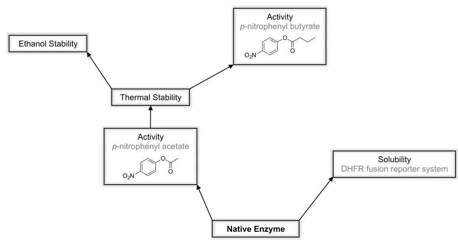
This project aimed to take a versatile catalytic framework in the !/" hydrolyse fold and engineer it for new and improved catalytic functions as well as enhanced physical properties. Multiple sets of directed evolution experiments were performed to alter the specificity of DLH and to enhance physical properties such as solubility, thermal stability and stability in organic solvent (Figure 1.9).
An initial aim of this project was to separately evolve DLH to have improved solubility and improved stability. Although protein solubility and stability are distinct features, the properties governing them are often considered to be highly correlated. That is to say, a stable protein likely also possesses good solubility, while unstable proteins generally lose their structure and form insoluble inclusion bodies and aggregates. There are of course exceptions to this, for example intrinsically disordered proteins (IDPs) are often highly soluble but lack three-dimensional structure.104-106 It was envisaged that side-by-side evolutions for improved solubility and stability
would yield sets of mutations that could provide insight into the more intricate differences between these two properties. Problems were encountered that delayed progress in both the solubility (discussed in Chapter 3) and stability evolutions. The stability evolution was hindered by lack of
N N
H202
S
C123 S
C123
H
O
O D171
H
S C123
O O
O2C
O
-O 2C
O
O2C
O
O
S C123
H
O H
O2C
OH
O
HO S C123
O2C O
O OH
HN N
H202
S C123 H
O
O D171
Resting State Tetrahedral Intermediate
!
$*!
[image:27.595.68.534.141.389.2]activity to the easily assayable p-nitrophenyl esters, which were used to monitor residual activity of the enzyme following heat exposure. Thus focus shifted to evolving and altering the catalytic properties of DLH, which opened new avenues of research (Chapters 4, 5 and 6) that were pursued in favour of the solubility and stability comparison.
Figure 1.9 Directions for the evolution of DLH
Overall several selection and screening methods were developed or optimised for the directed evolution experiments discussed in this thesis. These can now subsequently be employed to evolve other enzymes – in particular more complex and less well characterised !/" hydrolase fold enzymes. Additionally and perhaps more interestingly these studies aim to contribute to the global understanding of evolutionary biology, by observing how DLH evolves under different selection pressures.
1.7.
Thesis Outline
Three publications arose from the work contained within this thesis. They are reproduced in Chapters 4, 5, and 7 respectively.
Chapter 2. Description of routine experimental procedures used during completion of the work
presented in this thesis.
Chapter 3. Using the DHFR fusion reporter system to select for DLH variants with improved
solubility. This comprises initial testing and modification of the system to enable it to be used to select for enzymes with high solubility, previously it had only been used to select for improvements in natively insoluble enzymes. The results presented in this Chapter, although only preliminary, provide a solid foundation for the future continuation of this work.
Solubility
DHFR fusion reporter system
Native Enzyme Activity
p-nitrophenyl acetate
O O O2N
Activity
p-nitrophenyl butyrate
O O O2N
!
$+!
Chapter 4. Enhancing the activity and substrate specificity of DLH through directed evolution using
a three-stage process. This chapter is about Publication 1.
Chapter 5. Investigating of the role of compensatory surface mutations during the directed
evolution of DLH. This chapter is focused on Publication 2.
Chapter 6. Assessment of the solubility, stability and activity of DLH in the presence of organic
solvents. This focuses on the evolution of DLH for greater stability in the presence of ethanol with the eventual aim of evolving a transesterase, whether DLH itself can become capable of catalysing a transesterification or the methods can be used to more efficiently evolve other candidate enzymes in the future.
Chapter 7. Differences in protein structure caused by changes in the crystalline environment
assessed by crystallisation of DLH in the space group P212121 and C2. This chapter is about
Publication 3.
!
$.!
Experimental Methods
This chapter is designed to provide details on the routine experimental procedures used throughout the course of this research. Information on specific and developed protocols is given in the relevant chapters. All chemicals were purchased from Sigma-Aldrich unless otherwise specified and water was purified using a Milli-Q purification system (Millipore).
2.1.
Cell Strains and Expression Vectors
Throughout the course of this work, enzymes were synthesised using the native cellular machinery of bacteria. The requirements for cell based protein expression are a vector incorporating the DNA sequence for the protein of interest and a host cell. Escherichia coli is one of the most widely used hosts and DNA is most commonly introduced using a plasmid expression system.
2.1.1.
DH5
!
E.coli Strain
!
%7!
2.1.2.
The pCY76 Expression Vector
A plasmid must be able to produce desired target variants with ease and in high quality for it to be an effective carrier of a gene library. The vector pCY76 (par+, bla+, lacZpro, T7!10tir+) is a constitutive
protein expression system under the control of the lac promoter. It was developed in-house and its construction has been previously described.107 The pCY76 vector contains a partition element (par)
that aids in maintenance of plasmid copy numbers (stability). It also contains the bla gene encoding ampicillin resistance. The clcD gene encoding DLH was cloned into the multi-cloning site of pCY76 using NdeI and EcoRI unique restriction sites.
The pCY76 expression system was utilised for the expression of native and variant DLH enzymes during the process of library screening and preliminary testing. However, it was not suitable for purification, since the co-expressed "-lactamase – responsible for conference of ampicillin resistance – possesses a similar isoelectric point (pI) and is of similar molecular weight to DLH. Separation of these enzymes during the purification process proved a difficult task. As such, pJWL1030 a kanamycin selective expression vector was employed for this purpose.
2.1.3.
The pJWL1030 Expression Vector
The pJWL1030 expression vector was engineered from pCY76, with only one significant difference. Where pCY76 is encoded with an ampicillin resistance gene, pJWL1030 contains the gene for an aminoglycoside 3’-phosphotransferase enzyme conferring kanamycin resistance.
2.2.
Molecular Biology
2.2.1.
Bacterial Transformation
Plasmid DNA was transformed into bacterial cells by electroporation. In this process, an electric field is applied to the cells, weakening the phospholipid bilayer of the cell membrane. This creates temporary pores in the cell wall through which DNA and other charged molecules can permeate.
!
%$!
2.2.2.
Polymerase Chain Reaction
The polymerase chain reaction (discussed in Section 1.2.1.1) was used routinely for both general amplification and mutant library generation. Two models of thermocyler were used: an MJmini (Bio-Rad) and a Veriti® 96-well Veriflex™ PCR system (Applied Biosystems). General amplification was carried out using Phusion high fidelity polymerase (Thermo Scientific) in accordance with the manufacturer’s protocol. The PCR conditions were 20 cycles of 98 °C for 10 s, 55 °C for 15 s and 72 °C for 30 s or 2 minutes depending on whether the gene alone or full plasmid were being amplified.
2.2.2.1.
Error-Prone and StEP PCR
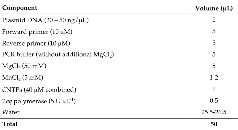
Perhaps the most common method of introducing genetic diversity for the directed evolution of enzymes is by random mutagenesis in the form of error-prone PCR. For error-prone PCRs carried out during the course of this work a combination of increased MgCl2 concentration and addition of
MnCl2 were used to control the error-rate. Taq polymerase (Bioline) lacking 3’-5’ exonuclease
[image:31.595.112.497.392.602.2]proofreading ability was used with an increased number of cycles. A typical error-prone PCR set-up is outlined in Table 2.1.
Table 2.1 Reaction set up for Error-prone PCR
Component Volume (µL)
Plasmid DNA (20 – 50 ng/µL) 1
Forward primer (10 µM) 5
Reverse primer (10 µM) 5
PCR buffer (without additional MgCl2) 5
MgCl2 (50 mM) 5
MnCl2 (5 mM) 1-2
dNTPs (40 µM combined) 1
Taq polymerase (5 U µL-1) 0.5
Water 25.5-26.5
Total 50
The PCR conditions were 30 cycles of 96 °C for 10 s, 55 °C for 10 s and 72 °C for 20 or 5 s. The longer extension time (20 s) was used for first round library generation in the directed evolution process. For subsequent rounds when shuffling was required, error-prone PCR was combined with a staggered extension process (StEP). The shorter extension time (5 s) was used to generate small gene fragments that anneal to different templates based and extend further during each cycle.108
!
%%!
example, a single point mutation to a CCU proline codon can alter it to encode for serine, threonine, alanine, leucine, histidine or arginine. In order to access codons for other amino acids, two point mutations (F, Y, C, Q, I, N, V, D and G) or even three (W, M, K, R and E) are required. Although this genetic bias is optimised toward the substitution of amino acids with those less likely to cause loss of function, it does dictate library size and diversity. The third bias is caused by the exponential nature of the amplification process. A mutation introduced in the first amplification cycle of an error-prone PCR will be significantly more abundant in the product molecules than mutations introduced in later stages. To partially combat this third bias, four separate error-prone PCRs were carried out at every stage (i.e. reactions were run in four separate tubes), then combined to construct the final working library.
2.2.2.2.
Site Saturation Mutagenesis
Site saturation and site directed mutagenesis were performed using a method adapted from Stratagene’s Quickchange® protocol. Primers containing the desired nucleotide change or randomisation were employed in a PCR to replicate the entire plasmid (Figure 2.1).
Figure 2.1 Schematic of the site saturation mutagenesis protocol adapted from Stratagene
The mutagenic primers used for site saturation mutagenesis experiments in this thesis utilised the NNK codon (where N = A, C, G or T and K = G or T). The genetic code is made up of sixty-four possible codons. For creation of a site saturated library, randomising the first two bases in the codon and limiting the third to either G or T, is more practical than complete randomisation of all three bases. Complete randomisation produces a theoretical library size of sixty-four. Use of the NNK codon reduces the library size by half to thirty-two, while still representing all twenty amino acids. Although for practical purposes an excess of the theoretical library needs to be screened to ensure almost complete coverage (discussed in Chapter 4, Section 4.4.2).
A typical site saturation or site directed PCR was performed in 200 µL PCR tubes as outlined in Table 2.2.
Template Plasmid DNA
!
%&!
Table 2.2 Reaction set up for site-saturation mutagenesis PCR
Component Volume (µL)
Plasmid DNA (20 – 50 ng/µL) 1
Forward mutagenic primer (10 µM) 2.5
Reverse mutagenic primer (10 µM) 2.5
Phusion high fidelity buffer 10
dNTPs (40 µM combined) 1
Phusion high fidelity polymerase (2 U µL-1) 0.5
Water 32.5
Total 50
The PCR thermocycling conditions were 30 cycles of 98 °C for 10 s, 55 °C for 15 s and 72 °C for 2 minutes. This was followed by a final extension at 72 °C for 10 minutes at the end of the cycling period.
2.2.3.
Restriction Enzyme Digestion
Plasmid expression vectors or PCR products were digested with 10 U of the appropriate restriction enzymes (New England Biolabs) in a solution (50 µL) with the manufacturer’s recommended buffer. The restriction endonuclease DpnI was added to the digestion of PCR products for the purpose of cleaving initial methylated template DNA. Digestion mixtures were incubated for three hours at 37 °C, then gel purified. To prevent recircularisation, digested plasmid expression vectors were treated with calf intestinal alkaline phosphatase (CIP) (New England Biolabs) for sixty minutes before purification.
2.2.4.
DNA Ligation
Ligation of the DLH genes into expression vectors was performed with 100 ng of vector and a 3-fold molar excess of insert. Ligation reactions were on the 20 µL scale with 0.5 U of T4 DNA ligase (Fermentas) in the supplied buffer. The reactions were carried out at 18 °C for 12 – 18 hours.
2.2.5.
DNA Purification
Plasmid DNA was routinely purified from overnight cultures (5 mL) using the Qiagen Miniprep® kit. The Promega® PCR purification kit was used to purify PCR products and digested or ligated DNA. Gel purification was performed by first subjecting the DNA sample to gel electrophoresis on a 1% agarose gel using RedSafeTM nucleic acid staining solution (iNtRON Biotechnology). The
!
%'!
2.2.6.
DNA Sequencing
The native and mutant DLH genes were sequenced on a AB 370xI DNA Analyzer (Applied Biosystems) at the ACRF Biomolecular Resource Facility at the JCSMR at the Australian National University. Samples were prepared by PCR using BigDye terminator (Applied Biosystems) as recommended by the facility (Table 2.3).
Table 2.3 Sample preparation for DNA sequencing
Component Volume (µL)
Plasmid DNA (200 – 300 ng/µL) 1
BigDye terminator 1
Sequencing buffer (10 mM MgCl2, 400 mM Tris, pH 9.0) 3.5
Primer (32 pM) 1
Water 13.5
Total 20
Thermocycling conditions were 96 °C for 5 mins, followed by 50 cycles of 96 °C for 10 s, 50 °C for 5 s and 60 °C for 4 minutes. The PCR extension products were prepared for analysis by mixing with a precipitation solution consisting of ethanol (62.5 µL), sodium acetate (3 µL) and water (14.5 µL). The combined precipitation mixture and sequencing products were incubated at room temperature for 15 minutes and then centrifuged at 3700 rpm (eppendorf 5804) for 30 minutes. After removal of the supernatant, the pellet was washed with 70 % (v/v) ethanol and centrifuged for a further 15 minutes. The supernatant was removed and the pellet air-dried in a BSB12 laminar flow hood (Gelaire) for 10 minutes before sequencing.
2.3.
Directed Evolution
A general introduction to the directed evolution of enzymes was provided in Chapter 1. Any directed evolution experiment can be divided into two main stages. The first stage is the introduction of genetic diversity, whether randomly or targeted. The second stage is selection or screening for the desired traits.
!
%(!
After library generation the screening or selection stages take place. Multiple screening methods have been employed during this course of this work depending on the desired trait and library size. These methods are described in the relevant subsequent chapters (Chapter 3, 4 and 6).
2.4.
Protein Analysis by SDS-PAGE
Polyacrylamide gels were used to evaluate solubility and expression in cell lysate and to test FPLC fractions during enzyme purification. The gels contained a 5 % (w/v) stacking region and a 15 % (w/v) separating region,109 both prepared from a 37.5:1 ratio of acrylamide to N,N
-methylene-bisacrylamide (Amresco). The gels were run using a SE250 Mighty Small II Mini Vertical Electrophoresis Unit (Hoefer) and visualised using Bio-Safe Coomassie Blue G250 stain (Bio-Rad).
2.5.
Enzyme Expression and Purification
Proteins were expressed constitutively from the pJWL1030 vector in LB media supplemented with kanamycin. Single colonies were inoculated into a 10 mL starter culture and grown for 8 hours before being used to seed a 1 L expression culture that was grown overnight at 37 °C. Cells were lysed either using a French Pressure Cell Press (SLM Aminco) or using rLysozyme (Novagen).
!
%)!
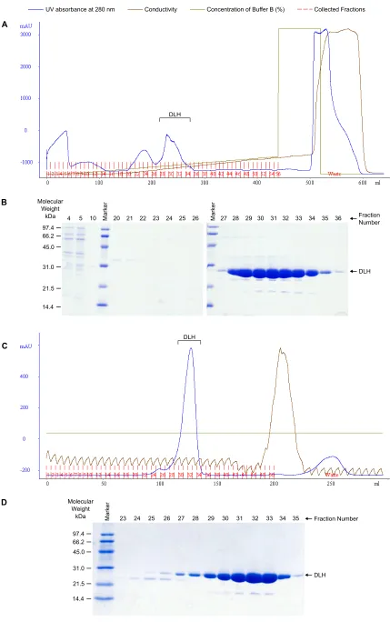
Figure 2.2 Purification of DLH C123S (A) elution profile using DEAE anion exchange and (B)
corresponding SDS-PAGE of selected fractions (C) elution profile using Sephadex G75 size exclusion and (D) corresponding SDS-PAGE
!
"
! " #$ %&'()
'
*$ *# ** *+ *! *" *, %&'() *. */
*-'
+$ +# +* ++ +! +" +,
#!0! *#0" +#0$ !"0$ ,,0* -.0! %12)342&'
5)6789
(:& ;'&3961<=
>4?@)'
:AB :AB
#
$
CD=&@E1'@&<3)=&9=*/$=<?==================F1<G4396H69I==================F1<3)<9'&961<=1J=K4JJ)'=K=LMN==================F122)39)G=;'&3961<E=
:AB
*" *+ *!
%&'()
'
*, *. */ *- +$ +# +*
#!0! *#0" +#0$ !"0$ ,,0* -.0! %12)342&'
5)6789
(:& ;'&3961<=>4?@)'
!
%*!
2.6.
X-Ray Crystallography
X-ray crystallography can provide a detailed description of the atomic and molecular structure of a crystalline material.110 This is an especially valuable tool for the study of large and complex
macromolecules such as proteins, where mechanisms and responses to stimuli are often hard to predict. During the course of this work X-ray crystallography was used to visualise and compare the structure of evolved DLH variants with the native enzyme.
X-ray crystallography is a well-established technique and has been successfully implemented for protein structure determination countless times since the later half of last century.111 The following introduction to X-ray crystallography is intended to outline the basic
methods of protein crystallisation and structure determination as relevant to this work.
2.6.1.
Crystallisation
The fundamental characteristic of the crystalline state is an extremely high degree of internal order.110 In other words, the molecules or atoms that comprise a crystal are arranged in an ordered
lattice that extends in all directions, where equivalent lattice points have identical environments.
Proteins can generally be encouraged to form crystals when combined with the appropriate reagents – i.e. buffers, salts, precipitants – and transitioned into a supersaturated solution. Since the theory of protein crystallisation is not fully understood, finding the appropriate conditions for crystallisation of a particular protein is typically a trial and error process.110 However, there are now
many commercially available protein crystallisation screens that enable rapid systematic searches to aid in this process.112
Techniques such as dialysis and free interface diffusion have been established for the crystallisation of proteins. However by far the most common is the vapour diffusion method in the form of a hanging drop.110 In these experiments, the protein is mixed with a precipitating solution in
!
%+!
Figure 2.3 Phase diagram showing the changes in protein and precipitant concentration over the
course of a protein crystallisation experiment (adapted from similar figures110, 113-115)
DLH readily crystallises in space group P212121 space group with unit cell dimensions 49 x
71 x 77 Å.92, 93, 116 This is a common space group for protein crystals, especially for monomeric
proteins like DLH.
The native and variant DLH enzymes were crystallised using hanging drop vapour diffusion. The conditions used for crystallisation of the native protein in space group P212121
differed only slightly to those previously reported.117 The hanging drop was 8 µL in size with a 5:3
ratio of protein to precipitant, where the precipitant consisted of 0.1 M sodium citrate, 1.2 M ammonium sulphate at pH 5.8. The variants crystallised under similar conditions with some requiring microseeding and slightly altered precipitant concentration (0.8 – 1.2 M ammonium sulphate). Crystals were harvested after 10-30 days growth at 4 °C and stored in a solution with elevated concentration of precipitant, 15 % (w/v) glucose and 2.5 % (v/v) glycerol were added for cryoprotection. Specific crystallisation conditions for the proteins crystallised as part of the work presented in the publication reproduced in Chapter 4 are detailed in Table 2.4.
Table 2.4 Conditions for crystallisation of native and evolved DLH variants
Condition Native A-6 S-2 S-3 B-1 B-4
Sodium citrate (M) 0.1 0.1 0.1 0.1 0.1 0.1
pH 5.8 5.8 5.8 5.8 5.8 5.8
Ammonium sulphate (M) 1.2 1.2 0.8 1.0 1.2 1.0
Temperature (C) 4 4 4 4 4 4
Micro seeding no yes no no no yes
Drop ratio (protein : precipitant) 5:3 3:1 3:1 3:1 5:3 3:1
Protein Concentration
Io
ni
c
St
re
ng
th
Metastable/ Growth Zone
UNDERSATURATED SUPERSATURATED
Precipitation Zone
Nucleation Zone
a
b c
STAGES OF A PROTEIN CRYSTALLISATION EXPERIMENT
!"
#"
$"
!
%.!
Separate structures of the native enzyme in the space groups P212121 and C2 were solved
and are the subject of the publication reproduced in Chapter 7. The conditions for crystallisation are given in the same publication.
2.6.2.
Data Collection and Structure Determination
Structures were solved as part of the work in Chapters 4 and 7, data collection and structure determination were performed as described in the publications reproduced in the respective chapters. All structures have been deposited in the PDB and the codes are listed in Table 2.5.
Table 2.5 Structures and their corresponding PDB codes
Enzyme Space group PDB Code
Chapter 4 DLH-C123S P212121 4U2B
A-6 P212121 4U2C
S-2 P212121 4U2D
S-3 P212121 4U2E
B-1 P212121 4U2F
B-4 P212121 4U2G
Chapter 7 DLH-C123S P212121 4P92
!
&7!
Improving Enzyme Solubility
3.1.
Introduction
Protein solubility along with stability is a major limiting factor when considering enzymes for utilisation in industrial or commercial practices. A protein that is soluble when expressed in its native cellular environment will not necessarily be soluble when expressed in a recombinant system. The cell may only require a small amount of protein, such that the protein never reaches its solubility limit within the cell. In many cases proteins receive assistance in folding (by chaperones) and will be less soluble in their absence. Often recombinant proteins – particularly those expressed at very high levels – will fail to fold correctly and insoluble protein will accumulate resulting in aggregates or inclusion bodies (Figure 3.1).118
Figure 3.1 Conformational changes and protein inactivation pathways
Codon substitution, reduction of growth temperature, addition of fusion partners and co-expression of chaperones are just some of the methods used to obtain higher yields of soluble protein.119 However success is far from guaranteed and new methods of improving protein
Folded Molten Globule Unfolded
Aggregated