“LIQUID BASED CYTOLOGY OVER CONVENTIONAL
PAP STAINING METHOD IN EVALUATING
CERVICAL SMEARS”
DISSERTATION SUBMITTED FOR
M.D. DEGREE EXAMINATION
BRANCH III PATHOLOGY
OF
THE TAMILNADU DR.M.G.R. MEDICAL UNIVERSITY
CHENNAI
TIRUNELVELI MEDICAL COLLEGE HOSPITAL
TIRUNELVELI
APRIL -2013
CERTIFICATE
This is to certify that the Dissertation “LIQUID BASED
CYTOLOGY OVER CONVENTIONAL PAP STAINING METHOD IN EVALUATING CERVICAL SMEARS” presented
herein by Dr. A. SANGEETHA is an original work done in the
Department of Pathology, Tirunelveli Medical College Hospital,
Tirunelveli for the award of Degree of M.D. (Branch III) Pathology
under my guidance and supervision during the academic period of 2010 -
2013.
The DEAN
CERTIFICATE
I hereby certify that this work embodied in the dissertation entitled
“LIQUID BASED CYTOLOGY OVER CONVENTIONAL PAP
STAINING METHOD IN EVALUATING CERVICAL SMEARS” is
a record of work done by Dr. A. SANGEETHA, in the Department of
Pathology, Tirunelveli Medical College, Tirunelveli, during her
postgraduate degree course in the period 2010-2013. This work has not
formed the basis for any previous award of any degree.
Dr.Arasi Rajesh MD(Guide), Dr.Sithy Athiya Munavarah MD,
Professor of Pathology, Professor and HOD of Pathology,
Department of Pathology, Department of Pathology,
Tirunelveli Medical College, Tirunelveli Medical College,
DECLARATION
I solemnly declare that the dissertation titled “Liquid Based
Cytology Over Conventional Pap Staining method In Evaluating Cervical Smears” is done by me at Tirunelveli Medical College
hospital, Tirunelveli.
The dissertation is submitted to The Tamilnadu Dr. M.G.R.Medical
University towards the partial fulfillment of requirements for the award of
M.D. Degree (Branch III) in Pathology.
Place: Tirunelveli Dr. A. SANGEETHA,
Date: Postgraduate Student,
M.D Pathology,
Department of Pathology,
Tirunelveli Medical College
ACKNOWLEDGEMENT
I take immense pleasure to acknowledge all those who have helped me to
make this dissertation possible.
I am grateful to the Dean, Tirunelveli Medical College and Medical
Superintendent of the Tirunelveli Medical College Hospital for permitting me
to undertake this study.
I express my profound sense of gratitude to Dr.Sithy Athiya Munavarah
MD, my respected Professor and Head of Department of Pathology, Tirunelveli
Medical College, Tirunelveli and my guide Dr. Arasi Rajesh M.D., for their
unstinted guidance and motivation.
I immensely thank Dr.K.Shantaraman,M.D; Dr.S.Vallimanalan, M.D;
Dr.K.Swaminathan, M.D; Dr. J. Suresh Durai, M.D., Professors of Pathology
for their constant support and encouragement. I profusely thank all the other
faculties and my postgraduate colleagues for their valuable support.
I sincerely thank the Professors and faculties of the Department of
Obstetrics & Gynaecology for providing me the patients for my study.
I also sincerely thank the Technicians and other members of the Department
of pathology and the Central Diagnostic Lab for their kind co-operation.
I thank all my family members for their encouragement and support during
this study.
CONTENTS
S.No Title Page.No
1 INTRODUCTION 1
2 AIMS & OBJECTIVES 5
3 REVIEW OF LITERATURE 6
4 MATERIALS AND METHODS 41
5 OBSERVATION AND RESULTS 50
6 DISCUSSION 65
8 SUMMARY & CONCLUSION 77
BIBLIOGRAPHY
APPENDIX
ABBREVIATIONS
LBC : Liquid Based cytology
MLBC : Manual Liquid based cytology
CS : Conventional smear
LP : LiquiPrep
CIN : Cervical intraepithelial neoplasia
NILM : Negative for intraepithelial lesion or
malignancy
NILM-IS : Negative for intraepithelial lesion or
Malignancy - inflammatory smear
ASC : Atypical squamous cells
ASCUS : Atypical squamous cells of undetermined
significance
AGC : Atypical glandular cell
ASC-H : Atypical squamous cells-cannot exclude HSIL.
LSIL : Low grade squamous intraepithelial lesions
HSIL : High grade squamous intraepithelial lesions
SCC : Squamous cell carcinoma
HPV : Human papilloma virus
1
INTRODUCTION
Cervical cancer is the third most common cancer among
women worldwide & second most common cancer in developing
countries, over half of which are fatal (Jemal A et al 2011)1. About 80%
of these cases and deaths occurred in developing countries (Ferlay et al
2010)2.This high mortality makes cervical cancer an important public
health problem. Cervical cancer is slow growing and hence has potential
for effective prevention through various screening procedures. Cervical
cytology has proved to be one of the most successful examples of cancer
screening in many developed countries and has resulted in significant
decrease in incidence and mortality from invasive cancer by detecting and
eradicating the pre invasive lesions (Clarke EA 1979)3(Hakama M et al
1985)4(Miller AB et al 1990)5 (Mathew A et al 2009)6.
Invasive cervical cancer is the end result of a long pathological
process that begins with precursor lesion called squamous intraepithelial
lesions .Early changes in the cervix in the form of CIN can be detected
years before invasive carcinoma develops and this is the basis of
effectiveness of cytological screening (Wright T et al 1994)7. The main
objective of cervical screening is to decrease worldwide incidence and
mortality of cervical cancer, by detecting and treating precancerous
2
The screening of cervical cytology smears was introduced in 1928
by Dr.George N.Papanicolaou when he reported the observation of
dysplastic/malignant cells in women with cervical cancer by sampling
vaginal smears. Subsequently Papanicolaou and Dr.Herbert Traut
identified cells of both invasive and pre invasive cervical neoplastic
lesions by cervical cytology. This test is now known as the conventional
Pap smear or Pap test (Papanicolaou et al 1941)8.
For Conventional cervical cytology cell samples taken from the
cervix by using Ayre’s spatula is smeared onto slides, fixed and stained
with Papanicolaou stain. Specificity of this test is 98 to 99%. But the
sensitivity, ranges from 50%-75% (Fahey MT et al 1995)9(Nanda K et al
2000)10. Several limitations of conventional pap test are identified such as
1) Inadequate transfer of cells to slide, 2) Un uniform distribution of
abnormal cells, 3) Presence of obscuring inflammation, blood and
overlapping of epithelial cells (Richart RM et al 1965)11.
Liquid based thin layer technology was introduced as a FDA
approved alternative method to conventional Pap in 1996 to address these
limitations. The first generation automated Liquid-based cytology (LBC)
involves rinsing the sampling device into a vial of fixative to form a
suspension of cells from which a monolayer of cells on a slide is
prepared. These slides can be read more quickly than Conventional
3
are two methods of FDA approved LBC technologies -ThinPrep and
SurePath (Lee RL et al 1997)12 (Monsonego J et al 2001)13(Fang-Hui
Zhao et al 2011)14.These new LBC techniques require an automated
instrument and so higher cost per test.
LiquiPrep, the second generation Liquid Based Cytology system
eliminates most of the instruments required by the first generation
techniques thereby offering a simpler method with lower costs for
cervical cancer screening. LiquiPrep system consist of fixative fluid vial,
a cleaning solution & a cell base that act as a membrane matrix to
produce a monolayer of cells(Geyer J et al 2004)15. Easy method of
preparation & high correlation of results obtained with CS makes this
method highly suitable for cervical cytology in developing countries
(Jongkolnee Settakorn et al 2008)16.
There are a few studies showing an Indigenous method of Liquid
based Cytology called Manual Liquid Based Cytology (MLBC). In this
method chemicals available in their own laboratory were used to prepare
fixative and polymer solution and use simple equipments to prepare
cervical smear slides. This is a low cost method of cervical pap smear
screening (Maksem et al 2001)17 (Maksem et al 2005)18 (Lee et al
4
The present study was undertaken to compare this low cost Manual
LBC with conventional pap cytology .In addition we also compared
second generation LiquiPrep system with conventional smears.
5
AIMS AND OBJECTIVES
Aim of this study is
1. To evaluate the efficiency of a new inexpensive Manual Liquid
Based Cytology.
2. To make a comparative morphological analysis of Conventional
Papanicolaou stained cervical smear with Manual liquid based
cytology smear.
3. To make a comparative morphological analysis of Conventional
Papanicolaou stained cervical smear with Second generation
LiquiPrep cytology smear.
4. To make a comparative analysis of the results of both the Liquid
Based Cytology methods with Conventional smears.
6
REVIEW OF LITERATURE
CARCINOMA CERVIXEpidemiology
Cervical cancer is one of the most common cancer among women
worldwide (WHO 2009)22.Majority of cervical cancer cases today occur
in the developing countries. According to National Cancer Registry of
ICMR the incidence in India is 14.42/100000 pop with mortality rate
2.83/100000 pop (ICMR 2004)23. Before the introduction of screening,
the rates of cervical cancer in Europe, North America and Japan were
very similar to those now seen in developing countries. Over past several
decades the incidence rate has declined in both white and African
American women. Since 2004, rates have decreased by 3.1% per year in
women 50 and above (American Cancer Society, Cancer Facts & Figures
2012)24.
Role of Human Papilloma Virus
Cervical cancer is unique among human cancers by being the first
to be found almost completely attributable to the effects of an infectious
agent(Thomison et al 2008)25.The most important risk factor for cervical
cancer is infection with a high-risk strain of human papilloma virus and
persistence of HPV infection .For his discovery of HPV as a cause of
7
More than 150 types of HPV exist. Of these, 15 are classified as
high-risk types (Walboomers J.M et al 1999)26 of which types 16 and 18
together with 31 contribute to 70% of cervical cancer cases (Munoz N et
al 2003)27. HPV infection of the cervical epithelium is usually transient
and produces cellular immune response that seems to be the most
important factor for regression. Any event inhibiting normal
differentiation of the epithelium or preventing normal sequence of viral
replication may lead to the development of persistent infections, which
can remain clinically latent or become active due to a compromised
immune status or other factors. Progression of this HPV infection to
cancer may be influenced by other factors including immune suppression,
high parity, cigarette smoking and long term use of oral contraceptives
(American Cancer Society,Cancer Facts & Figures 2012)24.
HPV mainly infects immature metaplastic squamous cells present
at squamo-columnar junction. But HPV replicates only in the maturing
squamous cells & result in a cytopathic effect ‘koilocytic atypia’. The
koilocyte is a superficial or intermediate mature squamous cell
characterized by densely stained peripheral cytoplasm and a large nuclei
with an undulating nuclear membrane and a rope-like chromatin pattern
with sharply outlined perinuclear vacuolation(Lee KR et al 1997)28.HPV
has to reactivate the mitotic cycle by interfering with the function of Rb
8
squamous cells . E7 viral protein binds with Rb gene and up regulates
cyclin E, thereby promotes the cell cycle. Whereas E 6 protein binds to
P53 and interrupt cell death (Schiffman M et al 2007)29.
GROSS ANATOMY OF CERVIX
The cervix is the narrow inferior segment of the uterus which
projects into the vaginal vault. It measures 3 cm in length and 2.5cm in
diameter. The cervix is traversed by the endocervical canal .It has 3
parts.
1. The Endocervix lined by mucous secreting columnar epithelium.
2. The Ectocervix lined by non keratinising stratified squamous
epithelium
3. The Squamocolumnar Junction (SCJ) - Due to metaplastic changes in
the columnar lining of the cervix, the position of SCJ varies
throughout the life. Before puberty the SCJ is usually located at the
external os; in the parous women it lies on the ectocervix; after the
menopause the SCJ is usually within the endocervical canal.
METAPLASTIC CHANGES IN THE CERVIX AND ITS PHYSIOLOGICAL BASIS
Exposure of endocervical epithelium to the acid pH of the vagina,
act as a stimulus for metaplastic changes in the columnar epithelium.
The process of metaplasia starts initially in the crypts and at the
9
epithelium will be replaced by squamous epithelium .In the cervix, the
area of the epithelium that has undergone metaplastic change is called
[image:18.595.92.337.169.699.2]the transformation zone ( Fox H et al 1987)30.
10
Three histological stages in metaplasia have been identified:
TABLE 1: HISTOLOGICAL CHANGES IN MEATPLASIA
STAGES CHANGES
STAGE 1 Reserve cell hyperplasia
STAGE 2 Immature squamous metaplasia
STAGE 3 Mature squamous metaplasia
Numerous studies have shown that the immature metaplastic
epithelial cells are susceptible to carcinogens and most, if not all
cervical cancers arise here ( Richart RM, 1973)31
11
CERVICAL CYTOLOGY
The contents of cervical smears from a normal cervix is categorized
as follows
1. SQUAMOUS EPITHELIAL CELLS DERIVED FROM THE ECTOCERVIX
The epithelial cells are classified based on cell size, shape,
cytoplasmic staining property and N:C ratio. The commonly used
Papanicolaou staining depends on pH , so the cytoplasm of superficial
cells does not always take up eosin and the intermediate or parabasal cells
is not always cyanophilic.
A. Superficial Cells
These are large angular cells measuring 50µm in diameter, shed
from the surface layer of fully mature epithelium. They contain abundant
cytoplasm that stains pink to orange with Papanicolaou stain and a single
round pyknotic dark nucleus measures less than 5µm with a nuclear
cytoplasmic ratio of 1:10(Boschaun H.W 1958)32.
B. Intermediate Cells
These cells originate from middle layer of cervical epithelium.
They are the most common cells seen in smears at post ovulatory time,
during pregnancy, or as a result of action of progesterone. These are large
angular cells measuring 30-50µm in diameter often with folding tendency
12
blue to greenish blue with Papanicolaou stain and a single round vesicular
nucleus measures 5-10µm with nuclear cytoplasmic ratio of 2:10. These
cells usually appear in clumps.
During pregnancy these cells assume boat shape with thickened
borders and eccentric nuclei due to effect of progesterone. These are
referred to as Navicular cells. The cytolytic action of Doderline’s bacillus
on the fragile cytoplasm of intermediate cells forms many stripped nuclei
(Bertalanffy F.D 1963)33.
C. Parabasal /Basal Cells
Basal cells are the smallest cells seen in normal smear, they are
commonly found in scrapped smears of an atrophic or deeply ulcerated
mucosa. These are round to oval cells of 10-12µm size (about the size of
leukocyte) seen in clusters. They have scanty deep blue cytoplasm and
a round uniform nuclei with coarse chromatin , occupying one third of
the volume of cytoplasm of the cell with N:C ratio of 8:10.Parabasal
cells are similar to basal cells but the size range from 15-30 µm with blue
13
FIG 3: CONTENTS OF NORMAL CERVICAL CYTOLOGY
2. GLANDULAR EPITHELIAL CELLS FROM ENDOCERVICAL CANAL
These cells may appear single or in sheets (palisade or honeycomb
appearance).They are uniform tall columnar cells of 10-25µm size with
sharp smooth borders & abundant blue cytoplasm with occasional large
cytoplasmic vacuoles. The nuclei are round to oval measuring 9-20µm in
size with fine chromatin often with prominent nucleoli (Gondos B et al
1972)34.
Sometime endocervical reserve cells can also be found .These are
young endocervical parabasal cells capable of multipotential
14
8-20µm in size with scant cyanophilic , finely vacuolated cytoplasm and
round to oval central nuclei with fine uniform chromatin, small nucleoli
and occasional mitosis.
3. METAPLASTIC CELLS FROM TRANSFORMATION ZONE A. Mature Squamous Metaplastic Cells
It resembles superficial or intermediate cell. These cells are usually
found in sheets adjacent to normal endocervical cells. These cells are
irregular in shape with well defined cell borders, abundant deep orange
cytoplasm and an irregular nuclei with vesicular chromatin.
The presence of metaplasia increases the length of squamo
columnar junction from which carcinoma arises. Metaplastic cells
themselves are not considered to be a precursor of cancer (Fetherston
W.C 1975)35. These cells must be differentiated from low grade invasive
squamous cell carcinoma cells in which cytoplasmic keratosis & nuclear
abnormalities are more.
B. Immature Squamous Metaplastic Cells
These cells resemble the parabasal cells found in atrophic smear.
They are usually found in sheets and group. These are round to oval in
shape that mould tightly against one another. The cytoplasm is dense,
stains deep blue, pink or orange, with centrally located vesicular nucleus.
15
smear by admixture of mature squamous epithelium. These cells are
usually seen in smears of pregnant women and those on OCP.
4. CELLS FROM THE ENDOMETRIAL LINING AND STROMA
Endometrial cells may be found in normal cervical smears
following menstruation, early pregnancy, postpartum period (Liu W et al
1963)36. They are found in tight clusters or in acinar pattern with a central
core of stromal cells. These cells are smaller than basal cells (8-10µm),
round to oval in shape with indistinct cell border. They have scant
transparent green to pink cytoplasm and round uniform central nuclei
having both fine and coarse chromatin resemble salt and pepper
appearance. They differ from endocervical cells by regularity of size of
nuclei, scantiness of cytoplasm, chromatin pattern and their exfoliation in
tight clusters (Boschaun H.W 1958)37.
5. COMMENSAL MICRO-ORGANISMS
Numerous organisms colonize in the vagina in the absence of
disease. They include lactobacillus which appears as blue staining rod
shaped organism 1-2µm in length, Diptheroids, Coliforms , Anaerobes
and Enterococci.
6. OTHER COMPONENTS OF CERVICAL SMEARS
This includes leukocytes, erythrocytes, histiocytes, spermatozoa,
16
SCREENING METHODS
Screening is search for unrecognized malignancy by means of
rapidly applied test to reduce the incidence and mortality from the
disease. Early detection and timely treatment of early cancer &
precancerous conditions provide the best protection against cancer.
Objective of the National cervical Cancer screening program is to reduce
the cervical cancer incidence and mortality by detecting and treating
precancerous lesions. There are various screening procedures for cervical
cancer that includes colposcopy, visual inspection, cervical cytology,
cervicography and HPV testing.
Among these Cervical cytology is a widely used screening test in
asymptomatic populations and in the follow-up of patients with cervical
carcinomas treated by either conservative surgery or irradiation(
Ducatman BS et al 2002)38. This can be done either by routine Pap test or
by Liquid Based Cytology method.
1. CONVENTIONAL CERVICAL CYTOLOGY
Conventional cervical cytology involves taking samples from
ectocervix & endocervix ,smearing onto glass slides, fixing and staining
by Papanicolaou stain ( Papanicolaou GN 1942)39. The Pap smear has
been utilized for cervical screening for more than 50 years and has
17
(Cramer DW 1974)40. In spite of this success, the Pap smear has a false
negative rate reported as 55 %( Coppleson LW et al 1994)41.
Errors are due to
1. Poor sampling and
2. Non representative samples
3. Inadequate transfer of the collected sample to the slides
The transfer of the collected material to the slide for smearing is
usually done in clinic and so is subjected to many variations in the
quality of fixation, amount of obscuring mucus, blood, inflammation,
thickness of the smear and diagnostic homogeneity of the final
preparation.
New generation of collection devices has greatly improved the
sampling techniques as it dependably removes large and representative
samples from the endocervix and ectocervix(Hutchinson M et al 1992)42.
Careful attention to technical factors is essential to achieve good
results. The smear should be promptly fixed and carefully stained.
Air-dried smears are grossly inadequate in this regard. Even if squamous cells
are rehydrated, they never exhibit the fine structural details of wet-fixed
smears. The glandular cells are even more distorted.
Few studies have been done in the past to improve the cervical
18
chemical depolymerisation of cervical mucus helps to produce a
monolayer sheets of cells.
2. LIQUID-BASED CERVICAL CYTOLOGY
Liquid-based cytology is an alternative to the conventional
Papanicolaou (Pap) cytology smear for early detection of cervical
abnormalities and cervical cancer. Liquid-based cytology tests purport to
improve the quality of cervical specimens and increase the detection of
cervical abnormalities (i.e. reduce the false-negative rate). With the
conventional Pap test, a portion of the cell sample is lost when the
sampling device is discarded and material such as blood and mucus may
get on the slide and impede diagnosis (Gay JD et al 1985)44(Goodman A
et al 1996)45. Liquid-based cytology tests provides more representative
portion of the cell sample and remove large portion of non diagnostic
material. In this method samples are collected using a special cytobrush.
The tip of the brush, which contains the sample is removed and placed
into a vial containing a fixative to get more representative samples. In the
laboratory blood, mucus and debris from the sample are also removed.
The sample is then mixed to an even homogenous mixture that is placed
on a glass slide to form monolayer sheets and stained with pap stain.
Liquid based methods add to the cost of a conventional Pap smear.
However ,several studies showed that LBC method improved the quality
19
detection of epithelial abnormalities (Austin RM et al 1998)46 (Baker et al
2002)47.
Two types of LBC are in use. The First generation LBC & Second
generation LBC.
A. FIRST GENERATION LIQUID BASED CYTOLOGY
Liquid-based cytology (LBC) was introduced in the mid-1990s. A
number of different LBC techniques are in use worldwide. These include
ThinPrep, SurePath, Cytoscreen, Cyteasy, Labonord Easy Prep,
Cytoslide, SpinThin, AutoCyte and PapSpin. Of these the first two
methods are approved by FDA and are widely used worldwide. Both have
also been used for nongynecological cytology (Yukihiro Kobayashiet al
2011)48.
i. Thin Prep Method
In ThinPrep method specimens are collected by using
Cervex-Brushes. Each brush is rinsed in a vial of PreservCyt solution which is
methanol based fixative and preservative fluid, by pressing it into the
bottom of the vial .The preservative liquid probably consist of buffered
cell mediums with a relatively low alcohol content as it must fulfill both
the requirements of a cell transport medium and a cell fixative.
Further processing of specimen is carried out in the ThinPrep
automated processor in which the clumps of cells and mucus are broken
20
filtered through a membrane filter with a pore size specifically designed
to trap epithelial cells while allowing contaminating red blood cells and
inflammatory cells to pass through. The epithelial cells collected on the
membrane filter are then transferred onto a glass slide in circle of 20mm
diameter. After that the slides are dried and stained by automated stainer.
This produces a relatively thin, monolayer-type preparation (Abulafia O
et al 2003)49 (Park et al 2001)50.
Numerous studies have been done to evaluate the efficacy of this
ThinPrep method. A study by Bernstein Sara J et al(2001)51 showed that
the ThinPrep test provide more number of adequate smears & detect more
cases of squamous intraepithelial lesions than CS. There is no difference
in the rate detection of ASCUS between the two methods.
Another comparative study by Annie N. Y. Cheung et al (2003)52
showed that TP method increases the number of satisfactory smear
compared to CS .It also increases the rate of detection of intraepithelial
lesions. This shows that TP method is highly effective method of cervical
cancer screening.
ii. Sure Path Method
This system works on the principle of density gradient. In this
method samples are collected with a broomlike device with a detachable
head .Head of the brush is removed from its stem and placed into a vial
21
are broken up by aspiration through a syringe. The cell suspension is then
layered on top of a density gradient and the red blood cells and
inflammatory cells are separated from the epithelial cells by density
gradient centrifugation.Then the cell pellet is resuspended and transferred
to a glass microscope slide in 13 mm circlular area(Colgan TJ et al
2004)53.
Numerous studies compared the diagnostic performance of
SurePath LBC technique with CS.A study by B.Kirschner et al (2006)54
showed that SurePath method reduces the rate of unsatisfactory smears .
But smears without endocervical component were increased.The
percentage of samples with atypical cells and cells suspicious for
malignancy were also increased. This study showed that TP method
detects more number of precancerous lesions.
Another comparative study by Maurice Fremont-Smith et al
(2004)55 showed that SurePath method detect more cases of intraepithelial
lesions than CS and provides more number of adequate smears.
Staining
The two system of LBC uses slightly different approach to
staining. With the thin prep method slides are stained by using automated
staining machine and protocols for staining is similar to that of
22
process and results are slightly different from the conventional cytology
with regards to cytoplasmic staining.
The following table depicts the difference between ThinPrep
&SurePath method(TABLE 2)
[image:31.595.50.547.312.659.2]
TABLE 2: DIFFERENCE BETWEEN THINPREP AND SUREPATH METHOD
THINPREP SUREPATH
Collecting Device Brush is washed in the
fixative and discarded
Bristle is detached
into the fixative
Name of Fixative PreserveCyt fluid CytoRich fluid
Fixative Component Methanol Ethanol
Vortex No vortex Vortex mixed
Gradient Centrifuge No gradient centrifugation gradient centrifugation
Sedimentation No sedimentation Sedimentation
Filter Filter used No Filter used
Staining Standard automated staining Integral part of
procedure
23
FIG 4: COMPARISION OF SUREPATH & THINPREP SLIDES
Few studies have compared the 2 LBC techniques in terms of
accuracy, rate of satisfactory cytology and sufficiency of residual material
for HPV DNA testing .A study conducted by Fang-Hui Zhao, et al
(2011)14 showed that both methods yield similar rate of detection of
cervical cancer. However, SurePath method provides greater reduction in
the rates of unsatisfactory smears and provides sufficient residual
material for HPV testing. This is because SurePath cell enrichment
process was able to handle significantly greater amount of mucus &
blood than ThinPrep membrane filtration process.
SUREPATH SLIDES-13mm
24
Advantages of LBC
Advantages of LBC over conventional cervical cytology are
1. More representative transfer of cells from the collection device to the
slide.
2. Reduces the rate of unsatisfactory cytology smears.
3. The availability of residual cellular material for ancillary studies.
4. Reduces the inflammatory cell background. Hence epithelial cell
morphology can be better evaluated.
5. A possible reduction in specimen interpretation time.
Disadvantages
The first generation LBC systems (ie:-ThinPrep and SurePath)
requires
1. Automated equipment, plastic devices, filters and vacuums.
2. High cost per slide.
3. Cytological interpretation differs from conventional methods and
users have got to be trained.
To address these limitations of the first generation Liquid Based
25
B.SECOND GENERATION LIQUID BASED CYTOLOGY
The second generation of liquid-based cytology system named
LiquiPrep ,eliminates most of the instruments required by the first
generation tests thereby offer a simpler method with lower costs for
cervical cancer screening( Jongkolnee Settakorn J et al 2008) 16.
LIQUI PREP (LP) SYSTEM
This has been introduced recently & it is designed to match the
features and benefits of first generation LBC but address the major issues
of instrumentation and cost.
LP system consist of
1. Specimen preservative
2. Specimen cleaner
3. Cell base reagent.
Procedure For Specimen Collection And Processing
Excess cervical mucus is removed using cotton swab and the
cervical brush is inserted into the cervical canal & rotated 3- 5times in
clockwise direction .The brush head is detached into the vial containing
5ml of preservative fluid which is an alcohol based fixative. The
specimen containing cervical brush and preservative is mixed with the
vortex to form a homogenous mixture. Then 4 ml of cleaning solution is
taken in to a tube and entire content of the fixative vial is poured in to the
26
the cells from the mucus and blood .The supernatant is discarded .To the
cell pellet, the cell base is added (4-5 volumes of cell button) and fully
suspended by vortex mixing .50 µl of mixture is pippetted onto a slide in
a circular motion (15-17mm) and the slides are dried at room temperature
and stained with Pap stain (Jongkolnee Settakorn et al 2008)16Hao
Deshou et al (2009) .56
There are various studies showing the efficacy of LP system. The
study by Roghaei MA et al (2010)57 showed that LP method increases the
number of satisfactory smears (62.4%) compared to CS (31.9%). This
study also states that, LiquidPrep is an inexpensive method, relying on
cell handling procedures. The number of cells transferred to the slide is
controlled by the cytologist.A study by Hao Deshou et al (2009) 56showed
that LP method detect more cases of intraepithelial lesions of cervix
compared to CS.
A comparative study conducted by Mahmood Khaniki et al
(2009) 58showed that LiquiPrep samples (94.7%) were more adequate
than CS (92.1%).The LiquiPrep method provided significantly higher
sensitivity (83% vs. 66%) than the CS to detect SIL at histology but the
difference in specificity was not significant (98% vs. 86%).
Another study conducted by M Tunc Canda et al (2010) 59showed
that LP method reduces the number of unsatisfactory smears and
27
detection of LSIL & HSIL was also increased with LP. The LP method
detected more squamous cell lesions than CS.
Alves et al (2004)60compared the different types of LBC
techniques based on the morphological details like cellular adequacy,
clean background, cell overlapping ,uniform distribution ,cytoplasmic
&nuclear changes and presence of inflammatory cells etc. This study
highlights that all these methods provide adequately preserved cellular
structure & choice of the method depends on cost & availability of the
procedure.
Advantages of LP Method
The LP method provides a clean background with better
preservation of cells compared to CS. The area for examination of slide
was also reduced thereby decreases the screening time. Cost comparison
of LP is higher than the CS but less expensive than 1st generation LBC
method. Ancillary studies particularly for HPV can be done on the same
residual samples. This feature makes LiquiPrep system apt for cervical
cytology screening in developing countries (Park et al 2007)61
(Jongkolnee Settakorn J et al 2008) 16(M Tunc Canda et al 2010) 59.
3. MANUAL LIQUID BASED CYTOLOGY
One of the major limitations of LP method compared to CS is its
higher cost. To overcome this, few studies have been done on manual
28
cytology smears are processed by using a fixative and cell encapsulating
polymer solution prepared in their own laboratory .The smears are
prepared with the use of simple equipment –a vortex and laboratory
centrifuge (Maksem et al 2001) 17(Maksem et al 2005) 18 (Lee et al 2006)
19
( Kavatkar et al 2008) 20 (Nandini et al 2012) 21.
In 2001 Maksem et al reported on the formulation of an
alcohol-agar solution for manual slide preparation. An aqueous blend of nutrient
agar, linear alcoholic alkoxylate - a surface wetting agent, polyethylene
glycol and reagent alcohol are mixed together in an appropriate ratio to
form a viscous solution that mixes with all types of cytology fixative to
form a uniform viscous suspension. On spreading across a glass slide, this
produces a monolayer sheet of cells. In his study cytology specimens
were fixed with commercially available fixative which is then transferred
into alcohol-agar in a test tube. The tubes were centrifuged at 600g for 10
minute and supernatant was discarded. Vortex mixing of cell pellet
produces a gel to sol transition to form a cell suspension, from which
smears are prepared. The slides showed un clumped monolayer sheets of
cells with good preservation of cellular morphology. He found that only
0.2% of smears are unsatisfactory which was solely attributed to
inadequate sampling. He also noted that there was 3 fold increase in the
detection of SIL & 45% reduction of ASCUS diagnosis compared to
29
This inexpensive method is based on Saccomano’s technique for
sputum processing. The difference of the MLBC from Saccomano’s
techniques involves substitution of vortex mixer for a mechanical blender
and addition of nutrient agar, glycerin and linear alcohol alkoxylate to a
PEG-alcohol solution (Maksem JA et al 2001) 17.
In 2005 Maksem et al again reported a technical improvement in
MLBC method. An improved polymer-Gel solution was prepared by
using DNA –grade agarose, PEG and 0.1% poly-L-lysine solution which
can be stable for 2 years18.
In his study he also found that most of the discrepancies between
Automated LBC & MLBC method may be related to
1. The size of the screened area
2. Number of slides examined
3. MLBC’S capacity to retain microbiopsies on glass slide.
In 2006 Lee et al conducted a split sample study to validate
MLBC method for cervical smear preparation. In his study, the cells
suspended in polymer solution were spreaded over the slide to cover a
circular area of 20-25 mm in diameter. Later the slides are air dried and
stained with Papanicolaou stain. He noted that there was 76.3% overall
agreement between MLBC & CS. In addition MLBC method was highly
30
Anita N Kavatkar et al (2008)20 their study prepared cervical
cytology smears using the manual method. The samples were fixed in a
fixative prepared in their own laboratory by using alcohol, water, sodium
chloride &10% formalin. After that specimen was vortex mixed and
centrifuged at 800g for 10 min. The supernatant was discarded and 2 ml
of alcohol-agar solution containing agarose, polyethylene glycol, alcohol,
and poly-L-Lysin was added. Again the specimen was vortex mixed and
3-6 drops of suspension was placed on a glass slide and allowed to
spread. On drying, polymer solution forms monolayer sheet of cells
which are sealed in to partly soluble membrane that hold the cells on to
the glass slide. They found that MLBC method was comparable to
conventional smears.
NM Nandini et al (2012)21 compared MLBC method with CS &
histopathology. They adopted the same method of Kavatkar et al for
preparation of fixative ,cell base & compared the morphological features
of the both preparation .They found that MLBC method detected more
precursor lesions by providing good morphological details, compared to
CS.
The best prevention programs should be determined regionally on
the basis of local resources and acceptability. Hence this low cost MLBC
can be used as screening method in resource limited settings (Schiffman
31
ASSESSMENT OF LIQUID-BASED CYTOLOGY SMEARS
The screening fields of the slide are round-shaped and much
smaller than the fields in conventional preparations. Screening methods
therefore differ from those used for conventional smear preparations
A. Adequacy Criteria For Liquid-Based Cytology Preparations
The minimum requirement in liquid-based samples is the presence
of 5000 well-preserved and well visualized squamous cells. In contrast,
cellular adequacy of conventional Pap smears is based on the assessment
of the cellular pattern on the slide; cell counts are not recommended in
CS. For LBC a minimum of 10 microscopic fields should be assessed
(Solomon D 2004)63.
The number of cells required per field = 5000/(area of
preparation/area of field. For both conventional smears and LBC an
adequate transformation zone component requires at least ten well
preserved endocervical cells. Satisfactory specimen reports should
include a comment on the presence or absence of endocervical
component (George G. Birdsong et al 2004) 64.
B. Cell Morphology of Liquid-Based Cytological Preparations In Comparison To Conventional Preparations
Liquid-based cytology preparations are very similar to those of the
32
1. The most important difference is the clear background of liquid
cytology preparations which enables an easy visual access to
abnormal cells to facilitate their interpretation.
2. There is less blood, fibrin and necrotic debris on the slides due to
their special processing.
3. Tumor Diathesis consisting of blood, fine granules of fibrin and
necrotic debris is found in a discrete pattern .Diathesis hang like
wallpaper on the surface of the cells and cell structures (“clinging
diathesis”).
4. Generally LBC preparations have less nuclear enlargement than
conventional smears due to immediate fixation .Naked nuclei from
autolysis may be reduced in number.
5. Squamous metaplastic cells in LBC preparations shows increased
N/C ratio due to rounding up of cells which may mimic HSIL.
Features favoring metaplasia are an increased N:C ratio less than
50% of cell, smooth nuclear contour and even distribution of
chromatin(Sherman ME et al 2001) 65.
6. In LBC preparations endometrial cells appear slightly larger with
more obvious nucleoli and enhanced chromatin details than
conventional smears. They appear above the plane of squamous
33
cytoplasmic vacuoles and bean shaped nuclei in a cleaner
background.
7. Atypical squamous cells denotes cytological changes favour SIL
which are qualitatively and quantitatively insufficient for a
definitive interpretation(Solomon D et al 2002)66.The interpretation
of ASC requires 3 Features
a. Squamous differentiation
b. Increased N:C ratio with 2-3 times the size of nucleus of
intermediate cells
c. Minimal nuclear hyperchromasia, irregularity &
multinucleation.
The appearance of ASC-US in CS and LBC is similar .In CS the
cells may appear large and flatter. In LBC smears, ASC-H cells
may appear small with nuclei 2-3 times the size of neutrophil
nucleus.
8. LSIL typically involves mature squamous cells with intermediate
or superficial type. Cells of HSIL have more immature type
cytoplasm .Overall cell size is smaller in HSIL as compared with
LSIL.
9. The squamous cell carcinoma should be recognized due to its
characteristic morphology and not due to tumour diathesis. LBC
34
(Clark SB et al 2002) 67.Tumour diathesis and invasive features
may be difficult to discern in LBC smears resulting in some
[image:43.595.84.527.239.532.2]cancers being interpreted as HSIL (Renshaw AA et al 2004)68.
TABLE 3: FEATURES OF KERATINIZING AND NON KERATINIZING SCC IN LBC PREPARATION
C. Cytomorphology of HPV Infection Using Liquid-Based Cytology
Both classical HPV sign ( koilocytes) and non classic signs such as
abortive koilocytosis, mild dyskeratosis, parakeratosis, mild nuclear
hyperchromasia, pointed nuclei, grooved nuclei, multinuclear cells,
keratohyalin-like granule cells, and condensed cytoplasmic filaments are
better appreciated in LBC preparations. These secondary HPV signs have
CORNIFIED TYPE(IN LBC) NONCORNIFIED TYPE(IN LBC)
There is no difference
in keratinized cells in
comparison to conventional
preparations
• Cytoplasm is more contracted
&denser
• The nucleus appears smaller
• Chromatin is distributed more
evenly
• Nucleoli are more prominent
• Malignant nuclear features are
35
a negative predictive value of 100 %. If they are missing, it is highly
probable that the woman is HPV negative (Bollmann et al. 2005) 69.
HPV DNA TESTING
HPV plays a central role in the development of carcinoma of
cervix.HPV DNA testing can be done on the residual material obtained
from LBC preparations.
Various methods of HPV detection are
1. Simple scoring of koilocytes
2. IHC staining
3. Dot –blot
4. Southern blot
5. In –situ-hybridization
6. The Hybrid Capture Assay
7. PCR
Hybrid Capture assay
This system works on the principle of nucleic acid hybridization
assay with signal amplification for the qualitative detection of DNA of
high-risk type. Since it is based on signal rather than amplification, it is
36
CYTOLOGICAL TERMINOLOGY
The terminology used in cervicovaginal cytology has evolved over
the course of years. The first system introduced in 1954 was the
Papanicolaou classes.
A.PAPANICOLAOU CLASSES
This system consist of five classes
Although this nomenclature fulfilled a very important role in the
establishment of the technique resulting in standardized format of
reporting, it was eventually abandoned because of the vagueness of the
information provided. It had also lacks equivalent terminologies for
histopathologically diagnosed lesions and does not mention about non
Neoplastic condition (Seybolt JF et al 1971)70.Then WHO terminology
was introduced.
Class Description
I Absence of atypical or abnormal cells
II Atypical cytology, but no evidence for malignancy
III Cytology suggestive of, but not conclusive for, malignancy
IV Cytology strongly suggestive of malignancy
V Cytology conclusive for malignancy
37
B.WORLD HEALTH ORGANIZATION TERMINOLOGY
The WHO terminology allows more precise correlation between
cytological and histopathological findings (Riotten et al 1973)71. It
includes a number of different entities. These are
1. Mild dysplasia
2. Moderate dysplasia
3. Severe dysplasia
4. Epidermoid carcinoma in situ
5. Epidermoid carcinoma in situ with minimal stromal invasion
6. Invasive epidermoid microcarcinoma
7. Invasive epidermoid carcinoma.
But there are many disadvantages in WHO terminologies. Studies
have shown high rates of intra-observer and inter-observer variation with
cervical cytology .Other limitations of the WHO terminology are that it
does not adequately deal with non neoplastic conditions nor with
specimen adequacy (Sherman ME et al 2001) 65.
As a result of better understanding of the pathogenesis of cervical
cancer, the cervical intraepithelial neoplasia (CIN) terminology was
38
C. CERVICAL INTRAEPITHELIAL NEOPLASIA (CIN)TERMINOLOGY
The CIN concept emphasizes that dysplasia and carcinoma in situ
represents different stages of the same biological process. It had a major
impact on how precancerous lesions are treated, since all types of cervical
cancer precursor are considered to form a biological and clinical
continuum.
The CIN terminology includes
1. CIN 1
2. CIN 2
3. CIN 3
The CIN terminology is still widely used in many countries for
reporting both histological and cytological diagnoses.
D. THE BETHESDA SYSTEM TERMINOLOGY
This was introduced in 1988, by the US National Institutes of
Health conference in Bethesda, Maryland to develop a new terminology
to provide better standardization and uniform reporting of Pap smears.
This terminology is known as The Bethesda System (TBS). On the basis
of experience obtained during the first three years of its use in 1991 the
Bethesda System was slightly modified. After the invent of role of HPV
in the pathogenesis of cervical neoplasia TBS was once again revised.
39
lesions were inconsistent. As a result, in April 2001 the third Bethesda
conference convened, to update the 10- year-old system and it was further
modified and The Bethesda System 2001 was developed. (Appendix 1)
(Solomon et al 2002) 66.
The overall structure of the TBS 2001 reporting system is similar
to the previous system (TBS 1991) but there are several important
changes. That includes
1. The report is considered to be an ‘‘interpretation’’ and not a
diagnosis.
2. The adequacy statement of ‘‘satisfactory but limited by’’ has been
dropped. The Pap test is now interpreted either satisfactory or
unsatisfactory for evaluation and not further classified according to
a limitation.
3. All negative Pap tests are reported under the general interpretation
of ‘‘negative for intraepithelial lesion or malignancy,’’ or
‘‘NILM.’’ This term may be compared with the finding of
organisms, reactive changes, and other benign findings, in contrast
to the previous system, whereby ‘‘within normal limits’’ /Benign
cellular changes was reported alone.
4. The categories of ‘Infection’ are changed to ‘Organism’.
5. The reporting of benign reactive changes is optional.
40
series of cervical cytology specimen from same patient. Some
studies showed a mild increase in the incidence of SIL in cases
interpreted as reactive compared to that reported as within normal
[image:49.595.49.564.295.632.2]limits. This helps in future studies (Mali SN et al 2001) 72.
TABLE:4 COMPARISION OF VARIOUS CYTOLOGICAL TERMINOLOGIES
PAPANICOLAOU CLASSES
WHO CIN THE BETHESDA
SYSTEM
Class 1 Within normal limits
Class 2 BCC ,ASC
Class 3 Mild dysplasia
Moderate dysplasia
Severe dysplasia
CIN I
CIN II
CIN III
LSIL
HSIL
Class 4 Carcinoma in situ CIN II
Class 5 Microinvasive
carcinoma
Invasive
carcinoma
Invasive carcinoma
41
MATERIALS AND METHODS
This study was conducted in the Department of Pathology at
Tirunelveli Medical College from August 2010 to April 2012.A split
sample study was done and approval of the Ethical committee of
Tirunelveli Medical College & Hospital was obtained.
In our study we proposed to conduct a comparative analysis of
cervical cytology by using (a) conventional Pap smear with manual liquid
based cytology and (b) conventional smears with LiquiPrep method.
Samples were collected from the patients attending the Gynaecology
Outpatient Department after obtaining consent. The patients presenting
with white discharge, post menopausal bleeding, unhealthy cervix on
speculum examination were included in our study(Appendix 2).Totally
150 samples were studied .100 cases were analyzed by MLBC .Of these,
50 case were subjected to different concentration of fixative & cellular
base for standardization. The remaining 50 cases were subjected to
comparative analysis of manual liquid based cytology with Conventional
pap smear. The other 50 cases were analyzed &compared by LiquiPrep
42
I. LIQUI PREP METHOD
A cervical brush provided by the manufacturer was used for
collecting specimens for LiquiPrep preparations .The brush was inserted
in the endocervical canal while the patient was in lithotomy position &
rotated to 360 degree 2-4 times .After that conventional smear was
prepared by touching the brush on to the slide. Then the bristle was
detached from the stem and put into the vial containing 5ml of alcohol
based fixative fluid and the sample was send to the laboratory for further
processing.
In the laboratory the sample was mixed with vortex till it becomes a
homogenous mixture which will take 5 -10 mins. Then 3ml of cleaning
solution was added to the specimen. This will remove the mucus and
blood from the specimen that obscures the cellular morphology. This
mixture was centrifuged at 800 rpm for 10 minutes. The supernatant was
discarded. To the cell pellet at the base of the tube, 1.5 ml of cellular base
was added. This mixture was once again mixed with vortex. With the
help of the micropipette 50 µl of the suspensions was taken and placed
over the slide in a circular manner. The slides are air dried and stained
43
II.MANUAL LIQUID BASED CYTOLOGY
For MLBC fixative and cell base were prepared in our laboratory.
Fixative
Fixative was prepared by using absolute alcohol, 10% formalin,
sodium citrate and sodium chloride.
Cell Base (Alcohol-Agar Polymer Suspension)
The main purpose of Cell base is to suspend the cells in a monolayer
sheets. This was prepared by using Agarose, Poly ethylene glycol,
Absolute alcohol and Poly-L-Lysine.
1. 1 gram of Agarose was added to 75 ml of deiodinized water in
a beaker and mixed to an even suspension .The suspension is
then boiled, until a yellow coloured clear suspension is
obtained.
2. Poly ethylene glycol(PEG)
3. Poly -L-Lysine –10 mg of L-lysine is dissolved in 100ml of
distilled water to obtain 0.1% solution.
44
STANDARDIZATION OF FIXATIVE & CELL BASE FOR MLBC STANDARDIZATION METHOD I
Initially we prepared a fixative and cell base in the following
ratio.20 cases were evaluated by using this fixative & cell base.
TABLE: 5 COMPOSITION OF FIXATIVE -METHOD I COMPOSITION OF FIXATIVE RATIO
10% formalin
Sodium citrate
Sodium chloride
Absolute alcohol
1 (5ml):1 (5ml):1 (5ml): 17 (85 ml)
TABLE: 6 COMPOSITION OF CELL BASE-METHOD I
Final volume of 100 ml is obtained by diluting it with 25 ml of
absolute alcohol
COMPOSITION OF CELL BASE RATIO
Agarose
PEG
L-lysine
45
STANDARDIZATION METHOD II
Later on glacial acetic acid was added to the fixative in an attempt
to remove the RBC’s & inflammatory cells in the background. We
adjusted the ratio of agarose and PEG in the cell base to hasten the
process of drying of smears. 20 cases were studied by using this fixative
[image:54.595.80.516.334.526.2]& cell base.
TABLE:7 COMPOSITION OF FIXATIVE –METHOD II
TABLE :8 COMPOSITION OF CELL BASE-METHOD II COMPOSITION OF
FIXATIVE
RATIO
10% formalin
Sodium citrate
Sodium chloride
Glacial acetic acid
Absolute alcohol
1(5ml):1(5ml):0.5(2.5ml):0.5(2.5ml):17(85ml)
COMPOSITION OF CELL BASE
RATIO
Agarose
L-lysine
PEG
46
STANDARDIZATION METHOD III
Then the concentration of alcohol was gradually increased in an
attempt to remove/ dissolve mucus, based on Saccomano’s method of
sputum processing. Concentration of agarose in the cell base was
increased to further decrease the drying time of the smears .10 cases were
subjected for standardizing this fixative & cell base.
TABLE: 9 COMPOSITION OF FIXATIVE -METHOD III COMPOSITION OF FIXATIVE RATIO
10% formalin
Glacial acetic acid
Absolute alcohol
1 ( 2.5ml):1 (2.5ml): 19 (95ml)
.
TABLE: 10 COMPOSITION OF CELL BASE-METHOD III
COMPOSITION OF CELL BASE RATIO
Agarose
PEG
3 (45ml):2(35ml)
The 50 cases of Manual LBC were analyzed in this study by
47
PROCEDURE FOR PREPARATION OF SLIDES
1. Samples are collected by using wooden spatula .The spatula was
inserted into cervical canal and rotated to 360 degrees. The head of
the spatula was broken into a vial containing 4 ml of fixative and
fixed for 1-4 hours.
2. The fixative solution with cervical scrape sample was mixed
thoroughly to obtain a homogenous mixture.
3. This mixture was then centrifuged at 800 rpm for 10 min
4. The supernatant was discarded and 1-2 ml of polymer solution was
added.
5. This was further mixed thoroughly to obtain a homogenous
suspension.
6. 2 drops of suspension was pippetted and placed over a glass slide.
With the help of another slide the drops were spread out in a
homogenous layer .Then the slides are pulled apart, so that the cells
will be equally represented on both slides.
7. The slides were then air dried and stained with Rapid Pap stain.
RAPID PAP STAINING
1. Dip the slides in nuclear stain- hematoxylin -2 min
2. Wash in Scott’s tap water buffer for 30 seconds.
3. Dip in rapid pap dehydrant I & II each for 30 seconds
48
5. Repeat dehydration for 30 seconds
6. Air dry the smears
7. Dip in xylene and mount in DPX
INTERPRETATION
Nucleus-blue
Keratinized cells-pink/orange
Squamous cells prior to keratinisation-sky blue/light green
RBC-salmon pink
WBC- blue
Mucus-blue/pink
MORPHOLOGICAL PARAMETERS ASSESSED
The morphological parameters studied includes
1. Cellularity(adequate/inadequate)
2. Clean background( present/absent)
3. Uniform distribution( present/absent)
4. Cellular overlapping( present/absent)
5. Inflammatory cell background( present/absent)
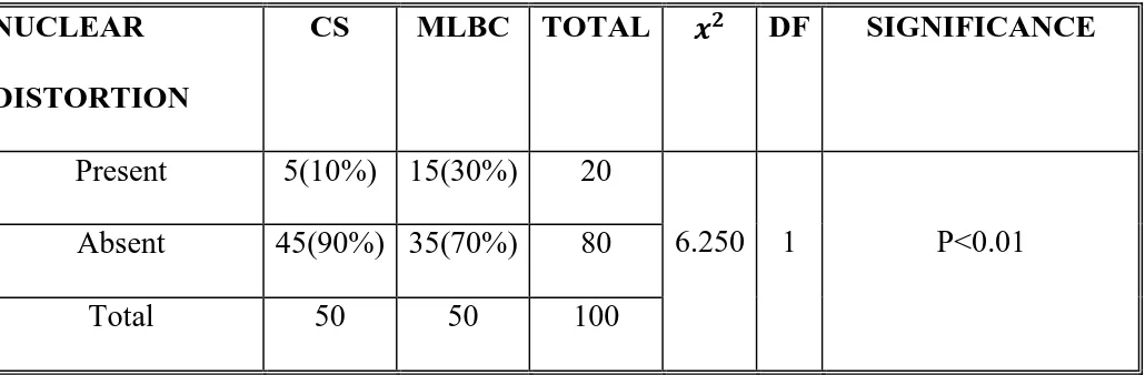
6. Cytoplasmic distortion( present/absent)
7. Nuclear irregularity( present/absent
49
The LiquiPrep slides were assessed on above criteria. MLBC smears
were also assessed with same criteria except for cellularity which was
graded in to 3 grades ( 1,2,3) based on number of cells in each 40X field.
GRADE 1- up to 150 cells- Inadequate for reporting
GRADE 2-150- 500 cells-Just adequate
OBSERVATION AND
This study was conducted in the Department of Pathology at
Tirunelveli Medical College
of cervical smears were
standardization of MLBC technique and excluded from the study
cases were analyzed by MLBC with comparative conventional smears
various morphological features
cases were subjected to LiquiPrep
smear study (CHART1)
CHART
The smears were
For reporting ,The Bethesda system
50
OBSERVATION AND RESULTS
This study was conducted in the Department of Pathology at
Tirunelveli Medical College from August 2010 to April 2012.
were collected. 50 of these cases were used for
standardization of MLBC technique and excluded from the study
by MLBC with comparative conventional smears
various morphological features and final interpretation. The remaining 50
s were subjected to LiquiPrep cytology along with conventional
(CHART1).
CHART: 1 DISTRIBUTION OF CASES
The smears were studied by using 7 morphological parameters.
The Bethesda system2001 was used in both methods.
STANDARDIZATION 50
MLBC vs CS, 50 LP vs CS, 50
This study was conducted in the Department of Pathology at
from August 2010 to April 2012. 150 cases
50 of these cases were used for
standardization of MLBC technique and excluded from the study. 50
by MLBC with comparative conventional smears for
The remaining 50
along with conventional
morphological parameters.
51
I. MANUAL LBC versus CONVENTIONAL SMEARS
For Manual LBC we had to standardize fixative and cell base
solution. We required 50 cases for standardization and the following
observations are made.
METHOD I OF STANDARDIZATION OF SOLUTION
In the first method we used the fixative & cell base in the ratio
depicted in TABLE 5 &6. The observations are
1. Cells shows shrinkage artifact.
2. Nuclear & cytoplasmic details are distorted and not clearly made
out.
3. Background shows abundant mucus & blood which obscures the
cellular details.
4. Smears do not dry quickly.
5. Smears easily washed off while staining.
METHOD II OF STANDARDIZATION
An attempt was made to remove the inflammatory background by
adding glacial acetic acid to the fixative. The ratio of agarose and PEG in
cell base was altered in an attempt to hasten the process of drying of
smears (TABLE 7&8).The observations are
1. Inflammatory cells in the background are reduced
2. Cell shrinkage artifact still present
3. Wisps of mucus in the background present
52
METHOD III OF STANDARDIZATION
The concentration of alcohol was increased in an attempt to
remove/ dissolve mucus, based on Saccomano’s method of sputum
processing. The concentration of agarose was increased, to further
decrease the drying time of the smears. (TABLE 9&10).
The observations are
1. Cell shrinkage is reduced.
2. Removes most of the mucus and all the inflammatory cells.
3. Smears dry well with the formation of membrane.
4. Smears does not get washed away.
I.STATISTICAL ANALYSIS OF CS AND MLBC RESULTS
The data regarding Conventional smears vs Manual Liquid based
cytology were compared and interpreted by χ2 (Chi- square) test. The
above procedure of statistical analysis and interpretations were made by
the statistical software IBM SPSS statistics 20. The P-values <0.05
(P<0.05) were treated as significant.
The CS and MLBC smears were compared in respect of cellularity,
clean background, uniform distribution, cell overlapping, inflammatory
back ground, nuclear distortion, cytoplasmic distortion and interpretation
of results.
53
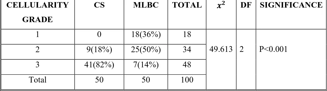
TABLE-11:COMPARISON OF CELLULARITY CELLULARITY
GRADE
CS MLBC TOTAL DF SIGNIFICANCE
1 0 18(36%) 18
49.613 2 P<0.001
2 9(18%) 25(50%) 34
3 41(82%) 7(14%) 48
Total 50 50 100
Of 50 cases, Grade 3 cellularity was seen in only 7 (14%) cases of
MLBC slides whereas 41 (82%) cases of CS showed grade 3 cellularity.
The difference in cellularity between the procedures was statistically
[image:62.595.35.572.107.257.2]significant (P<0.001) . (TABLE 11&CHART 2).
TABLE-12: COMPARISON OF CLEAN BACKGROUND CLEAN BACK
GROUND
CS MLBC TOTAL DF SIGNIFICANCE
Present 3(6%) 29(58%) 32
31.066 1 P<0.001
Absent 47(94%) 21(42%) 68
Total 50 50 100
In MLBC 29(58%) cases revealed clean back ground compared to
3(6%) cases in CS which was statistically significant (P<0.001).(TABLE
1 0%
36%
COMPARISON OF CELLULARITY (CS&MLBC)
PRESENT 6%
COMPARISON OF CLEAN BACKGROUND
54
CHART 2
CHART 3
2
3 18%
82%
36%
50%
14%
COMPARISON OF CELLULARITY (CS&MLBC)
CS MLBC
PRESENT
ABSENT 6%
94%
58%
42%
COMPARISON OF CLEAN BACKGROUND (CS &MLBC)
CS MLBC