Variations in the Branching Pattern and Dimensions of
Arch of Aorta
A Dissertation Submitted To
The Tamil Nadu Dr. M.G.R. Medical University
In partial fulfilment of the requirements
for the award of the degree of
M.S. (Branch V) Anatomy
CERTIFICATE
This is to certify that this dissertation entitled
“Variations in the
Branching Pattern and Dimensions of Arch of Aorta” is the bonafide
record of work done by Dr. N. Isai Vani under my guidance and
supervision in the Department of Anatomy during the period of her
postgraduate study for M.S. (Branch V) Anatomy from 2006 to 2009.
The Dean,
Professor and Head,
Thanjavur Medical College,
Department of Anatomy,
Thanjavur.
Thanjavur Medical College,
ACKNOWLEDGEMENTS
I express my heartfelt thanks to my teacher Dr. T.Sivakami M.S., Professor
and Head, Department of Anatomy, Thanjavur Medical College, Thanjavur, for her
advice and guidance in designing and enabling me to do this study with creative
suggestions and constructive criticisms.
I profoundly thank The Dean, Thanjavur Medical College, Thanjavur, for all
the facilities and encouragement provided to me for the completion of my thesis
in this prestigious institution.
My sincere thanks to Dr. S. Vijayalakshmi M.D., Professor and Head,
Department of Forensic Medicine, Thanjavur Medical College, Thanjavur, for
graciously giving me permission to collect the specimens from their department.
I am grateful to Assistant Professors Dr. R. Manoranjitham M.S.,
Dr. S. Sumathi M.S., Dr. K. Mohan M.S., Dr. M. Elangovan M.S. and Dr.
Kasuri Fathima D.G.O. for their valuable suggestions and help. I am very much
thankful to my senior and junior post graduates Dr. B. Santhi, Dr. S.
Kalaiyarasi and Dr. R. Nithiya Priya for their valuable help and co-operation. I
would also like to express my sincere appreciation to all the staff of the
Department of Anatomy, Thanjavur Medical College, Thanjavur.
I also thank all the authors who sent me e-copy / hard copy of their articles,
which helped me in writing my literature review. Special thanks to my husband
Dr. S. Manikandan M.D. and my parents for their inspiration, encouragement,
CONTENTS
CONTENTS
Page No.
Introduction 1
Aims and Objectives 4
Review of literature 5
Materials and methods 36
Results 43
Discussion 53
Conclusion 60
Summary 61
References 62
Introduction
The normal aortic arch curves smoothly upward into the superior
mediastinum, running from right to left and anterior to posterior , with its apex
approximately at the mid manubrium. The most common right to left branching
pattern of the aortic arch is brachiocephalic trunk, left common carotid and left
subclavian artery. In the complete formation of the aortic arch and its branches,
there is ample opportunity for anomalous development by persistence of certain
embryonic vessels and disappearance of others. These may include anomalies
pertaining to the arch per se., such as absent or double aortic arch, right aortic
arch or anomalies of branching patterns such as common trunk for
brachiocephalic and left common carotid artery, common trunk for left common
carotid and left subclavian artery, vertebral artery or retroesophageal right
subclavian artery as the fourth branch or combination of any of these.
Anomalous development of the aortic arch and its major branches
becomes clinically significant when these vessels encroach upon and constrict
adjacent mediastinal structures such as the esophagus and trachea, interfering
with respiration and deglutition (dysphagia lusoria).1,2 The various anomalies of
the aortic arch and its branches are often associated with other congenital
anomalies such as tetralogy of Fallot (ToF),3,4 tricuspid atresia, truncus arteriosus,
transposition of great vessels, ventricular septal defect (VSD),5 pulmonary
stenosis, double outlet right ventricle,4 patent ductus arteriosus (PDA),6 persistent
trigeminal artery, persistent proatlantal segmental artery, tracheoesophageal
addition to congenital malformations, pathological aneurysms and obstructions
can also occur.
Knowledge concerning the various anomalies of the aortic arch
derivatives is important to the clinician, radiologist and surgeon who treat patients
who have symptoms attributable to these anomalies. To the clinician and
radiologist, this knowledge aids in evaluating the findings in cases in which
symptoms may result from the presence of vascular anomalies. To the surgeon,
realization of the possible vascular configurations which may interfere with the
function of either esophagus or trachea, will serve as a guide while exploring the
region of the aortic arch and in determining the exact nature of the anomaly in a
given case. This determination will serve to guide the surgeon to perform those
procedures most suitable for the correction of the condition or for the relief of
symptoms resulting from it. Improvements in the methods of opacification of the
aorta and its branches leading to the exact diagnosis and localization of arterial
and aortic lesions have kept pace with the advances in vascular surgery, the
proper interpretation of which requires an awareness of these variations.8
Knowledge of dimensions of the aorta and its major branches assumes
great importance in cardiovascular surgical and stenting procedures. Attempts at
stenting in cases of anomalous aortic arches were associated with increased
failure rates and neurological complications.9 And in such cases the surgical
approach may have to be changed from the usual femoral to a brachial
approach.10 Dissecting aneurysm is more common when the vertebral artery
aortic arch is the proximal landing zone and the diameter of the stent-graft is
decided only according to the diameter of the aortic arch.12
A thorough knowledge of the aortic arch, variations of its branching
pattern and its dimensions is therefore necessary for the accurate diagnosis and
proper management of the various disorders associated with these anomalies.
Even though the variations in branching pattern and anomalies are documented
in the literature, not much is known about the dimensions of the normal as well as
the anomalous branches. This study is therefore undertaken to shed more light
Aims & Objectives
1. To study the anatomical variations in the branching pattern of arch of aorta in
cadaveric specimens by gross dissection.
2. To measure and analyse thevariations in the dimensions of normal and variant
Review of Literature
I. Arch of aorta
1. Normal anatomy
1.1. Extent
1.2. Course
1.3. Relations
1.4. Branches
1.5. Dimensions
1.6. Radiological anatomy
2. Variations
2.1. Embryology
2.2. Anomalies of the arch
2.3. Anomalies of branching pattern
3. Clinical significance
3.1. Historical perspective
3.2. Surgical significance
3.3. Radiological importance
II. Casting:
1. History
2. Methods
I. ARCH OF AORTA
1. Normal anatomy
The term “aorta” is derived from Greek aeiro, which means “to raise”.
Anatomically, the aortic arch is defined as the segment of aorta between a line at
a right angle proximal to the brachiocephalic artery origin and extending to a line
drawn at a right angle distal to the left subclavian artery.13
1.1. Extent
The arch of aorta begins posterior to the right half of sternal angle as a
continuation of ascending aorta. It ends by joining the descending aorta on the
disc between fourth and fifth thoracic vertebral bodies, in the same horizontal
plane as its origin.14 Topographically, it is that part of aorta that rises above the
plane dividing superior from inferior mediastinum. It lies in the superior
mediastinum opposite to the lower half of mediastinum sterni.15
1.2. Course
The arch first ascends diagonally back and to the left over the anterior
surface of trachea, then back across its left side and finally descends to the left of
fourth thoracic vertebral body, continuing as the descending thoracic aorta. It
ends in level with the sternal end of second left costal cartilage.16 It curves around
the hilum of left lung and its upper convexity reaches as high as the midpoint of
1.3. Relations
The arch is convex to the left as well as upwards, that is, it is convex in
two planes (Figure 1). Hence, the arch has 4 aspects15 – left anterior, right
posterior, concave lower. Five structures are related to the four aspects:
Right posterior aspect:
a) trachea
b) esophagus
c) left recurrent laryngeal nerve
d) thoracic duct and
e) vertebral column.
Left anterior aspect:
a) left lung and pleura
b) left phrenic nerve
c) left vagus nerve
d) cardiac nerves and
e) left superior intercostal vein
Concave lower aspect:
a) left bronchus
b) right pulmonary artery
c) ligamentum arteriosum
d) left recurrent laryngeal nerve and
Convex upper aspect:
a) brachiocephalic artery
b) left common carotid artery
c) left subclavian artery
d) thymus and
e) left brachiocephalic vein.
1.4. Branches
Three branches arise from the convex aspect of the arch:
♥ Brachiocephalic trunk
♥ Left common carotid artery
♥ Left subclavian artery
They may branch from the beginning of the arch or the upper part of
ascending aorta. The distance between these origins varies, the most frequent
being approximation of left common carotid artery to the brachiocephalic trunk.16
Brachiocephalic artery:
The brachiocephalic (innominate) artery, the largest branch of the aortic
arch, is 4-5 cm in length.16 It arises from the convexity of the arch posterior to the
centre of manubrium sterni. It then passes upward and to the right of trachea and
divides into right subclavian and right common carotid arteries behind the right
Left common carotid artery:
The left common carotid artery (LCCA) originates directly from the aortic
arch immediately posterolateral to the brachiocephalic trunk, and ascends until
level with the left sternoclavicular joint, where it enters the neck. It is 20-25 mm
long16 and it lies at first in front of the trachea, then it inclines to the left.
Left subclavian artery:
The left subclavian artery (LSCA) arises from the aortic arch below the left
common carotid artery and rises into the neck lateral to the medial border of
scalenus anterior, crosses behind this muscle and then descends towards the
outer border of first rib, where it becomes the axillary artery.16
1.5. Dimensions
The diameter of the aortic arch at its origin is 28 mm, but it is reduced to
20 mm at the end,16 after the issue of its large collateral branches. At the border
with the thoracic aorta, a small stricture (aortic isthmus), followed by a dilatation,
can be recognized. In foetal life, the isthmus lies between the origin of left
subclavian artery and the opening of ductus arteriosus.16
1.6. Ligamentum arteriosum (Botallo’s ligament)
This is a fibrous remnant of ductus arteriosus (DA). It passes from the
commencement of left pulmonary artery to the concavity of aortic arch, beyond
the point where the left subclavian artery branches off. It lies almost horizontally.
The left recurrent laryngeal nerve hooks around it. The superficial part of cardiac
plexus lies anterior to it, and the deep part is on the right, between the aortic arch
1.7. Radiographic anatomy
The shadow of the arch is easily identified in anteroposterior radiographs
and its left profile is sometimes called the “aortic knuckle”. The arch may also be
visible in left anterior oblique views enclosing a pale space, “the aortic window”,
in which shadows of the pulmonary trunk and its left branch may be discerned.16
Computed tomography (CT), magnetic resonance imaging (MRI) and digital
subtraction angiography (DSA) can be useful diagnostic tools because they
reveal the positions of vascular, tracheobronchial and esophageal structures and
their relationships to one another.19 Gadolinium enhanced three dimensional
magnetic resonance angiography, with multi planar reformations and sub volume
maximum intensity projection (MIP) is particularly helpful at depicting the origins
and courses of the branches of aortic arch.3
2. Variations
The aorta may vary in its position and extent without any other
irregularities. The height of the arch may be as high as third thoracic vertebra or
as low as fifth thoracic. The summit of the arch is usually about 2.5 cm below the
superiosternal border.16 It may reach the top of sternum.20 It is closer to the upper
border of sternum in infants and also in old age, because of dilatation of the
vessel.
2.1. Embryology
The first major intraembryonic vessels that develop in the 3-week embryo
are the dorsal aortae, which run along the axis of the embryo. As a result of
cranial portions of the dorsal aortae become arched and embedded in the
mesenchyme of the branchial arches. The paired dorsal aortae fuse to form a
midline descending aorta.21,22
Six pairs of arteries, called aortic arches, develop from the aortic sac,
pass laterally around the developing gut, and connect to the paired dorsal aorta.
During the sixth to eighth week of gestation, the six aortic arches along with the
seventh segmental dorsal artery from each dorsal aorta develop into the aortic
arch and its major branches (Figures 2 and 3).
In human embryos, all aortic arches are never present at the same time.
Their formation and remodelling (with the exception of the fifth arch) show a
pronounced craniocaudal gradient.23 The more cranial are in the process of
disappearing before the caudal ones are completed. The proximal portion of
aortic arch is derived from the left horn of aortic sac and the distal portion is
derived from left dorsal aorta. The arch of aorta between left common carotid and
left subclavian arteries is derived from the left fourth aortic arch.
The brachiocephalic artery is derived from the right horn of aortic sac. The
proximal segment of right subclavian artery is derived from right fourth aortic arch
and its distal part from a portion of right dorsal aorta and seventh intersegmental
artery. The distal part of left sixth aortic arch persists during intrauterine life as the
ductus arteriosus. The proximal parts of the third pair of aortic arches form the
The left subclavian artery is not derived from an aortic arch but from the
left seventh intersegmental artery. As development proceeds, differential growth
shifts the origin of left subclavian artery cranially and it comes to lie close to the
origin of left common carotid artery. Vertebral artery is formed by the
development of longitudinal anastamoses that link the dorsal cervical
intersegmental arteries. The intersegmental arteries regress, except for the
seventh, which becomes the proximal subclavian artery and which includes the
point of origin of vertebral artery in adults.
The transformation of the sixth pair of aortic arches explains why the
course of recurrent laryngeal nerves differs on the two sides. These nerves
supply the sixth pair of aortic arches on their way to the developing larynx. On the
right, because the distal part of the right sixth aortic arch degenerates, the right
recurrent laryngeal nerve moves superiorly and hooks around the proximal part of
right subclavian artery, a derivative of fourth aortic arch. On the left, the left
recurrent laryngeal nerve hooks around the ductus arteriosus formed by the distal
part of left sixth aortic arch. When this arterial shunt involutes after birth, the
nerve hooks around the ligamentum arteriosum and arch of aorta.24
Genomics:
Genetic factors determining aortic arch development and its perturbations
are now being elucidated. In 2001, McElhinney et al25 found that chromosome
(chr) 22q11 deletion was significantly more common in patients with abnormal
branching or laterality of the aortic arch than it was in those with a left sided arch
The internal organs of all vertebrates are symmetrically organised across
the left-right axis. The development of this asymmetry is controlled by a
molecular pathway that includes the signalling molecule Nodal and the
transcription factor Pitx2, proteins encoded by genes that are predominantly
expressed on the left side of all vertebrate embryos. At the genome level, cardiac
neural crest cells are known to play multiple roles during development of the
inflow and outflow tract of the heart and aortic arch. In 2002, Liu et al26 showed
that the Pitx2 gene contributes to aortic arch development, suggesting that a
major function of the Pitx2 mediated left right asymmetry pathway is to pattern
the aortic arches.
2.2. Anomalies of the arch
Recognition of anomalies of the aortic arch date back to 1737, when
Homell first described a double aortic arch.27 In view of the complex events that
occur in the development of the aortic arch and its branches, it is not surprising
that anomalies occur. When cardiac neural crest is removed from early avian
embryos, malformations involving the carotid arteries and arch of aorta result.23
Most irregularities are the result of persistence of parts of aortic arches that
usually disappear, or disappearance of parts that normally persist.24,28
A classification of major variations of the aortic arch given by Gross and Ware
(1946) and Neuhauser (1946) is as follows20:
I. Right aortic arch
A. Situs inversus viscerum
1. Anterior type (arch is anterior to the trachea and descending
aorta is on the right side)
2. Posterior type (aorta passes to the left behind the esophagus
and descending aorta courses to the right)
II. Double aortic arch
A. Both aortic limbs are patent
B. One aortic limb is obliterated
III. Anomalies right subclavian artery
The artery arises last from a normal aortic arch and crosses the
midline to its distribution on the right side (behind trachea & esophagus)
IV. Patent ductus arteriosus
V. Coarctation of the aorta (CoA)
Right sided aortic arch
This anomaly reproduces the normal vascular pattern seen in birds.20,29 A
right aortic arch arises from the persistence of the complete embryonic right
aortic arch and disappearance in the left arch of the segment caudal to the exit of
left subclavian artery. A right aortic arch was first described three centuries ago
by Fioratti and Aglietti (1763). Fifty five years later, Corvisart (1818) reported its
occurrence in a case of tetralogy of Fallot (Corvisart’s disease).4,5 Liechty, Shields
and Anson30 (1957) found one instance of right sided aortic arch among 1000
cadavers examined.
Right sided aortic arch has been found to be associated with other
anomalies such as coarctation of aorta, patent ductus arteriosus, pulmonary
stenosis, tricuspid atresia and ventricular septal defect .5 A right aortic arch with
Double aortic arch
A double aortic arch results when the right dorsal aorta persists between
the origin of seventh intersegmental artery and its junction with the left dorsal
aorta. This anomaly is characterized by a vascular ring around the trachea and
esophagus producing varying degrees of compression of these structures (Figure
5).21,24
The right aortic arch is most commonly dominant.21 The descending aorta
is usually in its normal position on the left, as is the ligamentum arteriosum.
Generally, each subclavian and common carotid artery arises independently from
its respective aortic arch. The brachiocephalic vessels do not develop. 20% of
cases have other anomalies, with VSD and ToF being most common.27 Double
aortic arch was first described by Homell in 1737. The first surgical correction of a
double aortic arch was performed by Gross in 1945 on a one year old boy with
chronic wheezing.27
Retroesophageal right subclavian artery
Abnormal origin of right subclavian artery is formed by the distal portion of
right dorsal aorta and seventh intersegmental artery. The right fourth aortic arch
and the proximal part of right dorsal aorta are obliterated. With shortening of the
aorta between left common carotid and left subclavian arteries, the origin of
abnormal right subclavian artery finally settles just to the right of left subclavian
artery. Since its stem is derived from the right dorsal aorta, it must cross the
midline behind the esophagus to reach the right arm (Figure 4).21
It is found in about 0.5-1.8% of the population27 and its incidence is 16 in
every 1000 autopsies.31 Although this anomaly is fairly common and always forms
tight enough to constrict the esophagus and trachea.24 This anomaly is especially
associated with three other anomalies – anomaly of right recurrent branch of
vagus, right thoracic duct instead of a left one and tracheoesophageal fistula.32
Patent ductus arteriosus
Patent ductus arteriosus is one of the most frequently occurring
abnormalities of the great vessels, especially in premature infants.21 Ductus
arteriosus is formed from the distal part of left sixth aortic arch. At birth, the
ductus constricts, probably as a result of initial exposure of oxygen sensitive
muscle cell in its wall to blood with high oxygen content. By one month, the DA is
normally obliterated to become ligamentum arteriosum.28
Closure of the ductus is not an instantaneous event. Anatomical patency
of DA is present in 65% of infants at the age of two weeks and slightly over 1% at
the age of one year. The lumen of a patent ductus may vary from a few mm to a
size equal to or greater than the aorta. Its length is usually about 1 cm, but may
be so short that there is practically an aortopulmonary window, or it may be as
long as 3 cm.6
Approximately 10% of patients with PDA have associated cardiovascular
anomalies. In some of these patients, the presence of a patent ductus serves as
a compensatory mechanism to sustain life. In others, the ductus is an isolated
Coarctation of aorta
Coarctation (from the Latin “arctare”, to make tight) refers to a congenital
or developmental anomaly of the aorta characterized by stenosis or obliteration
of a section of that vessel. This malformation was first described by Morgagni in
1760.1 In 98% of cases, the constriction is at the level of insertion of DA but may
be located anywhere between the arch and the bifurcation of aorta.1 Its incidence
according to post-mortem studies is 1:10006 and 0.3% in newborn infants.33 CoA
is associated with PDA in 10% of cases.6
Two types – preductal and postductal may be distinguished based on the
location of the constriction, either above or below the entrance of DA (Figure 6). It
results from the incorporation of oxygen sensitive muscle tissue from the DA into
the wall of the aorta. Normally, this muscle constricts when exposed to high
oxygen tension, closing the DA. If the muscle is also in the wall of the aorta, the
aorta constricts at that level. Eventually, chronic changes occur and the
constriction becomes permanent.28
Absence of aortic arch
It is one of the rare anomalies of aortic arch. Absence of aortic arch
means that there is no connection of any kind between the ascending and
descending aorta, the latter being the continuation of pulmonary trunk via a PDA.
Functionally it corresponds to the most severe form of “infantile type” (preductal)
coarctation. It is also called “Steidele’s complex”.34 Interruption of the aortic arch
can occur at any point between the embryonic aortic sac and the ductus
arteriosus. Based on the location of the absence of aortic arch, Celorie and
Patten have classified this defect into three types – A, B and C. The constant
Carson to suggest that these anomalies should be known as a “cardiovascular
triology”.35 This lesion has been produced in mice lacking the winged helix
transcription factor MF-1 (mesenchyme fork head-1).23
Recurrent laryngeal nerve in variations
The disposition of recurrent laryngeal nerves is normal in cases of CoA
and PDA. In cases of posterior subclavian artery, the right recurrent laryngeal
nerve is replaced by branches passing directly from vagus to the larynx. In cases
of double and right sided aortic arch, it passes around the arch instead of around
the right subclavian artery.
The position of left recurrent laryngeal nerve is normal when there is also
a left aortic arch or when DA is on the left side, joining the pulmonary artery with
a persistent left aortic root. When there is a left brachiocephalic artery and
ligamentum arteriosum is on the right side, it winds around the left subclavian
artery.36
2.3. Anomalies of branching pattern
The most commonly encountered branching pattern in humans consists of
three great vessels originating from the arch of aorta, brachiocephalic artery, left
common carotid artery and left subclavian artery. The final configuration of the
aortic arch and its branches is related to the development of the embryonic aortic
arches and the associated migration and merging of their branches.
Many variations occur in the number and position of vessels arising from
the aortic arch. More than 15 different aortic arch configurations have been
I – In this type, three branches leave the arch from right to left in the following succession: innominate, left common carotid and left subclavian.
II – Here both common carotids arise from the innominate reducing the number of branches to two.
III – There is an increase in the number of branches and the additional vessel is left vertebral.
IV – Here the left vertebral artery replaces the left common carotid being second in right to left succession and both common carotids arise from a common stem.
V – The left vertebral artery arises from the innominate, and the order of left common carotid and left subclavian arteries is reversed.
VI to VIII – The right subclavian artery arises as the last branch of aortic arch reaching the right upper extremity by passing dorsal to the esophagus. These types differ with respect to the origin of other vessels.
IX – A bi-innominate sequence in which paired vessels are the only derivatives of the
X & XI – In both these varieties the left vertebral artery arises from a common stem from which the left subclavian is also derived. However, in type X a regular innominate artery is present, whereas in type XI, the innominate shares a common trunk with left common carotid.
XII – Here, as in type III, an extra vessel arises from the aortic arch, but the additional vessel being arteria thyroidea ima.
XIII – In this, the usual three branches take origin from the aortic arch through a single trunk as an intermediary vessel.
XIV – Single stem for the usual three branches as in type XIII, with the left vertebral arising as a separate branch from the arch.
There may be as few as one or as many as six branches. The common
variant in which two vessels arise from the aortic arch is one where the left
common carotid artery has a common origin with the brachiocephalic trunk
(BCT). A similar but less common variant occurs when the left common carotid
artery originates directly from the brachiocephalic artery rather than as a common
trunk. Both variants of left common carotid artery have been referred in various
textbooks as a bovine type arch, through this term is most commonly ascribed to
the common trunk variety.37 A true bovine arch bears no resemblance to any of
the common human aortic arch variations. In cattle, a single great vessel
originates from the aortic arch. This large brachiocephalic trunk gives rise to both
subclavian arteries and a bicarotid trunk. The bicarotid trunk then bifurcates into
right and left common carotid arteries.38
A common variant with four vessels, is one in which the left vertebral
artery arises directly from the aortic arch between left common carotid and left
subclavian arteries. Another variant (1.1%) which differs slightly from the
preceding variety is one in which the left vertebral artery replaces the left
common carotid artery as the second stem in right to left succession while the left
common carotid artery arises as a common trunk with BCT.30 The anomalies of
branches arising from the aortic arch are due to variations in the extent of the
fusion process and abnormal absorption of some of the aortic arches in to the
aortic sac. Variations in the arterial morphology of aortic arch branches may also
be a consequence of alterations in tissue development. Certain growth factors
such as placental growth factor, fibroblast growth factor-2, angiopoietins and
vascular endothelial growth factor are released by the developing tissues to
pattern of arterial morphology may result from premature or late release of these
factors, or increase or decrease in the level of these factors. The vertebral
arteries provide blood supply to the posterior neck musculature,
rhombencephalon, mesencephalon, occipital lobe and part of temporal lobe of
the brain. A direct origin of vertebral artery from the arch of aorta may be a
reflection of variation in demand by these structures.39
3. Clinical significance
3.1. Historical perspective
In 1789, Bayford wrote a fascinating account of a woman, with a life long
history of severe dysphagia, who “fancied that she nourished within her a
voracious animal, and attributed all her uneasiness to the fury with which this half
starved monster fell upon each morsel in its passage to the stomach”. When at
the age of 62, “worn out with fatigue and famine she sunk into a grave”, he
carried out a post-mortem examination and published his delightful description
with drawings of an anomalous subclavian artery.29 Bayford’s term ‘dysphagia
lusoria’ (from lusus naturae – freak or prank of nature) is still employed to
dysphagia resulting from many vascular anomalies other than an aberrant
retroesophageal subclavian artery.27
The first successful correction of a congenital cardiac defect was reported
by Gross in 1939, when he successfully ligated a PDA.2 In 1945, it was Gross
again who first surgically corrected a double aortic arch.27 In 1957, DeBakey and
colleagues first described aortic arch replacement using ante grade brain
doing aortic arch surgery became considerably simpler. In 1963, Bernard
combined deep hypothermia with circulatory arrest and cardiopulmonary bypass
for aortic arch operations and dissection. But this became popular only after 1975
when Griepp reported a series of aortic arch replacements using deep
hypothermia and circulatory arrest. In 1983, Borst et al reported replacing the
aortic arch and leaving a tube graft lying free in the descending aorta, which they
called the “elephant trunk technique”. In 1990, Crawford, Svensson and
colleagues reported replacement of the entire aorta as a planned stage
procedure using a modified elephant trunk technique. Currently, aortic arch has
become relatively common and safe with a mortality risk of 2% and a stroke risk
of 2%.13 The first video assisted thoracoscopic surgery (VATS) vascular ring
division was performed in 1993 at Children’s Hospital, Boston, the same
institution where Gross had performed the open surgical division 48 years
earlier.13
3.2. Radiological significance
Congenital anomalies of the aortic arch and its branches produce
recognizable effects largely through distortion of esophagus and trachea.
Diagnosis could hardly progress until radiology contributed its essential quota.
The studies of Evans (1936) on the esophagus, and of Fray (1936) and of
Bedford and Parkinson (1936) on right sided aorta, laid the foundations of
radiological diagnosis, now advanced to a high degree of precision. The nature of
many of the anomalies of aortic arch in radiographs may be determined by
characteristic combination of great vessel shadows, lateral deviations of trachea
and esophagus, rounded pressure defects in the contour of trachea and rounded
Most of the anomalies of the aortic arch are usually asymptomatic.
Therefore, radiographic demonstration of such abnormalities is often a first
indication of their existence.42 Chest radiography is the first and most commonly
performed investigation. Many authorities consider barium esophagography to
be the most important study in patients with suspected vascular ring and it is
diagnostic in the vast majority of cases.19 CT can make apparent the size and
orientation of the aortic arch and determine the origin and course of aberrant
subclavian arteries.42
Multi detector CT (MDCT) can also provide exquisite images, but is rarely
used because of its radiation implications. Angiography, previously the imaging
procedure of choice, is now rarely required unless cardiac catheterization is
necessary for the investigation of associated cardiac abnormalities.19 Gadolinium
enhanced three dimensional angiography with multi planar reformations and sub
volume MIP is particularly helpful at depicting the origins and courses of branch
vessels.3 High resolution multi detector row CT angiography enables the display
of images from different views. These can be useful as a roadmap in
interventional and surgical and therapeutic decision making, especially in patients
with complex aortic arch anatomy.43
Foetal sonographic diagnosis of aortic arch anomalies
Recognition of trachea, three vessels branching from the aortic arch,
ductus arteriosus and descending aorta in the axial views of the upper
mediastinum is necessary for a complete foetal cardiac assessment. Clues to
aortic arch anomalies include abnormal position of the descending aorta,
between the ascending aorta and main pulmonary artery in the three vessel view
and an abnormal vessel behind the trachea with or without a vascular ring or loop
around the trachea. Meticulous attention to anatomical landmarks will lead to
successful prenatal diagnosis of important vascular rings, making early postnatal
management possible.44
3.3. Surgical significance
Knowledge of variations of great vessels like the aortic arch and its
branches is of vital interest to surgeons because even a minor accidental injury of
the vessels may cause sudden massive haemorrhage, shock & fatalities in the
operation theatre.45
Percutaneous carotid revascularisation is used with increasing frequency
to treat carotid atherosclerotic disease. Classically the procedure is performed
from the femoral approach, with a long sheath placed into the common carotid
artery, and angioplasty and stenting performed through this. Congenital
anomalies of the aortic arch increase the technical difficulty of this procedure. In
2003, Shaw et al 10 reported three cases of bovine arch associated with increase
in the level of case difficulty when performing left carotid stenting from the
femoral approach. According to them, the right upper extremity (radial or brachial)
approach is preferable when there is a left carotid target lesion in the presence of
a bovine arch.
In the study conducted by Fagolli GL9 in 2007, it was concluded that
bovine arch was associated with increased failure and neurological complication
rate in carotid stenting procedures. Therefore, the presence of an aortic arch
accomplish such stenting procedures and in the presence of arch anomalies,
stenting should be reserved for patients with high surgical risk.
Retroesophageal right subclavian artery is important to the angiographer
who uses the right axillary, brachial or radial approach to the ascending thoracic
aorta. The presence of the anomaly is suspected in cases in which
catheterization of the ascending aorta proves difficult.46
The usual clamping techniques used in surgical aneurysms of the aortic
arch could not be performed in cases where the left common carotid artery arose
from the brachiocephalic artery. In such cases, it is necessary to implement total
circulatory arrest in order to repair the aneurysm completely.47
According to Bernardi and Detori (1975),49 the abnormal origin of vertebral
arteries “may favour cerebral disorders because of alterations in cerebral
hemodynamics”.48 In 2005, Dudich et al49 postulated that anomalous vertebral
artery origin may be an independent risk factor for arterial dissection; the longer
extra cranial course may lead to increased vulnerability of the vessel wall to
shear stress resulting in intimal tear and dissection.
Komiyama et al11 in 2007 retrospectively examined the angiograms of
1109 patients to evaluate arterial dissection of intracranial or extracranial
vertebral arteries and the origin of vertebral artery from the aorta or subclavian
artery. They concluded that left vertebral artery of aortic origin is associated with
a predilection for vertebral artery dissection in comparison to left vertebral artery
of subclavian artery origin. Shear stress may be larger in the vertebral artery of
aortic origin than in the vertebral artery of subclavian artery origin, possibly due to
pulsatile flow whereas vertebral artery of subclavian artery origin may receive
damped flow due to the presence of the proximal subclavian artery.
The true value of detecting anomalous origins is the diagnostic gain prior
to the surgery of supra aortic arteries. Detailed knowledge of an anomalous origin
of supraaortic arteries is also important in patients who have to undergo four
vessel angiography in an emergency to rule out the possibility of intracranial
aneurysm after subarachnoid haemorrhage. If the detection of a vertebral artery
in the normal position is not possible, the presence of a variant of vertebral artery
arising from the aortic arch must be taken into consideration.50
Anatomy of the vertebral artery and its variations is critical for surgeons to
help avoid serious complications during surgery in the brain, neck and chest
regions.51 Anomalous vertebral artery might constitute a risk factor for the
development of a saccular aneurysm at the origin of a posterior communicating
artery, with increased hemodynamic risk or vertebrobasilar insufficiency.52
In 2006, Kahraman et al53 measured the diameters of the various branches of
the aortic arch in patients with coronary artery ectasia and compared them to a
control group. They concluded that in the coronary artery ectasia group, the
diameters of all arteries arising from the aortic arch were significantly larger than
the corresponding arteries in the control group.
Occlusive disease of branches of the aortic arch
That obstruction of arteries at the point of origin from the aortic arch can
cause circulatory disturbances in the head or arms has long since been
1881.40 In Japan in 1908, Takayasu54 first reported a syndrome characterized by
loss of pulsations in the radial arteries, absence of detectable blood pressure in
the arms, syncope and visual disturbances. Caccamise and Whitman are
accredited with reporting the first authentic case in the US (1952) and called the
syndrome “pulseless disease”.54 Pulseless disease is also known as
anisophygmia, pulsus incongruens or pulsus differens and Takayasu’s disease.
The syndrome caused by obstruction of the branches of aortic arch was called
“the aortic arch syndrome” by Ross and McKusick in 1953.55 Anomalous aortic
arches might contribute to vascular occlusion in aneurysm of aorta. Martorell and
Fabre in 1944 and again in 1954 best described this syndrome of occlusion of the
supra aortic branches, which has been frequently referred to as “Martorell
syndrome”.54
II. Casting
1. History
The earliest example of casts being made of an anatomical cavity are
those of cerebral ventricles by Leonardo da Vinci (1452-1519). He used molten
wax for his work, but as he had no method of hardening the brain before making
the injection, the casts obtained bore little resemblance to the actual cavities. The
introduction by Jan Swammerdam (1637-1680) of molten wax injection masses
for filling arteries, veins and ducts, pointed the way to the preparation of corrosion
casts, in which, the original tissues are corroded away by means of some
reagent which does not attack the cast. But casts made of wax were very fragile.
with which Govert Bidloo (1649-1713) succeeded in making casts of the bronchial
tree.56 The technique was refined and popularised by Swammerdam’s student
Ruysch, during the following fifty years, but the interest in vascular casting was
greatly diminished by the middle of the 18th century. Hyrth in 1873 published a
most elegant monograph in corrosive preparations. His illustrations clearly
demonstrate injections of minute macroscopic blood vessels.57
2. Methods
A notable advance in the technique for the preparation of corrosion casts
was made when Paul Schiefferdecker in 1882 described a method using celloidin
(cellulose nitrate) dissolved in ether. Celloidin casts have to be kept in fluid as
when celloidin becomes dry it crumbles. In 1899, Carl Storch substituted celluloid
(cellulose acetate) for celloidin, so that it was no longer essential to preserve the
casts in fluid. In 1903, Mme.Krasuskaja recommended the substitution of acetone
for ether as the solvent. In 1936 Narat and others described the use of a vinyl
resin dissolved in acetone, as a substitute for cellulose acetate. The appearance
in 1948 of cold setting synthetic unsaturated polyester resins opened up new
possibilities in the sphere of anatomical casting. They make it possible to produce
beautiful, rigid, coloured casts of any large anatomical cavity. But the extent of
the injection is impossible to control while using synthetic resins.56
With the advent of modern chemistry, elastic materials like latex came into
use, and more recently, different materials like silicon rubber, polyester or acrylic
resins were introduced.58,59 According to requirements, coloured or radio opaque
materials were added.60 Measurement of the casted structures with silicon
EMV used Batson’s methylmethacrylate corrosion compound no. 7 for his
specimens.61 Live casting of the vascular tree of an animal using silicon was
performed by Sobin SS in 1965 and Xue Mei Li in 2003.57,62
Gaillord P et al63 embedded wax copies of methyl methacrylate corrosion
casts of cervicocranial arteries with liquid silicon modified into transparent blocks.
These blocks after wax evacuation, contained hollow reproductions of the original
vascular trees. The models were connected to a pulsatile pump and their
compatibility with various imaging techniques and endovascular treatment
materials was evaluated. The models were compatible with DSA, CT, MRI and
transcranial Doppler sonography. They provided a realistic endovascular
environment for the simulation of interventional stent procedures.
3. Advantages
The in vitro vascular models are easy to handle, respect human
morphological characteristics and are fairly reproducible. They have been shown
to provide realistic experimental settings for hemodynamic studies and may also
represent an interesting alternative to animal models for the evaluation of
endovascular treatment methods, with the exception of biocompatibility studies.63
The resilience and non adhesiveness of the silicon rubber allowed casts
to be extracted without recourse to corrosion. The surrounding tissues were then
available for conventional pathological examinations. Thus, silicon casting is a
relatively a simple technique with important applications, not practicable using
earlier casting materials for the study of vascular morphology and for the post
Materials and Methods
Design
Comparative study.
Time of the study
The study was conducted from August 2006 to June 2008.
Setting
Dissection laboratory of the Department of Anatomy, Thanjavur Medical
College and autopsy room of the Department of Forensic Medicine, Thanjavur
Medical College.
Inclusion criteria
Specimens from adult human cadavers aged more than 20 years of both
sexes in the Department of Anatomy and from autopsied cadavers in the
Department of Forensic Medicine during the period August 2006 to June 2008
were included in the study.
Exclusion criteria
Specimens from cadavers aged less than 20 years.
Parameters studied
Position of the aortic arch in relation to oesophagus:
- right / left
Number of branches arising directly from the arch of aorta
Anomalies present
Diameters of the human aortic arch and its branches at various levels
(Figure 8)
Taper ratio
Sample size
100.
Statistical analysis
Data were entered in Microsoft Excel spread sheets. One way ANOVA
with Tukey’s post hoc test was used for comparison between groups. Repeated
measures ANOVA was used to analyse the parameters measured at various
levels in the same specimen. This was done using GraphPad Instat version 3.01
for Windows (GraphPad Software, San Diego, California, USA). P<0.05 was
A – ascending aorta 25 mm proximal to the origin of brachiocephalic artery.
B – proximal to the origin of brachiocephalic artery.
C – distal to the origin of brachiocephalic artery.
D – distal to the origin of left subclavian artery.
E – descending aorta 25 mm distal to the origin of left subclavian artery.
R1 – origin of brachiocephalic trunk.
R2 – 15 mm distal to R1.
L1 – origin of left common carotid artery.
L2 – 15 mm distal to L1.
S1 – origin of left subclavian artery.
S2 – 15 mm distal to S1.
T1 – distance between adjacent margins of R1 and L1.
T2 – distance between adjacent margins of L1 and S1.
Procedure
The heart with the aortic arch and its branches were removed en bloc
from cadavers in the Department of Anatomy, Thanjavur Medical College and at
autopsy in the Department of Forensic Medicine, Thanjavur Medical College
from 100 adult human cadavers in the age group of 20 – 70 years, during the
study period.
The specimen was removed from the cadaver as described in
from the mentum to the xiphisternum and the skin was reflected. Transverse cut
was made through the manubrium sterni immediately inferior to its junction with
the first costal cartilage. Then the second and subsequent ribs and intercostals
spaces were cut inferiorly, to the level of xiphisternal joint. The inferior part of
the sternum was gently elevated and the pleura was cut to expose the lungs.
The root of the lungs were cut from above downwards after pulling the lungs
laterally from the mediastinum, and removed. A cut was made over the middle
of each clavicle and the first rib and the manubrium sterni was turned upwards.
The veins were then displaced to expose the aortic arch and its branches. The
diaphragm was cut around the inferior margin of the heart and the branches of
the aortic arch along with trachea and oesophagus were cut as high as possible
in the neck.
The heart with the aortic arch and its branches thus removed were
washed thoroughly in running water and preserved in a solution containing 10%
formalin and thymol after serially numbering them from 1 – 100. The specimen
were then dissected and studied with regard to any anatomical variations in
their branching pattern. The diameters of the aorta and its branches were
determined at various levels and metric analysis was performed by use of a
movable calliper with 0.05 mm of possible error. All values were expressed as
mean ± SEM.
From the values obtained for the diameter of the aortic arch, taper ratio
was calculated by using the formula,12
Taper ratio = A – E x 100 A
Calculated taper ratio was analysed for statistical significance between the
three groups.
Confirmation
In specimens sharing a common trunk for the brachiocephalic and left
common carotid artery, the presence of the common ostium was confirmed
either by direct examination of the ostium on its luminal side by making a slit on
the posterior surface of the aortic arch or by making luminal silicon casts of
those specimens.
Luminal casting
The specimen was washed thoroughly in running water and flushed with
spirit to remove blood and secretions and allowed to air dry. General purpose
purpose and its canula was introduced into the descending thoracic aorta with
its tip towards the aortic arch. The cut ends of the branches of the aortic arch
and the pulmonary vessels were tightly secured with silk.
Now silicon was injected retrograde into the aorta. The injection was
stopped when the aorta and all its branches were filled up with silicon. The
canunla was removed and the free end of the descending thoracic aorta was
ligated. It was allowed to cure for 24 hours. The surrounding tissues were then
removed from the cast by careful dissection to reveal an anatomical replica that
illustrates clearly the common origin of brachiocephalic and common carotid
arteries.
Results
Hundred aortic arches were dissected and studied with respect to the
branches arising from them and the specimens were divided into various types
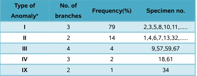
based on Anson’s classification32 (Table 1). In the present study, five types of
branching pattern were observed.
Type I – aortic arch with three branches – brachiocephalic, left common carotid and left subclavian arteries in that order (Photographs 3 and 4).
Type II – common trunk for BCT and LCCA (Photographs 5 and 6).
Type III – left vertebral artery as an additional branch from the arch in between the origins of LCCA and LSCA (Photograph 7).
Type IV – coexistence of Type II and Type III anomalies (Photograph 8).
[image:47.595.108.438.522.649.2]Type IX – left brachiocephalic trunk from the arch dividing into LCCA and LSCA (Photograph 9).
Table 1. Classification of branching pattern of aortic arch in 100 cadaveric specimens.
Type of Anomaly*
No. of
branches Frequency(%) Specimen no. I 3 79 2,3,5,8,10,11,...
II 2 14 1,4,6,7,13,32,...
III 4 4 9,57,59,67
IV 3 2 18,61
IX 2 1 34
IV – coexistence of Type II and Type III anomalies; Type IX – left brachiocephalic trunk from the arch dividing into LCCA and LSCA.
Photograph 3. Aortic arch in situ with normal branching pattern (Type I).
Photograph 4. Aortic arch with normal branching pattern (Type I).
Photograph 5. Aortic arch with common stem for brachiocephalic and left common carotid arteries (Type II).
A. B.
Photograph 6. Luminal casting of Type II branching pattern of aortic arch.
A. B.
Photograph 7. Left vertebral artery arising from the aortic arch
between left common carotid and left subclavian arteries (Type III).
A. B.
C. D.
Photograph 8. Coexistence of Type II and Type III anomalies in the same
specimen (Type IV).
Photograph 9. Aortic arch with common stem for left common carotid and left
subclavian arteries (Type IX). A. Anterior view B. Posterior view showing the
window made for confirming the common ostium.
A. B.
The relative proportions of the various types of aortic arch branching is
depicted in the following pie chart (Figure 9).
Figure 9. Frequency of types* of aortic arch branching pattern in 100 human adult
cadavers.
*Based on Anson’s classification. Type I – aortic arch with three branches – brachiocephalic, left common carotid and left subclavian arteries in that order; Type II – common trunk for BCT and LCCA; Type III – left vertebral artery as an additional branch from the arch in between the origins of LCCA and LSCA; Type IV – coexistence of Type II and Type III anomalies; Type IX – left brachiocephalic trunk from the arch dividing into LCCA and LSCA.
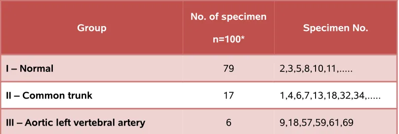
Dimensions of the aortic arch and its branches were measured at various
levels. For purpose of comparison of these dimensions, the specimens were
divided into three groups (Table 2).
The first group includes Type I aortic arch with three branches –
brachiocephalic, left common carotid and left subclavian arteries in
that order. Since this type occurs more frequently, it is designated
The second group includes those specimens in which the
brachiocephalic and left common carotid arteries share a common
trunk of origin. Also included in this group are two specimens in
which the common trunk was present in addition to the anomalous
aortic origin of left vertebral artery. Because there is only one
instance where the left common carotid and left subclavian artery
share a common left brachiocephalic trunk, this specimen was
also included in the common trunk category.
The third group includes specimens in which aortic origin of left
vertebral artery existed as an isolated anomaly or along with a
common trunk for the brachiocephalic and left common carotid
arteries.
Table 2. Grouping of anomalies of branching pattern of arch of aorta in 100 cadaveric specimens.
Group No. of specimen n=100*
Specimen No.
I – Normal 79 2,3,5,8,10,11,...
II – Common trunk 17 1,4,6,7,13,18,32,34,...
III – Aortic left vertebral artery 6 9,18,57,59,61,69
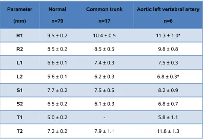
[image:50.595.113.517.485.621.2]The dimensions of the three branches of aortic arch (at their origin from
the arch and 1.5 mm away from the origin) and the distances between them were
[image:51.595.114.510.258.530.2]measured and their mean values in the three groups calculated (Table 3).
Table 3. Dimensions of the branches of aortic arch and the distance between its
three branches. Parameter (mm) Normal n=79 Common trunk n=17
Aortic left vertebral artery
n=6
R1 9.5 ± 0.2 10.4 ± 0.5 11.3 ± 1.0*
R2 8.5 ± 0.2 8.5 ± 0.5 9.8 ± 0.8
L1 6.6 ± 0.1 7.4 ± 0.3 7.5 ± 0.3
L2 5.6 ± 0.1 6.2 ± 0.3 6.8 ± 0.3*
S1 7.7 ± 0.2 7.5 ± 0.5 8.2 ± 0.9
S2 6.5 ± 0.2 6.1 ± 0.3 6.8 ± 0.7
T1 5.0 ± 0.2 - 5.8 ± 1.1
T2 7.2 ± 0.2 7.9 ± 1.1 11.8 ± 1.3
Values are mean ± SEM.*P < 0.05, compared to normal by one way ANOVA followed by Tukey’s post hoc test. R1 – origin of BCT, R2 – 15 mm distal to R1, L1 – origin of LCCA, L2 – 15 mm distal to L1, S1 – origin of LSCA, S2 – 15 mm distal to S1, T1 – distance between adjacent margins of R1 and L1, T2 – distance between adjacent margins of L1 and S1.
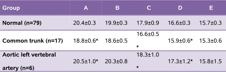
The diameters of the aortic arch at multiple sites in the three groups from
its origin to its termination were estimated and variations in diameter of aortic
Table 4. Diameters of the aortic arch (mm) at multiple levels in the three groups.
Group A B C D E
Normal (n=79) 20.4±0.3 19.9±0.3 17.9±0.9 16.6±0.3 15.7±0.3
Common trunk (n=17) 18.8±0.6* 18.6±0.5 16.6±0.5
* 15.9±0.6* 15.3±0.6
Aortic left vertebral
artery (n=6)
20.5±1.0* 20.3±0.8 18.3±1.0 *
17.3±1.2* 15.8±1.5
Values are mean ± SEM. A – ascending aorta 25 mm proximal to the origin of BCT, B – proximal to the origin of BCT, C – distal to the origin of BCT, D – distal to the origin of LSCA, E – descending aorta 25 mm distal to the origin of LSCA. Total number of specimens does not add to 100 as two specimens have both anomalies.
Figure 10. Diameter of aortic arch at different levels.
Values are mean ± SEM. p>0.05, when diameters were compared between groups by one way ANOVA followed by Tukey’s post hoc test. A – ascending aorta 25 mm proximal to the origin of BCT, B – proximal to the origin of BCT, C – distal to the origin of BCT, D – distal to the origin of LSCA, E – descending aorta 25 mm distal to the origin of LSCA.
Figure 10 illustrates the trend of decreasing diameter of aortic arch from
its proximal to distal level in the three groups. The mean taper ratio calculated
from the diameter at the proximal and distal end of the aortic arch was 22.57
18.88 ± 1.3% (Group II) and 23.4 ± 3.4% (Group III) and they do not differ
significantly (Figure 11).
Figure 11. Taper ratio of aortic arch.
Discussion
In the present study conducted on cadaveric specimens, the branching
pattern of the aortic arch described as normal (Type I) was observed in 79 of the
100 specimens. The most common anomaly was the presence of a common
stem for the brachiocephalic and left common carotid arteries (Type II) which was
observed in 14% of the specimens. The next common anomaly was observed in
4% of specimens, in which the left vertebral artery arose directly from the aortic
arch in between the origins of left common carotid and left subclavian arteries
(Type III). In two of the specimens both the above mentioned anomalies
co-existed (Type IV). And one specimen had a common stem for the left common
carotid and left subclavian arteries (Type IX). The frequency of the various types
of the branching pattern of the aortic arch in previous studies and the present
study is given in table 5.
Type II
The most common anomaly in our study is Type II. This is the type of
branching found in most apes.20
Preservation of distal blood flow and cerebral protection are of major concern
during surgical repair of aneurysms of the brachiocephalic trunk, where the aorta
is sequentially or partially clamped. If the left common carotid artery arose as a
common stem with the brachiocephalic trunk such clamping techniques could not
be used and in order to repair the aneurysm completely total circulatory arrest
Classically, percutaneous carotid angioplasty and stenting is performed from
the femoral approach with a long sheath placed into the common carotid artery.
In 2003, Shaw JA10 observed that stenting could not be performed in the usual
manner in Type II anomalies and carried out the technique with some
modifications. In one case, because of angular take off of left common carotid
artery, a 7 Fr shuttle sheath could not be advanced into the common carotid and
a more flexible 6 Fr was substituted. In another case, despite attempts with
various catheters from the femoral approach, adequate access to the common
carotid could not be achieved. Hence, a radial approach was chosen and stenting
procedure was performed successfully.10
From the above two studies it becomes clear that Type II anomaly plays a
pivotal role in deciding the choice of approach to stenting and the necessity of
total circulatory arrest in aortic aneurysm repair. Therefore the presence of such
an anomaly must be confirmed before any surgery on the aortic arch is
attempted.
Type III
The next most common pattern is one in which the left vertebral artery arose
as a direct branch from the aortic arch (Type III). This is observed in 6% of the
specimens. Dissecting aneurysms of the left vertebral artery is more common
when it arose directly from the aortic arch than when it arose from the subclavian
artery.11 Two possible mechanisms have been postulated for this :
i) vertebral artery of aortic origin may receive direct arterial pulsatile flow
whereas vertebral artery of subclavian origin may receive damped flow due to the
ii) the longer extracranial course may lead to increased vulnerability of the
vessel wall to shear stress resulting in intimal tear and dissection.11
Table 5. Frequency (%) of various types of branching of aortic arch as reported in scientific literature.
S.No Author Year Type I Type II Type III Type IV Type IX 1 Thompson65
(n=500) 1893 82.4 10.2 5.4 0.4
-2 Adachi 65
(n=516) 1928 83.3 10.9 4.3 0.6
-3 William JD
65 Negro(n=79) White(n=80) 1932 53.7 75.0 42.4 19.4 2.8
-4 William JD
66 Negro(n=137) White(n=111) 1935 56.9 75.9 37.0 17.8 2.0 2.6 1.8 0.5
-5 Anson32
(n=1000) 1957 64.9 27.1 2.5 1.1
-6 Wright NL67
(n=100) 1969 85 7 8 -
-7 Gupta68
(n=100) 2005 77 7 7 3 2
8 Present study
(n=100) 2008 79 14 4 2 1
Type I – aortic arch with three branches – brachiocephalic, left common carotid and left subclavian arteries in that order; Type II – common trunk for BCT and LCCA; Type III – left vertebral artery as an additional branch from the arch in between the origins of LCCA and LSCA; Type IV – coexistence of Type II and Type III anomalies; Type IX – left brachiocephalic trunk from the arch dividing into LCCA and LSCA.
Anomalous vertebral artery origin also represents a potential pitfall in
diagnostic cerebrovascular imaging. It may be wrongly assumed to be occluded
or diseased either by eluding catheterization during angiography or by lying
angiography, MR angiography or Doppler sonography.52 A variant of the
subclavian steal syndrome can occur when proximal stenosis of the subclavian
artery is combined with a vertebral artery of aortic origin. Brain ischemia is known
to occur in such cases particularly with ipsilateral limb exercise, as the extra
blood supply to the upper limb on the affected side is maintained at the expense
of blood flow to the brain via vertebral artery.39
It is to be noted here that some types of variation in the aortic arch and its
branches encountered as surgical problems, did not occur among the dissection
room specimens. This is an expected circumstance, since these atypical patterns
are likely to be associated with compression of the esophagus or trachea or with
tetralogy of Fallot. The former condition if left untreated results in early death.
Similarly life expectancy in untreated cases of the latter sort is poor.30
Aortic dimensions:
The diameter of the aortic arch shows a decreasing trend from its proximal to
distal portion. This decrease is evident in all three groups (Figure10). The
quantity of blood reaching the distal end of the arch is much reduced as all
branches of the arch are given off proximal to this level. In order to maintain the
pressure in the aorta inspite of this reduced blood flow, the diameter also
decreases. This correlates with similar studies conducted by Xu SD12 and Meurs
HV.70 This tapering of the aorta from its proximal to distal end assumes great
importance in designing stent-grafts used in endovascular repair of aortic
dissection. Most currently available commercial stent-grafts used in the treatment