Copyright (C 1994,American Society for Microbiology
ICP34.5 Mutants of Herpes Simplex Virus
Type 1 Strain
17syn+ Are
Attenuated
for
Neurovirulence in Mice and for
Replication
in
Confluent
Primary
Mouse
Embryo Cell Cultures
CYNTHIA A. BOLOVAN,' NANCY M. SAWTELL,2AND RICHARD L.THOMPSON'*
Department of Molecular Genetics, Biochemistry and Microbiology, University ofCincinnati Medical Center,
Cincinnati, Ohio45267-0524,' and Children'sHospital Medical Center, Cincinnati, Ohio 452292
Received 19April1993/Accepted 23 September 1993
Ina recent report, theneurovirulence of herpes simplex virus type 1 (HSV-1)was mapped to the ICP34.5 gene (J.Chou, E. R. Kern, R. J. Whitley, and B. Roizman, Science 250:1262-1266, 1990). In this report, specific mutations withinICP34.5 were constructed in HSV-1 strain 17syn+todetermine the effects of these mutations in afullyneurovirulent isolate. It was found that termination of the ICP34.5 gene after the N-terminal 30 amino acids resulted in a mutant, 17termA, whichwas 25- to 90-fold reduced in neurovirulence. This reduction of
neurovirulence was associated with restricted replication of the mutant virus in mousebrain. The reduced
replication phenotype was also evident in the trigeminal and dorsal rootgangliafollowing inoculation at the
periphery. 17termA was capable of replicating with wild-type kinetics in mouse footpads, and therefore the
restriction seen in neural tissues was not due to a generalized replication defect in mouse cells.Significantly,
replication of the mutant was also restricted in the mouse cornea in vivo and in confluent primarymouse embryo cells and mouse 10T1/2 cells in vitro. However, 17termA replicated with much greater efficiency in subconfluent mouse embryo cells,suggesting that thephysiologicalstateof the cell may beanimportantfactor
for productive replicationof this mutant. Restoration of the ICP34.5 gene to the mutant resulted in a virus
which displayed wild-type neurovirulence andreplication kinetics in all cells and tissues tested.
Herpessimplex virus type I (HSV-1), which is ubiquitousin
the humanpopulation, is the leading causeofacute sporadic
fatal viralencephalitisinthe UnitedStates andresponsible for
over 200,000 cases ofblindness per year (8, 11). It has been
demonstrated thatvirusisolates varywidelyintheircapacityto
induce such severediseasestatesinexperimental animals (6).
The molecularbasis for this variation islargelyunknown,but
theviralgeneswhichcontributetoefficientreplicationinselect
tissuesin vivo are offundamental importance.
Several lines of evidencedemonstrate that alocus associated
with neurovirulence of HSV-1 maps to sequences in the long
internal and terminal repeat sequences. Viruses containing
mutations which map tothis region have beenreported tobe
as much as 1 million-fold reduced for neurovirulence when
assayed by intracranial inoculation of mice (18, 31, 32, 38).
Onlyone HSV-1 open reading frame(designated ICP34.5
[1]
or RL1 [7]) within the neurovirulencelocus defined by these
mutants has so far been identified. This genomic region also
containscis-actingsequences importantfor cleavage and
pack-aging of the HSV genome and for the regulation of the
immediate-early transactivatorprotein ICPO (3, 5, 16-18).
Recently, Chou et al. (2) reported that in HSV-1 strain F,
premature termination of the ICP34.5 protein resulted in a mutant (designated R4009) which replicated 4-fold less well
than the parental strain F in Vero cell cultures and was
reduced for neurovirulencein mice by more than 100,000-fold
compared with strain F(PFU/50% lethal dose
[LD511]
>107).
However,the relative contribution of the ICP34.5 gene prod-uct to neurovirulence is still in question, as HSV-1 F yields a
*Corresponding author. Mailing address: Department of Molecular
Genetics, Biochemistry and Microbiology, University of Cincinnati MedicalSchool, 3110Medical Sciences Building, 231 Bethesda Ave-nue, Cincinnati, OH 45267-0524. Phone: (513) 558-0063. Fax: (513) 558-8474.
PFU/LD5O
ratio 103-fold higher than that of other wild-typeHSV-1 strains in adult mice (6), and the genetic background
which containsamutationmaygreatly influencethephenotype
observed.
Here we report the results of analysis of two isogenic
ICP34.5 mutants of the neurovirulent HSV-1 strain 17syn+
(PFU/LD50
c 10). Deletion of eight codons of ICP34.5centered around amino acid 30 had no detectable effect on
neurovirulence or viral replication. Introduction of stop
codons at this site, exactly the same mutation as was
engi-neered into R4009, resulted in a 25- to 90-fold reduction in
neurovirulence for mice
(PFU/LD50
= 250 to 900). Thisphenotype could be attributed to a diminished capacity to
replicate in brain tissue. The replication defect displayed by
thismutantwasnotrestrictedsolelytothenervoussystembut
was also evident on the surface of the eye in vivo and in
primary mouse embryo cells (MEC) in culture under certain
conditions.However,themutantreplicated efficiently in other
tissues in vivo and in subconfluent MEC cultures in vitro.
When the wild-type ICP34.5 sequence was recombined into
the premature termination mutant, wild-type levels of both
neurovirulence and replicative capacity were restored. The
implicationsof these results withregardtothepathogenesisof
herpetic infections are discussed.
MATERLALS
ANDMETHODSMice. Four-week-old male Swiss Webster mice (Charles
River)were used for these studies. Animals were maintained
in American Association for Laboratory Animal
Care-ap-proved areas.
Cells. Rabbit skin cells were cultured in Eagle's minimal
essential medium(MEM)supplemented with 5%newborncalf
serumandantibiotics(250Uofpenicillinperml and 250
pLg
ofstreptomycin per ml) in a 5% CO2 atmosphere at 37°C.
48
on November 9, 2019 by guest
http://jvi.asm.org/
(a}p TRL UL IRL IRS us TR
(b)
-III
I
I
'JI
ICP345
(C)
BSEIat 125,769bp17 /n+wid 1YPE
-OdE I
eA
--W
I
G4cMeE.Csa
rnefmA
17dsIA
1.0 KB
BdE I
DEED
FIG. 1. Schematic diagram of the HSV-1 strain 17syn+ genome. (a) HSV-1 genome in the prototypic P-isomer arrangement. Unique long (UL), unique short(Us),terminal repeat long(TRL),internalrepeat long(IRL), internal repeat short(IRS),andterminal repeat short(TRs)regions areshown. Arrowheads indicate locations of ICP34.5. (b)BamHIS+Q region of the genome. Location anddirection of ICP34.5 and theprimary latency-associated transcript(LAT) sequence are indicated. The relevant restriction sites used for construction of the mutations are indicated with corresponding base pair numbers(4). (c) Mutations constructed within virus isolates 17termA and17del8A.
Primary MEC cultures were prepared from 16-day Swiss Webster mouse embryos as described previously (9, 36) and
cultured in MEM supplemented with 10% fetal calf serum
under the sameconditions. Murine C3H/1OT1/2 cells (ATCC CCL 226) were cultured in Dulbecco's MEM supplemented
with 10% fetal calfserum andsameantibiotics and under the
sameincubationconditions.Mediaandserum werepurchased
from GIBCO/BRL (Gaithersburg, Md.).
Virus.HSV-1 strain 17syn+wasobtained from J. H. Subak-Sharpe, Medical Research Council Virology Unit, Glasgow, Scotland (38). Restriction endonucleasefragment names and
basepairdesignations arebasedonthesequenceof thisstrain
as compiled by McGeoch and colleagues (7, 21, 22). Viral
stocks were generated by routine passage in rabbit skin cell cultures asdescribed (36).
Construction of virus mutants. The three isogenic virus mutantsused in these experimentswereconstructed fromthe
parental strain 17syn+. Methods for constructing recombi-nantsare described elsewhere (33, 35, 37, 38). Enzymeswere
purchased from GIBCO/BRL and used as instructed by the
manufacturer.Briefly, the 17syn+BamHIfragment S+Q (bp 123459 to 129396) was cloned in pUC19. The plasmid was
linearized at the unique BstEII site (bp 125769 in the virus genome) andsubjected tolimited BAL 31 nuclease digestion
asdescribedpreviously (26). The digested endswerefilled with theKlenowfragmentof DNApolymeraseIandligatedwith T4 ligase. Clones which contained deletionswere initially
identi-fied by restriction fragment length polymorphism (RFLP) analysison6%polyacrylamidegels. Sequencing by the Sanger
method (27)wasusedtoidentifyaclonecontainingadeletion ofeightcodons (codons 28to 35). Unit-length 17syn+ DNA
wascotransfected with the altered BamHIS+Qfragment, and
a recombinant viruscontaining the deletion in bothcopies of the ICP34.5 gene was identified by RFLP Southern blot
analysis, using a nick-translated 32P-labeled BamHI S+Q
fragment probe aspreviously described (35). Following three
rounds of plaque purification, the resulting mutantviruswas
designated 17del8A (Fig. 1).
To construct mutant17termA,a20-bp linker(GTAACCTA
GACTAGTCTAGC anditscomplementarysequence GTTA
CGCTAGACTAGTCTAG) was inserted into the BstEII site
(bp 125769) within the BamHI S+Q fragment. The
oligonu-cleotidesequences weresynthesizedonanAppliedBiosystems
380B DNA synthesizer. This isthe same linker sequence as used to generate mutant R4009in thestrainFbackground(2)
and results in the premature termination of the ICP34.5
protein. The termination is predictedtooccur afterthe initial
30 amino acids. This clone was then used in cotransfection
experiments as describedabove, and viral isolates containing
the mutationin bothcopies of the ICP34.5 genewere
identi-fiedby RFLP Southern blot analysis forthe SpeI sites
intro-ducedby the linker. The restoredvirus17termARwas
gener-ated bycotransfections of 17termA viralDNAwith wild-type
17syn+ BamHI S+Q sequence (38). Viruses in which both
copies of the ICP34.5 gene were repaired were identified by
RFLPSouthern blot analysis for the loss oftheSpeI sites.All
mutantviral genomes were furtheranalyzed by Southern blot
analysis withavarietyof labeledprobesspanningthemajority
of the HSV-1 genomefollowingdigestionwith threerestriction
endonucleases
(BamHI,
EcoRI, and Asp718) as previouslydescribed indetail (35). Nounexpected genomeperturbations
were detected (data not shown). The genomic structures of
viruses 17del8Aand 17termA are shownschematicallyinFig. 1.
Extract preparation andWestern immunoblotanalysis for detection ofICP34.5.Extractsofrabbit skincellsinfectedwith
HSV-1atamultiplicity of infection(MOI)of10wereprepared
by homogenization in 10 mM Tris (pH 7.6) containing 0.4%
NonidetP-40.Nuclei and insolublematerialwereremovedby
centrifugation, and extracts were boiled in sodium
dodecyl
sulfate, loaded onto 10%
polyacrylamide gels,
on November 9, 2019 by guest
http://jvi.asm.org/
[image:2.612.136.479.69.303.2]sed, and transferred to nitrocellulose as recently described
(28). The uniformity of transfer was evaluated by staining the
nitrocellulose with Ponceau S, which was subsequently
re-moved by washing in phosphate-buffered saline. Western blot
analysisof the transferred proteins was carried out in a 10-well
blotting chamber (Hoefer Scientific),using a three-step
biotin-avidin-alkaline phosphatase system. The primary antibody
(78.3; a generous gift of A. MacLean and M. Brown, MRC
Virology Unit, Glasgow, Scotland) was a total serum from a
rabbit hyperimmunized with a peptide consisting of10 repeats ofthe sequence PAT, a repeat which is predicted to be present
5 timesintheICP34.5 protein of strain 17syn+ and 10 times in
that ofstrain F(20).Theprimary antibody was used at a 1:400
dilution. The secondary antibody, biotinylated goat anti-rabbit
immunoglobulin G (Vector, Burlingame, Calif.) was diluted
1:500, and the alkaline phosphatase conjugate was diluted 1:400. Blots were developed with the chromogenic substrate Immuno Select (GIBCO-BRL) as specified by the manufac-turer. To demonstrate specificity, 100 1l of the primary antiserum was preincubated with 10
[tg
ofthe peptide for 30 min at 37°C.Neurovirulence assays. Mice (five per dilution) were
inocu-latedintracranially in the left brain hemisphere(36) with serial
10-fold dilutions ranging from 107 to l0"'PFU of virus in 0.03
ml ofMEM. Animals were maintained for 21 days and scored
fordeath byencephalitis.
PFU/LD5O
ratios were calculated bythe method of Karber as described in reference 15.
Replication kinetics. Single-step and multistep replication
kinetics experimentswere performed at 37°C on confluent or
subconfluent primary MEC or IOT1/2 cell cultures as
previ-ously described (36). Insingle-step experiments, the MOI was
10PFUper cell; formultistepexperiments,the MOI was 0.01
(MEC) or0.005 (1OT1/2 cells) PFU per cell.
Mice (three per time point) were inoculated intracranially
with 5 x 1(5 PFU in a 0.03-ml volume. At indicated times
postinfection (p.i.), three mice infected with each virus were
sacrificed, and brains were collected and stored at -80'C.
Brain tissueswere pooled, homogenizedas 10%suspensionsin MEM, clarified at 5,000 x g for 5 min, and assayed for virus
titer on rabbit skin cell monolayers (36).
Virus replicationwas assayed following footpad inoculation
of micewith 10" PFU in 0.05 ml aspreviously described (32).
At indicated times p.i., mice were sacrificed (three mice per
time point), and hind feet and dorsal root ganglia were
collected andstoredat -80°C. Virus content wasthen assayed
bystandardtechniques (36).
Virus replication following eye inoculation was assayed as
previously described (36). Mice were inoculated bilaterally
following corneal scarification with 10" PFU in 15
[l
per eye.Animals were sacrificed at various times p.i. (three mice per
time point), and eyes and trigeminal ganglia were collected,
stored at -80°C, and assayed for virus content as described above.
RESULTS
Construction of specificICP34.5 mutants. Deletions within the ICP34.5 gene may disrupt the packaging and
recombina-tion signals of HSV-1 (2, 5, 18, 31, 39) as well as having the
potential to perturb the promoter of the required gene ICPO
(16, 17).The ICP34.5 mutations in this study were designed to
minimally perturb the viral genome. A clone of the 17syn+
BamHI S+Q fragment was mutated at the BstEII site at bp
125769 onthe HSV-1 genome. One mutation was engineered
by limited BAL31 deletion mutagenesis, resulting in a 24-bp
deletion centered around theBstEII site and the loss of eight
1 2
+ 9
a'..
3 ,:
._ _
Ax,
SM9bp_
24
bp_-a*
123bp_
0b) 1 2
+
I
0
:'iL
39bp -_ _ "I 246bp- *
123bp
FIG. 2. Genomic structure of 17del8A. (a) Viral DNAs were isolated, digested withBamnHI andS1ial,separatedby electrophoresis on 6% polyacrylamide gels, transferred to nitrocellulose bycapillary blotting,andprobedwith 32P-labeled BaniHI S+Q as described in the Materialsand Methods. Theexpecteddeletion of24 bpresides within a 260)-bp SmaI fragment (arrowhead). Thewild-type fragment is 284 bp (21). (b) Thesameanalysiswasperformedonvirusrecovered from the brains of infected mice asdescribed in thetext.
codons from the ICP34.5 open reading frame. It was reasoned that such an in-frame mutation might not disrupt ICP34.5
function, and such a mutant could serve as a control for any
secondary effects that mutations at this site could have on
neighboringgenes or cis-acting signals. A second mutation was
made by introducing a 20-bp linker (GTAACCTAGACTA GTCTAGCC) into the BstEIl site. This is precisely the same mutation as engineered into the strain F mutant R4009 and results in the introduction of stop codons in all three frames of both DNA strands at this site. The exact nature of the mutations introduced into the BamHI S+Q fragment was confirmed by DNA sequence analysis (data not shown). The design of each mutation is shown schematically in Fig. 1. Recombinant viruses which contained the mutations in both copies of theICP34.5gene were produced by cotransfection of genomic DNA from HSV-l strain 17syn+ and the mutated
BarnHI S+Q fragments. Mutant isolates were identified by
Southern blot RFLP analysis and plaque purified. Figure 2 depicts the results from a Southern blot of the deletion mutant isolates. Lanes I shows thehybridizationof 17syn+ sequences, with a wild-type SmaI band at 284 bp. Lanes 2 shows mutant
17del8A, with the 24-bp deletion at theBstEII site within this
SmaI fragment causing a band shift from 284 to 260 bp. The mutation is present in both copies of the long repeat
se-quences,since there is nowild-type 284-bp banddetectable.As
can also be seen, all other restriction fragments present
comigrate withbands in the 17syn+ lane. No other
perturba-tions in the mutant viral genomes were detected (data not shown).
Figure 3shows the results of genomic analysis of17termA.
Lane 1 shows the pattern obtained with 17syn+ DNA. The
BamHI S+Q band migrates at 5.8kb,with the Q and S bands
migrating as a doublet at 3.3 and 2.5 kb. Lane 2 contains the mutant 17termAsample. The expected shift of the S+Q band from 5.8 kb to a doublet at 3.5 and 3.3 kb, representing the
addedSpeIsites, is indicative of the stop codon insert. The Q
bandcomigrates at 3.3 kb, with the S band truncated to 2.3 kb.
on November 9, 2019 by guest
http://jvi.asm.org/
[image:3.612.352.522.73.250.2]+ E
1 2
6.5kb_
2.3 *M
2.0
-C E
3 4
FIG. 3. Genomicstructure of 17termA. Viral DNAswereisolated,
digested with BamHI and SpeI, electrophoresed, transferred, and probed with 32P-labeledBamHI S+Q as described in Materials and Methods. Lane 2 reveals the added SpeI site within the insertion
sequence.Sizes of markers (lambda phage DNA HindIll digests) are
indicatedatthe left.Inlanes 3 and4,thesameanalysiswasperformed onviral DNA recovered from infected mouse brains asdescribed in
Materials and Methods.
Sincenowild-type 17syn+ bandswerepresentinthis lane, the mutation was present in both copies of ICP34.5. Cotransfec-tions were performed with mutant genomic DNA and wild-type 17syn+ BamHI S+Q to restore the ICP34.5 gene, and
wild-type viruses were identified by Southern blot analysis as
described above and plaque purified. The resulting isolate 17termAR containedan intact ICP34.5gene atboth loci, and its restriction endonuclease fragment pattern was
indistin-guishable from that of 17syn+ (datanot shown).
l7del8A and 17termAweretested for the abilitytoreplicate in rabbit skin cell cultures. Each of these isolates replicated with wild-type kinetics and yielded wild-type titers of 108 PFU/ml, and therefore noreplication defectswere detectable
(datanot shown).
Detection of ICP34.5 in infected cell lysates. Immunoblot analysis was performed to determine whether 17termA was
indeedatruncationmutantofICP34.5. A polyclonal antiserum raised against 10 repeats of the peptide sequence PAT was
used to detect ICP34.5 in thecytoplasmic fraction of infected rabbit skin cells. Aprevious reportdemonstrated thatarabbit
serum raised against this peptidewasspecific for the ICP34.5 gene product (1).
As shown inFig. 4, this antiserum detectedaprotein withan
apparentmolecular size of41 kDa inlysates from cells infected with HSV-1 strain F (lane A). The apparent molecular size of
A B C D E F
-43kd
FIG. 4. Detection ofICP34.5 by Western immunoblotanalysis of infected rabbit skin cell extracts. Protein extracts were prepared,
electrophoresed, transferred, and analyzed for the presence of the
ICP34.5antipeptide antibodyasdescribed in Materials and Methods. Themigrationof molecularweight standards (GIBCO-BRL) is indi-cated atthe right. Lane A, HSV-1 strain F extract. Theposition of ICP34.5 is indicatedbythearrowattheleft. LaneB, HSV-1 strain F
extract reacted with antiserum preadsorbed with peptide. Lane C, 17termAextract.Lane D,17termAextractwithpreadsorbed primary antibody.LaneE, 17termARextract.Thepositionof theICP34.5 band is indicated bythe arrowatthe right.The decreasedmobilityof this protein comparedwith that of strain F is discussed in thetext.LaneF, 17termARextractreacted withpreadsorbedantiserum.
TABLE 1. NeurovirulenceofICP34.5mutants"
Virus PFU/LDS,, ratio
isolate Expt I Expt 2
17syn+ 9.8 ± 5.2 ND
17termA 948 ± 300 244 ± 90
17del8A 3.7 ± 2.0 1.3 ± 0.7
17termAR 3.3 ± 1.6 ND
' Mice were inoculated intracranially in the left brain hemisphere (36) with serial 10-fold dilutions ranging from107to10"PFU of virus in 0.03 ml of MEM. Animals were maintained for 21 days and scored for death by encephalitis. Ratios were calculated by the method of Karber as described in reference 15. ND, not determined.
thisprotein is inclose agreementwith the43kDaobtained in
a previous report (1). This protein was not detected in
mock-infected extracts orinfected cell extractsincubated with
preimmune serum (not shown). Preincubation of the serum
with peptide (PAT)IO eliminated this reactivity (lane B). No
specific proteinband was detectedin 17termAextracts
regard-less of whether the serum waspreincubated with peptide(lanes C and D). Aband of39 kDa wasspecifically detected in the 17termAR extract, and as above, this reactivity waseliminated
by incubation with peptide (lanesE and F, respectively). On
similarblots,aproteinof the same apparentmolecular sizewas
detected in extracts from cellsinfected with17syn +.Thefaster
migration ofthisprotein is consistent with the molecularsize
predicted from the DNAsequenceof strain 17syn+ and most
likely reflectsthefact that only5 repeatsofthe PATrepetitive
amino acidsequence are present,comparedwith 10 instrainF
(1, 7, 20). These data indicate that the insertion sequence
resulted intruncation of the ICP34.5 protein after the first30
amino acid residues as predicted, since no ICP34.5 protein
could be detected in extracts infected with 17termA. It is
unlikelythat a truncated form of theproteinwasproduced,as
there are noadditional methioninecodons in the openreading
frame (7, 20).
Neurovirulence phenotypes of the ICP34.5 mutants. The
neurovirulence of the mutants was quantified by
PFU/LD50
ratiosgeneratedin intracranially inoculated mice. Theparent
strain 17syn+ served as a positive control. l7del8A yielded
ratiosthat wereindistinguishable from wild-type ratios (Table
1). Thus, deletion of these eight amino acids fromthe ICP34.5
reading frame didnotaffect theneurovirulence phenotype. In
addition, this finding demonstrated that this region of the
genome can beperturbed minimally without adversely
affect-ing the transcription of neighboring essential genes or the
efficientcleavageand packaging of theviral genome.
In contrast to the more than 100,000-fold reduction in
neurovirulence reported for a premature termination mutant
of ICP34.5 in strain F, this same mutation in strain 17syn+
background resulted in a25- to 90-fold increase in the PFU/
LD5t)
ratio(Table 1). It should be noted, however, that a fewmiceconsistentlysurvivedinoculationof104PFUof 17termA;
this is not the casewith 17syn+, inoculation of 102 PFU of
which issufficient to kill 100% ofthe mice. Inaddition, mice
inoculated with 17termA survived longer than those infected
with equivalent titers of 17syn+ (data not shown). The fact
that the mutation in ICP34.5wasresponsibleforthe decrease
in neurovirulence observed was confirmed by analysis ofthe
rescued virus 17termAR. This virus produced a PFU/LD5O
ratioindistinguishable from that of 17syn+ (Table 1),and the kinetics of death of the animals were the same (data not shown).
The reason for the difference in neurovirulence phenotype
on November 9, 2019 by guest
http://jvi.asm.org/
[image:4.612.111.241.73.153.2] [image:4.612.317.560.85.157.2]between 17termA and R4009 is not yet known, but one
possibilitywasthatour mutantviral stockswerecontaminated
with virus which carriedatleastoneintact copy of the ICP34.5
gene. Ithasbeenshownthatreplicationinmousebrain tissue
isapowerful selectivepressureforvirulent virusisolates(38),
and such agents could amplify in vivo, resulting in a lethal
infection.To testfor thispossibility, viruswasrecovered from
the brain tissueofmoribund mice inoculatedwith low titersof
17termA or 17de18 10 to 13 days p.i. Such isolates would be
highly enriched for wild-type virus if the mutant stock were
contaminated with low levels of 17syn+ (38). The genomic
DNA of the recovered virus was compared with that of
wild-typeDNAby Southern blotRFLPanalysis.Noreversions
or rearrangements to wild-type sequence were detected
(17del8A [Fig. 2b]; 17termA [Fig. 3]). Clearly, the
neuroviru-lence displayed by these mutants could not be attributed to
contamination withwild-type virus.
It was not possible to determine a
PFU/LD5O
ratio for17termA following inoculation of peripheral tissues of the
mouse. No mice died after inoculation of full-strength viral
stockoneither thefootpadorthe eye.Furthermore, the mice
did not display any signs of central nervous system disease
(hunched posture, roughened fur, ataxia, or convulsions). Therefore,thePFU/LD5Oratio of 17termAwas>6 x 10⁷ after
footpad inoculation, compared with aratio of _-103for strain
17syn+ (32).
Replicationkinetics in vivo. Itis possiblethat the
nonneu-roinvasiveand reducedneurovirulencephenotypes of 17termA
arethe result ofageneralized replicationdefect in anymouse
cell.Viralreplicationkineticanalysis inmousetissuewasused
todetermine the anatomical location of the restriction of the
ICP34.5 termination mutant. Micewere inoculated eitheron
both rear footpads or in both eyes as described above, and
peripheraland ganglionictissueswere analyzed forinfectious
virusat24-hintervals. The resultsarepresentedgraphicallyin
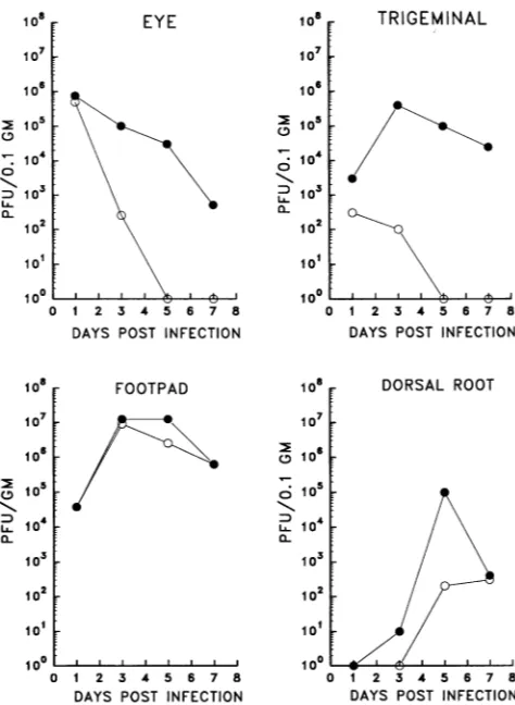
Fig.5.
In the mouse footpad, 17termA replicated with kinetics
indistinguishable from that of 17syn+. Both isolates reached
titers of >107 PFU/g by day 3 p.i. Therefore, no generalized
replication defect was evident in this tissue. In contrast, the mutant was severely restricted at the level of the dorsal root
ganglia. Infectious viruswas not detecteduntil day5 p.i., and the amount present was reduced by 3 orders of magnitude comparedwith the parent strain. Whether thevirus detected wasproducedinneurons orother cell types within theganglia was not determined.
Intheeye,mutant17termAdidnotreplicateefficiently.The
titer recoveredwas reducedby3 orders ofmagnitude 3 days
p.i., and virus couldnolonger be recovered from theeyeafter
day3. In contrast,17syn+wasrecoveredthrough day7.Within the trigeminal ganglia, both 17termA and 17syn+ were
de-tectedonday1p.i., althoughtheyieldof 17termAwasreduced
by 1 log. By day 3, 17termAwas reduced by3 logs compared
with 17syn+. 17termAwas not recovered from thetrigeminal
ganglia after day 3, although 17syn+ was recovered through
day 7p.i.
For acute viral replication kinetics in brain, mice were
inoculated intracranially with 5 x 10⁵ PFU and assayed for
virusyieldsin brain tissue over 5 days(Fig. 6). Two ordersof
magnitudeless viruswasrecovered frommice inoculated with
17termA 48hp.i.Thereare notimepoints for17syn+ after48
h,sincenomiceinthisgroupsurvived beyondthispointatthis
inoculation titer. While 17termAcontinuedtoreplicateinthe
brainpastthis time and is lethal at this inoculation titer, the
replicationkineticssuggestthat thepathology inducedmaybe
quite
different from that ofawild-typestrain.Furtheranalysis108
106
>i05
. 0
io
I07
Dl103
0~
102
101 100
0
I0a
10F TRIGEMINAL1o7
[106
'1046
m 103
0 14
101
100
DAYS POST INFECTION
108
i07
106
m 5
L 10
104
102
101 100
FOOTPAD
0
0
CL
0 1 2 3 4 5 6 7 8
DAYS POST INFECTION
I
-
/-I/ A
0 1 2 3 4 5 6 7 8
DAYS POST INFECTION
0 1 2 3 4 5 6 7 8
DAYS POST INFECTION
FIG. 5. Virusreplicationkineticsinvivo.Micewereinoculatedby corneal scarification of both eyes with 105 PFU per eye. At the indicated timesp.i.,three mice infected with each virusweresacrificed, and the eyes and trigeminal ganglia were removed and stored at
-80°C. The tissues were pooled, homogenized, clarified, and then assayedfor virus titers asdescribed in Materials and Methods. For virus replicationinthe mouse footpadand dorsal rootganglia,mice wereinoculatedby footpadabrasiononboth hind feet with105PFU per foot. Infected tissues were pooled and processed as described aboveatthe indicated timepoints p.i.0,17syn+; 0,17termA.
willberequiredtodetermine whetheronlyasubpopulationof
the cellsorcertainregionswithin the brainarepermissive for
this virus.
Replication kinetics in primaryMEC. Thein vivo
replica-tion kinetic data demonstrated that 17termA isfully
replica-tioncompetent inatleastsome mousetissuesinvivo.
There-fore, 17termAwas notreplication defective in mousecells in
general. Itwas ofinterest that in addition to the restriction
seenintheperipheral and centralnervoussystemtissues,this mutant also failed to replicate efficientlyin the eye. Cellson
theeyesurfaceare alargely quiescent population (19),as are
several cell typesinthegangliaand brain (10, 23).Apossible
explanationfor these observations is thatafactor(s)presentin
actively dividingcellscancomplementthe ICP34.5 termination mutant phenotype. Totestthispossibility, replicationkinetics
were examined in primary cultures of MEC underconfluent
(largely quiescent) andsubconfluent(actively dividing) condi-tions.
Multistepkinetics (MOI = 0.01 PFU percell)werecarried
out in subconfluent primary MEC cultures (about 40%
con-fluence at infection). Asshown in Fig. 7, 17termAreplicated
on November 9, 2019 by guest
http://jvi.asm.org/
[image:5.612.322.559.69.393.2]2 4 _ 6
FIG. 6. Virusreplication kinetics in mouse brain. Mice were inoc-ulated intracranially with5 x 105PFU.At the indicated time points p.i., three mice infected with each virus weresacrificed,and the entire brains were removed andstored at -80C until assayed. The brains were pooled and homogenized as a 10% suspension in cell culture media and assayed forvirus content as described in Materials and Methods. *, 17syn+; 0, 17termA.
efficiently in these cultures. Viral yields of 17termA were
reduced by about 10-fold compared with 17syn+ levels, but the
rateof virusreplicationwasthesame. In contrast, the
replica-tion ofmutant 17termA wassignificantly restrictedinconfluent
MECcultures. By 24h,yields of thismutantwere reduced by
1 log compared with wild-type levels; by 48 h, yields were
reduced 100-fold. Thus,theyield of17termA inthesecultures
was <1 PFU percell,whereas the yield of 17syn+ wasabout
100 PFU per cell. Takentogether, theseresults suggest that a
functionpresentinactivelydividingMEC was able to
compen-sate, atleast partially, for thereplication restriction seenwith thismutant within confluentprimary MEC cultures.
Confluent mouse 1OT1/2 cellsarerestrictive forreplication
of 17termA. Primary MEC cultures contain manydiverse cell
types,and it ispossiblethatthesevaryin theirpermissivityfor
replication of the ICP34.5 mutant (9).Therefore, the
replica-tion of 17termA was examined in a defined cell line. Mouse
10T1/2cells were chosenbecause theyarevery contact
inhib-ited (24). Multistep kinetics werecarriedoutinthese cultures
atanMOI of 0.005 PFU per cell.Strains17syn+, 17del8A,and
17termAR were used as controls. As seen in Fig. 7, the
replication of 17termA was severely restricted in these cells.
Lessthan 100 PFU/ml was recovered at the peak of 3 days p.i.
Virus production in these cultures was therefore less than
0.001 PFU per cell. In contrast, all three control viruses
demonstratedsimilarkineticsof replication and reached 106to
107 PFU/ml(-100 PFU percell) by 3 days p.i.
The restriction seen in these cultures was evident even at a
very high MOI. Confluent cultures of 10T1/2 cells were
infected with50 PFU percell and assayedover a30-h period.
17termA produced <104 PFU/ml (burst size, <0.1 PFU per
cell). Incontrast, the control viruses rapidly reached titers of
>107 PFU/ml, withaburstsize of>100PFU percell (data not
shown). Taken together with data presented above, these
resultsdemonstrate that ICP34.5 nullmutants arerestrictedin
avariety ofcell types and notsolely in neuronal cells as has
been suggested (2, 4). Furthermore, they suggest that the
physiologicalstateof the cell may be important in overcoming
the restriction of replication ofICP34.5 mutants.
DISCUSSION
While several lines of evidence have shown that functions
important for the neurovirulence phenotype of HSV-1 and
HSV-2 map to thelongterminalrepeatof the HSVgenome(2,
18, 31, 35, 38), the relative contributions ofcis-acting signals
andopenreadingframe (ICP34.5RLI)encoded inthisregion
are stillunclear. In thisstudy,weconfirm the resultsreported
by Chou et al. that the ICP34.5 gene plays a role in HSV-1
neurovirulence (2),although clearlythis gene does notplayas
predominantarole in strain 17syn+ as wassuggested forstrain
F (2, 4).
Inaddition,wehaveextendedthese findings by
demonstrat-ing that the phenotype is not displayed solely in neurons as
suggested (4) but also in other cell types both in vivo and in
vitro. Strain 17syn+ and two isogenic mutants designed to
affect onlythe ICP34.5 reading framewere analyzed. Mutant
17del8A contains a 24-bp deletion in both copies of the
ICP34.5 gene centered around the BstEII site at 595 and
125769bpontheHSV-1 genome.Thisdeletionresulted in the
lossof aminoacids 28 to 35from theprotein. 17del8Awasfully
neurovirulentandreplicatedwithwild-type kinetics in all cells
andtissues tested. Therefore, asmall deletion atthis location
SUBCONFLUENTMEC CELLS
0--
/-D 1 2 3 4 5
DAYSPOST INFECTION
106
> 10
10
102
101
CONFLUENT MEC CELLS
D 1 2 3 4 5
DAYS POSTINFECTION
107
106
1o5
> 10 X 10
i02 101 100
CONFLUENT 1OT1/2 CELLS
1 2 3 4 5
DAYS POST INFECTION
FIG. 7. Virusreplication kinetics.PrimaryMEC cultures in35-mm-diameter tissueculture disheswere infectedatan MOIof0.01 PFU per
cell. At the indicated timepoints, duplicatecultureswereharvested and stored at -80°Cuntilassayedfor virus titers. 1OT1/2cellcultureswere
maintained under the sameconditions but infected atan MOIof 0.005PFU percell.0, 17syn+;0, 17termA; FL, 17del8A; 0, 17termAR. 1o5
> 10
3
106 10
m 1 0
"Io
DO
l'. o 6
4
5
---- --Q
100
on November 9, 2019 by guest
http://jvi.asm.org/
[image:6.612.122.237.80.232.2] [image:6.612.130.481.543.695.2]did not adversely effect theexpressionofneighboring genes, or
the function of the packaging signals, to a detectable extent.
Mutant 17termA contains an insertion of 20 bp in both
copies of the ICP34.5 gene at the BstEIIsites.Thisis the same
mutation as was recently reported in mutant R4009 in the
HSV-1 strain F background and results in the premature
termination of ICP34.5 after the amino-terminal 30 amino
acids(2). It was found that 17termA was 25- to90-fold reduced
in neurovirulence, as assayed by intracranial inoculation of
mice (PFU/LD50 = 250 to 900). Restoration of the ICP34.5
gene,as determined both by genomic structureandproduction
ofthe protein, resulted in isolate 17termAR, which wasfully
neurovirulent and wild type in all other characteristics tested,
indicating that the mutant phenotype was due to the ICP34.5
disruption specifically. In contrast, R4009 wasreported to be
completely nonneurovirulent
(PFU/LD50
> 10 ) (2). Thus,17termA is at least 10,000-fold more neurovirulent than
R4009.
The reason for thedifference in neurovirulence phenotype
between 17termA and R4009 is not yetknown, but thereare
several possibilities. In this study,neurovirulence was assayed
in adult outbred Swiss Webster mice, while the studies with
R4009 were performed in weanling BALB/c mice (2).
How-ever,it isunlikely that this accounts for the difference seen,as
BALB/c mice are more susceptible to HSVinfection than are
Swiss Webster mice (6, 36), and weanling mice are
consider-ably more susceptible than adult mice(34). Unknown
second-sitemutations in the strain F-based mutant mayhave
contrib-uted to its avirulent phenotype. Such mutations may be present
in strain F itself or may have been introduced during the
construction of R4009. This latterpossibility was not tested by
repair of theICP34.5 gene to determine whether this restored
virulence to the mutant (2). Evidence exists that strain F is
itself less virulent than strain 17syn+ in mice following
in-tracranial inoculation. The PFU/LD50 ratio of strain F in
4-week-old BALB/c mice inoculatedintracranially is >104.In
contrast, several other wild-type laboratory HSV-1 isolates,
including 17syn+, and all early-passage clinical isolates tested
yielded ratios of -10 (6). It therefore seems likely that the
neurovirulence displayed by ICP34.5 mutants is the result of a
combination of factors and depends on the geneticbackground
inwhich the mutation is expressed.
The phenotype of R4009 is similar to that of deletion
mutations reported in this locus, including 1716 on the strain
17syn+ background and R3616 on the strain F background.
Theboundaries of the deletions reported todate also include
the direct repeat 1 of the cleavage and packaging signals,
upstream regulatory regions of the ICPO gene promoter, or
both(2, 5, 16-18). ICPO is required for efficient replication of
thevirus,andreducedlevels of this criticalregulator may affect
the course of infection (12, 25, 29). It is perhaps significant
that, where tested,these mutants replicated less well than their
parent strains in any cell type, including the cells in which they
were produced (2).
17termA was replication competent in actively dividing
primary MEC (mostly fibroblasts [9]) but more severely
re-stricted inconfluent MEC. This mutant also failed to replicate
efficiently in confluent mesodermally derived mouse 1OT1/2
cellsfollowing infection even at an MOI as high as 50 PFU per
cell. The failure of the mutant to efficiently replicate in such cultures is likely due to the mutation introduced into the
ICP34.5 gene, as restoration of this gene resulted in isolate
17termAR, which displayed wild-type replication kinetics both
in vitro and in vivo. Whether this restriction to replication of
themutantis the result of shutoff of protein synthesisfollowing
viral DNA replication aswas seenin R4009-infected SK-N- SH
neuroblastoma cells (4) has not yet been determined. These
results suggest that the physiological state of the cell is an
important factorin permissivityfor ICP34.5 mutants. Further
experimentation should reveal whether permissivity is
associ-atedwith a specific stage in the cell cycle or with cell cycling
time.
Despite the quantitative difference in neurovirulence
be-tween the mutants, it is clear that the replication of ICP34.5
null mutants is restricted in the central nervous system. In
addition, 17termAwascompletely avirulent followingfootpad
inoculation with a
PFU/LD50
of >108, compared with aPFU/LD50
of _103for strain 17syn+ (32). While the mutantreplicated with wild-type kinetics in the footpad, this reduction
inneuroinvasiveness could be attributedtoa severerestriction
in replication at the level of the dorsal root ganglia. Clearly
then, this locus is required forefficient replication within both
the central nervous system and, perhaps more biologically
relevant, the peripheral nervous system.
Chou and Roizman have proposed that the role of the
ICP34.5 protein is to permit virus replication in sensory
neuronsbypreventingapoptosis(4).Ourfindings demonstrate
the requirement for this protein for productive infection of
cells at the body surface, particularly the cornea. This result
along with results obtained in cultured cells demonstrates that
the effect of ICP34.5 is not neuronspecific and suggests a more
generalized function for ICP34.5. This protein may increase
the numberand/or types of cellsatthebody surface whichcan
be productively infected by allowing replication of the virus in
quiescent cell types. This could lead to anincreased
transmis-sion rate and increase the chance of infection ofneurons and
subsequent establishment oflatency. This geneproduct may be
particularly important for infections of the eye leading to
recurrent keratitis and blindness. It has recentlybeen
demon-strated that viral replication occurs in sensory neurons during reactivation from latency in vivo (28). A potential function of
ICP34.5 may be to permit replication in neurons during a
reactivation event leading to recurrent disease and transmis-sion of the virus to new hosts.
McGeoch and Barnett (20) reported that 63 amino acids
near the carboxy terminus of RL1 (ICP34.5) share a high
amino acid identity with the mouse gene myD116 (14).
Re-cently, an openreading frame in African swine fever virus with
significant sequence identity to the same region of myD116
and ICP34.5 wasdescribed (30). The function of myD1 16 is not
known, but its expression is induced in a myeloid leukemia cell
line upon differentiation mediated by interleukin 6(13). It is of
interest that African swine fever virus replicates in mature macrophages (30). It is possible that ICP34.5 serves as an agonist or antagonist of a cellular factor such as myD116. The
presence or absence of such a cellular factor maycompensate
for the replication restriction of ICP34.5 mutants within some
cells but not in quiescent cells at the body surface or in the
peripheral and central nervous systems.
ACKNOWLEDGMENTS
Thiswork was supported by Public HealthService grants NS 25879 from the National Institute of Neurological and Communicative Disorders and Stroke and Al 32121 from the National Institute of Allergy and Infectious Diseases.
We thank C. Colmenares, J. Stringer, and S. Stringer forcritically reading the manuscript and T. Suter for expert technical assistance.
REFERENCES
1. Ackermann, M., J. Chou, M. Sarmiento, R. A. Lerner, and B. Roizman. 1986.Identification by antibody to a synthetic peptide of a protein specified by a diploid gene located in the terminal
on November 9, 2019 by guest
http://jvi.asm.org/
repeats of the Lcomponent ofherpes simplex virus genome. J. Virol. 58:843-850.
2. Chou, J., E. R. Kern, R. J. Whitley, and B. Roizman. 1990. Mapping of herpes simplexvirus-I neurovirulence to yl 34.5, a genenonessential forgrowthin culture. Science 250:1262-1266. 3. Chou, J.,and B.Roizman. 1985. Isomerization ofherpes simplex
virus 1 genome:identification of the cis-acting and recombination sites within the domain of theasequence.Cell 41:803-811. 4. Chou, J., and B. Roizman. 1992. The yl 34.5 gene of herpes
simplex virus 1 precludes neuroblastoma cells from triggering total shutoff of protein synthesis characteristic of programmed cell death in neuronal cells. Proc. Natl. Acad.Sci. USA89:3266-3270. 5. Deiss, P. L., J. Chou, and N. Frenkel. 1986. Functional domains within theasequenceinvolved in the cleavage-packaging of herpes simplex virusDNA.J. Virol.59:605-618.
6. Dix,R.D.,R. R.McKendall,andJ.R.Baringer. 1983. Compar-ative neurovirulence of herpes simplex virus type I strains after peripheral or intracerebral inoculation of BALB/c mice. Infect. Immun. 40:103-112.
7. Dolan, A.,E.McKie,A. R.MacLean,and D.J.McGeoch. 1992. Status of theICP34.5 gene inherpes simplex virus type 1 strain 17. J.Gen.Virol. 73:971-973.
8. Fenner, F., B. R. McAuslan, C. A.Mims, J.A. Sambrook, and D.0.White. 1974.Thebiologyof animalviruses, 2nd ed., p. 393. Academic Press, Inc., New York.
9. Freshney,R.I. 1983. Culture of animal cells, p. 99-118. Alan R. Liss, Inc., New York.
10. Jacobson, M. 1991. Developmental neurobiology, 3rd ed., p. 41-93. PlenumPress, New York.
11. Joklik,W.K.,H.P.Willet, D. B. Amos, and C.M.Wilfert. 1992. Zinsser microbiology, 20th ed., p. 955. Appleton and Lange, Norwalk, Conn.
12. Leib, D. A.,D. M.Croen,C. L.Bogard, K. A. Hicks,D. R.Yager, D. M.Knipe, K. L.Tyler,and P. A.Schaffer. 1989. Immediate-early regulatory gene mutants define different stages in the establishment andreactivation ofherpes simplexvirus latency. J. Virol. 63:759-768.
13. Lord, K. A., B. Hoffman-Liebermann, and D. A. Leibermann. 1990.Complexityof the immediateearlyresponseofmyeloidcells to terminal differentiation and growth arrest includes ICAM-1, jun-B,and histone variants.Oncogene5:387-396.
14. Lord, K. A., B. Hoffman-Leibermann, and D. A. Leibermann. 1990.Sequence of myD116 cDNA:anovelmyeloiddifferentiation primary response gene induced by IL6. Nucleic Acids Res. 18: 2823.
15. Lynn, D. E. 1992. A BASIC computer program for analyzing endpointassays.BioTechniques 12:880-881.
16. Mackem, S., and B. Roizman. 1982. Differentiation between a promoter and regulator regions ofherpes simplex virus 1: the functional domains and sequence ofamovable otregulator.Proc.
Natl. Acad. Sci. USA79:4917-4921.
17. Mackem, S., and B. Roizman. 1982. Structural features of the herpes simplex virus ox gene 4, 0, and 27 promoter-regulatory sequenceswhich confer aregulationonchimericthymidinekinase genes. J.Virol. 44:939-949.
18. MacLean, A. R., M. Ul-Fareed, L. Robertson, J. Harland, and S.M. Brown. 1991.Herpessimplexvirus type 1 deletion variants 1714 and 1716 pinpoint neurovirulence related sequences in Glasgow strain 17+ between immediateearlygene 1 and the 'a' sequence. J.Gen.Virol.72:631-639.
19. McDevitt, D. S. 1982. Cell biologyof the eye, p. 99. Academic Press, New York.
20. McGeoch,D.J.,and B. C. Barnett. 1991.Neurovirulence factor. Nature (London)353:609.
21. McGeoch,D.J., C.Cunningham, G. McIntyre,and A. Dolan.1991. Comparative sequence analysis of the long repeat regions and
adjoining parts of the long unique regions in the genomes of herpes simplex viruses types I and 2. J. Gen. Virol. 72:3057-3075. 22. McGeoch, D. J., M.A.Dalrymple, A. J. Davison,A.Dolan,M. C. Frame,D.McNab,L.J. Perry, J.F.Scott,andP.Taylor. 1988. The complete sequence of the long unique region inthe genome of herpessimplexvirus type 1.J.Gen. Virol. 69:1531-1574. 23. Pardee,A. B. 1989.G,eventsandregulation of cellproliferation.
Science 246:603-608.
24. Reznikoff, C. A., D. W. Brankow, and C. Heidelberger. 1973. Establishment andcharacterization ofaclonedline of C3Hmouse embryo cells sensitive to postconfluence inhibition of division. Cancer Res. 33:3231-3238.
25. Sacks,W. R.,and P. A. Schaffer. 1987. Deletion mutantsin the gene encoding the herpes simplexvirus type 1 immediate-early protein ICPO exhibit impaired growth in cell culture. J. Virol. 61:829-839.
26. Sambrook, J., E. F. Fritsch, and T. Maniatis. 1989. Molecular cloning:alaboratorymanual, 2nded. ColdSpringHarbor Labo-ratoryPress, ColdSpringHarbor, N.Y.
27. Sanger, F., S. Nicklen,and A.R. Coulson. 1977. DNAsequencing with chain-terminating inhibitors. Proc. Natl. Acad. Sci. USA 74:5463-5467.
28. Sawtell,N. M.,and R. L.Thompson. 1992. Rapidinvivo reacti-vation ofherpes simplexvirus in latentlyinfected murine gangli-onicneuronsafter transienthyperthermia.J.Virol. 66:2150-2156. 29. Stow,N.D.,and E.C. Stow.1986. Isolation and characterization of aherpes simplexvirus type 1mutantcontainingadeletionwithin the gene encoding the immediate early polypeptide vmwllO. J. Gen. Virol. 67:2571-2585.
30. Sussman,M.D.,Z.Lu, G.Kutish,C. L.Afonso,P.Roberts,and D. L. Rock. 1992. Identification ofan African swine fever virus genewithsimilarityto amyeloiddifferentiationprimaryresponse gene and a neurovirulence-associated gene of herpes simplex virus. J. Virol.66:5586-5589.
31. Taha, M.Y., S. M.Brown, G. B. Clements, and D.I. Graham. 1990.The JH2604 deletion variant ofherpes simplexvirus type 2 (HG52) fails to produce necrotizing encephalitis following in-tracranial inoculation of mice. J. Gen. Virol. 71:1597-1601. 32. Thompson,R.L.,M. L.Cook,G.B.Devi-Rao,E. K.Wagner,and
J. G. Stevens. 1986. Functional and molecular analysis of the avirulent wild typeherpessimplexvirus type 1 strain KOS. J. Virol. 58:203-211.
33. Thompson,R.L., G.V.Devi-Rao, J. G.Stevens,and E. K.Wagner. 1985. Rescue of a herpes simplex virus type 1 neurovirulence function withaclonedDNAfragment.J. Virol.55:504-508. 34. Thompson, R. L., M. Nakashizuka, and J. G. Stevens. 1986.
Vaccinepotentialofalive avirulentherpes simplexvirus.Microb. Pathog. 1:409-416.
35. Thompson,R.L.,S. K.Rogers,and M. A. Zerhusen.1989.Herpes simplexvirus neurovirulence and productive infection of neural cells is associated with afunction which maps between 0.82 and 0.832 mapunitson the HSV genome.Virology 172:435-450. 36. Thompson, R.L., andJ.G. Stevens. 1983. Biological
character-ization ofaherpes simplexvirusintertypic recombinant which is completelyand specificallynon-neurovirulent.Virology 131:171-179.
37. Thompson,R.L.,andE. K.Wagner. 1988. Partialrescueofherpes simplexvirus neurovirulence witha3.2 kb clonedDNAfragment. Virus Genes 1:261-273.
38. Thompson,R.L.,E. K.Wagner,andJ.G. Stevens. 1983.Physical location ofaherpes simplexvirustype-I genefunction(s) specif-icallyassociated witha10million fold increase in HSV neuroviru-lence.Virology 131:180-192.
39. Varmuza, S. L., andJ. R. Smiley. 1985. Signals for site-specific
cleavageof HSVDNA:maturation involvestwoseparatecleavage
events atsites distaltotherecognitionsequences. Cell 41:793-802.