Copyright © 1998, American Society for Microbiology
Construction and Characterization of a Temperature-Sensitive
Human Immunodeficiency Virus Type 1 Reverse
Transcriptase Mutant
MINGJUN HUANG,† RALF ZENSEN, MICHAEL CHO,
ANDMALCOLM A. MARTIN*
Laboratory of Molecular Microbiology, National Institute of Allergy and Infectious Diseases, Bethesda, Maryland 20892
Received 21 August 1997/Accepted 20 November 1997
A temperature-sensitive (ts) human immunodeficiency virus type 1 (HIV-1) reverse transcriptase (RT)
mutant was generated by charged-cluster-to-alanine mutagenesis. The mutant virus, containing three charged
residues within the RT finger domain changed to alanine (K64A, K66A, and D67A), replicated normally at 34.5
but not 39.5°C. Quantitating virus particle production by p24 antigen capture or virion-associated RT activity
and virus infectivity by the MAGI cell assay, we found that (i) mutant virions produced at the permissive
temperature were indistinguishable from wild-type virus in assays performed at the nonpermissive
tempera-ture, suggesting that the ts mutation did not impair early steps in the virus replication cycle and that the
mutant RT enzyme was not ts; and (ii) virus particle production in cells transfected with the ts mutant at the
nonpermissive temperature was comparable to that of wild-type virus. However, the particle-associated RT
activity and infectivity of mutant virions produced at the nonpermissive temperature were greatly reduced
when assays were conducted at the permissive temperature. These results are consistent with an irreversible
ts event affecting RT that occurs during virus particle production. Radioimmunoprecipitation analyses revealed
that both p66 and p51 RT subunits were absent from mutant virions generated at 39.5°C. The presence of
normal levels of HIV-1 integrase in mutant particles produced at the nonpermissive temperature was
incon-sistent with defective Gag-Pol synthesis or Gag-Pol incorporation into progeny virions. Furthermore, wild-type
levels of the mutant Pr160
gag-polwere detected in virions produced at the nonpermissive temperature when the
HIV-1 protease was inactivated by site-specific mutagenesis. Taken together, these results are most consistent
with a ts defect affecting the degradation or aberrant processing of the mutated RT during its processing/
maturation within nascent particles.
The conversion of viral genomic RNA into unintegrated
double-stranded linear DNA molecules during the early phase
of the productive virus infection is the signature of Retroviridae.
This reaction is catalyzed by reverse transcriptase (RT),
en-coded by the highly conserved pol gene found in retroviruses
and eukaryotic retrotransposable elements. In the case of
hu-man immunodeficiency virus type 1 (HIV-1), the mature RT is
a heterodimer (13), consisting of 66- and 51-kDa subunits that
are colinear at their N termini. The larger subunit contains
DNA polymerase and RNase H domains, whereas p51 lacks
the C-terminal RNase H domain. Retroviral RTs possess two
enzymatic activities: (i) a DNA polymerase that can use RNA
or DNA templates and (ii) RNase H, which degrades genomic
RNA present in DNA-RNA intermediates.
Crystal structures of unligated HIV-1 RT (44) or RT
com-plexed with the non-nucleoside inhibitor Nevirapine (28) or an
18/19-mer oligonucleotide (23) have been reported. The
struc-ture of the polymerase domain of the 66-kDa component
within the heterodimer has been likened to a right hand,
pos-sessing finger, palm, thumb, and connection domains.
Al-though the same subdomains are present in p51, they differ in
arrangement: both the DNA binding groove and polymerase
active site of p66 are missing from p51.
As is the case for other retroviruses, the pol gene of HIV-1
encodes a high-molecular-weight Gag-Pol precursor
(Pr160
gag-pol) that is incorporated into nascent particles as a
result of its noncovalent association with the Gag precursor
polyprotein Pr55
gag(40, 47, 54). Processing of Pr160
gag-polby
the HIV-1-encoded protease (PR) during virus budding from
the cell membrane gives rise to the mature, virion-associated
PR and integrase (IN) proteins as well as the p66 and p51
forms of the RT heterodimer (25). Subsequent to
adsorp-tion, fusion, and entry into a newly infected cell, the
parti-cles are partially uncoated and commence synthesizing
DNA copies of their genomic RNAs as they traverse the
cytosol. Thus, the generation of full-length linear viral
DNAs by RT during the early phase of infection reflects the
successful completion of multiple biosynthetic, metabolic,
and enzymatic reactions in both the virus-producing and
newly infected cells.
The identification and characterization of RT mutants has
proven to be invaluable for integrating structural and
func-tional properties of this critical HIV-1 protein. Both in vitro
mutagenesis of the HIV-1 RT and studies of drug-resistant RT
variants have delineated several functionally important
do-mains, the majority of which involve its DNA-polymerizing
activity (2–9, 11, 16, 22, 24, 26, 30, 32–34, 36, 43). We have
attempted to create conditional RT mutants affected in one or
more of the myriad of processes and functions that must be
completed during a productive virus infection. The
charged-cluster-to-alanine mutagenesis approach, previously used to
generate temperature-sensitive (ts) mutants of cellular and
vi-ral genes (12, 19, 50, 55), was employed to alter exposed
re-gions of the HIV-1 RT. Such mutants may exhibit impaired
interaction with other cellular or viral proteins or be unable to
* Corresponding author. Mailing address: Laboratory of Molecular
Microbiology, NIAID, NIH, 4 Center Dr., Building 4, Room 315,
Bethesda, MD 20892-0460. Phone: (301) 496-4012. Fax: (301)
402-0226. E-mail: [email protected].
† Present address: Laboratory of Antiviral Drug Mechanisms, SAIC
Frederick, NCI-FCRDC, Frederick, MD 21702.
2047
on November 9, 2019 by guest
http://jvi.asm.org/
stably associate with the viral RNA template or the DNA
product of the reverse transcription reaction.
In this report, we describe the construction and
character-ization of a ts mutation affecting the HIV-1 RT. When charged
residues at positions 64, 66, and 67 within the finger domain of
RT were changed to alanine, the resultant virus replicated at
the permissive (34.5°C) but not the restrictive (39.5°C)
tem-perature. Further characterization of the ts mutant indicated
that progeny virions produced at 39.5°C possessed no RT
ac-tivity, contained no detectable or only background levels of p66
and p51, and were not infectious. In contrast, the mutant (mt4)
particles produced at 34.5°C possessed RT activity when
as-sayed at 34.5 or 39.5°C, contained the RT heterodimeric
pro-tein, and were infectious in MAGI cells at both temperatures.
The ts step in the life cycle of mt4 occurs in virus-producing
cells subsequent to the incorporation of Pr160
gag-pol, very likely
during the processing of the Gag-Pol precursor in nascent
particles.
MATERIALS AND METHODS
Construction of RT mutants.Recombinant plasmids were constructed and used to transform bacteria by standard procedures. The full-length infectious molecular clone of HIV-1LAI(pLAI [41]) was used in these experiments as the wild-type control and as the source of mutant derivatives. A 2,219-bp BclI-EcoRI fragment (nucleotides 2511 to 4730) from pLAI was subcloned into M13mp18. Oligonucleotide-directed mutagenesis was performed as previously described (29) to construct 33 charged-cluster-to-alanine mutations within the RT coding region of the pol gene. We defined a charged cluster as containing at least two charged amino acid residues within a group of five consecutive residues. In most mutants, at least two charged residues within a cluster were changed, although occasionally only one residue was altered. Each set of mutations created a novel
BstUI restriction endonuclease site which allowed the presence of the introduced
mutations to be readily identified. A 1,057-bp BclI-PflMI (nucleotides 2511 to 3568) or a 1,162-bp PflMI-EcoRI (nucleotides 3568 to 4730) fragment, carrying the desired mutation, was excised from the mutagenized DNA and cloned back into pLAI. The presence of putative mutations in viral mutants that failed to replicate following transfection of CEM (12D7) cells and the absence of unde-sired changes in subcloned pol gene segments were confirmed by DNA sequenc-ing.
Cell culture, transfection, and infection.HeLa cells were maintained in Dul-becco’s modified essential medium (DMEM) supplemented with 5% fetal bovine serum, 100 U of penicillin G sodium per ml, and 0.1 mg of streptomycin sulfate per ml at 37°C unless otherwise specified. HeLa cells were transfected by the calcium phosphate precipitation method as previously described (14). Forty-eight hours posttransfection, culture supernatants were tested for RT activity and p24 Gag protein as outlined below. Transfected cell supernatants were filtered through a 0.45-mm-pore-size filter for use as virus stocks.
CEM (12D7) cells (45) were propagated in RPMI 1640 medium supplemented with 10% fetal bovine serum, 100 U of penicillin G sodium per ml, and 0.1 mg of streptomycin sulfate per ml. CEM (12D7) cells were transfected using DEAE-dextran or infected with cell-free virus preparations as previously described (14). For infections, an amount of the wild-type HIV-1LAI, prepared at 37°C from transfected HeLa cells, equivalent to 106 32P-RT cpm was used to infect 53106 CEM (12D7) cells. This HIV-1LAIinoculum contained approximately 5 ng of p24 Gag protein; this amount of ts mutant virus was also used in infectivity assays.
The CD4-positive long terminal repeat–b-galactosidase-expressing HeLa (MAGI) cell indicator line (27) was obtained from the AIDS Research and Reference Program, Division of AIDS, National Institute of Allergy and Infec-tious Diseases, and maintained in DMEM supplemented with 5% fetal bovine serum, 100 U of penicillin G sodium per ml, 0.1 mg of streptomycin sulfate per ml, 0.2 mg of G418 sulfate per ml, and 0.1 mg of hygromycin B per ml. Cells were seeded at a density of 43104per well in a 24-well plate 24 h prior to infection. Samples of virus (5 ng of p24 in 150ml) were adsorbed to cells in the presence of 3mg of DEAE-dextran for 3 h at 34.5°C, 2 h at 37°C, or 1 h at 39.5°C prior to the addition of 1 ml of medium. Following incubation for 72 h at 34.5°C, 48 h at 37°C, or 40 h at 39.5°C, the cells were fixed and stained with 5-bromo-4-chloro-3-indolyl-b-D-galactopyranoside (X-Gal) as previously described (27).
RT and p24 assays.The RT assay was performed as reported previously (51), with the addition of 0.8 mM EDTA to the reaction cocktail. Briefly, 10ml of cell-free supernatant from infected-transfected cells was added to 50ml of RT cocktail for 3 h at 37°C unless otherwise specified. A 5-ml aliquot was spotted onto DE-81 paper, washed three times with 23SSC (13SSC is 0.15 M NaCl plus 0.015 M sodium citrate), and counted in a liquid scintillation counter.
The amount of p24 Gag protein in cell-free supernatants was determined by enzyme-linked immunosorbent assay (Coulter Immunology), using the instruc-tions and standards supplied by the manufacturer. Samples from transfected CEM (12D7) cultures were diluted 2,000- to 4,000-fold in RPMI 1640 medium
containing 10% fetal bovine serum, and samples from transfected HeLa cultures were diluted 5,000- to 10,000-fold in DMEM containing 5% fetal bovine serum so that they were in the linear range of the assay.
Metabolic labeling and radioimmunoprecipitation.The procedure used for metabolic labeling of transfected HeLa cells has been previously described (21), although the incubation temperatures were modified for these experiments. Cells were initially transfected at 37°C for 3 h. The cultures were then incubated at 34.5°C for 54 h or at 39.5°C for 40 h and then labeled for 16 h at 34.5°C or 12 h at 39.5°C.
Transfected CEM (12D7) cells were incubated at 3660.5°C, and the medium was changed at 2-day intervals up to the time when syncytium formation was first observed (usually around 11 days posttransfection). Cells from 6-ml cultures (approximately 107cells) were pelleted, washed, and plated in 25-cm2tissue culture flasks in 2 ml of methionine-free RPMI 1640 medium supplemented with 10% fetal bovine serum. [35S]methionine (500mCi) was added, and the cells were incubated for 16 h at 34.5°C or for 12 h at 39.5°C. Following the labeling period, the cells were harvested and pelleted by a brief spin in a microcentrifuge. The cell-free supernatants were filtered through a 0.45-mm-pore-size filter, and viri-on-associated proteins were collected by pelleting for 30 min at 35,000 rpm in an SW50.1 rotor (Beckman).
Pulse-chase experiments were initiated when syncytia became visible in trans-fected CEM (12D7) cells maintained at 3660.5°C (approximately on day 10 or 11). Cells from 6 ml of CEM (12D7) cultures were used for each pulse-chase experiment. The remaining steps were performed as described previously (53) except that the incubation temperature was maintained at either 34.5 or 39.5°C. At the conclusion of each pulse and chase period, the CEM (12D7) cells were pelleted and the supernatants were filtered. To obtain virion-associated proteins, 200ml of cell-free supernatant was pelleted for 99 min at 14,000 rpm in a Tomy high-speed microcentrifuge. The cell-free, virion-free supernatant fractions from the microcentrifugation were also collected after each spin.
Immunoprecipitation was carried out as previously described (52) with the indicated antibodies. AIDS patient sera and a sample of a rabbit anti-HIV-1 RT polyclonal antibody (anti-RT Ab 1) were obtained from National Institute of Allergy and Infectious Diseases AIDS Research and Reference Reagent Pro-gram (human HIV immune globulin, catalog no. 192; rabbit HIV-1 RT anti-serum, catalog no. 634). Another anti-HIV-1 RT rabbit polyclonal antibody (RT Ab 2) was purchased from Intracell (Cambridge, Mass.), and anti-HIV-1 IN monoclonal antibody 35 was generously provided by Stephen Hughes. All precipitates were subjected to sodium dodecyl sulfate-polyacrylamide gel electrophoresis (SDS-PAGE) analysis in 10% acrylamide-AcrylAide (FMC) gels. The gels were fixed in methanol and acetic acid, treated with 1 M salicylic acid, dried at 100°C, and placed on Biomax MR film (Kodak).
Immunoblot analysis of virion-associated proteins. Cell-free supernatants from transfected HeLa cells were pelleted as described above. Equivalent amounts of each virus preparation, as measured by p24 content, were subjected to SDS-PAGE (10% gel) and then transferred to nitrocellulose membranes. Binding of an anti-HIV-1 RT rabbit polyclonal antibody (Intracell) to virion-associated RT subunits was visualized by chemiluminescence using horseradish peroxidase-conjugated goat anti-rabbit immunoglobulin G as described in the protocol of the manufacturer (Pierce).
RESULTS
Construction and properties of an RT ts HIV-1 mutant.
The
charged-cluster-to-alanine mutagenesis approach (12, 19, 50,
55) was used to generate potential ts mutations affecting HIV-1
RT coding sequences. This mutagenesis protocol, outlined in
Materials and Methods, involved the substitution of alanine
residues in charged regions of the RT coding segment of the
pLAI infectious molecular clone, which was derived from the
HIV-1
LAIisolate (41). pLAI DNAs, containing different
mu-tagenized RT regions, were transfected into HeLa and CEM
(12D7) cells, and progeny virus production at 34.5, 37, and
39.5°C was monitored by RT activity released into the medium.
None of these RT mutations conferred a ts phenotype,
al-though several resulted in mutant viruses that had lost
infec-tivity for CEM (12D7) cells at all temperatures tested (Table
1). One of these (mt1), with alterations affecting amino acids
64 through 67 of RT, failed to establish a spreading infection in
CEM (12D7) cells but did generate wild-type levels of progeny
virions following transfection of HeLa cells, as determined by
p24 antigen capture assay (data not shown). Interestingly, the
virion-associated RT activity measured at 39.5°C was
approx-imately fourfold lower than that determined at 34.5°C.
Postu-lating that the mt1 mutation might be located within a
poten-tial ts region of the HIV-1 RT and that the four-residue alanine
on November 9, 2019 by guest
http://jvi.asm.org/
substitution might be too incapacitating, three additional
mu-tants (mt2 to mt4) were constructed. As shown in Table 2, a
lysine-to-alanine substitution at residue 65 (mt1 and mt3)
re-sulted in loss of infectivity at all temperatures, and
replace-ment of only residues 66 and 67 (mt2) resulted in a virus with
a wild-type phenotype. Only mt4, with alanine substitutions at
amino acids 64, 66, and 67, exhibited a consistent ts phenotype
following infection of CEM (12D7) cells.
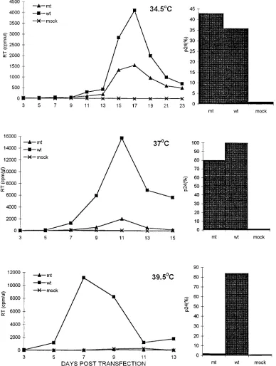
The infection kinetics of the HIV-1 mutant mt4 and its
wild-type HIV-1
LAIparent at 34.5, 37, and 39.5°C in
trans-fected CEM (12D7) cells are presented in Fig. 1. The spread of
wild-type virus throughout the infected cultures, monitored by
both peak RT activity and p24 antigen released into the
su-pernatant medium, was greatest at 37°C, somewhat diminished
at 39.5°C, and markedly reduced at 34.5°C. In contrast, mt4
exhibited virtually no infectivity at 39.5°C, as measured by RT
activity or p24 production. At the permissive temperature of
34.5°C, mt4 infectivity, as measured by p24 antigen capture,
was indistinguishable from that of the wild-type HIV-1
LAI.
When monitored by release of RT activity into the medium,
the spread of mt4 through the CEM (12D7) cell cultures
rel-ative to wild-type HIV-1
LAIwas inversely related to incubation
temperature.
A ts defect is observed during the production of mt4
parti-cles at the nonpermissive temperature.
To ascertain whether
the observed loss of mt4 infectivity at 39.5°C reflected impaired
synthesis, processing, assembly, or release of progeny virions at
the restrictive temperature, HeLa cells, which lack surface
CD4 and therefore support only the postintegration steps of
the virus life cycle, were transfected with wild-type or mt4
mutant viral DNA at various temperatures, and the properties
of the particles released into the medium were examined
(Ta-ble 3). The RT activity associated with mt4 particles produced
at 34.5°C was readily detected when assayed at 34.5, 37, and
39.5°C, although the levels measured were consistently two- to
threefold lower than wild-type virus levels. In contrast, the RT
activity associated with mt4 produced at 39.5°C was 34- to
57-fold lower than that of the wild-type HIV-1
LAI, irrespective
of the temperature at which the RT assay was conducted. Thus,
mutant particles produced at the nonpermissive temperature
displayed greatly reduced RT activity. However, equivalent
numbers of mt4 and HIV-1
LAIprogeny virions, as measured by
p24 antigen levels in the culture supernatants, were released
from the transfected HeLa cells at the permissive and
nonper-missive temperatures (Table 3).
A similar phenotype was observed in the single-cycle MAGI
cell infectivity assay. HIV-1 mutant mt4 produced at 34.5°C
displayed infectivities indistinguishable from those of wild-type
virus at all assay temperatures, whereas mutant particles
gen-erated at 39.5°C exhibited only background levels of
b
-galac-tosidase activity compared to HIV-1
LAI(Table 4). Because
reverse transcription of the viral genome is a required step for
infectivity in MAGI cells, these results indicate that the RT
incorporated into mt4 at the permissive temperature is fully
capable of directing the synthesis of viral DNA in infected cells
at the nonpermissive temperature.
mt4 particles produced at the nonpermissive temperature
lack RT protein.
When it became apparent that the ts
pheno-type of HIV-1 mutant mt4 was a property of progeny virions
produced at 39.5°C but not at 34.5°C, the synthesis, processing,
and incorporation of RT and other pol gene products were
examined at both temperatures to ascertain which step(s) in
the virus life cycle was temperature dependent. Cultures of
CEM (12D7) cells, transfected for several days with wild-type
or mt4 viral DNA at the semipermissive temperature of 36.5°C,
were metabolically labeled with [
35S]methionine for 16 or 12 h
at 34.5 or 39.5°C, respectively. Cell-associated proteins were
immunoprecipitated with serum from an AIDS patient (Fig.
2A); virion-associated proteins were immunoprecipitated with
the same patient serum (Fig. 2A), two anti-HIV-1 RT
poly-clonal antibodies (Fig. 2B), or an anti-HIV-1 IN monopoly-clonal
antibody (Fig. 2C). All immunoprecipitated proteins were then
resolved by SDS-PAGE.
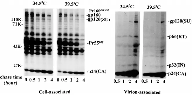
The electropherogram of virion-associated proteins, present
in particles produced at 39.5°C and immunoprecipitated with
AIDS patient serum, revealed the presence of multiple HIV-1
structural proteins in both the wild-type and mutant virus
prep-arations (Fig. 2A). Notably missing or present at near
back-ground levels in samples of mt4 produced at the nonpermissive
temperature was the 66-kDa subunit of RT. To confirm the
absence of particle-associated RT protein, the same samples of
HIV-1
LAIand mt4, produced at 39.5°C but
immunoprecipi-tated with two different polyclonal antibodies directed against
HIV-1 RT, were similarly analyzed. As shown in Fig. 2B, p66
was immunoprecipitated from wild-type virions but not from
the mutant particles with both anti-RT antibodies. Anti-RT Ab
2 also immunoprecipitated the 51-kDa RT subunit from the
wild-type virus sample but significantly reduced amounts of
p51 from the preparation of mutant virions (Fig. 2B, right).
The Pr160
gag-polprecursor is synthesized and incorporated
into mt4 particles produced at the nonpermissive
tempera-ture.
Several explanations can be entertained to account for
the absence of the heterodimeric RT protein in mutant virions
generated at the nonpermissive temperature. It is possible that
the Gag-Pol precursor polyprotein is not synthesized at 39.5°C
or is so unstable that none of the proteins encoded by the pol
gene are assembled into progeny virions. From the data
pre-sented thus far, this cannot be the case. The HIV-1 Pr160
gag-pol [image:3.612.53.290.88.163.2]precursor becomes incorporated into nascent virions and is
processed into the mature PR, RT, and IN proteins during or
immediately following release of particles from productively
infected cells. The presence of fully active protease in mt4
TABLE 1. RT mutants unable to infect CEM (12D7) cells at
any temperature
aMutant no. Amino acid changes
1...K64A, K65A, K66A, D67A
3...K64R, K65A
26...K527A, K528A, E529A
29...D218A, K219A, K220A
32...R72A, K73A
32a...W71F, R72A
aAmino acids are numbered with respect to the first residue of the HIV-1 RT protein. Infectivity was monitored by measuring the RT released into the me-dium from transfected CEM (12D7) cells during a 2-week (39.5 and 37°C) or 3-week (34.5°C) observation period.
TABLE 2. Replication of RT mutants in CEM (12D7) cells
Provirus Amino acid at residue
a: Replicationbat:
64 65 66 67 34.5°C 37°C 39.5°C
Wild type
K
K
K
D
Yes
Yes
Yes
mt1
A
A
A
A
No
No
No
mt2
K
K
A
A
Yes
Yes
Yes
mt3
R
A
K
D
No
No
No
mt4
A
K
A
A
Yes
Yes
No
aNumbered with respect to the first residue of the RT protein.
bMonitored by measuring RT released from transfected CEM (12D7) cells during a 2-week (39.5 and 37°C) or 3-week (34.5°C) observation period.
on November 9, 2019 by guest
http://jvi.asm.org/
[image:3.612.47.291.619.701.2]particles produced at the nonpermissive temperature can be
inferred from the presence of several virion-associated Gag
cleavage products, including p24 (Fig. 2A). The AIDS patient
antiserum used in that experiment also immunoprecipitated a
32-kDa protein from both wild-type and mutant virus particles
with an electrophoretic mobility consistent with HIV-1 IN,
another product of the processed Pr160
gag-pol. Confirmation
that this was indeed the IN protein was obtained by
immuno-precipitating mt4, generated at 39.5°C, with an anti-HIV-1 IN
monoclonal antibody (Fig. 2C); a prominent 32-kDa band was
observed in samples of both wild-type and mutant virus
parti-cles.
[image:4.612.106.494.73.587.2]The presence of the protease and IN proteins but not the RT
heterodimer in mutant virions produced at the nonpermissive
FIG. 1. Replication kinetics of mt4 (mt) and its wild-type HIV-1LAIparent (wt) in CEM (12D7) cells. CEM cells were transfected with the indicated proviral DNAs as described in Materials and Methods. Cells were incubated at 34.5°C (top), 37°C (middle), or 39.5°C (bottom) and were split 1:3 (37 and 39.5°C) or 1:2 (34.5°C) every 2 days. RT activity in the supernatant was monitored at the indicated times (left panels). Release of p24gaginto the medium was also determined at the peak of RT production (right panels). The amount of p24gagdetected in duplicate samples of supernatant medium collected from cells transfected with the wild-type HIV-1LAI proviral DNA at 37°C was arbitrarily set to 100%.
on November 9, 2019 by guest
http://jvi.asm.org/
temperature suggested that RT was either aberrantly
pro-cessed or proteolytically degraded after the incorporation of
the Pr160
gag-polprecursor into budding virions. This possibility
was first examined by pulse-labeling mt4-infected CEM (12D7)
cells, maintained at 34.5 or 39.5°C, for 30 min followed by a 4-h
chase. Figure 3 shows that cell-associated Pr160
gag-polwas
syn-thesized at both temperatures and that increasing levels of p66
RT and p32 IN appeared in the progeny virions released from
cells at 34.5°C. At 39.5°C, however, little if any p66 was
asso-ciated with virus particles, even after only 30 min of chase, nor
was free, non-virion-associated p66 shed into the supernatant
medium at the restrictive temperature (data not shown).
To definitively demonstrate that the Gag-Pol precursor
en-coded by mt4 was incorporated into nascent particles at 39.5°C,
the PR coding sequence was inactivated in the context of both
the wild type and the temperature-sensitive mutant, and
prog-eny virions released from HeLa cells were examined for the
presence of unprocessed Pr160
gag-pol. A preliminary
experi-ment revealed that HeLa cells, labeled with [
35S]methionine
for 12 h, 40 h following transfection with cloned mt4 DNA at
the nonpermissive temperature, released virus particles
con-taining little or no p66 and p51 (Fig. 4A). An Asp-to-Asn
substitution was then introduced at residue 25 of PR into both
HIV-1
LAIand mt4 cloned DNAs, which were then transfected
into HeLa cells maintained at 39.5°C. This mutation in the
HIV-1 PR had previously been shown to eliminate HIV-1 PR
activity (35). As expected for HIV-1 PR
2mutants, the
cell-associated and virion-cell-associated samples contained
unproc-essed Pr55
gag(Fig. 4B). More importantly, both the HIV-1
LAI
and mt4 progeny virions contained nearly equivalent amounts
of the Pr160
gag-pol(Fig. 4B, right), confirming that the Gag-Pol
precursor encoded by mt4 was, in fact, incorporated into virus
particles at 39.5°C.
DISCUSSION
The charged cluster of amino acids (KKKD) mutated by
alanine substitution in this study (residues 64 to 67) is situated
in the finger domain of the 66-kDa subunit of HIV-1 RT,
within a loop connecting beta sheets
b
3 and
b
4 (23, 28, 44).
Three of these residues are also included within a highly
con-served retroviral RT motif (IKKK), consisting of a highly
hy-drophobic amino acid (I or V) followed by at least two basic
amino acids. For lentiviruses and the Mason-Pfizer monkey
virus, three lysines follow an initial isoleucine. From structural
analyses, it has been proposed that this region of RT is
in-volved in the interaction of the enzyme with the primer (8).
[image:5.612.313.544.73.397.2]Several mutations affecting HIV-1 RT residues 64 to 67 have
been previously reported. For example, the substitution of
[image:5.612.50.291.82.178.2]FIG. 2. Radioimmunoprecipitation analysis of HIV-1 proteins produced in transfected CEM (12D7) cells at the nonpermissive temperature. CEM cells were transfected with the indicated DNAs (wt, HIV-1LAIproviral DNA; mt, mt4 proviral DNA; mock, pUC19) and were maintained at 3660.5°C. About 11 days posttransfection, the cells were shifted to 39.5°C and metabolically labeled at this temperature with [35S]Met for 12 h as described in Materials and Methods. (A) Cell-associated or virion-associated HIV-1 proteins were immunoprecipitated with serum from an AIDS patient. Virion-associated proteins were also subjected to immunoprecipitation with anti-HIV-1 RT Ab 1 and Ab 2 (B) or with an anti-HIV-1 IN monoclonal antibody (C). All immunoprecipitated proteins were resolved on SDS–10% polyacrylamide-AcrylAide gels. The positions of the gp160 envelope glycoprotein precursor, the gp120 surface (SU) envelope glyco-protein, the 66- and 51-kDa RT subunits, the 55-kDa Gag precursor [Pr55gag], the 32-kDa IN [p32(IN)], and the 24-kDa capsid protein [p24(CA)] are indicated at the right; the positions of molecular weight standards are shown on the left.
TABLE 3. RT and p24
gagreleased from transfected HeLa cells
Provirus temp (°C)Culture
RT activity (cpm/ml)
assayed ata: Relative p24gag
level (%)b
34.5°C 37°C 39.5°C
mt4
34.5
10,778
15,984
10,118
50
Wild type
34.5
26,277
34,090
28,632
53
mt4
37
6,185
9,238
5,978
93
Wild type
37
34,444
43,100
35,414
100
mt4
39.5
449
680
406
60
Wild type
39.5
17,498
23,128
23,128
65
aAfter incubation of transfected cells at the indicated culture temperatures for 48 h, cell-free supernatants were collected and monitored for RT activity at the indicated assay temperatures. Each value is the average of duplicate experi-ments.
bp24 levels were determined from the same cell-free supernatants used for the RT assays. The value for p24gagreleased from cells transfected with wild-type virus at 37°C was arbitrarily set at 100%. Each value represents an average of duplicate experiments.
TABLE 4. MAGI cell assay of wild-type and mt4 mutant viruses
Virus stock Preparationtemp (°C) No. of blue cells assayed at a:
34.5°C 37°C 39.5°C
mt4
34.5
395
23,900
3,900
Wild type
34.5
583
23,200
4,010
mt4
39.5
2
13
10
Wild type
39.5
513
5,910
5,350
aMAGI cells were infected with similar amounts (approximately 5 ng of p24gag) of the indicated virus preparations. Only cells containing dark blue nuclei were counted as positive; each value is the average from two independent experiments.
on November 9, 2019 by guest
http://jvi.asm.org/
[image:5.612.50.291.622.693.2]arginine for lysine 65 results in
.
95% loss of RNA-dependent
DNA-polymerizing activity, as measured in bacterial lysates
containing 66-kDa homodimeric forms of RT (5, 6); this
mu-tation had no detectable effect on RNase activity. In contrast,
mutagenesis of lysines 64 and 66 (K to R) had only modest or
no significant effects, respectively, on either polymerizing or
RNase H activity (5, 6, 26). More recent analyses of a
lysine-to-arginine substitution at codon 65 (K65R) of the HIV-1 RT,
associated with the emergence of resistance to 2
9
,3
9
-dideoxy-cytidine (ddC), 2
9
,3
9
-dideoxyinosine (ddI), and the (
2
)
enan-tiomer of 2
9
,3
9
-dideoxy-3
9
-thiacytidine (15, 17, 18, 56), revealed
no loss of infectivity of the resultant virus when assayed in
MT-4 cells or activated peripheral blood mononuclear cells. As
noted in Table 2, any RT mutant containing an
alanine-for-lysine substitution at residue 65 (viz., mt1 or mt3) was
repli-cation incompetent in CEM (12D7) cells. These results suggest
that for the establishment of a spreading virus infection, a
positively charged amino acid must be present at this position
of the HIV-1 RT; an uncharged alanine substitution causes
loss of infectivity.
Mutant mt4 particles produced at the nonpermissive
tem-perature contain little or no detectable p66-p51 heterodimeric
RT protein. This property is very similar to that described for
an RT variant that emerged following exposure to
(alkylami-no)piperidine bis(heteroaryl)piperizines in vitro. This G190E
RT revertant exhibited marked reductions in both
particle-associated RT activity and heterodimeric RT protein levels,
the latter measured by immunoblotting with a polyclonal
an-tibody to the HIV-1 RT (39). One could argue that our
inabil-ity to detect the 66- and 51-kDa RT subunits in mt4 particles
produced at 39°C merely reflected the failure of the antibodies
used to bind to the mutant RT proteins. This seems highly
unlikely because the same results were obtained with a variety
of antibody preparations (AIDS patient serum, two different
rabbit anti-HIV-1 RT polyclonal antibodies, and two different
anti-HIV-1 RT monoclonal antibodies [data not shown]) in
both immunoprecipitation and immunoblotting assays.
Because the 66- or 51-kDa RT subunits have unique
con-formations within the precursor homodimer and mature RT
heterodimer, the ts mutant mt4 described in this study may be
useful in delineating the role(s) of each subunit during its
assembly into a stable and functional viral polymerase. At
present, we can only speculate about the mechanism
respon-sible for the absence, and presumed degradation, of the two
RT protein subunits in mt4 produced at the nonpermissive
temperature. Nonetheless, several aspects of this
temperature-dependent phenomenon are unambiguous. First, Pr160
gag-polcontaining the ts RT mutation is incorporated into progeny
virions at 39°C. Second, as long as the mutant RT remains a
component of the Gag-Pol precursor, it is stably maintained
within virus particles produced under nonpermissive
condi-tions. Third, the mutant p66-p51 heterodimeric RT, formed at
34.5°C, is biochemically and functionally stable at 39°C. This
last property, in conjunction with the results of a pulse-chase
experiment (Fig. 3) showing that cell-associated Pr160
gag-polis
converted to detectable 66-kDa particle-associated RT within
[image:6.612.134.465.69.235.2]FIG. 3. Pulse-chase analysis of mt4 viral protein expression and release from transfected CEM (12D7) cells at permissive and nonpermissive temperatures. CEM (12D7) cells were transfected with mt4 proviral DNA and maintained at 3660.5°C for approximately 11 days. The cells were then split, pulse-labeled with [35S]Met for 30 min, and chased in unlabeled medium for the indicated times at either 34.5 or 39.5°C. At each time point, equal aliquots were removed and separated into cell-associated and virion-associated fractions (the cell-associated fraction was collected at the end of the pulse period since a preliminary experiment indicated that negligible amounts of viral proteins had been released into the medium at that time). Lysates from each fraction were immunoprecipitated with AIDS patient sera and then resolved on SDS–10% polyacrylamide-AcrylAide gels. The positions of the HIV-1 marker proteins (described in the legend to Fig. 2) and molecular weight standards are shown.
FIG. 4. Analysis of wild-type and ts mutant viral proteins produced in trans-fected HeLa cells. HeLa cells were transtrans-fected with the indicated plasmids (wt, HIV-1LAIproviral DNA; mt, mt4 proviral DNA; PR2, PR-defective proviral
DNA; PR2/mt, mt4 proviral DNA with a mutated PR; mock, pUC19). (A)
Forty-eight hours posttransfection, viral particles were harvested from the cells incubated at either 34.5 or 39.5°C. Virion-associated proteins were analyzed by immunoblotting using the anti-HIV-1 RT polyclonal antibody described in Ma-terials and Methods. (B) Forty-eight hours posttransfection, the HeLa cells were metabolically labeled with [35S]Met at 39.5°C for 12 h. Cell-associated or virion-associated proteins were immunoprecipitated with an AIDS patient serum and resolved on SDS–10% polyacrylamide-AcrylAide gels. The markers used are described in the legend to Fig. 2.
on November 9, 2019 by guest
http://jvi.asm.org/
a 30-min chase period at 34.5 but not 39°C, indicates that the
mutant p66-p51 heterodimer is very rapidly degraded.
Extracts from bacteria expressing the HIV-1 RT contain the
p66-p66 homodimer, which is in equilibrium with p66
mono-mer, as well as a p66-p51 heterodimer. The latter is a bacterial
protease cleavage product of either monomeric or dimeric p66
or both (10, 20, 31, 37, 38, 42, 46, 48). All of these structures
possess unique physical and chemical properties (42). For
ex-ample, the dissociation constant for the p66-p51 heterodimer
has been reported to be 10
29M or lower, compared to 10
25M
for the p66-p66 homodimer. Furthermore, because the site
that must be cleaved to generate p51 is situated within a
rel-atively inaccessible region of the N-terminal
b
sheet of RNase
H, it has been proposed that the p66-p66 homodimer assumes
a structurally asymmetric, and possibly less stable,
conforma-tion, thereby exposing the C-terminal RNase domain on one of
the subunits for digestion by PR (28, 49). During virus
repli-cation, p66-p51 heterodimers arise as a result of cleavage of
HIV-1 Pr160
gag-polby the viral PR. It is very likely that other
intermediates are also generated during the processing
reac-tion and that the presence of the ts mutareac-tion further
destabi-lizes one of these RT intermediates, rendering it less resistant
to degradation by the HIV-1 PR and/or cellular PRs at the
nonpermissive temperature.
The charged-cluster-to-alanine mutagenesis strategy would
be expected to alter residues located on solvent-exposed
pro-tein surfaces and possibly interfere with electrostatic
interac-tions with other molecules. In the case of the human growth
hormone, this mutagenesis approach resulted in the
identifi-cation of residues involved in hormone binding (1). In general,
charged-cluster-to-alanine mutagenesis has also resulted in
high frequencies of ts cellular and viral mutants (12, 19, 50).
For example, 9 of 26 substitutions affecting the poliovirus
RNA-dependent RNA polymerase resulted in conditional
mu-tants defective in RNA synthesis (12). This is to be contrasted
with the results reported in this study, where only 1 of 33
HIV-1 RT mutants was ts, and a previous report describing a
single conditional HIV-1 IN mutant of the 24 constructed (55).
It is not clear why so few ts mutants of HIV-1 pol gene products
have been obtained by this mutagenesis strategy. However, it is
interesting to note that the charged-cluster-to-alanine ts HIV-1
mutants affected a step(s) involving virion assembly, not the
catalytic functions of RT or IN. Instead, the conditional
phe-notype exhibited by HIV-1 mutant mt4 described in this report
most likely reflects the altered interaction of the RT precursor
and RT subunits with other virion components and results in
the degradation of an RT intermediate subsequent to its
cleav-age from the Gag-Pol precursor. Less can be said about the
nature of the defect in the previously reported ts HIV-1 IN
mutant (55). In contrast to our results, the IN protein was
detected in virus particles produced at the nonpermissive
tem-perature. Since neither mutation impairs enzymatic activity,
one could speculate that, like the ts RT mutant, the mutated
virion-associated IN protein may be unable to assemble into
functionally active IN oligomers, form stable associations with
viral and cellular DNA targets, or associate with other viral and
cellular proteins during the early phase of productive infection.
ACKNOWLEDGMENTS
We are grateful to Bachoti Rao for sequencing the different ts RT
mutants and thank Eric Freed and Stephen Hughes for suggestions
and comments on the manuscript.
REFERENCES
1. Bass, S. H., M. G. Mulkerrin, and J. A. Well. 1991. A systematic mutational analysis of hormone-binding determinants in the human growth hormone
receptor. Proc. Natl. Acad. Sci. USA 88:4498–4502.
2. Beard, W. A., S. J. Stahl, H. R. Kim, K. Bebenek, A. Kumar, M. P. Strub,
S. P. Becerra, T. A. Kunkel, and S. H. Wilson.1994. Structure/function studies of human immunodeficiency virus type 1 reverse transcriptase. Ala-nine scanning mutagenesis of an alpha-helix in the thumb subdomain. J. Biol. Chem. 269:28091–28097.
3. Beard, W. A., and S. H. Wilson. 1994. Site-directed mutagenesis of HIV reverse transcriptase to probe enzyme processivity and drug binding. Curr. Opin. Biotechnol. 5:414–421.
4. Beard, W. A., D. T. Minnick, C. L. Wade, R. Prasad, R. L. Won, A. Kumar,
T. A. Kunkel, and S. A. Wilson.1996. Role of the helix clamp in HIV-1 reverse transcriptase catalytic cycling as revealed by alanine-scanning mu-tagenesis. J. Biol. Chem. 271:12213–12220.
5. Boyer, P. L., A. I. Ferris, and S. H. Hughes. 1992. Cassette mutagenesis of the reverse transcriptase of human immunodeficiency virus type 1. J. Virol.
66:1031–1039.
6. Boyer, P. L., A. L. Ferris, and S. H. Hughes. 1992. Mutational analysis of the fingers domain of human immunodeficiency virus type 1 reverse transcrip-tase. J. Virol. 66:7533–7537.
7. Boyer, P. L., A. I. Ferris, P. Clark, J. Whitmer, P. Frank, C. Tantillo, E.
Arnold, and S. H. Hughes.1994. Mutational analysis of the fingers and palm subdomains of human immunodeficiency virus type-1 (HIV-1) reverse tran-scriptase. J. Mol. Biol. 243:472–483.
8. Boyer, P. L., C. Tantillo, A. Jacobo-Molina, R. G. Nanni, J. Ding, E. Arnold,
and S. H. Hughes.1994. Sensitivity of wild-type human immunodeficiency virus type 1 reverse transcriptase to dideoxynucleotides depends on template length; the sensitivity of drug-resistant mutants does not. Proc. Natl. Acad. Sci. USA 91:4882–4886.
9. Chao, S.-F., V. L. Chan, P. Juranka, A. H. Kaplan, R. Swanstrom, and C. A.
Hutchison III.1995. Mutational sensitivity patterns define critical residues in the palm subdomain of the reverse transcriptase of human immunodefi-ciency virus type 1. Nucleic Acids Res. 23:803–810.
10. Clark, P. K., A. L. Ferris, D. A. Miller, A. Hizi, K. W. Kim, S. M.
Deringer-Boyer, M. L. Mellini, A. D. Clark, G. F. Atnold, and W. B. Lebherz III.1990. HIV-1 reverse transcriptase purified from a recombinant strain of E. coli. AIDS Res. Hum. Retroviruses 6:753–764.
11. DeClerq, E. 1994. HIV resistance to reverse transcriptase inhibitors. Bio-chem. Pharmacol. 47:155–169.
12. Diamond, S. E., and K. Kirkegaard. 1994. Clustered charged-to-alanine mutagenesis of poliovirus RNA-dependent RNA polymerase yields multiple temperature-sensitive mutants defective in RNA synthesis. J. Virol. 68:863– 876.
13. di Marzo Veronese, F., T. D. Copeland, A. L. DeVico, R. Rahman, S.
Oros-zlan, R. C. Gallo, and M. G. Sarngadharan.1986. Characterization of highly immunogenic p66/p51 as the reverse transcriptase of HTLV-III/LAV. Sci-ence 231:1289–1291.
14. Freed, E. O., and M. A. Martin. 1994. Evidence for a functional interaction between the V1/V2 and CD4 domains of human immunodeficiency virus type 1 envelope glycoprotein gp120. J. Virol. 68:2503–2512.
15. Gao, Q., Z. Gu, M. A. Parniak, J. Cameron, N. Cammack, C. Boucher, and
M. A. Wainberg.1993. The same mutation that encodes low-level human immunodeficiency virus type 1 resistance to 29,39-dideoxyinosine and 29,39 -dideoxycytidine confers high-level resistance to the (2) enantiomer of 29,39 -dideoxy-39-thiacytidine. Antimicrob. Agents Chemother. 37:1390–1392. 16. Ghosh, M., P. S. Jacques, D. W. Rodgers, M. Ottman, J.-L. Darlix, and
S. F. J. Le Grice.1996. Alternations to the primer grip of p66 HIV-1 reverse transcriptase and their consequences for template-primer utilization. Bio-chemistry 35:8553–8562.
17. Gu, Z., Q. Gao, H. Fang, H. Salomon, M. A. Parniak, E. Goldberg, J.
Cameron, and M. A. Wainberg.1994. Identification of a mutation at codon 65 in the IKKK motif of reverse transcriptase that encodes human immu-nodeficiency virus resistance to 29,39-dideoxycytidine and 29,39-dideoxy-39 -thiacytine. Antimicrob. Agents Chemother. 38:275–281.
18. Gu, Z., R. S. Fletcher, E. J. Arts, M. A. Wainberg, and M. A. Parniak. 1994. The K65R mutant reverse transcriptase of HIV-1 cross-resistant to 29,39 -dideoxycytidine, 29,39-dideoxy-39-thiacytine, and 29,39-dideoxyinosine shows reduced sensitivity to specific dideoxynucleoside triphosphate inhibitors in vitro. J. Biol. Chem. 269:28118–28122.
19. Hassett, D. E., and R. C. Condit. 1994. Targeted construction of tempera-ture-sensitive mutations in vaccinia virus by replacing clustered charged residues with alanine. Proc. Natl. Acad. Sci. USA 91:4554–4558. 20. Hizi, A., C. McGill, and S. H. Hughes. 1988. Expression of soluble,
enzy-matically active, human immunodeficiency virus reverse transcriptase in E. coli and analysis of mutants. Proc. Natl. Acad. Sci. USA 85:1218–1222. 21. Huang, M., J. M. Orenstein, M. A. Martin, and E. O. Freed. 1995. p6Gagis
required for particle production from full-length human immunodeficiency virus type 1 molecular clones expressing protease. J. Virol. 69:6810–6818. 22. Jacobo-Molina, A., and E. Arnold. 1991. HIV reverse transcriptase
structure-function relationship. Biochemistry 30:6351–6361.
23. Jacobo-Molina, A., J. Ding, R. G. Nanni, A. D. Clark, Jr., X. Lu, C. Tantillo,
R. L. Williams, G. Kamer, A. L. Ferris, P. Clark, A. Hizi, S. H. Hughes, and E. Arnold.1993. Crystal structure of human immunodeficiency virus type 1
on November 9, 2019 by guest
http://jvi.asm.org/
reverse transcriptase complexed with double-stranded DNA at 3.0 Å reso-lution shows bent DNA. Proc. Natl. Acad. Sci. USA 90:6320–6324. 24. Jacques, P. S., B. M. Wohrl, M. Ottmann, J. L. Darlix, and S. F. J. Le Grice.
1994. Mutating the “primer grip” of p66 HIV-1 reverse transcriptase impli-cates tryptophan-229 in template-primer utilization. J. Biol. Chem. 269: 26472–26478.
25. Kaplan, A. H., M. Manchester, and R. Swanstrom. 1994. The activity of the protease of human immunodeficiency virus type 1 is initiated at the mem-brane of infected cells before the release of viral proteins and is required for release to occur with maximum efficiency. J. Virol. 68:6782–6786. 26. Kim, B., T. R. Hathaway, and L. A. Loeb. 1996. Human immunodeficiency
virus reverse transcriptase. Functional mutants obtained by random mu-tagenesis coupled with genetic selection in Escherichia coli. J. Biol. Chem.
271:4872–4878.
27. Kimpton, J., and M. Emerman. 1992. Detection of replication-competent and pseudotyped human immunodeficiency virus with a sensitive cell line on the basis of activation of an integratedb-galactosidase gene. J. Virol. 66: 2232–2239.
28. Kohlstaedt, L. A., J. Wang, J. M. Friesman, P. A. Rice, and T. A. Steitz. 1992. Crystal structure at 3.5 Å resolution of HIV-1 reverse transcriptase complex with an inhibitor. Science 256:1783–1790.
29. Kunkel, T. A., J. D. Roberts, and R. A. Zakour. 1987. Rapid and efficient site-specific mutagenesis without phenotypic selection. Methods Enzymol.
154:367–382.
30. Larder, B., D. Purifor, K. Powell, and G. Darby. 1987. AIDS virus reverse transcriptase defined by high level expression in E. coli. EMBO J. 6:3133– 3137.
31. Larder, B., D. Purifor, K. Powell, and G. Darby. 1987. Site-specific mutagen-esis of AIDS virus reverse transcriptase. Nature (London) 327:716–717. 32. Larder, B. A. 1995. Viral resistance and the selection of antiviral
combina-tions. J. Acquired Immune Defic. Syndr. Hum. Retrovirol. 10(Suppl. 1):S28– S33.
33. Larder, B. A., S. D. Kemp, and D. Purifor. 1989. Infectious potential of human immunodeficiency virus type 1 reverse transcriptase mutants with altered inhibitor sensitivity. Proc. Natl. Acad. Sci. USA 86:4803–4807. 34. Le Grice, S. F. J., T. Naas, B. Wohlgensinger, and O. Schatz. 1991.
Subunit-selective mutagenesis indicates minimal polymerase activity in heterodimer-associated p51 HIV-1 reverse transcriptase. EMBO J. 10:3905–3911. 35. Loeb, D. D., R. Swanstrom, L. Everitt, M. Manchester, S. E. Stamper, and
C. A. Hutchison III.1989. Complete mutagenesis of the HIV-1 protease. Nature (London) 340:391–400.
36. Lowe, D. M., A. Aitken, C. Bradley, G. K. Darby, B. A. Larder, K. L. Powell,
D. J. Purifoy, M. Tisdale, and D. K. Stammers.1988. HIV-1 reverse tran-scriptase: crystallization and analysis of domain structure by limited prote-olysis. Biochemistry 27:8884–8889.
37. Lowe, D. M., V. Parmor, S. D. Kemp, and B. A. Larder. 1991. Mutational analysis of two conserved sequence motifs in HIV-1 reverse transcriptase. FEBS Lett. 282:231–234.
38. Muller, B., T. Restle, S. Weiss, M. Gautel, G. Sczakiel, and R. S. Goody. 1989. Co-expression of the subunits of the heterodimer of HIV-1 reverse transcriptase in Escherichia coli. J. Biol. Chem. 264:13975–13978. 39. Olmsted, R. A., D. E. Slade, L. A. Kopta, S. M. Poppe, T. J. Poel, S. W.
Newport, K. B. Rank, C. Biles, R. A. Morge, T. J. Dueweke, Y. Yagi, D. L. Romero, R. C. Thomas, S. K. Sharma, and W. G. Tarpley.1996. (Alkylami-no)piperidine bis(heteroaryl)piperizine analogs are potent, broad-spectrum nonnucleoside reverse transcriptase inhibitors of drug-resistant isolates of human immunodeficiency virus type 1 (HIV-1) and select for drug-resistant variants of HIV-1IIIBwith reduced replication phenotypes. J. Virol. 70:3698– 3705.
40. Park, J., and C. D. Morrow. 1992. The nonmyristylated Pr160gag-pol polypro-tein of human immunodeficiency virus type 1 interacts with Pr55gagand is incorporated into viruslike particles. J. Virol. 66:6304–6313.
41. Peden, K., M. Emerman, and L. Montagnier. 1991. Changes in growth properties on passage in tissue culture of viruses derived from infectious molecular clones of HIV-1 LAI, HIV-1 MAL, and HIV-1 ELI. Virology
185:661–672.
42. Restle, T., B. Muller, and R. S. Goody. 1990. Dimerization of human immu-nodeficiency virus type 1 reverse transcriptase. A target for chemotherapeu-tic intervention. J. Biol. Chem. 265:8986–8988.
43. Richman, D. D. 1993. Resistance of clinical isolates of human immunodefi-ciency virus to antiretroviral agents. Antimicrob. Agents Chemother. 37: 1207–1213.
44. Rodgers, D. W., S. J. Gamblin, B. A. Harris, S. Ray, J. S. Culp, B. Hellmig,
D. J. Woolf, C. Debouck, and S. C. Harrison.1995. The structure of unli-gated reverse transcriptase from human immunodeficiency virus type 1. Proc. Natl. Acad. Sci. USA 92:1222–1226.
45. Ross, E. K., A. J. Buckler-White, A. B. Rabson, G. Englund, and M. A.
Martin.1991. Contribution of NF-kB and Sp1 binding motifs to the repli-cative capacity of human immunodeficiency virus type 1: distinct patterns of viral growth are determined by T-cell types. J. Virol. 65:4350–4358. 46. Rowley, G. L., Q. F. Ma, I. C. Bathurst, P. J. Barr, and G. L. Kenyon. 1990.
Stabilization and activation of recombinant human immunodeficiency virus-1 reverse transcriptase-P66. Biochem. Biophys. Res. Commun. 167:673–679. 47. Smith, A. J., N. Srinivasakumar, M.-L. Hammarskjold, and D. Rekosh.
1993. Requirements for incorporation of Pr160gag-polfrom human immuno-deficiency virus type 1 into virus-like particles. J. Virol. 67:2266–2275. 48. Tanese, N., V. R. Prasad, and S. P. Goff. 1988. Structural requirements for
bacterial expression of stable, enzymatically active fusion proteins containing the human immunodeficiency virus reverse transcriptase. DNA 7:407–416. 49. Wang, J., S. J. Smerdon, J. Jager, L. A. Kohlstaedt, P. A. Rice, J. M.
Friedman, and T. A. Steitz.1994. Structural basis of asymmetry in the human immunodeficiency virus type 1 reverse transcriptase heterodimer. Proc. Natl. Acad. Sci. USA 91:7242–7246.
50. Wertman, K. F., D. G. Drubin, and D. Botstein. 1992. Systematic mutational analysis of yeast ACT1 gene. Genetics 132:337–350.
51. Willey, R. L., D. H. Smith, L. A. Lasky, T. S. Theodore, P. L. Earl, B. Moss,
D. J. Capon, and M. A. Martin.1988. In vitro mutagenesis identifies a region within the envelope gene of the human immunodeficiency virus that is critical for infectivity. J. Virol. 62:139–147.
52. Willey, R. L., J. S. Bonifacino, B. J. Potts, M. A. Martin, and R. D. Klausner. 1988. Biosynthesis, cleavage and degradation of the human immunodefi-ciency virus type 1 envelope glycoprotein gp160. Proc. Natl. Acad. Sci. USA
85:9580–9584.
53. Willey, R. L., T. Klimkait, D. M. Frucht, J. S. Bonifacino, and M. A. Martin. 1991. Mutations within the human immunodeficiency virus type 1 gp160 envelope glycoprotein alter its intracellular transport and processing. Virol-ogy 184:319–329.
54. Wills, J., and R. C. Craven. 1991. Form, function, and use of retroviral gag proteins. AIDS 5:639–654. (Editorial.)
55. Wiskerchen, M., and M. A. Muesing. 1995. Identification and characteriza-tion of a temperature-sensitive mutant of human immunodeficiency virus type 1 by alanine scanning mutagenesis of the integrase gene. J. Virol.
69:597–601.
56. Zhang, D., A. M. Caliendo, J. J. Eron, K. M. DeVore, J. C. Kaplon, M. S.
Hirsch, and R. T. D’Aquila.1994. Resistance to 29,39-dideoxycytidine con-ferred by a mutation in codon 65 of the human immunodeficiency virus type 1 reverse transcriptase. Antimicrob. Agents Chemother. 38:282–287.