0022-538X194/$04.00+0
Copyright © 1994,AmericanSocietyforMicrobiology
Identification of
Factor-Binding
Sites
in
the Duck
Hepatitis
B
Virus Enhancer and In Vivo Effects of Enhancer Mutations
CHEN
LIU,1'2
WILLIAMS.MASON,`*
ANDJOHN B. E. BURCH1FoxChase Cancer Center, Philadelphia, Pennsylvania 19111,1 and GraduateGroupinPathology, Biomedical Graduate Group, University of Pennsylvania, Philadelphia, Pennsylvania 191042
Received1 November 1993/Accepted29December 1993
HepatitisB viruses(hepadnaviruses) can causechronic,productiveinfections of
hepatocytes. Analyses
of the enhancers and promoters oftheseviruses incell lines have suggested arequirement
of these elements for liver-enriched transcription factors. In this study, a minimum of seven factor-binding sites on the duck hepatitisBvirusenhancer weredetectedbyDNaseIfootprintingusing
duckliver nuclearextracts.Among
the sites that weretentatively identifiedwere oneC/EBP-,oneHNF1-,andtwoHNF3-binding
sites. Mutations of theHNF1- and HNF3-likesites,whicheliminated factorbinding,asassessedbyboth DNaseIfootprinting
and competitive gel shift assays,wereevaluatedfortheireffectsonenhanceractivity. Using
aconstruct in which humangrowth hormonewasexpressed from theviral enhancerand coregenepromoter,wefoundthat all of themutations,either alone or incombination,reducedexpressiontwo-tofourfold in LMH chickenhepatomacells. The mutations in the HNF1 site and one of the HNF3 sites,wheninserted intotheintactviralgenome, alsosuppressedvirus RNAsynthesisin
primary
hepatocytecultures.Viruscarryingthe latterHNF3mutation was also examined for itsabilitytoinfect andreplicatein ducks. Nosignificantinhibition of virusreplicationwas observedin a short-termassay;however, virus with the HNF3mutationwasapparentlyunabletogrowin thepancreas,a second site of duckhepatitisBvirusreplication in theduck.
Hepatitis Bviruses (hepadnaviruses) are highlyspecificfor replication in the liver.Specificityis believedtoreflectboth the need of the virus torecognizereceptorsspecifictothesurface ofliver cells and therequirementfor the liver'scomplementof transcription factors for the balanced synthesis of viral mRNAs.A number of studies on transcription from human hepatitisB virus(HBV) DNAfragments transfected into cell lines of diverse origin have in fact supported the idea that transcription of viral DNA to produceat leastsome ofthese mRNAs is highly dependenton the presence oftranscription factors that are either specific to or enriched in the liver. However, none of these reports indicated whether the
tran-scription factors and HBV transcriptional control elements that have been identified in cell lines are relevant to virus infection and replication in vivo. We therefore undertook a
study which addressed both the nature of the transcription
factors present in liver extracts and the ability of virus with altered transcription factor-binding sites to replicate in pri-mary hepatocyte cultures or in the liver. These studies were carriedoutwith duckhepatitisBvirus(DHBV) and its natural
host, the domestic duck.
The template for DHBV RNA synthesis is a unit-length
covalently closed circular (CCC) DNA, initially formed from theinfectingviralgenomeandsubsequently amplified to ca. 5 to 25 copies per cell by the reverse transcription pathway of viralDNAsynthesis (10, 27, 37, 47, 49, 52, 54). Three mRNAs areknowntobeproduced from this CCC DNA in hepatocytes infected with DHBV (5). Studies with transfected cell cultures have also tentatively identified some of the cis elements involved in the transcription of these RNAs (5, 13, 35, 45). These include the core, pre-S, and S promoters and a single
enhancer, located upstream of the core promoter, that
acti-*Correspondingauthor. Mailing address: Fox Chase Cancer
Cen-ter, 7701 Burholme Ave., Philadelphia, PA 19111. Phone: (215) 728-2402. Fax: (215)728-3616.
vatestranscription from thecoreand S promoters butnotthe pre-S promoter. Comparisons oftranscription in cell lines of hepatic andnonhepatic origin suggest thattranscription from the core and S promoters is liverspecific, whereas transcription from thepre-S promoter may haveabroader tissuespecificity.
Moreover, it is clear that part of the restriction oftranscription
from thecoreandS promoters in cell cultures is dueto their dependenceonthe viral enhancer andapreferenceofthe viral enhancer for cells of hepatic derivation. In extending these studies to the liver, the normal site of DHBV transcription, threequestions have been addressed: Whattranscription fac-tors in nuclear extracts of duck liver bind to the DHBV enhancer sequence and which of these factors are liver en-riched? Are the respective factorbinding sites important for enhancer activity in a liver derived cell line? Are the same
transcription factors/sites important for virus replication in primary cultures of duck hepatocytes and in ducklings?
Induck liver extracts,atleast sevenfactor-binding sites were apparent within the DHBV enhancer by DNase I footprint analyses. We tentatively identified the factors that bound to several of these sites; thus, the DHBV enhancer apparently contained one C/EBP (28,29) factor-binding site, two HNF3
(26) binding sites, one HNF1 (11, 12, 39) binding site, and threesites whichappeared to bind GATA factors (16, 18). We nextexamined whether selected factor-binding sites, for HNF1 and HNF3, were important for DHBV enhancer activity. Mutations that destroyed the ability of the individual sites to bind HNF1-like or HNF3-like factors that were present in nuclear extracts caused a two- to fourfold drop in enhancer-mediatedexpression from the viral core promoter, as assessed in acell lineofliver origin. We then inserted these mutations into the viral genome and assessed the effects on virus repli-cation in cell lines and in primary hepatocyte cultures. None of themutations hadamajor effect on virus replication following transfection of a liver cell line. Moreover, the viral particles producedby the mutants seemed to be as infectious as those produced following transfection with the wild type.
2286
on November 9, 2019 by guest
http://jvi.asm.org/
HEPADNAVIRUS TRANSCRIPTION 2287 Virus replicationwasthen studied inprimaryduck
hepato-cyte cultures. Viruses having a mutation of either the HNF1 siteorofoneofthe HNF3 sites(HNF3/2) replicatedalmost as efficiently as the wild type, as determined by the intracellular levels of viral RNA and the accumulation in hepatocytes of intermediates in viral DNAsynthesis,whereas mutation at the other HNF3 site (HNF3/1) reduced virus replication two- to threefold. Incontrast,replicationofvirus withmutationsin the two HNF3 sites, or in both the HNFI and HNF3 sites, was reduced about 10-fold.
Finally, we examinedwhether a selected mutant virus, with anHNF3/1 sitemutation,wasable to replicate in thedomestic duck. The mutantvirus appeared to replicate in the liver as well as the wild-type virus did, as revealed by viral DNA accumulation in the liver and bylevels of virusreleased into the serum. However, unlike the wild-type virus, which repli-catesin a subpopulation of cells ofthe pancreas, virus with a mutation at the HNF3/1 site was unable to replicate in the pancreas.
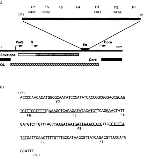
A)
F7 F6 F5 F4 F3 F2 Fl
C/EBP HNF3/1 HNF1 (HNF3/2)
2172 - 2351
Envelope
Core POL
B)
2171
ACCCCAACCATGGCGCATATCCCATATCACCGGCGGGAGCGCAG
F7
TGTTGCITTIMCAAAGGTCAGAGATATACATGTTCAGGAACTA1T
F6 F5 F4
MATERIALSAND METHODS
Preparation of duck tissue nuclear extracts. Nuclear ex-tracts were prepared from tissues of 14-day-old Pekin ducks (Anas platyrhynchos domesticlis). To prepare a liver nuclear extract, the liverwasperfused with Swimm-77 medium in the presence of1 mMphenylmethylsulfonyl fluoride (PMSF) and 1 mM leupeptin as protease inhibitors. The liver was then removed and placed in ice-cold homogenization buffer. All subsequent steps were performed at 4°C. The procedure for making nuclear extracts was modified from that of Tian and Schibler (50). Briefly, the tissue was homogenized, using a tissuehomogenizerdesignedtopreventairintake, ina homog-enization buffercontaining2 M sucrose, 10 mM
N-2-hydroxy-ethylpiperazine-N'-2-ethanesulfonicacid(HEPES; pH 7.6),25 mM KCl, 1 mM EDTA, 10% glycerol, 5% nonfat milk, 0.15 mM spermine, 0.5 mM spermidine, 0.5 mM PMSF, 0.5 mM dithiothreitol, 1 p.gofleupeptinperml, and 1 pLgofaprotinin perml. The homogenatewascentrifuged for35 min at24,000 rpm in a Beckman SW27 rotor. The pelletwas washed with NE-1 buffercontaining250 mM sucrose, 15 mMTris-HCI (pH 7.6), 140 mM NaCl,2 mM MgCI2, and the proteinase inhibi-tors as described above. The nuclear pellet was then
resus-pended in NE-2 buffer, which is similar to NE-1 buffer but contains 350 mM KCl, and wasthentransferred to a Dounce homogenizer. The nucleiwere lysedby 20 strokes with pestle A. The viscous lysates were ultracentrifuged for 90 min at 45,000 rpm in an SW6OTi rotor. The supernatant wasfinally
dialyzed with buffercontaining 25 mM HEPES (pH 7.9), 40 mM KCl, 0.1 mMEDTA, 1 mMdithiothreitol, 10% glycerol,
and 0.1 mM PMSF. The crude extract was aliquoted,
quick-frozen in liquid nitrogen, and stored at -80°C. Nuclear extracts from kidney, lung, heart, and spleen were made directly from the organs without perfusion. The protein con-centration for the nuclearextracts was about 2 to 3 p.g/pl. It should be notedthattwomeasures werefoundtobe critical for makinghigh-qualityextracts,one beingtheuse of nonfat milk as a proteinase inhibitor andthe other that nuclear
proteins
are releasedwith 350 mMsalt.
Gelmobility shift assay and DNase I footprinting.The gel
mobility shift assay was
performed
as describedpreviously
(15). Briefly, 1-ng aliquots of 32P-labeled, double-stranded
oligodeoxynucleotides representing different regions of the enhancer region of DHBV
(Fig. 1)
were incubated at roomtemperature for 20 min with livernuclearextract
prior
to5%polyacrylamide gel electrophoresis and
autoradiography.
TCTGATTCAACT|{ WGTTrGCCATAAGCGTIATCAGACGTTACCATG
F2 Fl
GCATTl
2361
FIG. 1. Locations of transcription factor-binding sites along the DHBVgenome. (A) The DHBV enhancer(En),aspreviously defined (35), is shown along with the locations of DNase I footprints defined in the current study and the apparent identities of transcription factors which contributed, in part, to their generation. (B) The DNA sequence of the DHBV enhancer and of therespective footprints shown in panel A.
When a gel mobility shift competition assay was performed, the nuclear extract was incubated with 100 ng of either the specific or the nonspecific double-stranded competitor
oli-godeoxynucleotide for 15 min at room temperature. The double-stranded HNF3-specific competitors, TGT3 andTTR,
defining HNF3 sites from the mouse albumin gene (24) and mouse transthyretin gene (33), respectively, were kindly pro-vided byKen Zaret (Brown University, Providence, R.I.).The humanHNFI competitor (defined byasitein the
otl-antitryp-sin gene) was synthesized according to a published sequence (39), aswere the DNAs eG (33) and USF (19).The DHBV-specific double-stranded probes used in the gel shift
experi-ments included HNFI, HNF3/1, HNF3/2, mutant HNF1
(mHNF1),
mHNF3/1, and mHNF3/2. The latter three oligo-nucleotides were also used for mutagenesis of the DHBV enhancer.The sequencesofall these DNAsarelisted inTable1.
DNaseIfootprintingassays wereperformedasfollows.One nanogram of a
32P-end-labeled
DNA fragment (DHBV en-hancerpositions 2172 to 2361)was used as a probein a total reactionvolumeof50.1l.
The different nuclearextracts(20to 30p.g
each)werefirst incubated in reaction buffer containinga final concentration of 12 mM Tris-HCI (pH
8.0),
100 mM KCI, 1 mMMgCl,,1%
polyvinyl alcohol,4pLg
ofpoly(dI-dC),
1 mM
CaCl,
5 mMNaCI,
0.1 mM bovine serum albumin (BSA), 0.1 mM dithiothreitol, and 1% glycerol for 15 minonice; then the probewas added, and incubation continued for another 45 min. The reaction mixturewas removed from ice and leftat room temperature for 2 min, and the volume was
adjusted to 100
Rl
with reaction buffer. Different amounts of DNase Iwereadded,and thereactionmixtureswereincubatedm
VOL.68? 1994
on November 9, 2019 by guest
http://jvi.asm.org/
[image:2.612.329.560.84.337.2]TABLE 1. Sequencesofoligonucleotides used in factorbindingassays
Oligonucleotide(specificity) Sequence(5'-33')
HNF1(human otl-antitrypsin)... GATCACCTTGGTTAATATTCACCAGCA
TGT3(HNF3, mouse
albumin)
... CCGAACGTGTTTGCCTTGGCCTTR(HNF3,mouse transthyretin)... TGATTCTGATTATTGACTTAGTCA
eG(HNF3,mousealbumin)... CCAGGGAATGTTTGTTCTTAAATACCAT
USF(adenovirus majorlate
promoter)
... TAGGTGTAGGCCACGTGACCGGGTGTTCCTGHNF1 (DHBV, nt2272-2294)... TTTAGCCAAGATAATGATTAAAC
mHNF1...TTTAGCCAAAATTATGATCAAACC
HNF3/1 (DHBV,nt 2210-2236)... AGCGCAGTGTTTGCTTTTTCAAAGGTC
mHNF3/1...AGCGCAGTATTCGCCTTCTCAAAG
HNF3/2(DHBV,nt 2312-2338)... GATTCAACTTTTGTTTGCCATAAGCGT
mHNF3/2...GATTCAACCTTCGTCTGTCATAAG
for2minat roomtemperature.Stoppingbuffercontaining0.1 mM Tris-HCl (pH 8.0), 0.1 mM NaCl, 10 mM EDTA, 1% sodiumdodecyl sulfate(SDS), 100pgofproteinase Kperml,
and50,ugof salmon sperm DNA per mlwasthen added. This reaction mixturewasincubatedfor 30 minat37°C,andnucleic acids were then purified by phenol extraction. DNA was precipitated with ethanolat roomtemperature.TheDNA was collected and resuspended in sequence gel loading buffer
containing 95%formamide and loadedonto an8%
polyacryl-amide sequencing gel. G+A and A sequence ladders of the sameDNAusedin thefootprintingassaywere createdbythe method ofMaxam and Gilbert (38).
Site-directed mutagenesis. The single-site mutations were made by using the BioRad Muta-Gene Phagemid in vitro mutagenesis system as recommended by the manufacturer. Briefly, DHBV DNA fragment BamHI (1658)-EcoRI (3021)
was cloned between BamHI and EcoRI sites in the pBlue-scriptSK(+) polylinker. The recombinant
pBluescript(SK+)-DHBV DNAwas used to transform Escherichia coli CJ236,
and the cultures were grown in LB containing 100 ,ug of
ampicillin per ml. The cultures were then infected with the helper phage M13K07. The single-stranded, circular, minus-strandDNA wasextracted from the supernatant. Two hundred nanograms of the single-stranded DNA was then annealed with 10 ng of the minus strand of themutant phosphorylated oligonucleotide mHNF3/1, mHNF3/2, or mHNF1 (Table 1),
and T7 phage DNA polymerase was used to synthesize a
full-length plus strand from theseprimers. Following ligation
with bacteriophage T4 ligase, the DNAs were used to trans-formE.coli JM101. The presence of theexpected mutations in the cloned DNAs wasconfirmed by DNA sequencing.
Tomake human growth hormone(hGH) expression vectors inwhich hGH transcription is under control of the wild-type or mutant DHBV enhancer (single mutated factor-binding site) andcorepromoter,a DHBVfragment spanning the enhancer andcorepromoterwasproduced by cleavage withNcoI(2351)
andBstEII (1847), gel purified, and then cloned into plasmid pGH-P6 (35). The mutant DNA fragments were also
ex-changed with the wild-type NcoI-to-BstEII fragment in the DHBV genome ofpCMVDHBV-9 vector (55), in which the viral pregenome is expressed under the control of the cyto-megalovirus immediate-early promoter.
To insert mutations of both HNF3 sites or the HNF3 and HNF1sites, the PCR approach was used. Briefly, two flanking
oligonucleotideswhich corresponded to the DHBV sequence from nucleotides (nt) 1745 to 1765 and the hGH/DHBV
junction of pGH-P5
(5'-GCCTFGGGATCTCGGCCGCT
TAAG-3', nt 2312 to 2526), respectively, were synthesized
(35).The mHNF2 oligonucleotide (see above) and the
oligo-nucleotide spanning the hGH/DHBV junction of pGH-P5
wereusedtoproduce PCR fragment I, using plasmid pGH-P5 as the template. The minus-strand mHNF3/2oligonucleotide and a DHBV sequence-specific nucleotide (5'-AACGCAAT TAGCCACGCTGTC-3', nt 1745 to 1765)wereused to pro-duce PCR fragment II, using as the template pCMVDHBV containing the mHNF3/1 mutation. PCR fragments I and II werethen mixed inequal molar ratio and amplified by using as primers theDHBV-specific oligonucleotide (nt 1745to 1765) and theoligonucleotidespanning thehGH/DHBVjunction of pGH-P5. To create atriplemutantcombiningthe HNF1 and HNF3 site mutations, the HNF1 oligonucleotide (see above) and the hGH/DHBVjunction oligonucleotide were used to produce PCR fragment I, with pGH-mHNF3/2 as the tem-plate. PCR fragment II was produced from the template
pCMVDHBVmHNF3/1, using as primers the minus-strand mHNF1 oligonucleotide and thehGH/DHBV junction oligo-nucleotide. Theresulting fragmentsweredigestedwith BstEII and NcoI and cloned into appropriate expression vectors as described above.
Transient transfection ofLMHcellcultures.Plasmid DNAs were prepared by the alkaline lysis procedure and further
purified by isopycnic centrifugation in cesium chloride (36).
Fivemicrograms of each of the hGH expression plasmid DNAs wascotransfected with 5 pLgof pRSV-SEAP(secreted alkaline
phosphatase) (4) DNAinto LMH cellsby the calcium phos-phatecoprecipitationmethod essentiallyasdescribed by Con-dreay et al. (9). The culture fluidswereharvested 72 h after
transfection, and hGH and SEAPwere assayed as described previously (35). The tissue culture fluids were changed every 24 h. The hGH expression levels presented in results were nor-malized to SEAP levels in the culture fluids.
Generation ofviral particles by transfection ofLMH cell cultures and infectionofduckhepatocyte cultures. Ten micro-gramsof each of the pCMV-DHBV DNA constructs described above wastransfected intoLMHcells. The culturefluids were harvested from 3 days to9daysposttransfection.Thetiter of virus particles in the pooled culture fluids was estimated by using the partial pronase digestion procedure designed by R. Lenhoff and J. Summers (University of New Mexico). This procedure is based on the observation that intact virus particles are resistant to pronase digestion, whereas viral DNA is released from viral nucleocapsids by pronase digestion under thesameconditionsandis therefore susceptible to digestion by added DNase I. Briefly, the culture fluids were clarified by
centrifugation at 5,000 rpm for 10 min. To precipitate virus
particles, polyethylene glycol was then added to 1 ml of supernatantto afinal concentration 10% (wt/vol). The
result-ing pelletwasresuspended in 500 ,ug of pronase solution per ml andincubated at 37°C for 1 h. ThenMgCl2wasadded to 20 mM andDNase Iwasadded to 5 ,ug/ml, and the mixture was
on November 9, 2019 by guest
http://jvi.asm.org/
HEPADNAVIRUS TRANSCRIPTION 2289
incubated at 37°C for 15 min. The reaction product was then subjected to 1% agarose gel electrophoresis, and standard Southern blot analysis was carried out to detect virion DNA. Virion DNA was quantitated with an AMBIS scanning image analyzer.
The infectivity of virus particles produced by transfected LMH cells was assayed in primary hepatocyte cultures. The primary hepatocyte cultures were prepared from the livers of 2-week-old duckling as described previously (52). Hepatocyte monolayers on 60-mm-diameter tissue culture dishes were infected with approximately 107virions. One day later and at daily intervals thereafter, the culture medium was changed with L15 tissue culture medium. Suramin (100
,ug/ml)
was included in the medium beginning at 1 day postinfection to block secondary rounds of infection (43). The hepatocyte monolayers were harvested at 8 days postinfection and either stored at -80°C for subsequent extraction of viral nucleic acids or fixed with 75% ethanol-25% acetic acid forimmuno-fluorescencemicroscopy to detect hepatocytes expressing viral core (nucleocapsid)protein. The preparation of tissue sections from ducklings was done as described by Jilbert et al. (27).
Extraction andanalyses of viral DNA and RNA. Total viral DNA and viral CCC DNA were extracted as described by Summers etal.(47) and subjected to Southern blotting follow-ing gelelectrophoresis in 1.5% agarose essentially as described previously (53). To extract viral RNA, one dish of frozen hepatocytes was thawed at room temperature for 5 min, and then 3 ml oflysisbuffer containing 0.2MNaCl, 0.2 MTris-HCl
(pH 7.6), 1.5 mM MgCl2, 2% (wt/vol) SDS, and 200 ,ug of proteinaseKper ml was added. The lysate was collected and homogenized with a Dounce homogenizer and then incubated at45°Cfor 1 h. The lysate was next passed through a 21-gauge needle to shearcell DNA. Following adjustment of the NaCl concentration of the lysate to 0.5 M, 30 mg of oligo(dT)-cellulose was added. The mix was rocked gently at room temperature for 30 min. The oligo(dT) with bound RNA was collected by centrifugation and washed twice with 0.5 M
NaCl-Tris-HCl (pH 7.6)-10 mM EDTA-0.1% (wt/vol) SDS. The oligo(dT) was then transferred to a 0.4-,um-pore-size Millipore filter unitand washed with low-salt binding solution
containing 100 mM NaCl, Tris-HCl (pH 7.6), 10 mM EDTA, and0.1% SDS. The RNA was eluted with 10 mM Tris-HCl (pH 7.6)-10 mM EDTA-0.1% SDS. The RNA was precipi-tatedby addition of 2.5 volumes of ethanol and 1/10 volume of 3 M sodium acetate. Viral RNA was detected by Northern
(RNA) blot analysis following electrophoresis on a 1.0% agarosegelcontainingformaldehyde (36). DHBV RNAs were detected byusingagenome-length minus-strandriboprobe.
RESULTS
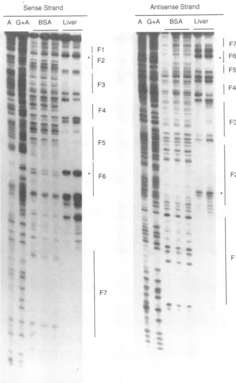
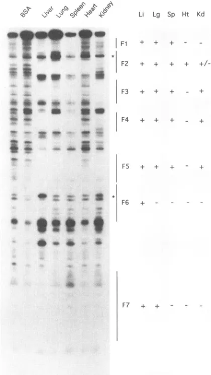
Atleastsevenfactors presentinduck liverextracts bind to theDHBV enhancer. Asafirst step inidentifyingtranscription
factors that interact with the DHBV enhancer, DNase I
footprinting assays were carried out with extracts from a
variety of ducktissues,including liver,lung, spleen, heart, and kidney. At least seven footprints, labeled Fl to F7, were
identifiedinaduck liverextract (Fig. 1 and2).Amongthese, all seven showed some tissue restriction, though only F6
showed ahigh degree of tissue
specificity
(Fig. 3). Sequence comparisons suggested that F3 might correspond to atran-scription factor HNF1-binding site (11, 39), that F2 and F6
might correspond to HNF3-binding sites (26), and that F7
mightcorrespondto aC/EBP-binding site (28).The fact that F2showedabroadertissue distribution than F6suggestedthat F2might be the consequence ofmorecomplexfactor
interac-Sense Strand
A G+A BSA Liver
Fl | F2
F3
I
F4F5
* F6
Antisense Strand
A G+A BSA Liver
F7
. F6
F5
F4
F3
F2
Fl
F7
&:
4 4:v I
2
4w
4w
FIG. 2. DNase I footprinting of the DHBV enhancer with duck liver nuclear extract. Locations of the footprints along the DHBV enhancer fragment were determined by reference to A and G+A sequence laddersgenerated with the same DNA fragment. For the liver extract and BSA controls, the different lanes represent digestions with increasing (from left to right) amounts of DNaseI.Hypersensitive sites indicativeofHNF3binding are indicated by asterisks.
tions than F6, despite the suggestion that both could have
HNF3-binding activity. The sequence at Fl, F2, and F3 suggested thepossible presence of GATA factor-binding sites
(16, 18) on the DHBVenhancer.
Gel shift assays support the identification of potential
HNF1
and HNF3 factor-binding sites within the DHBV en-hancer.Toobtain additional evidence thatspecificsitesiden-tified by DNase I footprinting could have resulted from the binding of transcription factors homologous to those
previ-ously identified in other species, competitive gel shift assays werecarriedoutwith duckliver extracts. The rationaleforthe gel shift assays is that bindingto aspecificDHBVsequence, if due to such a transcription factor, would be blocked by an excess of DNA fragment containing a consensus-binding se-quence for that factor but not by consensus-binding sites of othertranscriptionfactors. Ouranalyses focusedonHNF1and HNF3, which appear, from studies with other
species,
to controlsomeliver-specifictranscription during developmentas wellasinthe adult liver(57)and which thereforerepresented
VOL.68, 1994
on November 9, 2019 by guest
http://jvi.asm.org/
[image:4.612.328.559.79.453.2]Probe:HNF1(DHBV) Li Lg Sp Ht Kd
'I
H e
r. .'
Competitor:
+ + +
+ + +
+ + +
+
F7 + +
-FIG. 3. Tissuespecificityof factorsbindingtothe DHBVenhancer.
Nuclearextractswere preparedasdescribed in Materials and
Meth-ods. Locations of DNase Ifootprintsonthesense strand(seeFig. 2)
and theirtissue specificityareshown at the right.The tissue
assign-mentsof F7camefromdata from the other DNA strand(notshown). Li,liver; Lg, lung; Sp, spleen; Ht, heart; Kd, kidney.
potentially important factors in the facilitation of DHBV transcription.
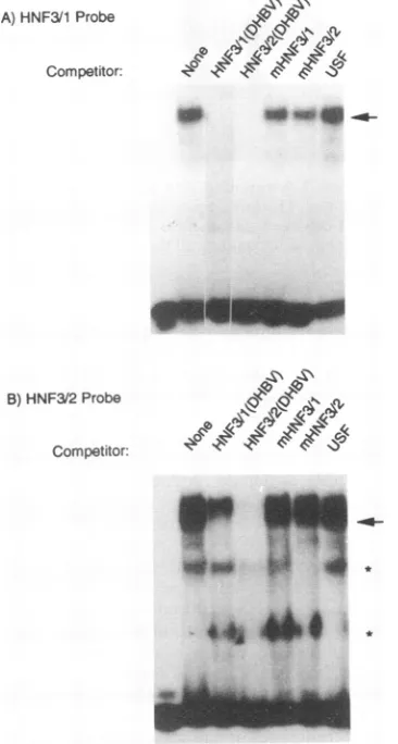
A competitive gel shift assay using a radiolabeled probe
spanningthe F3footprint (Fig. 1),acandidateHNF1-binding site, is shown in Fig. 4. Two prominent bands representing protein-DNA complexes were apparent in the absence of competitor DNA; however, in the presence of competitor
homologous tothe labeled probe, theuppercomplex (arrow)
was no longer detected and thefaster-migrating complexwas
reduced.Weinferredthat theuppercomplexwasindicativeof
an HNF1-like complex, since itwascompletelyeliminated by
competition with a DNA containing the well-characterized
HNFI-binding site of the human oLl-antitrypsinpromoter(39) butnotbyaDNAcontainingthebinding site for transcription
factor USF (44) (Fig. 4). The fact that the faster-migrating complex (indicated byx in Fig. 4)was not eliminated by the addition of the ot1-antitrypsin sequence suggested that it was
due to binding byan unrelated factor.A preliminary
experi-ment (data not shown) indicated that this factor existed
FIG. 4. Competitive gel shiftassays indicated that a duck HNF1 binds to the region of DNase I footprintF3. The radiolabeled and unlabeled competitorDNAsequenceswere aspresentedin Table 1. Theupperband(arrow) is apparentlydue to interaction withaduck HNF1, as indicated by competition with homologous, unlabeled
competitororwithanHNF1-binding site of distinctsequencefromthe humaneI1-antitrypsingene.The middle bandmaybe duetobinding by
anunrelatedtranscription factor. HNF1bindingwasnotcompetedfor by a DNA with a mutant binding site or by the unrelated USF oligonucleotide.
exclusively in the liver, but the identity of this factor is presently unknown. The band marked with an asterisk is apparentlyanartifact of thecompetitionreaction,as asimilar
bandwas also observed in othercompetitions (see Fig.5 and
7).
The results of competitive gel shift analyses of the two
candidateHNF3-bindingsites(Fig. 1) is shown inFig.5.One
major protein-DNA complexwasdetected(arrowinFig. 5A) with a probe spanning F6. This complex was reduced by
addition ofhomologous competitoraswellasbyacompetitor
spanning F2,the other candidateHNF3-bindingsite.Effective competition for binding was also obtained with the TTR
sequence, representing the high-affinity HNF3-binding site from thetransthyretinpromoter(33),butcompetitionwasless
effective with eG or TGT3 DNA, both of which contain
lower-affinity HNF3bindingsites from the albumin enhancer (26). Withtheprobespanning F2,two major complexeswere
resolved by gel electrophoresis (Fig. 5B). These complexes
were effectively competed for with the homologous
competi-tor. However, only one ofthese complexes (indicated by the
arrow in Fig. 5B) was competed for by an excess of a
competitor spanning F6orby eG, TGT3, andTTR competi-tors, which all contain previously identified HNF3-binding sites. The othercomplexwasnotcompetedforbytheseDNAs. Nocompetitionwasobserved withuseofthe USF competitor.
Inaddition, therewere someotherfast-migrating radiolabeled
complexes (Fig. SB). These complexeswere not reproducibly detected; therefore, we believe that these complexes were
nonspecific. Taken together with the sequence homologies, thesegelshiftresultssuggestedthatfootprintsF2 andF6(Fig. 2)weredue inpart tobindingofaduck transcriptionfactoror
factors homologous toHNF3.The site atF6 is referredto as
HNF3/1, and the siteat F2is referred toasHNF3/2.
Totesttherelevance of thefactor-binding sites inenhancer
I~~~-Admi
;=C:; ~F2
.ebS
F3
_
9RR,S$tt
*s
_ iS CAww fw -xl.; *~~F
_ : _ z. 0 _ :A~F
m-F7'
on November 9, 2019 by guest
http://jvi.asm.org/
[image:5.612.327.535.78.270.2] [image:5.612.73.283.82.455.2]HEPADNAVIRUS TRANSCRIPTION 2291 HNF3
En C L
HNF311
GTGTTrGCTTrr
Pol: V F A F
GTATTC:GCS
T-r.
mHNF3I1
HNF1
1--HNF1
HNF3 HNF312
AAG ATA ATG ATT ACT mGTT TGC
K I M I T F V C
AAAATI ATG AT<: ACfi TrQ GTGTGI
[image:6.612.328.560.78.175.2]mHNFI mHNF3/2
FIG. 6. Third-base mutations introduced into the HNF1- and HNF3-binding sites. Only the mutated region is shown; the sequences ofthe DNAs used for competitive gel shift assays are presented in Table 1. En, enhancer.
Toevaluate further the ability of these mutated binding sites to bind the respective transcription factor, these oligonucleo-tides were used as probes in gel mobility shift assays. These radiolabeledoligonucleotides were again found to be unable to bind the corresponding transcription factor (data not shown). Finally, when inserted into the viral enhancer, mutations at all
A)HNF3/1Probe w
Competitor:
40 lomw~~~~~~~~~~~~~~~~~~~~~~~~~~~~
FIG. 5. Competitive gel shiftassays indicated thatduck HNF3(s)
bindstoregionsF2andF6ofthe DHBVenhancer. Theradiolabeled
andunlabeledcompetitorDNAsequences were aspresentedinTable
1. (A)The HNF3/1 probe from region F6. The arrowindicates the
apparentcomplexwith HNF3, as revealed by effective competition with the homologous, unlabeled DNAaswellastheHNF3/2sequence
andthe TTR DNAcontainingtheHNF3-bindingsite from themouse
transthyretin gene. Partial competition by the lower-affinity HNF3-binding sequences of eG and TGT3 was also observed. (B) The
HNF3/2probe from region F2.TheHNF3complex, assuggestedby
the effectivecompetitionwith DNAscontaining heterologous
HNF3-binding sites, isindicatedbythearrow.The samplesin the leftmost lanes inpanelsAand Bcontainedprobebutnoliverextract.
function, especially duringvirusreplication,we designed mu-tants (Fig. 6) thatchanged the viral DNA sequence without changing the coding capacity of the viral pol gene, which overlaps the enhancer. Before inserting these mutations into the viralgenome,therespectiveoligonucleotides (see Materi-alsandMethods)werefirsttested for theabilitytocompetein the gel shift assay. As expected, the mutant HNF1 sequence
did notblockbinding of thewild-type sequence in theslowly moving complex (as indicated by arrow in Fig. 4), which appeared tocontain an HNF1 transcription factor. Competi-tionwas observed, however, with the faster-moving complex,
whichindicated that themutantoligonucleotides retained the binding activitytothe unknown factor. Our resultsindicated, therefore, that themutantDNA bound HNF1 lessavidlythan the wild-type DNA. Similar results were obtained with the mutations of theHNF3/1-andHNF3/2-binding sites (Fig. 7).
B) HNF3/2Probe
Competitor:
FIG. 7. MutationsatHNF3/1 andHNF3/2 abolishedcompetition
for factorbindingingelshiftassays.CompetitionassaysusingHNF3/1 and HNF3/2 as radiolabeled probes are shown in panelsA and B, respectively. Effectivecross-competition for duck HNF3binding (ar-row)wasobserved with thewild-typebut notthemutatedDNAs.The
samplesinthe leftmost lanesinpanelsAand Bcontainedprobebutno
liverextract. A)HNF3/1 probe
Competitor:
B) HNF3/2 Probe
Competitor:
VOL.68, 1994
S\ -Z
,I.b 101,
AN A.
a
Ioe
lp
AN .AN
4lb-
4lb-1/1
on November 9, 2019 by guest
http://jvi.asm.org/
[image:6.612.96.274.84.437.2] [image:6.612.355.538.320.662.2]Enhancer Core Promoter hGH
2351 2531
RelativehGH
Activity
1.0 WT
0.29 mHNF3/1
0.37 mHNF3/2
0.33 mHNF3/1+2
0.47 mHNF1
0.24 mHNF3+mHNF1
0.10 EnhancerDeletion
Mutant
~~i FS
F6(HNF3/1)
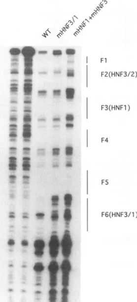
FIG. 8. DNase I footprinting wtth wild-type and mutant DHBV
enhancer sequences. Thefootprintingassayswerecarriedoutwith the
wild-type (WT)enhancer,anenhancercontainingthe HNF3/1
muta-tions,andanenhancercontainingallthree of the mutations shown in
Fig. 6.
three sites either eliminated or altered the respective
foot-prints in aDNase Ifootprintingassay (Fig. 8).
Mutations reduced theactivityof the DHBVenhancerina
transient expression assay. To evaluate the effects of
muta-tions in the DHBV enhancer inacellline ofhepaticorigin,we
first carried out a transient expression assay in which the DHBV enhancer andcorepromoterwereinsertedupstreamof thegene forhGH,andhGHproductionwasmeasured
follow-ingtransfection of theLMHline of chickenhepatomacells(2,
9, 35). Deletion of the entire enhancer (35) reduced hGH
productionabout 10-fold (Fig. 9). Mutation ofthe HNFI site reduced expression abouttwofold, and mutations of either of the HNF3 sites reduced expression about threefold. Mutation ofbothHNF3sitestogethercausednoadditional reduction in
expression,andmutation of both of these sites,togetherwith the HNF1 site, caused o'nly a slight additional decrease in
activity, toca. 25% of the control level.
Themutantenhancerswere nextplacedinto the intact viral genome, andvirusexpressionwasassayedintransfectedLMH
cells.Twodifferenttypesofplasmidconstructswereexamined.
In one, the DHBV genome wascloned at the uniqueEcoRI
site (locatedinthe viral coregene;Fig. 1)asatandem dimer.
[image:7.612.316.551.79.256.2]Intheother,DHBVwas inserteddownstream of the cytomeg-alovirus immediate-early promoter, which then directed syn-thesis of viral pregenomic RNA (9). With both types of construct, the effects of the enhancer mutationson the intra-cellular accumulation ofintermediates in viral DNAsynthesis
FIG. 9. DHBV enhancer mutationssuppressed expression ofthe hGHgenefrom the DHBVcorepromoter:Structures of the various
constructs are shown schematically at the left, with the sites of mutations indicated(x,HNF3 sitemutation;*,HNF1 sitemutation). The level of hGH expression is shown after normalization toSEAP production from the cotransfected control plasmid. WT, wildtype.
andonvirus releasewere marginal (datanotshown). In view of these results and the results of the transient expression
assays (Fig.9), it appeared that DHBV enhancer functionin LMH cellswasnotcompletely dependentonintact HNF1-and
HNF3-binding sites.
Effects offactor-binding site mutations on DHBV
replica-tion in
primary
hepatocyte cultures. Preparations ofwild-typeand mutantviruses releasedby transfected cultures ofLMH cellswereusedtoinfectprimarycultures of duckhepatocytes. The inoculawereadjustedtocontain equalamountsofDNA containing viral particles. Suramin was added to the culture mediumbeginning the dayafter infection to blocksecondary rounds of infection (43). The fraction of infected cells was
evaluated by immunofluorescent staining ofmonolayers with
serum reactive to DHBV core antigen. A similar numberof infected cells was observed by 6 days postinfection with the
wild-typevirusorwith virusescontaining multiple mutationsof
these three factor-binding sites. An approximately threefold-lower percentage of core antigen-positive hepatocytes was
detected with the immunofluorescence assayfollowing
inocu-lation of cultures with viruscontaining mutationsinmorethan onefactor-binding site, andthosehepatocytes thatwere core
antigen positivehadagenerallylowersignal thanseenwiththe
wild-typevirusorthesingle-sitemutants.Thisfindingsuggests
thatthe immunofluorescenceassay maybegivingan
underes-timate ofthe relative number ofhepatocytes infected by the double andtriplemutants.
Tofurther evaluatetheeffects of thedifferent mutationson
virus infection, hepatocyte monolayers were extracted, and
totaland CCCDNA and viral RNAlevelswerequantitated.
As shown inFig. 1OA, mutationatanyof thethree sitescaused, atmost,atwofoldreduction intotal viralDNAaccumulation. Bycontrast,monolayers infected withviruses with mutationsat
more thanonebinding site accumulatedca. 10-fold lessviral
DNAthan cells infectedwiththewild-type virus.Lessereffects
onCCC DNA levelswereobserved,thelevel of accumulation being reduced only 3- to 4-fold even in infections for which total viral DNAlevelswerereducedca.10-fold(Fig. lOB). The
relatively larger amount of CCC DNA could result from
S
1847
Fl
F2(HNF3/2)
F3(HNFl)
F4
v
v mki.m
-x x
mmimilmm
v * y
on November 9, 2019 by guest
http://jvi.asm.org/
[image:7.612.107.246.84.390.2]HEPADNAVIRUS TRANSCRIPTION 2293
A) TotalDNA
RC DL
-Ss
-__
_ _ E; *
. . _g.- kE -4
-40_ _ l _ lS1,S
$
-_ ! _ ~~~~~~~~~~~~~~~~~~~~~~~~~~~~~~Il
_ |
F~~~~~~~~~~~~~~~~~~~-a_|_! t~~~~~~~~~~~~~~~
1.0 0.46 1.0 0.1 0.7 0.08
B) CCC DNA
RC DL
ccc
Ss
1.0 1.0 1.3 0.36 1.0 0.36
C)mRNA
3.5kb 2.3kb 2.0kb
-1.0 0.26 1.1 0.150.35 0.26
FIG. 10. Effects ofmutations in the DHBV enhancer uponvirus
expressionininfectedprimaryduckhepatocytecultures.Southern blot
assaysforDHBV DNA induck hepatocyte culturesat6days
postin-fection are shown in panels Aand B, and a Northern blot assay is shown in panel C. (A) Total cell DNA, including DNA replication intermediates; (B)DNAextraction enrichedfor CCCDNA; (C)viral RNAs ininfectedhepatocytes.Percentexpressionoftotal viralnucleic acidsas comparedwiththewild-type (WT)virus infection is shown below the lanes in each panel. RC and DL, relaxed circular and double-stranded linear3-kbpviralDNAs, respectively.
selectivecycling of newlysynthesizedviral DNAtothenucleus
to elevateCCC DNAlevels (47, 48).
The effects of the mutationsonviralRNAaccumulationare
shown in Fig. 10C. As predicted by the transient expression
assayin LMHcells(Fig. 9),theeffectsonviralRNAsynthesis
in duck hepatocytes were only modest. Mutation of HNF3/2 had no apparent effect on RNA synthesis in the cultures, whereas synthesiswas apparentlyreduced two- tofourfold in cells infected byother mutant viruses.
Effects of enhancer mutations on DHBV replication in
ducks. Adifficultyinassessingtheregulationof viral transcrip-tion either in cell lines or in primary cell cultures that have
been derived from a particular tissue arises because of the possibility that the regulation maybe different in situ. It has
been reported that liver-specific transcripts (e.g., albumin
[image:8.612.102.262.81.450.2]mRNA [7, 8]) maybesignificantly reduced whenhepatocytes
TABLE 2. Virus with anHNF3/1 mutation can replicate in the duck liver but not the pancreas
DHBVcore Ag
Duck Virus Viremia expression
no.
Liver Pancreas
4 WT + + +
603 WT + + +
604 WIT + + +
608 WT + + +
609 WT + + +
1752 WT + + +
7 mHNF3/1 + +
8 mHNF3/1 + +
9 mHNF3/1 + +
10 mHNF3/1 + +
611 mHNF3/1 + + +
614 mHNF3/1 + +
616 mHNF3/l + +
1755 mHNF3/1 + +
1757 mHNF3/l + +
are maintained in vitro. We have therefore assessed the capacity ofoneofour mutantviruses, with a mutated HNF3/1 site, to replicate in ducklings. Ducklings at ca. 3 days of age wereinoculatedintravenously withca.
107
virions per duck. At 2 weeks postinfection, serum samples were collected and assayed for viremia; in addition, the ducksweresacrificed,and the liver and pancreas were monitored for virus replication. DHBV isknown toinfect and presumablyreplicatein asmall fraction of exocrine cells in the pancreas andprobably inthe majority of endocrine cells of thepancreas(21, 22, 24,37). The experimental resultsaresummarized inTable 2. Therewas no significant difference between the viremias obtained with the wild-type andmutantviruses,though therewassomevariation betweenducklings.Todetermine theextentof infection in the liver and pancreas, tissuesectionswere assayed forviral core antigen expression by immunofluorescence microscopy. As illustrated in Fig. 11, core antigen was expressed in the majority of hepatocytes in livers infected by eitherthe wild-type ormutantvirus. In contrast, viral core antigenexpressionwas notdetected inthepancreassectionsofeight ofnineofthe mutant-infected ducks, though it was detected in pancreas fromallsixducklings infected with wild-type virus. ViralDNAs were also isolated from the liver and pancreas tissues oftwo ducklings infected with wild-type virus and four ducklings infected with the mutantvirusand subjected toSouthern blot analyses (36a). In agreementwith the results of the immuno-fluorescence assays, we observed similar levels of viral DNA replication intermediates intheliversofwild-type and mutant virus-infectedducks. However,replicative formsof viralDNA were detectedonly in the pancreas extracts of ducks infected withwild-type virus,not in pancreas extractsfrom the mutant virus-infected ducks, including the duck that showed core
antigen-positive pancreas cells.
To rule out the possibility ofcontamination with wild-type
virus or of reversion of the mutant to wild type as an
explanationof theefficienthepatic infections,virion DNAwas
extractedfromthe seraof bothonewild-typeandtwo mutant
virus-infected ducks, and viral enhancer sequences were
am-plified by PCR. The purified DNA was then
sequenced
through the region of the original mutation, from 50 bases upstreamto30 bases downstream ofthe
original
mutation.No changeinsequence asaresult ofin vivo passagewasdetected (data notshown).Vot-. 68, 1994
on November 9, 2019 by guest
http://jvi.asm.org/
[image:8.612.321.562.97.276.2]WT
mHNF3/1
FIG. 11. Virus witha mutantenhancer(mHNF3/1) replicatedin the liver butnotthe pancreasof infected ducks. Viralcoreantigenexpression
wasdetectedby immunofluorescencemicroscopy,usingarabbitantibodyreactivetothisviralprotein.Virtuallyall of thehepatocyteswerecore
antigen positive, though therewasconsiderablevariation insignalintensitybetween individualcells. About1% oftheexocrinepancreascellswere
coreantigen positive after infection with thewild-type(WT)butnotthemutantvirus.
We nextcarriedout apreliminaryexperimenttodetermine whether the pancreaswas able to support replication of the HNF3/1 mutant virus when the ducks were maintained for
longer than 2 weeks. One group of ducks was infected with
either wild-type virus or mutant virus. Two ducklings were
inoculated intravenouslywiththe mutant virus and twowere
inoculated with the wild-type virus as described above. By 1 weekpostinoculation, all fourduckswere viremic. The
duck-lings were sacrificed at 6 weeks postinfection, at which time
theywereagaintestedfor viremiaaswellasfor infection of the
liverand pancreas.With the mutant virus, viremia had fallento a level that was significantly reduced in the spot test, and
though the core antigen was still detectable in the liver, the
signal intensity had fallen to very low levels compared with levels found for the
wild-type
virus-infected ducks. Again, by the immunofluorescence assay, infection of the pancreaswas detectedonly in thetwoducks infectedby thewild-type virus.DISCUSSION
Althoughthetranscriptional control elements of HBV have beenintensively studied, these studies have depended on the useofeithertumorcells ofhepatic origin(3, 14, 25, 46, 51, 56) orliver extract from a species such as the rat (42) which is not ahost for HBV. To evaluate the role of cis elements and host transcription factors in viral transcription, it is important
ultimatelytocarryoutstudies with liver from the natural host. We therefore chose to study liver-enriched transcription fac-torsaffecting the replication of a hepadnavirus by using DHBV
and tissues from its natural host, the domestic duck. This required, first of all, optimization of procedures for prepara-tion of nuclear extractsfrom duck tissues, including but not limited to the liver. Usingfootprinting analysesand gelshift assays with such extracts, a C/EBP-binding site, an
HNF1-binding site, and two HNF3-binding sites were tentatively
identified. InDHBV, the sequence (Fig. 1) of theC/EBPsite wasGCAAT, and that of the HNF1 sitewasAGATAATGAT TAAAC. The presence of theC/EBP-andHNF1-bindingsites in the DHBVenhancerwasalso recently reported by Lilien-baum et al. (34). The sequence TGTTTGC at both of the
HNF3-bindingsites(11)whichwehave describedwasdefined as a new consensus binding site by Jackson et al. (26). This
consensus sequence is found in all hepadnaviruses and for someliver-specifichost genes(3).
Inadditiontobinding sites forthewell-knowntranscription factors described above, consensus sequences were found in
footprintsFl,F2,and F3forthe GATAfamilyoftranscription factors. The GATAcoresequence wasinitially defined as the
binding site for an erythroid cell-specific transcription factor
(GATA-1),whichhas been showntohaveanessential role in the activation ofanumberof erythroidcell-specific genes(16,
17, 40,41). Several additional GATA factors havenow been cloned andcharacterized.Ithasbeen found that these factors have broad spectra of function during development (41). GATA-2and GATA-3 havehigh-levelexpression inmastcells and T lymphocytes (30, 32), respectively. Two new GATA
transcription factors (GATA-4 and GATA-5) have recently beencloned fromachickenliver cDNAlibrary (4a).
on November 9, 2019 by guest
http://jvi.asm.org/
[image:9.612.106.506.73.372.2]HEPADNAVIRUS TRANSCRIPTION 2295 ering that the avian liver does not have a hematopoietic
function during embryogenesis, it seems likely that GATA factors also have a role in gene expression in hepatocytes. Indeed, mouse (1) and frog (31) homologs of GATA-4 have also recently been identified and shown to be expressed in normal endodermally derived tissues aswell asin the heart.
With respect to the number of factors in liver extracts orin otherextracts thatbind tothe DHBVenhancer,it isimportant
to note that our footprinting studies defined a minimum of seven factor-binding sites, and our gel shift assays indicated that there are additional overlapping binding sites that may function with, or in addition to, the sites that we have described. This finding raises the possibility of a level of functional redundancy and may explain why the binding site mutations that we created had such small effects on viral transcription. In making factor-bindingsitemutations,we had to avoid altering the amino acid sequence of the viral poly-merase. Therefore, the only mutations that we madewere in the third bases of the respective codons (Fig. 6). This essen-tially conservative approach to mutagenesis may also have preserved binding by factors other than the ones under inves-tigation. For instance, while it was apparent that as a conse-quence of mutation, the different sites completely lost the
ability to bind HNF1 or HNF3, possible binding of other factors wasclearly seen by gel shift analysis.
Because of the presumed importance of liver-enriched HNF1 and HNF3 transcription factors during liver
develop-ment and hepatocyte differentiation (6, 57), we selected the respectivebindingsites in DHBVforastudy oftranscriptional
control during virus replication. Though mutations at the HNF1 site and the two HNF3 sites seemed to make thevirus enhancer slightly defective in the LMH liver cell
line,
asexpectedfromtheassayswith the hGH reporter gene,
only
oneof the three single mutants, with mutations in one of two
HNF3-binding sites(HNF3/1),was significantlydifferent from the wild type with regard to its ability to function in
primary
hepatocyte cultures. Interestingly,the viruswith this mutation was able to replicate in duck liver almost as well as the wild-type virus, but it was defective in the
ability
to infect and/ortoreplicate in the pancreas. Inthisregard,
itshould be noted that the HNF3/1 site was the only site defined in this study for which factorbindingappearedto becompletely
liverspecificamongthe severalorganextractsthatweretested
(Fig.
3); thus, we might have
anticipated,
apriori,
that this site would be especially important forreplication
in the liver. However, in vivo evaluation of the mutantvirus indicated that thiswas notthecasebut raised thehypothesis
that the sitewascritical for replicationin thepancreas.
Unfortunately,
we wereunsuccessful in several attempts to prepare nuclear extracts
from the pancreas with which to test this
hypothesis.
There-fore, itisequallypossible that a factor other than HNF3 may bindtotheHNF3/1 site toprovide
thepancreatic specificity.
It haslongbeenknownthat about 1% ofpancreatic
cellsareable tosupport DHBVreplication.
Some of thesecells,
located intypicalacini, have been
positively
identifiedasexocrinecellsby
Halpern and colleagues
(21,
23),
though
themajority
ofcells located in the aciniwerenotpositively
identified.Inadditiontoexocrine cells, Halpern and
colleagues
(20, 24)
have also demonstratedthatvirtually
allendocrinecells,
locatedin (xand , islets,are targetsofDHBVinfection. Aswe were unabletolocate endocrine isletsin thetissue sections thatwereavailable
foranalysis,wedonotknowwhether theHNF3/1 sitemutation also affected virus
expression
in the endocrine cellcompart-ment. In any case, it would not be
surprising
if HNF3 wereimportantfor DHBV
replication
inpancreatic cells,
inasmuch as liver and pancreas are derived from endoderm, which isinfluenced
by
HNF3during embryogenesis (57).
A direct assessmentof HNF3expression
inthe pancreas wouldhelp
to resolve thisissue,
butso far, HNF3 has notbeen clonedfromanavian
species.
Irrespective
of therole ofHNF3 intranscrip-tion of
DHBV,
it should be noted that the DHBV results indicate that exocrine pancreascells,
at least those whicharc targetsofinfection,
haveacomplement
oftranscription
factorsmaterially
different from that ofhepatocytes.
One
important
butunresolvedquestion
is whetheranysingle
factor-binding
site isreally important
forvirusreplication
in the liver.Atleast foroneof the twoHNF3sites inthe DHBV enhancer(HNF3/1),
this seemed not to be the case in ashort-term assay.
ACKNOWLEDGMENTS
We aregrateful to G. Mark, J. Pugh, J. Taylor,and C. Seegerfor helpfulcontributionsduringthecourseofthiswork,toR. Lenhof and J. Summers forcommunicatinganovelprocedurefor thetitration of viral particles, toC. Aldrich andJ. Saputellifortechnical assistance, and to KathyTruesdale forhelpinpreparation of themanuscript.
This workwas supported by USPHS grants AI-18641, CA-06927, GM-35535,and RR-05539 from the National Institutes of Health and by an appropriationfrom the Commonwealth ofPennsylvania.
REFERENCES
1. Areci, R.J.,A. A.J. King, M.C. Simon,S. H. Orkin, and D. B. Wilson. 1993. Mouse GATA-4: a retinoic acid-inducible GATA-binding transcription factor in endodermally derived tissue and heart.Mol. Cell.Biol. 13:2235-2246.
2. Aufiero, B., and R. J. Schneider. 1990). The hepatitis B virus X-gene product trans-activatesboth RNApolymerase II andIII
promoters. EMBO J. 9:497-504.
3. Ben-Levy, R., 0. Faktor, I.Berger, and Y. Shaul. 1989.Cellular factors that interactwith thehepatitisBvirusenhancer. Mol. Cell.
Biol.9:1804-1812.
4. Berger, J.,J.Hauber,R.Geiger,and B. R. Cullen. 1988.Secreted alkaline phosphatase: a powerful new quantitative indicator of geneexpressionin eukaryoticcells. Gene66:1-10.
4a.Burch, J.Unpublisheddata.
5. Buscher, M.,W.Reiser,H.Will,andH. Schaller.1985.
Transcripts
and the putative RNA pregenome of duck
hepatitis
B virus:implicationsforreverse transcription. Cell 40:717-724.
6. Cascio, S., and K. S. Zaret. 1991. Hepatocyte differentiation initiates during endodermal-mesenchymal interactions
prior
to liver formation.Development 113:217-225.7. Clayton,D.F.,andJ.E.Darnell,Jr.1983.
Changes
inliver-specific
comparedtocommongene
transcription during
primary
culture ofmouse hepatocytes.Mol. Cell. Biol. 3:1552-1561.
8. Clayton, D. F., A. L. Harrelson, and J. E. Darnell, Jr. 1985.
Dependenceofliver-specific
transcription
on tissueorganization.
Mol.Cell. Biol. 5:2623-2632.
9. Condreay,L. D.,C. E.Aldrich,L.Coates,W.S.Mason,and T. T. Wu. 1990.Efficient duck
hepatitis
Bvirusproduction by
anavianlivertumorcell line. J. Virol.64:3249-3258.
10. Condreay, L. D., T. T. Wu, C. E. Aldrich, M. A. Delaney, J. Summers, C. Seeger, and W. S. Mason. 1992.
Replication
of DHBV genomeswithmutationsatthesites of initiation of minus-andplus-strandDNAsynthesis.
Virology
188:208-216.11. Costa, R., D. Grayson, and J. E. Darnell, Jr. 1989.
Multiple
hepatocyte-enriched nuclear factors function in the
regulation
oftransthyretin
andetI-antitrypsin
genes. Mol. Cell. Biol. 9:1415-1425.12. Courtois, G.,J.Morgan,L.Campbell,G.Fourel,andG. Crabtree. 1987.Interaction ofa
liver-specific
nuclear factor with thefibrogen
and ol-antitrypsinpromoters. Science238:688-692.13. Crescenzo, C. B., J. Pillot, A. Lilienbaum, M. Levrero, and E. Elfassi. 1991. Identification of a strong enhancer element
up-streamfrom thepregenomicRNAstartsite of the duck
hepatitis
Bvirus genome.J.Virol.65:3882-3886.
14. Dikstein, R., 0. Faktor, and Y. Shaul. 1990. Hierarchic and
cooperative
binding
of theratliver nuclearprotein
C/EBPattheVol. 68, 1994
on November 9, 2019 by guest
http://jvi.asm.org/
hepatitis B virus enhancer.Mol. Cell. Biol. 10:4427-4430. 15. DiPersio, C. M., D. A. Jackson, and K. S. Zaret. 1991. The
extracellular matrix coordinately modulates liver transcription
factors andhepatocyte morphology.Mol. Cell. Biol. 11:4405-4414.
16. Evans, T., and G. Felsenfeld. 1989. The erythroid-specific
tran-scriptionfactoreryfl:a newfinger protein.Cell58:877-885.
17. Evans, T., and G. Felsenfeld. 1991. trans-activation of a globin
promoterinnonerythroidcells. Mol. Cell. Biol. 11:843-853. 18. Evans, T., M. Reifman, and G. Felsenfeld. 1988. An erythroid
specific DNA binding factor recognizes a regulatory sequence
common toall chickenglobingenes. Proc.Natl. Acad. Sci.USA 85:5976-5980.
19. Gregor, P. D., M. Sawadogo, and R. G. Roeder. 1990. The adenovirusmajorlatetranscriptionfactor USFisamember of the
helix-loop-helixgroupofregulatory proteinsand bindstoDNAas
adimer. Genes Dev. 4:1730-1740.
20. Halpern,M.S., J. Egan,W. S.Mason,andJ.M.England. 1984.
Viralantigen in endocrine cellsof thepancreaticislets and adrenal cortexofPekin ducks infected with duck hepatitis B virus. Virus Res. 1:213-223.
21. Halpern,M.S., J. Egan,S. B.McMahon,and D. L. Ewert. 1985. DuckhepatitisB virus istropicfor exocrine cells of thepancreas. Virology146:157-161.
22. Halpern,M. S., J. M.England, D. T. Deery, D. J. Petcu,W. S.
Mason,and K. K. Molnar. 1983. Viral nucleic acidsynthesisand
antigen accumulation in pancreas and kidney of Pekin ducks infected with duck hepatitisB virus. Proc. Natl. Acad. Sci. USA 80:4865-4869.
23. Halpern, M. S., J.-M. Lien, J. Egan, W. S. Mason, and J. M.
England. 1985. Duck hepatitis B virus replication in the Pekin
duck,p. 37-48. In K.Nishioka,B. S.Blumberg, N.Ishida,and K. Koike (ed.), Hepatitisviruses and hepatocellularcarcinoma. Ac-ademic PressJapan, Inc., Tokyo.
24. Halpern, M. S., S. B. McMahon, W. S. Mason, and A. P. O'Connell. 1986. Viral antigen expression in the pancreas of DHBV-infectedembryosandyoungducks.Virology150:276-282. 25. Hu,K. Q., and A. Siddiqui. 1991. Regulation of the hepatitisB
virus gene expression by the enhancer element I. Virology 181:
721-726.
26. Jackson,D.A., K. E.Rowader,K. Steven,C. Y.Jiang,P.Milos,
and K.S. Zaret. 1993. Modulation ofliver-specific transcription by
interactions between hepatocyte nuclear factor 3 and nuclear
factor 1 binding DNA in close apposition. Mol. Cell. Biol.
13:2401-2410.
27. Jilbert,A. R.,T.-T. Wu, J. M. England,P. de la M. Hall,N. Z.
Carp,A. P.O'Connell,and W. S. Mason. 1992.Rapid resolution of duckhepatitisBvirus infections occursafter massive
hepato-cellularinvolvement. J.Virol. 66:1377-1388.
28. Johnson, P.,W.Landsschulz,B.Graves, and S. McKnight. 1987.
Identification of a rat liver nuclear protein that binds to the
enhancer core element of three animal viruses. Genes Dev. 1:133-146.
29. Johnson,P. F.1990.Transcriptional activators in hepatocytes. Cell
GrowthDiffer. 1:47-52.
30. Joulin, V.,D.Bories, J. F. Eleouet, M.C. Labastie, S. Chretien,
M. G. Mattei, and P. H. Romeo. 1991. A T-cell specific TCR 8
DNAbinding protein is amember ofthe human GATA family.
EMBO J.10:1809-1816.
31. Kelley, C., H. Blumberg, L. I. Zon, and T. Evans. 1993. The GATA-4gene isa noveltranscription factor expressed in
endo-cardiumof thedeveloping heart. Development 118:817-827.
32. Ko, L.J., M. Yamamoto, M.W. Leonad, K. M. George, P. Ting, andJ.D.Engel.1991.Murine and humanT-lymphocyte GATA-3
factors mediate transcription through a cis-regulatory element
withinthe humanT-cell 8geneenhancer. Mol.Cell. Biol. 11:2778-2784.
33. Lai, E., V. R. Prezioso, E. Smith, 0. Litvin, R. H. Costa, and
J.E.J.Darnell. 1990.HNF-3A,ahepatocyte-enriched
transcrip-tionfactor ofnovel structure,isregulated transcriptionally. Genes
Dev. 4:1427-1436.
34. Lilienbaum, A.,B.Crescenzo-Chaigne, A.A. Sall, J. Pillot, and E. Elfassi.1993. Bindingofnuclear factorstofunctionaldomains of theduckhepatitisBvirusenhancer. J. Virol.67:6192-6200.
35. Liu, C., L. D. Condreay, J. B. Burch, and W. Mason. 1991. Characterization of the core promoter and enhancer of duck hepatitis Bvirus. Virology 184:242-252.
36. Maniatis, T., E. F. Fritsch, and J. Sambrook. 1982. Molecular cloning: a laboratory manual. Cold Spring Harbor Laboratory, ColdSpring Harbor, N.Y.
36a.Mark, G., and C. Liu. Unpublished data.
37. Mason, W. S., M. S. Halpern, J. M. England, G.Seal, J. Egan, L. Coates, C.Aldrich, and J. Summers. 1983. Experimental trans-mission of duckhepatitis B virus. Virology 131:375-384. 38. Maxam, A. M., and W. Gilbert. 1980. Sequencing end-labeled
DNA withbase-specified chemical cleavages. Methods Enzymol. 65:499-560.
39. Monaci, P., A. Nicosia, and R. Cortese. 1988. Two different liver-specific factors stimulate in vitro transcription from the human(xl-antitrypsin promoter. EMBO J. 7:2075-2087. 40. Orkin, S. H. 1990. Globin gene regulation and switching. Cell
63:665-667.
41. Orkin, S. H. 1992.GATA-binding transcription factors in hema-topoietic cells. Blood 80:575-581.
42. Patel, N. U., S. Jameel, H. Isom, and A.Siddiqui.1989. Interac-tions between nuclear factors and thehepatitisB virus enhancer. J. Virol. 63:5293-5301.
43. Petcu, D. J., C. E. Aldrich, L.Coates, J.M. Taylor, and W. S. Mason. 1988.Suramin inhibits in vitro infectionbyduckhepatitis B virus, Rous sarcomavirus, and hepatitis delta virus. Virology 167:385-392.
44. Pognonec, P., and R. G. Roeder. 1991. Recombinant 43-kDa USF binds to DNA and activatestranscription in a manner indistin-guishablefrom that of natural 43/44-kDa USF. Mol. Cell. Biol. 11:5125-5136.
45. Schneider, R., and H.Will. 1991. Regulatorysequencesofduck hepatitisBvirus C genetranscription. J. Virol. 65:5693-5701. 46. Siegrist, C. A., B. Purand, P. Emery, E. David, P. Hearing, B.
Mach, and W. Reith.1993. RFX1 is identifiedtoenhancerfactor C and functions as a transactivator of the hepatitis B virus enhancer. Mol. Cell.Biol. 13:6375-6384.
47. Summers, J., P. M. Smith, and A. L. Horwich. 1990.Hepadnavirus envelopeproteins regulate covalentlyclosedcircularDNA ampli-fication. J. Virol. 64:2819-2824.
48. Summers, J., P. M. Smith, M. J. Huang, and M. S. Yu. 1991. Morphogeneticandregulatory effects ofmutationsin theenvelope proteins ofanavianhepadnavirus. J. Virol. 65:1310-1317. 49. Tagawa, M., M.Omata, and P. L. Marion. 1990. Open reading
framesonplus strandgenomeofduckhepatitisBvirus. Gastro-enterol.Jpn. 2:20-22.
50. Tian, J. M., and U. Schibler. 1991. Tissue-specific expressionof the geneencoding hepatocytenuclear factor 1 mayinvolve hepa-tocytenuclearfactor 4. Genes Dev.5:2225-2234.
51. Trujillo, M. A., J.Letovsky,H. F. Maguire, C. M. Lopez, and A. Siddiqui. 1991. Functionalanalysis ofaliver-specificenhancer of thehepatitisBvirus.Proc.Natl.Acad.Sci.USA 88:3797-3801. 52. Tuttleman, J., J. Pugh, and J. Summers. 1986. In vitro
experimen-tal infection of primary duck hepatocyte cultures with duck hepatitisBvirus. J. Virol. 58:17-25.
53. Wahl, G. M., M. Stern, and G. R.Stark 1979.Efficienttransfer of largeDNAfragments fromagarosegels to diazobenzyloxymethyl paper andrapid hybridization by using dextran sulfate. Proc. Natl. Acad.Sci. USA 76:3683-3687.
54. Wu, J. Y., Z. Y. Zhou, A. Judd, C. A. Cartwright, and W. S. Robinson. 1990. The hepatitis B virus-encoded transcriptional trans-activator hbxappearstobeanovelprotein serine/threonine kinase. Cell 63:687-695.
55. Wu, T.-T., L. D. Condreay, L. Coates, C. Aldrich, and W. S. Mason.1991.Evidence that less-than-full-lengthpol gene products
arefunctional inhepadnavirus DNA synthesis. J. Virol. 65:2155-2163.
56. Yee, J. K. 1989. A liver-specific enhancer in the core promoter region of human hepatitis B virus. Science 246:658-661. 57. Zaret, K. S. 1994. Genetic control of hepatocyte differentiation, p.
53-63. In I.Arias(ed.),Theliver: biologyandpathobiology, 3rd ed. RavenPress, New York.