Dissertation
On
ACUTE INTESTINAL
OBSTRUCTION
Submitted to
THE TAMIL NADU DR. M.G.R. MEDICAL UNIVERSITY CHENNAI.
M.S. BRANCH II
GENERAL SURGERY
DEPARTMENT OF GENERAL SURGERY
MADRAS MEDICAL COLLEGE &
GOVERNMENT GENERAL HOSPITAL
CHENNAI – 600 003.
ACKNOWLEDGEMENT
I am deeply indebted to my beloved and kindhearted Professor
Dr.P.S.VENKATESWARAN, M.S., M.Ch., Professor and Head of the
Department of General Surgery, Government General Hospital, Chennai- 600 003 for his invaluable help, guidance and encouragement in preparing this study.
I am very thankful to Prof. RAJKUMAR WILLIAMS, M.S., our
surgical unit chief for his constant inspiration and immense help to me thoroughout my course of study.
I wish to thank Prof. KALAVATHI PONNIRAIVAN, MD., Dean,
Government General Hospital, Chennai – 600 003 for guiding me to complete this study.
I also thank my Assistant Professors in our unit who have guided me throughout my course with ample encouragement.
My heartful thanks and appreciation to all my fellow post graduates for their constant help and encouragement in completing this study.
CONTENTS
Chapter
No. Title Page No.
1. INTRODUCTION 1
2. AIM OF THE STUDY 3
3. HISTORICAL DATA 4
4. REVIEW OF LITERATURE 8
5. MATERIALS AND METHODS 43
6. OBSERVATIONS & RESULTS 44
7. DISCUSSION 49
8. SUMMARY AND CONCLUSION 51
9. BIBLIOGRAPHY 52
.
CERTIFICATE
This is to certify that “ACUTE INTESTINAL OBSTRUCTION” is a
bonafide work done by
Dr. KATHIRVEL.C M.S., post graduate student, Department of General
Surgery, Government General Hospital, Chennai - 3 under my guidance and supervision in fulfillment of regulations of The Tamilnadu Dr. M.G.R. Medical University for the award of M.S. Degree Branch II, (General Surgery) during the academic period from March 2003 to February 2006.
Prof. P.S. VENKATESWARAN M.S., M.CH.,
Professor and Head
Department of General Surgery Government General Hospital Chennai 600 003.
Prof. RAJKUMAR WILLIAMS
M.S.,
Chief Surgical Unit V
Department of General Surgery Government General Hospital Chennai 600 003.
Prof. KALAVATHI PONNIRAIVANM.D.,
The Dean
INTRODUCTION
Acute intestinal obstruction is one the of main surgical emergency problems
which a general surgeon has to face every day irrespective of day & night. It is quite
an exiting experience to examine, investigate, diagnose, explore and look in to the
abdominal cavity where it would reveal the puzzling conditions. A surgeon adds this
experience to his knowledge every day from each and every case.
Acute intestinal obstruction can result from a variety of causes. Success
in the treatment of acute intestinal obstruction depends largely upon early
diagnosis, skillful management and the appreciation of the importance of
treating the pathological effects of the obstruction just as much as the cause
itself.
The abdomen is said to be a magic box and so long as its lids remains
unopened heaven alone knows what lies within it. But every attempt should be
made to arrive at a provisional diagnosis with the available investigatory
modalities before embarking on surgery. The rapid onset and progress of the
clinical features and the spread with which the morbidity set in endangers the
patients as well as the relatives. Inspite of the recent advances still this
condition holds a major share of mortality due to so many practical factors
which provided the scope to study and analyse this interesting subject.
Since life to death is an one way traffic, it is not harm to open an acute
is better to open and see, rather than wait and see. This dissertation is
undertaken because early diagnosis and early interference is of immense value
AIM OF THE STUDY
To Analyse ,60 cases of acute intestinal obstruction and their
presentation.
¾ To identify the causes of acute intestinal obstruction.
¾ To identify the factors modifying the prognosis of the patient.
HISTORICAL HIGHLIGHTS AND PIONEERS IN
TREATMENT
The historical aspects of acute intestinal obstruction is very interesting
if it is known by its evolution.
¾ Praxagoras incised strangulated loop and established artificial anus in
3rd BC.
¾ Hippocrates and Celsus (460BC) used the pattern of Egyptian treatment
which was the administration of purgatives and enema once in a month
to clear the bowels as a prophylactic measure . Other methods practiced
were.
1. Percutaneous puncture in distended colon.
2.Cupping the abdomen.
o Ambrose Pare (1510 – 1590)
A French physician who identified the bowel obstruction first time and
reported a patient who died of twisted bowel.
¾ Marinus Sanctus gave many pounds of metallic mercury to pass
through the obstruction by weight - 16th century .
¾ Mery in 1701 excised gangrenous bowel and made colostomy.
¾ Littre 1713 suggested the possibility of proximal decompression.
¾ 1776 Pillare successfully made a caecostomy for a case of cancer
rectum.
¾ 1793 Duct performed first successful sigmoid colostomy .
¾ 1836 Diffexi Back resected the small bowel & anastomosed .He had a
major role in the study of suturing the bowel.
¾ 1839 Amussat found that a colostomy could be made in the left lumbar
region for ca rectum.
¾ 1800 Lancrances sutured the traumatic wounds of the colon .
¾ 1813Tracers Lambert- Famous for his lamberts suture.
¾ 1833 Lissrang and Reynard did the resection and anastomosis for
cancer rectum.
¾ 1846 – 1892 Paul & Black proved that exteriorisation of the Colon is
more useful than primary anastomosis.
¾ 1892 Murphy introduced button method of anastomosis 1908 Paul and
¾ Praleer and Keep 1908 introduced the aseptic anastomotic principle.
¾ Homes Hurtl 1908 - Surgical stapling was developed by him in Austria
and the modifications performed by Vonpetz and Friedrich of Ulm
(1934).
¾ Ein Horn (1910) & Wangestein (1913) introduced the Levin’s duodenal
tubes.
¾ Abbot & Miller 1934 - Introduced the long intestinal tubes.
¾ Lantor 1946 & Graffon Smith 1952 – Familiarise the Ryles tube as
gastric suction tube.
REVIEW OF LITERATURE
Classification of Intestinal Obstruction
According to causes
• Dynamic or mechanical
• Adynamic or paralytic ileus
Mechanical Obst - may be
• Extra luminal - Simple
• Intramural - Strangulating
• Intraluminal
Special varieties of strangulation
• Intussusceptions
• Volvulus - Simple
- Compound
• Closed loop obstruction
According to clinical presentation
• Acute
• Chronic
Pathophysiology
Each day the GIT secretes and reabsorbs 8-12 litres of intestinal,
gastric, pancreatic and biliary secretion .
Food & drink - 2000ml
Salivary gland -1000ml
Gastric juice -1500ml
Pancreatic juice - 1500ml
Bile - 500ml
Intestines -2000ml
Most of the fluid and electrolytes load (90%) is absorbed in the small
intestine and about 1.5lt reach the caecum. The colon normally absorbs the
water & sodium and secretes potassium ,so that stool water is about 200ml.
The normal adult man has mean intestinal gas volume of about 100ml
and excretes a variable amount ranging from 300 – 2000ml daily.
The composition which is highly variable contains N2 = 24 – 80% CH4 = 0 – 26%
O2 = 0.1-2.5%
H2 = 0.6 – 50%
With mechanical obstruction intestinal fluid accumulates and the
intestine distends. Its volume increases in the square of radius. Thus the volume
of a intestine of 2 cm diameter approximates to 300ml.This will increase to
1300ml if the same segment dilates to a diameter of 4 cm.
Major disruption of orderly balance of secretion and absorption begins
with intestinal obstruction.
Two phases were recognized
1. In early intestinal obstruction. (<12hrs) water and electrolyte accumulates in the lumen because of decrease in absorption.
2. By 24hrs second phase begin; intraluminal water and electrolyte accumulates rapidly.
In simple mechanical intestinal obstruction the nondistended proximal intestine spared initially and the net increase in intestinal volume is minimal because intraluminal fluid is dispersed and
absorbed. As obstruction persists and the proximal intestine dilates absorption decrease and the secretion increase. This isoosmolar fluid
tends to cause an isoosmolar volume contraction which is further aggravated by vomiting producing sequestration of fluid in the third
space.
Motility of the intestine
Early in the course of intestinal obstruction small intestinal contractile
abdominal pain The intestines contract vigorously to propel the intestinal
contents distally past the obstruction with sustained increase in intraluminal
pressure, contractile activity gradually decreases and ends. After a period of
quiescence (Fatigue) this cycle recur.
Later in the course of obstruction as the proximal intestine dilates
progressively, it becomes sluggish. Contractions become sluggish and absent
producing absence of bowel sounds (silent abdomen).
Blood Flow
Intestinal blood flow is related inversely to intraluminal pressure. When
intraluminal pressure increases capillary blood supply decreases and blood
flow interrupted in smaller calibre vessels. These alterations of mucosal Blood
flow and overall total blood flow are especially pertinent to closed loop
Bacteriology
In the absence of obstruction, jejunum and proximal ileum of the small intestine contains less number & more of gram + facultative organism(<104 /ml).In contrast in the distal ileum, coliform (gram negative )and anaerobes bacteroides species dominate (105-108
/gm). Colonic bacterial count is about 109 - 1012 /gm.
With the establishment of intestinal obstruction,the microflora of the
small intestine changes dramatically not only type of organism but also in
absolute numbers of organisms proximal to the obstruction.
Coliforms multiply profusely, reaching concentration of 109 - 1010/ml
consisting mostly of Escherichia coli, Bacteroides fragilis, Streptococcus
fecalis, Klebsiella spp., Proteus spp., Pseudomonas spp. and clostridium spp.
The fecal flora proliferates in direct proportion to the duration of the
obstruction and to the extent of intestinal distension.
Distal to the obstruction intestine retains less number of bacteriae. In
any case with established intestinal obstruction, preoperative antibiotic
prophylaxis is indicated and the peritoneal spillage of intestinal content should
be avoided during surgery.
Systemic Effects Of Intestinal Obstruction
The effects of intestinal obstruction related to site, extent and duration of
obstruction ,involves the isotonic contraction, dehydration that accompanies
sequestration of extra cellular fluid in the intestinal and peritoneal
compartment.
Cardiovascular effects manifest as tachycardia & hypotension secondary
to cardiac irritability due to catecholamines and hypovolemia. Respiratory
compromise occurs with severe abdominal distension and secondary to
aspiration of vomitus. Acute renal failure or renal shut down may occur.
When strangulated obstruction supervenes the fore-mentioned systemic
effects are magnified and hemorrhage occur both into the intestinal wall and in
to the lumen. Intestinal infection may precipitate metabolic acidosis and sepsis
with associated vascular collapse due to transmigration of gram negative
bacteriae and systemic inflammatory response syndrome.
Clinical presentation
Cardinal symptoms of intestinal obstruction are nausea & vomiting,
colicky abdominal pain, constipation or obstipation and abdominal distension.
Pain
Crampy abdominal pain and distension are absent when the proximal
small intestine is able to decompress in a retrograde manner into the stomach,
with more distal obstruction pain is episodic, crampy, often diffuse, poorly
With ileal obstruction, the quiescent interval between spasm is 1-3min,
in more distal obstruction 10-15 min may separates the episodes of pain. These
episodes of colic occur simultaneously with borborgymi and the patient
doubles up with pain. This is in contrast to peritonitis in which patient lie still.
The onset of constipation is a late development indeed the patient may
continue to have bowel movement and to pass feces as the distal bowel
empties.
Vomiting
In early stages of obstruction, the vomiting contains undigested food
particles. With time it becomes obvious when the obstruction becomes
complete and the intestine is dilated. If the vomiting turns feculent that
indicates late & established intestinal obstruction.
With closed loop obstruction or with incarceration of intestine in a
hernial orifice, a relentless vomiting may be the initial symptom. This
represents an abdominal reflex related to acute unrelieved intestinal obstruction
and strangulation. In large bowel obstruction vomiting will be a late feature.
Distension
Distension is minimal or absent in upper small bowel obstruction and
mesenteric vascular occlusion. It is delayed in colonic obstruction. Severe
Constipation
In large bowel obstruction constipation will be an earlier feature.
Obstipation occurs in the very late stage.
Pyrexia
Fever may signify
1. Onset of strangulation
2. Intestinal perforation
3. Presence of inflammation due to IL – 1, 6, 8 - associated with
obstructive disease, abscess due to diverticulitis or localized perforation
of an obstructed bowel, colonic cancer and inflammatory bowel disease.
4. Hypothermia has sinister significance and may be due to severe
hyponatraemia and end stages of septic shock.
Abdominal tenderness
Localised tenderness in the abdomen or an external hernia indicates
strangulation with overt infarction and /or perforation. The development of
rigidity with rebound tenderness signifies onset of peritonitis.
Water ; Salt depletion and hematological changes
Water and salt depletion result in increased haematocrit, loss of skin
turgor, dry tongue, poor capillary refilling & sunken eyes there is a rise in
Percussion
Percussion reveals tenderness with resonant note in cases of dilated
intestines. Free fluid in the abdomen detected.
Auscultation
Reveals obstructed bowel sounds with abnormal borborgymi and rushes
that coincides with abdominal colic. With late and unrelieved obstruction
bowel sounds will be absent.
Digital Rectal Examination
Distal growth may be palpable. In cases of proximal obstruction rectum
will be empty, dilated and roomy. Sometimes intussusception may be felt per
rectally.
Radiological studies
The accuracy of diagnosis of intestinal obstruction by roentgenography
is 55-80%.
Gas shadow
When the jejunum, ileum or the colon distended with the gas each of its
characteristic appearance allows it to be distinguished radiologically. the diameter of
the viscus is no criterion as to whether it is small or large bowel. Obstructed small
intestine is revealed by relatively straight segments that generally lie more or less
caecum by rounded gas shadow in the right iliac fossa. Jejunum characterized by its
valvulae conniventes that arise from antimesenteric to mesenteric border spaced
regularly giving rise to concertina effect.
Ileum – the distal ileum is characterless.
Large intestine
Except caecum, large intestines shows haustral folds. Haustral folds
unlike valvulae conniventes are spaced irregularly and are not placed opposite
to one another. Caecal diameter > 6cm precludes impending perforation.
Fluid levels
Infants under the age of 2 yrs few fluid levels in small intestine are
normal. In adults 2 inconstant fluid levels, are taken as physiological. One is at
the duodenal cap and the other is in the terminal ileum. In intestinal obstruction
, it takes a little time for the gas to separate from the fluid. So fluid levels
appears later than gas shadows. When paralysis of intestines has occurred fluid
levels become more conspicuous and more in numbers and by the time fluid
levels are more pronounced obstruction is advanced. The number of fluids level
proportionate to the degree of obstruction and to its site obstruction, low in the
colon does not commonly give rise to fluid levels in small intestine but in
cases of long standing obstruction this may occur due to incompetency of
No radiographic criteria reliably and consistently indicates strangulation.
Fixation of bowel, loop, thickens of volvulae conniventes, increasing
intraluminal fluid, low gastric emptying, reduced bowel activity and
intraperitoneal fluid accumulation strongly suggest impending devitalisation
and merit close follow up examination, clinical correlation. In the acutely
obstructed patient bowel wall gas with or without mesenteric portal vein gas or
pneumoperitoneum give clear cut evidence that strangulation has taken place.
Gall stone impaction may be evident in the radiography and gas in the
biliary tree indicate that bilio enteric fistula has occured.
Contrast studies
Antigrade Approach
Contrast given through NG tube insipissation and impaction does not occur
because of dilution of the contrast agent in the obstructed intestine. Water soluble
contrast studies can be useful in suspected perforation cases.
Retrograde approach
Preferred in patients with a presumed distal small bowel obstruction, for
suspected colonic obstruction or in acutely ill patients for whom emergency
Diagnostic Colonoscopy
Considered in patients suspected of
1. Non strangulated sigmoid volvulus.
2. Distal colonic strictures and colonic growths.
3. Chronically ill patients with caecal dilation suggestive of pseudo
obstruction.
a. In each of these situation colonoscopy offer therapeutic as well as
diagnostic benefits.
The finding of a corkscrew tapered luminal narrowing at colnoscopy
suggest a volvlus and often derotation using endoscopy is possible. Endoscopic
procedures also have been developed to stenting of obstructed bowel for
palliation or for preparation of bowel for surgery. Also it is useful in colonic
stricture cases for dilatation and for taking biopsy in cases of colonic growths.
CT of the Abdomen
CECT is useful in patient with subacute obstructive symptoms suspected
of having malignancy and is also useful in patient with early postoperative
obstruction of the large bowel in those patients associated with abdominal mass
or clinical sings of infarction.
Finding from CT scan includes
2. Soft issue edema associated with inflammation or infection.
3. Intramural or extra intestinal gas.
4. Abnormal fluid collection.
5. Abnormalities of retroperitoneum.
6. Any growth arising from the bowel.
Management
Supportive Management
Nasogastric suction by Ryles tube, intra venous fluid management with NS or
RL. The amount needed varies from patient to patient and is influenced by clinical
findings, biochemical and hematological parameters.
1. IV line with wide bore cannula and 50 percent crystalloids and 50 percent
colloids are used.
2. Gastric decompression with Ryles tube
3. Urinary catheter to monitor the urine output.
4. Blood sugar, Urea, creatinine, electrolyte, Hb% and grouping should be done.
5. Hourly abdominal girth, pulse rate, temp, respiratory rate & BP monitored.
6. Xray chest, Abdomen erect & supine views.
7. Broad spectrum antibiotics covering gram positive, gram negative aerobes and
anaerobes.
Type of surgery is according to the cause of the obstruction. Early
diagnosis and intervention is a must to prevent future complication and hence
timing of surgery is very important feature.
Strangulating obstruction
Vascular supply to a segment of the intestine is compromised.
Pathophysiology
Strangulation may be due to extraneous compression of the mesenteric
arcade (by an adhesive band or hernial orifice).
Most often the venous outflow obstruction starts first.
Less commonly local pressure necrosis occurs in cases of an obstructive
adhesive band or by a hernial orifice.
Venous obstruction results in vascular engorgement, edema and local
venous hypertension which leads arterial compromise that starts tissue
hypoxia. Capillary integrity lost which causes intramural haemorrhage and
stasis that lead to secondary vascular thrombosis and further anoxia. The
increased intraluminal pressure with compromised vascular supply leads to
mucosal infarction and necrosis. Trans mural migration of gram negative
Diagnosis
Patient present in extreme shock, hypothermia, acidosis and diffuse
peritonitis.
Phosphate level ↑ in serum, urine and peritoneal fluid.
CRP, LDH , Amylase and Alkaline Phosphatase levels increase.
Intra Operative differentiation between viable and nonviable intestine.
Functional intestinal obstruction (ileus )
Ileus occurs due to failure in enteric nervous system.
Management
The incidence of this condition greatly reduced by routine NG
aspiration and withholding timely by mouth after surgery until normal bowel
sounds and/or passage of flatus returns.
1. The primary cause must be removed.
2. Normal bowel activity will return if offending factor is relieved.
3. Close attention to maintain the circulatory blood volume and correction
of both fluid & electrolyte imbalance.
4. Surgical treatment is necessary if the ileus is secondary to a life
Specific small intestinal obstruction
I. Hernia
Inguinal and femoral hernias account for 80% of cases though the
proportion of femoral and umbilical hernias in which strangulation is greater
than that of inguinal hernia .The constricting agent being in umbilical hernia is
the facial defect in the abdominal wall.
Obstructed and strangulated hernias require emergency surgery. The
Constricting ring is divided, non viable bowel requires resection and
anastomosis, hernial defect repaired.
II. Adhesions - congenital and acquired
1. Adynamic areas – variations of anatomy
2. Foreign bodies – Talc, Starch
3. Inflammatory – chronic disease
Prevention
1. Washing the peritoneal cavity.
2. Avoidance of excessive packing gauze.
3. Covering of anastomosis and raw peritoneal surface by greater
omentum.
4. Leaving raw peritoneal areas unsutured.
Treatment
1. Adhesiolysis(division of bands).
2. Stricturoplasty.
3. Resection and anastomosis of the bowel if it is non viable.
Intussusception
Telescoping of a segment of intestine into an adjacent one. Most
common in children. In adult it may be due to polyp, Meckels
diverticulum or submucosal lipoma.
Treatment
Manual reduction of the bowel, If not viable resection and anastomosis.
Volvulus
It is a twist or rotation of a loop of intestine about its mesenteric attachment. It is therefore a sudden obstruction of closed
loop variety if the rotation is complete, ischaemia or total vascular occlusion may occur.
Operation is the untwisting of the loop and the causative band should be divided.
Internal hernia
A portion of the small intestine passes into one of the retroperitoneal
fossa or into a cengenital defect in the mesentry (Stammer’s hernia – hernia
through the mesocolic defect).
Treatment - Division of the constriction.
Stricture
Usually due to TB, Crohn’s disease and ischemic stricture.
Tuberculosis
- Ulcerative & Hyperplastic
Ulcerative
Multiple transverse ulcer in the terminal ileum, overlying serosa is
thickened, reddened and covered with tubercles. Strictures which are the cause
of intestinal obstructions.
Hyperplastic
Occurs usually in the ileocecal region(ileocecal mass) and the regional
lymphnodal involvement occurs.
Treatment
1. Stricturoplasty for supple strictures.
2. Dense wide strictures and multiple strictures in a short segment
of bowel – Resection and anastomosis.
3. Limited ileocecal resection (limited Right Hemicolectomy).
Miscellaneous
Obstruction may be due to worms, bolus of food (tricho bezoar, phyto
bezoar), foreign body and gall stone.
Embolism and Thrombosis of SMA
Possible sources are from left atrium in cases of atrial fibrillation, mural
Primary thrombosis due to athero or arteriosclerosis.
Treatment
• Embolectomy in early cases.
• If bowel viability is in question resection and anastomosis.
• Vascular grafting can also be done.
Sigmoid colon volvulus
Volvulus is defined as twisting of a hollow viscus organ either in
longitudinal axis or its mesenteric attachment.
Predisposing factors
1. Narrow attachment of the mesocolon.
2. Long pelvic mesocolon
3. Overloaded colon – providing the twisting force to the lumen of the
bowel. The dietary and bowel habits of the person plays a major role
in its causation.
4. Adhesions.
Clinical diagnosis
The triad of abdominal pain, distension and constipation are the
predominant signs and symptoms.
• In old age, present as acute large bowel obstruction with.
o Distended flanks
o Visible large bowel loops
o Empty and ballooned rectum on digital rectal examination
o Frimann-dhal sign on X-ray
• It is difficult to differentiate between viable and gangrenous bowel in
volvulus clinically. Understanding this is very important because one of
the methods of management of this is conservative approach and
obviously this cannot be undertaken if gangrene has already set in.
• Clinical features that suggest the presence of gangrene are severe pain,
deteriorisation in general condition of the patient with tachycardia,
hypotension, marked abdominal tenderness and absent bowel sounds.
DIAGNOSIS
A plain Abdomen X-ray erect film is most useful in establishing the
diagnosis. The significant radiological features include
1. Inverted `U’ sign
2. Liver overlap sign : Haustral margins overlapping the lower border
of liver shadow.
3. Left flank overlap sign : Haustral margins overlap the dilated
4. Frimann-Dahl sing : The two limbs of the loop converge interiorly
giving rise to 3 white lines representing the outer walls and the two
adjacent inner walls. It is usually on the left side of the pelvis.
5. Huge amount of air accumulates in the loop giving an air fluid ratio
> 2:1.
Bursel and Bakes conducted a study and concluded the following are
the most significant findings.
1. Apex of the loop ↓ (L) Hemidiaphragm
2. Inferior convergent to left
3. Left flank overlap sign
The least specific signs are
1. Distended sigmoid loop
2. Air fluid ratio > 2 : 1
This is because a similar picture seen in
1. Distended but non twisted sigmoid colon
2. Pseudo volvulus : Distended transverse colon looping down.
Treatment
1. When there are no signs of gangrene or peritonitis, resuscitation
Derotation easily accomplished by using a rigid proctoscope but a
flexible sigmoidoscope or colonoscope might also the effective.
2. A rectal tube may be inserted to maintain decompression.
3. The risk of recurrence is high with this kind of management.
4. For this reason elective colectomy has to be done after stabilizing the
patient with bowel preparation.
5. Clinical signs of gangrene or perforation mandates emergency
surgery. Similarly presence of necrotic mucosa, ulcer and dark blood
noted on endoscopy suggest strangulation and is an indication for
operation.
6. If bowel is non viable 3 options of surgery
a. Resection and primary anastomosis
b. Paul Mickulicz exteriorisation.
c. Hartmann’s procedure
Caecal Volvulus
This is more common in female individuals. Clock wise twist commonly
occurs. Patients will present with abdominal pain, distension, constipation and
vomiting. Plain X-ray abdomen erect will show bird’s beak appearance.
Barium enema also diagnose the condition.
Treatment
2. If the bowel is non viable - Right hemicolectomsy.
Compound volvulus
This is otherwise called as ileosigmoid knotting. Commonly this
condition present as acute intestinal obstruction. Most of the time bowel is not
viable hence resection and anastomosis of the ileum and Hartmann’s
procedure for sigmoid colon has to be carried out.
Malignant Obstruction
In the west up 90% of patients suffer obstruction secondary to
carcinoma. But the converse is not true. Only 15% of large bowel malignancies
present with obstruction. The risk of obstruction by a colorectal malignancy
varies with the site of malignancy.
In decreasing order of risk.
1. Splenic flexure – up to one half go in for obstruction.
2. The rest of the colon except rectum – 1/5th risk
3. Rectal carcinoma – 1/10th risk.
However, because of the unequal percentage distribution of malignancy
in different parts, distal colonic malignancy, being the most common. In
clinical practice approximately 1/4th of all malignant large bowel obstruction
DIAGNOSIS
Based on
1. Clinical fracture
Abdominal pain, distension, constipation, alternating bowel habits and
vomiting, Abdomen pain is present in 90% of cases.
2. Plain abdominal X-ray
Gaseous dilatation of the large bowel proximal to the site of obstruction
and a distal cut off. Caecal ballooning due to distal colonic growths.
3. Thin contrast enema
It demonstrates the site of obstruction.
MANAGEMENT
The aim in the management of patients with malignant obstruction is to
relieve the obstruction with low mortality and morbidity to ensure adequate
clearance where possible to ensure long term survival, but also provide good
palliation in the reminder. It is generally accepted that for obstruction proximal
to splenic flexure, resection and primary anastomosis is optimal therapy. An
internal bypass is justified in patients with an irresectable tumours and in high
risk patients with extensive distant spread of disease.
The controversy arise in the more common more distal lesions. Most
to anastomose dilated, edematous, unprepared bowel, so the therapy is initially
to decompress the bowel. At a second operation obstruction is resected and
anastomosis done. Presently it is considered staged surgery may result in poor
long term prognosis, that has led on to increased performance of primary
resection and anastomosis following intra operative colonic wash outs.
1) Decompression And Delayed Resection
Decompression can be achieved by
a) Blow hole ostomy or
b) A full fledged laparotomy and ostomy construction.
Disadvantages
1. The procedure by itself is followed by a mortality risk of around
16% partly because only high risk patients are chosen for this
procedures. Mortality is usually secondary to CVS complication.
2. Once created, the decompression may not be adequate in 5%, hence
a future specific procedure may still carry a high mortality.
3. The stoma itself suffer problems of retraction, prolapse, necrosis etc.
4. More than half the number of patients initially intended for a future
corrective procedure, but never made it either due to poor general
condition or disease progression or poor compliance for the next
surgical procedure.
Other methods of decompression.
a. Laser lumanisation
b. Transmural stents
II RESECTION AND DELAYED ANASTOMOSIS
The tumour is resected and the proximal bowel is brought to the surface
as end colostomy, while the distal stump is closed (Hartmann’s procedure).
Disadvantages
1. Morbidity and mortality secondary to intra abdominal sepsis.
2. The stoma may necrose and retraction occurs in up to 20% of
patients.
3. Reversal of the procedure is a major task due to adhesions.
4. Disease progression and decreased long term prognosis exist.
III RESECTION AND PRIMARY ANASTOMOSIS
The ideal management of malignant obstruction is to remove the tumor
and restore bowel continuity in one sitting. However segmental resection of left
sited lesion involves anastomosis in unprepared, dilated and edematous bowel
which would give mortality rate of up to 50% from anastomotic leakage.
To minimize these risks peroperative bowel preparation is employed.
A large foley catheter is introduced into the cecum either through the
appendix stump or through the terminal ileum across the ileocecal valve. Warm
saline is run in an antegrade fashion and feces removed distally. This method is
useful if fecal load is a viscous fluid.
2. Simple decompression of flatus and extrusion of solid feces is better
than lavage if the load is solid as the lavage would make the feces fluid and
difficult to manage.
Using the above procedures, resection and primary anastomosis is found
to have morbidity and mortality rates atleast equal to that of other procedures.
Advantages
1. Stoma and its associated problems avoided.
2. Decreased hospital stay.
Added proximal decompression
Proximal decompression not known to decrease the incidence of
dehiscence.
Subtotal colectomy
Advantages
1. The entire unprepared bowel is removed.
3. Obstructing carcinoma has increased risk of synchronous malignancy
in proximal bowel multiple colonic polyposis.
Disadvantage
1. Diarrhoea is more common
2. Colonic nutrition function is lost
Indication
1. > 50 year of age
2. Positive family history
3. Obstructing carcinoma
MATERIALS AND METHODS
The study was conducted in Madras Medical College Government General
Hospital during the period of 2004 August 2005 August. The study included 60
patients taken at random. The cases included those patients who were diagnosed
preoperatively as acute intestinal obstruction by clinical, biochemical and radiological
parameters. The details of the patient included their name, age, sex, IP, peroperative
diagnosis, operative procedure and any complications.
On admission the patients diagnosed as acute intestinal obstruction were
resuscitated with nasogastric aspiration, intra venous fluids, Preop, perop and
postop antibiotics. Those patients with postoperative infection, appropriate
OBSERVATIONS AND RESULTS
Total number of cases analysed as acute intestinal obstruction during
the period 2004 August to August 2005 taken at random- 60
Small intestine – 51
ETIOLOGY
1. Obstructed inguinal hernia – 23
2. Adhesive intestinal obstructed – 10
3. Incisional hernia – 4
4. Umblicial hernia – 4
5. Ileocecal TB – 1
6. Femoral hernia –1
7. Ileal volvulus – 1
8. Intussusception – 1
9. Ca caecum & ascending colon – 1
10. Ca descending colon – 2
11. Ca Transverse colon – nil
12. Ca sigmoid – 2
13. Ca rectum – 1
14. Sigmoid Volvulus – 2
15. Ileal knotting – nil
16. Stricture ileum – 1
17. Meckels diverticulitis – 1
18. Internal hernia - nil
19. Ischaemic enteritis - 2
SEX INCIDENCE
Males – 49
Females – 11
Ratio ; 4.5 : 1
AGE INCIDENCE
Age No.of Cases
12-20 1
21-30 9
31-40 10
41-50 18
51-60 14
61-70 4
71-80 4
INCIDENCE OF STRANGULATION
Total no. of cases – 15
* 25%
Causes No. of cases
1. Strangulated hernia 7
2. Adhesive Intestinal Obstruction 3
3. Incisional hernia obstructed 2
4. Ischaemic enteritis 2
Mortality – 11.6%
No. of cases – 7
Morbidity – 41.7%
DISCUSSION
¾ In this study, 60 cases of acute intestinal obstruction taken at random
were analysed. Small intestinal involvement is in 51 cases and large
intestine in 9 cases.
¾ Obstructed inguinal hernia was the most common cause of acute
intestinal obstruction which is 38.3% of cases.
¾ Adhesive intestinal obstruction was the second most common cause
which is 16.7% of cases (the most common cause of intestinal
obstruction in world wide).
¾ Males are affected more commonly than females.
Ratio is 4.5 : 1
¾ Commonest age group in which intestinal obstruction occurred is 41 –
50 yrs.
¾ Mortality was 11.6% and was associated more commonly in older age
¾ Morbidity was 41.7%. Wound infection was the main cause of
morbidity and was associated with patients who presented with
SUMMARY AND CONCLUSION
• Incidence of involvement of small intestine in Acute intestinal
obstruction is 85% and large intestine 15%.
• Most common cause of small intestinal involvement in Acute Intestinal
obstruction is obstructed inguinal hernia followed by Adhesive intestinal
obstruction (the most common cause of intestinal obstruction in world
wide).
• Males are affected more commonly than females.
• As age increases risk of strangulation increase.
• Strangulation was associated with increased mortality and morbidity.
•
Gangrene bowel was diagnosed mostly by clinical features•
By this study, that has been carried out, our conclusion is intestinalobstruction due to hernia were more common in our population than the
western population where post operative adhesive obstruction is the
commonest cause. May be because of illiteracy, ignorance and
delaying of the surgery for the hernia is the reason for, hernia being the
MECKLE’S DIVERTICULITIS
OBSTRUCTED RIGHT INGUINAL HERNIA
OBSTRUCTED INGUINAL HERNIA -
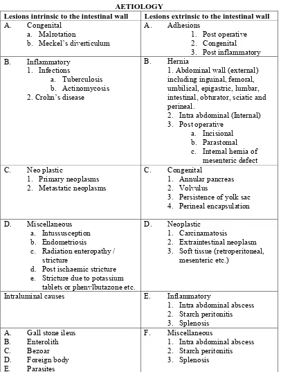
AETIOLOGY
Lesions intrinsic to the intestinal wall Lesions extrinsic to the intestinal wall
A. Congenital a. Malrotation
b. Meckel’s diverticulum
A. Adhesions
1. Post operative 2. Congenital
3. Post inflammatory B. Inflammatory
1. Infections
a. Tuberculosis b. Actinomycosis 2. Crohn’s disease
B. Hernia
1. Abdominal wall (external) including inguinal, femoral, umbilical, epigastric, lumbar,
intestinal, obturator, sciatic and perineal.
2. Intra abdominal (Internal) 3. Post operative
a. Incisional b. Parastomal c. Internal hernia of
mesenteric defect C. Neo plastic
1. Primary neoplasms 2. Metastatic neoplasms
C. Congenital 1. Annular pancreas 2. Volvulus
3. Persistence of yolk sac 4. Perineal encapsulation
D. Miscellaneous a. Intussusception b. Endometriosis
c. Radiation enteropathy / stricture
[image:57.612.105.511.65.602.2]d. Post ischaemic stricture e. Stricture due to potassium
tablets or phenvlbutazone etc.
D. Neoplastic 1. Carcinamatosis
2. Extraintestinal neoplasm 3. Soft tissue (retroperitoneal,
mesenteric etc.)
Intraluminal causes E. Inflammatory
1. Intra abdominal abscess 2. Starch peritonitis 3. Splenosis
A. Gall stone ileus B. Enterolith C. Bezoar D. Foreign body E. Parasites
F. Miscellaneous
CAUSES OF ADYNAMIC OBSTRUCTION
1. Reflux Inhibition of Intestinal Motility
due to increased sympathetic discharge with hyperpolarisation of smooth muscle cells which becomes non responsive to both neural and hormonal stimulation.
a. Abdominal operations b. Pneumonia
c. Crush injuries d. Fracture spine
e. Retroperitoneal haemorrhage or exudates f. Hyperextension of spine (plaster jacket) etc. 2. Meatabolic abnormalities
a. Hypokalemia b. Ureaemia
c. Various ketoacidosis d. Hyponatremia 3. Intra peritoneal sepsis
due to both reflux inhibition of intestinal motility and direct effect of bacterial toxins on the myenteric nerve plexuses.
4. Mesenteric vascular disease
a. Arterial embolism b. Arterial thrombosis c. Venous thrombosis d. Various vasculitis
e. Low flow state due to decreased cardiac output and reflex mesenteric vasoconstriction (non occlusive vascular insufficiency)
5. Drugs
DIFFERENTIATION BETWEEN VIABLE AND NONVIABLE INTESTINE
Intestine Viable Non viable
Circulation Dark colour becomes lighter; mesentery bleeds if pricked.
Dark colour remains ; No bleeding if mesentery is pricked.
Peritoneum Shiny Dull and Lusterless
Musculature Firm pressure rings may or may not disappear
Flabby, thin and friable. pressure rings persist. Peristalsis May be present No peristalsis.
DIAGNOSIS OF LEVEL OF OBSTRUCTION
Level of Obstructi
on
Onset Pain Dehydrati
on Disten tion Radiograp hic finding 1. High small Bowel Sudden Mostly upper abdomin al; variable
Extreme Absent May show
gasless abdomen or distention of duodenum r proximal jejunal loop. 2. Low small bowel More
gradual Central abdomin al severe colicky
Less
marked Moderate; central in position small loops of small bowel in supine film as lying transversely ; Fluid levels in erect film. 3. Large
bowel Usually insidio us Central or lower abdomen ; Colicky or generaliz ed discomfo rt due to
Slight Progressive; Extreme in late stage Mostly peripheral except volvulus
Gas seen in the colon mainly
proximal to the
distensio
n enema
`
APPROXIMATE ELECTROLYTE CONCENTRATIONS OF GIT27
Site Na+ K+ Cl– HCO3
Saliva 50 15 30 40
Stomach 55 15 115 10
Bile 145 5.2 90 35
Pancreas 141 4.6 77 90
Jejunum 120 5.0 100 –
Ileum 117 5.0 106 25
MECHANICAL – INTESTINAL OBSTRUCTION CLASSIFICATION
ETILOGY OF MECHANICAL – INTESTINAL OBSTRUCTION IN ADULTS
LESIONS INTRINSIC TO THE INTESTINAL WALL
LESIONS EXTRINSIC TO THE INTESTINAL WALL CONTINUED.
A.Congential
1. Malrotation 2. Meckel’s
diverticulum
2. Intra-abdominal (internal)
a. Congenital, including paraduodenal, foramen of Winslow, diaphragmatic, measentric defect, paracecal, intersigmoid, broad ligament. B. Inflammatory
1. Infections a. Tuberculosis b. Diverticulitis 2. Crohn’s disease
3. Eosinophilic granuloma
3. Postoperative a. Incisional b. Parastomal
c. Wound dehiscence
d. Internal hernia of mesenteric defects
4. Acquired C. Neoplastic
1. Primary neoplasma a. Benign b. Maligant 2. Metastatic neoplasms
C. Congenital
1. Annular pancreas 2. Volvulus
3. Persistence of yolk sac 4. Peritoneal encapsulation
D. Traumatic 1. Hematoma 2. Ischemic stricture
D. Neoplastic
1. Carcinomatosis
2. Extraintestinal neoplasm 3. Soft tissue recurrence (eg. Retroperitoneal, mesenteric) E. Miscellaneous
1. Intussusception 2. Endometriosis 3. Radiation stricture 4. Retroperitoneal fibrosis 5. Postischemic stricture
[image:61.612.111.510.239.726.2]6. Stricture due to potassium tablets or phenylbutazone 7. Intramural hematoma in
patients on oral
E. Inflammatory
1. Intra-abdominal abscess 2. Starch periotoritis 3. Splenosis
F. Miscellaneous
1. Superior mesenteric artery syndrome (Wilky’s syndrome).
anticoagulants 3. Sclerosing peritonitis due to practolol
LESIONS EXTRINIC TO THE INTESTINAL WALL
A. Adhesions
Postoperative (commonest ) Congenital Bands Postinflammatory B. Hernia
Abdominal wall (external), including inguinal, femoral, umbilical, ventral, epigastric, lumbar, interstitial, obturator, sciatic, speigilean and perineal
INTRA LUMINAL OBSTRUCTION
A. Gallstone illus B. Enterolith C. Bezoar D. Foreign body
E.Balloons of intestinal tubes F. Parasites including Ascaris and tapewarm
G. Cholestyramine
H. Intraluminal diverticulum
BIBLIOGRAPHY
1. Maingot’s Abdominal operations, 10th edition.
2. Bailey and love-short practice of surgery 24th edition.
3. Essential surgical practice – SA Cuschieri 4th edition.
4. Schwartz’s principles of surgery – 8th edition, 2005.
5. Hamilton Bailey’s emergency surgery.
6. Surgery of the colon and rectum – R.John Nicholes.
7. Lee Mc Gregor’s Synopsis of surgical anatomy – 12th edition.
8. R.J. Last – Anatomy- Regional and Applied – 10th edition.
9. Sabiston Text Book of Surgery – 16th edition.
10. Shackelford’s surgery of the Alimentary tract- 5th edition.
11. CSDT – 11th edition.
12. Mastery of surgery Nyhus– 4th edition.
14. ACS surgery principles and practice –2005.
15. Skandalakis surgical anatomy- 2004.
16. Surgery of the Anus, Rectum & Colon – 1999, 2nd Edition, Michael