FOUR COMMERCIALLY AVAILABLE
SELF-LIGATING BRACKETS
–A FINITE ELEMENT STUDY
Dissertation submitted to
THE TAMILNADU DR. M.G.R.MEDICAL UNIVERSITY
In partial fulfillment for the degree of
MASTER OF DENTAL SURGERY
BRANCH V
ORTHODONTICS AND DENTOFACIAL
ORTHOPAEDICS
I would like to take this opportunity to express my gratitude to
everyone who has helped me through this journey.
I would like to start with my very respected and beloved professor,
Dr. N.R. KRISHNASWAMY, M.D.S., M.Ortho RCS. (Edin), Diplomat of
Indian board of Orthodontics, Professor and Head, Department of
Orthodontics, Ragas Dental College and Hospital, Chennai. I consider myself
extremely lucky to have had the opportunity to study under him. He has
always been a source of inspiration to perform better not only in academics
but also in life. I would like to thank him for having taken interest in my
study and providing his valuable insight.
I am privileged to express my extreme gratefulness to my respected
Professor Dr. S. VENKATESWARAN, M.D.S., D.N.B. (Ortho), for being
a constant source of support and supervision, which stimulated, enthused
and encouraged me in the preparation of this main dissertation.
postgraduate teacher and guide Dr. SHAHUL HAMEED FAZEE MDS
(Associate Professor) Department of Orthodontics, Ragas Dental College and
Hospital, Chennai, for his valuable guidance and suggestions, tireless pursuit
for perfection, immense and constant support, encouragement and keen
surveillance for the minute details throughout this dissertation. I thank him
for all the help that have been conferred upon me without which this
dissertation would not have come true.
My sincere thanks to Prof. Mr. KANAGARAJ, Chairman &
Dr. RAMACHANDRAN, Principal, Ragas Dental College for providing me
with an opportunity to utilize the facilities available in this institution in
order to conduct this study.
I would also like to acknowledge Dr. ANAND (Reader),
Dr. JAYAKUMAR (Reader), Dr. SHAKEEL (Reader ), Dr. REKHA
(Reader ), Dr. RAJAN(Sr. Lecturer), Dr. SHOBANA (Sr. Lecturer),
Dr. PRABHU (Sr. Lecturer) and Dr. BIJU (Sr. Lecturer) for their support,
Shakthi, Dr. Siva subramanium, Dr. Vijay, Dr. Aarthi, Dr. Ashwin, Dr.
Ravanth, Dr. Deepak , Dr. Vishal, Dr. Vikram, Dr. Gayathri, Dr. Regina,
Dr. Manali, Dr. Murali, Dr. Saptharishi , and Dr. Femin for all their
support and for cooperating with me to conduct this study.
I am extremely thankful to Versetia Technologies for conducting the
finite element analysis & Mr. Arun Raj of Primus Design for helping me
with the modeling of the brackets for the study.
My thanks to
Mr.Ashok,
and Mr.
Rajendran
for helping me with the
photographs for the study.
I would like to thank Mrs.Marina, Sister
Lakshmi
,
Sister
Rathi,
Sister
Kanaka,
Ms.Haseena, Mr. Mani, Mr. Bhaskar, Ms. Divya, Ms.Banu,
support and an eternal source of energy in every endeavour of mine. She has
been there with me through the most challenging times and helped me
complete the study as her own. Without her support and love, this course and
this study would have just been a dream.
S.NO TITLE PAGE NO.
1. INTRODUCTION 1
2. REVIEW OF LITERATURE 5
3. MATERIALS AND METHODS 41
4. RESULTS 47
5. DISCUSSION 49
6. SUMMARY AND CONCLUSION 60
7. BIBLIOGRAPHY 62
1
INTRODUCTION
The term “torque” has two different but related meanings for the
orthodontist. On one hand it refers to the bucco-palatal root inclination, which
can be measured on the lateral headfilm as the incisor inclination to the
anterior cranial base or the maxillary plane, while on the other it describes the
activation generated by torsion of the archwire in the bracket slot.88
Correct buccolingual inclination of anterior teeth is considered
essential for providing good occlusal relationships in orthodontic treatment.
Inclination of the maxillary anterior teeth is particularly critical in establishing
an esthetic smile line, proper anterior guidance, and a Class I canine and molar
relationship5.
Orthodontist’s define torque around the dental arch such that the x-axis
follows the curve of the arch. Torque, in this sense, would be rotation
perpendicular to the long axis of the tooth. This could be generated by a
rotation through a moment or couple of forces.4
The completely programmed bracket system created by
Andrews (1989)3, was designed with the objective of using arches without bends. However, in spite of incorporating ideal torque characteristics in the
structure of such brackets, in some cases it is necessary to apply additional or
individual torque on some teeth. This would be necessary due to several
factors38: mechanical side-effects such as variations in bracket slot and archwire dimension113, morphological differences in the buccal faces of
teeth,20, 74, 75,111 changes in the position of the brackets, 71,119 different methods
2
wire and the bracket slot13,26, variations in the bracket designs28, properties of
the materials constituting the brackets39,44,92 and wires94 and differences
between the value of the torque informed by the manufacturer and the real
value of the torque in the bracket base38.
Self-ligating brackets introduced by Dr. Jacob Stolzenberg (1935)98
are ligature-less bracket systems that have a mechanical device built into the
bracket to close off the edgewise slot. They are generally smoother for the
patients because of the absence of wire ligature and also do not require as
much chair time.9,11,41 The precision arm or the sliding fourth wall accurately
locks the archwire within the dimensions of the slot providing robust ligation
and controlled tooth movement.
The proclaimed chief advantages of self-ligating systems over
conventional appliances include, (a) decrease in treatment duration,57,89, (b)
anchorage conservation,109 (c) asepsis,28 (d) patient comfort.33,36,97
Self-ligating brackets are broadly classified into Active and Passive
self-ligating brackets;
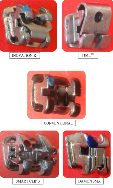
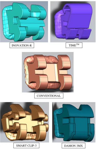
1) Active systems - those that have a spring clip that presses against
the archwire, such as the InOvation-R (GAC Intl, NY), TimeTM
(American Orthodontics, USA )
2) Passive systems-those in which the self-ligating clip does not press
against the wire such as Damon 3MX (Ormc, USA), SmartClip-3
3
The finite element method (FEM) is a powerful computer simulation
tool, which has been successfully applied to the mechanical study of stress and
strain and solving problems in the mechanics of solids and structures.44,53 This
makes it practical to elucidate the biomechanical components such as
displacements, stress and strain included in the living structures from various
external forces.
In the finite element method, the entire region of the structure is
divided into a set of elements that are connected by points called nodes.105
Element types are decided and each element is assigned its material properties
(Young’s Modulus and Poison’s Ratio). The forces and boundary conditions
are defined to stimulate loads and constraint of the structures. The structural
response is computed and then presented for display.
The FEM has some distinct advantages over other methods of stress analysis.
1) Compared to classical analytical methods, it is able to model much
more closely structures of irregular geometries and non-homogeneous
or anisotropic material properties and overcomes difficulties inherent
in conventional experimental methods.46
2) FEM has the potential for the equivalent mathematic modeling of a
real object of complicated shape with different material properties.
Thus FEM offers an ideal method of accurate modeling of
tooth-periodontium system with its complicated 3 Dimensional geometry.119
3) The force systems that are used in an orthodontic patient can be
complicated, FEM makes it possible to analytically apply various force
4
Clinically, torque control is often required in the maxillary incisors for an
ideal inter-incisal angle, adequate incisor contact, and sagittal adjustment of
the dentition in order to achieve an ideal occlusion.5
Although the self-ligating edgewise bracket was introduced to
orthodontists 75 years ago, recent advances in bracket technology have
resulted in a number of new self-ligating bracket systems and greater interest
in their use. Much of this interest is in response to information comparing the
benefits of self-ligating systems with conventional edgewise brackets. Often,
this information comes from marketing materials and non-refereed sources
claiming that self-ligating bracket systems provide superior treatment
efficiency and efficacy.22
Because of the complexity of the experimental configuration, only a
handful of experimental studies have been presented upon torque expression
until now, moreover numerical analyses have not been carried out for torque
expression in different self-ligating brackets on the tooth and its supporting
structures.1,24,35,43
Therefore the aim of the present study was to investigate the torque
expression of different self-ligating brackets (active and passive) with
various archwire combinations on the tooth and its supporting structures
5
REVIEW OF LITERATURE
Torque can be defined from a mechanical or from a clinical point of view.
Mechanically, it refers to the twisting of a structure about its longitudinal axis,
resulting in an angle of twist. Torque is a shear-based moment that causes
rotation. Clinically, in orthodontics, it represents the buccopalatal crown/root
inclination of a tooth, and it is an orthodontic adaptation used to describe rotation
around an x-axis. When applied in an orthodontic archwire/bracket interaction, it
describes the activation generated by twisting an archwire in a bracket slot2.
Depending on magnitude of torsion, the stiffness or resilience of the wire
cross section, wire size, edge bevel and manufacturer tolerance, bracket slot size
and manufacturer tolerance, engagement angle of the wire in the bracket slot,
experimental measurement technique, bracket placement as related to tooth
morphology8,29 and inclination of the tooth, the archwire moves the root of a tooth
through the alveolar bone via localized pressure and tension generated by torsion
in the archwire5.
Considering the above factors the review of literature for this study is
categorized into two groups:-
1) FEM studies in orthodontics and,
6 1) FEM STUDIES IN ORTHODONTICS
Tanne et al (1987)100 investigated the stress levels induced in the periodontal tissue by orthodontic forces using the three-dimensional finite
element method and concluded that during tipping movement, stresses
non-uniformly varied with a large difference from the cervix to the apex of the root.
Tanne et al (1988)104 investigated the relationship between moment to force (M/F) ratios and the centers of rotation by use of the finite element method
(FEM). They concluded that the center of resistance was located at 0.24 times the
root length measured apical to the level of alveolar crest. The centers of rotation
varied with the M/F ratios following a curve of hyperbola. The M/F ratio was
-9.53 for root movement (C, at the incisal edge), - 8.39 for translation, and -6.52
for tipping around the apex. It was found that even a small difference in the M/F
ratios produced clinically significant changes in the centers of rotation.
Tanne et al (1991)106 investigated the nature of initial tooth displacements associated with varying root lengths and alveolar bone heights. The results
showed that moment-to-force values at the bracket level for translation of a tooth
decreased with shorter root length and increased with lower alveolar bone height.
In addition, apico-gingival levels of the center of resistance shifted more
gingivally to the cervix, or the alveolar crest with a shorter root. However, the
relative distances of the centers of rotation from the alveolar crest in comparison
with the alveolar bone heights were constant at 0.4 mm, with variations in the root
7
alveolar bone height affect the patterns of initial tooth displacements both in the
center of resistance and the centers of rotation and also in the amount of
displacement, forces applied during orthodontic treatment should take into
consideration the anatomic variations in the root length and alveolar bone height
so as to produce optimal and desired tooth movement.
McGuinness et al (1992)69 conducted a finite element analysis (FEA) to determine the stress induced in the periodontal ligament in 3 dimensions when a
maxillary canine tooth is subjected to and orthodontic force similar to that
produced by an edgewise appliance. The findings suggested that even with the
perfect edgewise mechanics it would be difficult to obtain canine movement by
pure translation or bodily movement.
Cobo et al (1993)19 determined the stress that appears in tooth, periodontal ligament and alveolar bone, when a labiolingual force of 100 gm is
applied in a labiolingual direction in a midpoint of the crown of an inferior
digitalized canine, and its changes depending on the degree of loss of the
supporting bone. After applying the labiolingual force in the canine, a progressive
increase of the stress in the labial and lingual zones of the tooth, periodontal
membrane and alveolar bone was observed when the alveolar bone was reducing.
In the mesial and distal zones, no compensating forces appeared which could
provoke a tooth rotation during the tipping movements.
Katona et al (1994)56 developed a finite element model (FEM) of an orthodontic bracket bonded to enamel with GIC. The primary purpose of this
8
within the cement layer. The results indicated that peak stress values increase as
the load deflection angulation increases. If the tensile load is inadvertently applied
entirely on one wing of the bracket, the stress components nearly doubled in
magnitude.
Ghosh et al (1995)37 generated finite element models for selected ceramic brackets and graphically displayed the stress distribution in the brackets when
subjected to arch wire torsion and tipping forces. Six commercially available
ceramic brackets, one monocrystalline and five polycrystalline alumina, of twin
bracket design for the permanent maxillary left central incisor were studied.
Three-dimensional computer models of the brackets were constructed and loading
forces, similar to those applied by a full-size (0.0215 × 0.028 inch) stainless steel
arch wire in torsion and tipping necessary to fracture ceramic brackets, were
applied to the models. The brackets with an isthmus connecting the wings seemed
to resist stresses better than the one bracket that did not have this feature. The
design of the isthmus for the Transcend (Unitek/3M, Monrovia, Calif.) and
Lumina (Ormco, Glendora, Calif.) brackets were found to be acceptable as well.
The Starfire bracket ("A" Company, San Diego, Calif.) showed high stresses and
irregular stress distribution, because it had sharp angles, no rounded corners, and
no isthmus. The finite element method proved to be a useful tool in the stress
analysis of ceramic orthodontic brackets subjected to various forces. This analysis
9
Cobo et al (1996)18 studied the stress that appears in the tooth, the periodontal ligament, and the alveolar bone, when a couple and horizontal forces
were applied to obtain the bodily movement of a lower digitalized canine and its
changes depending on the degree of loss of the supporting bone. The analysis of
tensions was carried out by means of the finite element method (FEM) with no
bone loss and after reducing the support bone 2, 4, 6, and 8 mm. When the bone
loss is 2 mm, an increased stress in the levels next to the alveolar crest is already
apparent. After 4, 6, and 8 mm of bone support reduction, a change of the sign
and an increment of the magnitude of stress in the lowest levels occurs.
Middleton et al (1996)73 reported an initial time-dependent (continuous/dynamic) finite element model for tooth movement that uses newly
developed software, the results being cross-referenced against historical data.
These early results, from a two-dimensional mathematical model of a loaded
canine tooth, suggest that the remodeling process may be controlled by the
periodontal ligament rather than the bone. In the finite element model, bone was
found to experience a low strain of 1 × 10⁻⁵, whereas the periodontal ligament
experienced a strain of 0.1 when the "tooth model" is loaded. Only this latter
figure is above the threshold usually reported to be necessary to initiate the
remodeling process.
Puente et al (1996)86 analyzed the distribution of the stress on dental and periodontal structures when a simple tipping dental movement or torque
movement is produced. A tridimensional computer model based on finite element
10
constructed on the average anatomical morphology and 396 isoparametric
elements were considered. The three principal stresses (maximum, minimum and
intermediate) and Von Mises stress were determined at the root, alveolar bone and
periodontal ligament (PDL). It was observed how the distribution of stress is not
the same for the three structures studied. In all loading cases for bucco-lingually
directed forces, the three principal stresses were very similar in the PDL. The
dental apex and bony alveolar crest zones are the areas that suffer the greatest
stress when these kind of movements are produced.
Raboud et al (1997)87 conducted a numerical method to provide quantitative insight into three dimensional effects for typical appliance designs.
Concluded, that the out-of-plane effects are independent of the in-plane behavior
so that the usual forces and moment to force ratios are maintained.
Jeon et al (1999)52 simulated the stress response in the periodontium of the maxillary first molar to different moment to force ratios, and to determine the
moment to force ratio for translational movement of the tooth by means of the
finite element method. Their results demonstrated the sensitivity of the
periodontium to load changes. The stress pattern in the periodontal ligament for a
distalizing force without counterbalancing moments showed high concentration at
the cervical level of the distobuccal root due to tipping and rotation of the tooth.
After various counter rotation as well as counter tipping moments were applied,
an even distribution of low compression on the distal side of the periodontal
ligament was obtained at a counter tipping moment to force ratio of 9:1 and a
11
concentration was observed on the root surface at the furcation level in contrast
with anterior teeth reported to display high concentration at the apex. This result
may suggest that the root morphology of the maxillary first molar makes it less
susceptible to apical root resorption relative to anterior teeth during tooth
movement.
Thomas et al (1999)107 reported that the tests commonly used for the evaluation of orthodontic adhesives measure tensile and shear bond strength. The
two methods were compared with finite element analysis using a
three-dimensional model and the effect of misalignment of the tensile and shear forces
were calculated. Applying a shear load produced significant compressive and
tensile stresses in the adhesive layer. Under ideal conditions of shear loading, the
induced tensile stress is over 5 times the induced shear stress. The model showed
that a tensile load induces predominantly tensile stresses in the adhesive layer.
The calculations indicate that the tensile test method is a robust testing method
with low sensitivity to misalignment of the applied load.
Geramy (2000)33 studied the behavior of initial tooth displacements associated with alveolar bone loss situations when loaded by a force of 1 N. The
results revealed that the moment/force ratio (at the bracket level) required for
producing bodily movement increases in association with alveolar bone loss.
Bone loss causes center of resistance movement toward the apex, but its relative
distance to the alveolar crest decreases at the same time. Center of rotation of the
tipping movement also shifted toward the cervical line. Among the many
12
the presence of alveolar bone loss in the adult cases. Alveolar bone loss causes
center of resistance changes as a result of the alterations in bone support. This
necessitates modifications in the applied force system to produce the same
movement as in a tooth with a healthy supporting structure.
Jeon et al (2001)53 studied the use of finite element method to simulate the effect of alveolar bone loss on orthodontically induced stress in the
periodontal ligament of the maxillary first molar. An anterior force of 300 g was
applied at the center of the buccal crown surfaces of teeth with normal bone
height and with bone loss that ranged from 2.0 to 6.0 mm. The results showed that
force magnitude required lowering from 80% (2-mm bone loss) and gradually to
37% (6-mm bone loss) of the initial load applied to the tooth without bone loss.
The counter tipping moment (gram-millimeters) to force (gram) ratio should
increase from 9 (no bone loss) to nearly 13 (6-mm bone loss) to maintain the
same range of stress in the periodontal ligament as was obtained without bone
loss. A linear relationship was observed between the amount of bone loss, the
desired reduction in force magnitude, and the increase in M/F ratio. The results of
this study indicate that a combination of force reduction and increased M/F ratio
is required to achieve uniform stress in the periodontal ligament of a tooth with
bone loss.
Knox et al (2001)58 evaluated the influence of bracket base mesh geometry on the stresses generated in the bracket-cement-tooth continuum by a
shear/peel load case. When the double-mesh bracket base was considered, the
13
superficial (coarse) mesh layer and an increase in the stresses recorded in the
deepest (fine mesh) layer when compared with the single-layer designs in
isolation. Modification of single-mesh spacing and wire diameter influences the
magnitude and distribution of stresses within the bracket-cement-tooth
continuum. The use of a double mesh design results in a reduction in the stresses
recorded in the most superficial mesh. Mesh design influenced stress distribution
in this study, primarily by determining the flexibility of the bracket base.
Rudolph et al (2001)91 conducted a study to determine the types of orthodontic forces that cause high stress at the root apex. The material properties
of enamel, dentin, PDL, and bone and 5 different load systems (tipping, intrusion,
extrusion, bodily movement, and rotational force) were tested. The finite element
analysis showed that purely intrusive, extrusive, and rotational forces had stresses
concentrated at the apex of the root. The principal stress from a tipping force was
located at the alveolar crest. For bodily movement, stress was distributed
throughout the PDL; however, it was concentrated more at the alveolar crest.
They conclude that intrusive, extrusive, and rotational forces produce more stress
at the apex. Bodily movement and tipping forces concentrate forces at the alveolar
crest, not at the apex.
Melsen (2001)72 studied the tissue reaction to a force system generating translation of premolars and molars in the five Macaca fascicularis monkeys is
described. Three force levels, 100, 200, and 300 cN were applied for a period of
11 weeks. Based on these results and a finite element model simulating the
14
distribution generated by orthodontic forces is suggested. The direct resorption
could be perceived as a result of lowering of the normal strain from the
functioning periodontal ligament (PDL) and as such as a start of remodelling, in
the bone biological sense of the word. Indirect remodelling could be perceived as
sterile inflammation attempting to remove ischaemic bone under the hyalinised
tissue. At a distance from the alveolus, dense woven bone was observed as a sign
of a regional acceleratory phenomena (RAP). The results of the intrusion could,
according to the new hypothesis, be perceived as bending of the alveolar wall
produced by the pull from Sharpey’s fibres.
Schneider et al (2002)93 studied the optimal force system for bodily movement of a single-root tooth with an orthodontic bracket attached. This was
achieved by the use of the numerical finite element method, including a distinct
mechanical bone-remodeling algorithm. This algorithm works with equilibrium
iterations separated in 2 calculation steps. Furthermore, a parametric 3-
dimensional finite element model, which allows modifications in the root length
and its diameter, is described. For different geometries, the ideal
moment-by-force ratios that induce a bodily movement were determined. The knowledge of
root geometry is important in defining an optimal force system.
Geramy (2002)34 investigated the stress components (S1 and S3) that appear in the periodontal membrane (PDM), when subjected to transverse and
vertical loads equal to 1 N. A further aim was to quantify the alteration in stress
that occurs as alveolar bone is reduced in height by 1, 2.5, 5, 6.5, and 8 mm,
15
maxillary central incisor were designed. The models were of the same
configuration except for the alveolar bone height. The results showed that alveolar
bone loss caused increased stress production under the same load compared with
healthy bone support (without alveolar bone resorption). Tipping movements
resulted in an increased level of stress at the cervical margin of the PDM in all
sampling points and at all stages of alveolar bone loss. These increased stress
components were found to be at the sub-apical and apical levels for intrusive
movement.
Kang et al (2003)55 analyzed the relationship between the critical contact angle and the torque angle in an orthodontic bracket and archwire assembly in 3
dimensions. Three-dimensional mathematical models were created with geometric
bracket-archwire parameters that included 2 slot sizes, 3 bracket widths, and 3 to
4 wire sizes. From this, 3-dimensional mathematical equations (3DMEs) for the
critical contact angle and the maximum torque that result in critical contact angles
of 0º were derived and calculated. For all bracket-archwire combinations, the
critical contact angle decreased as bracket width, torque angle, and wire size
increased. Therefore, all bracket-archwire parameters except slot height had an
effect on the critical contact angle. In addition, the effect of a beveled edge was
investigated in some archwires. The results of this study provide theoretic and
experimental bases for clinical orthodontic practice and indicate that torque angles
16
Toms et al (2003)108 sought to determine the importance of using nonlinear mechanical properties and non-uniform geometric data in computer
predictions of periodontal ligament stresses and tooth movements. Predictions of
the maximum and minimum principal stresses and von Mises stresses in the PDL
were determined for extrusive and tipping forces. The results indicated that
biofidelic finite element models predicted substantially different stresses in the
PDL for extrusive loading than did the uniform thickness model, suggesting that
incorporation of the hourglass shape of the PDL is warranted. In addition,
incorporation of nonlinear mechanical properties for the PDL resulted in dramatic
increases in the stresses at the apex and cervical margin as compared with the
linear models.
Cattaneo et al (2003)14 conducted a FEA that allowed them to simulate the displacement of a molar in relation to the well-defined morphology of the
maxilla. When the molar was loaded with occlusal forces, the stresses were
transferred predominantly through the infrazygomatic crest. This changed when
mesial and distal displacements of the molars were simulated. In the model with
mesial molar displacement, a larger part of the bite forces were transferred
through the anterior part of the maxilla, resulting in the buccal bone being loaded
in compression. In the model with distal molar displacement, the posterior part of
the maxilla was deformed through compression; this resulted in higher
compensatory tensile stresses in the anterior part of the maxilla and at the
zygomatic arch. This distribution of the occlusal forces might contribute to the
17
Kojimaa et al (2005)59 discussed a method that allowed the simulation of more complex tooth movements. A 3-dimensional finite element method was
used to simulate the orthodontic tooth movement (retraction) of a maxillary
canine by sliding mechanics and any associated movement of the anchor teeth.
Absorption and apposition of the alveolar bone were produced in proportion to the
stress of the periodontal ligament. The canine tipped during the initial unsteady
state and then moved bodily during the steady state. It became upright when the
orthodontic force was removed. The anchor teeth moved in the steady state and
tipped in the mesial direction. The decrease in applied force by friction was about
70%. The tipping of the canine decreased when the wire size was increased or
when the applied force was decreased. They suggested that this method might
enable one to estimate various tooth movements clinically.
Ziegler et al (2005)120 studied the elastic properties of the periodontal ligament (PDL) in eight multi-rooted teeth were examined in a combined
experimental and numerical study in six minipigs. The initial tooth movement of
the mandibular primary molars surrounded by the periodontium was registered
three-dimensionally (3D) in an optomechanical measuring system. The
dissections were then embedded in resin and cut in transverse sections. Based on
these sections, 3D finite element (FE) models were constructed and numerically
loaded with the same force systems as used in the experiment. There was no
significant difference in the material parameters determined for specimens with
18
parameters of the PDL, determined in previous investigations of single-rooted
human and pig teeth.
Kojimaa et al (2006)60 developed a comprehensive mechanical, 3-dimensional, numerical model for predicting tooth movement. Tooth movements
produced by wire bending were simulated numerically. The teeth moved as a
result of bone remodeling, which occurs in proportion to stress in the periodontal
ligament. With an off-center bend, a tooth near the bending position was
subjected to a large moment and tipped more noticeably than the other teeth.
Also, a tooth far from the bending position moved slightly in the mesial or the
distal direction. With the center V-bend, when the second molar was added as an
anchor tooth, the tipping angle and the intrusion of the canine increased, and
movement of the first molar was prevented. When a wire with an inverse curve of
spee was placed in the mandibular arch, the calculated tendency of vertical tooth
movements was the same as the measured result. In these tooth movements, the
initial force system changed as the teeth moved. Tooth movement was influenced
by the size of the root surface area. Concluded, that tooth movements produced by
wire bending could be estimated.
Kojima et al (2006)61 studied the combined effect of friction and an archwire’s flexural rigidity on canine movement in sliding mechanics, and to
explain how to select a suitable archwire and force level for efficient bodily
movement. As the frictional force decreased, both the net force acting on and the
moving speed of the canine increased. The elastic deformation of the archwire
19
to tipping, although there was no clearance between the archwire and the bracket
slot. When a light wire was used, wire deformation increased, and the canine
experienced greater tipping.
Jayade et al (2007)51 evaluated the magnitudes of initial and subsequent sequential deactivational third order moments generated in rectangular twisted
archwires in order to judge their biologic acceptability. A finite element study was
carried out with the MSC Patran/Nastran interface. Required twists were applied
at the appropriate locations to derive the applied and reactionary moments both
initially and during the time needed for complete deactivation. The results
indicated that a round-tripping possibility does exist in certain clinical procedures.
Furthermore, the moments produced could be quite high, thereby enhancing the
possibility of root resorption. They concluded twists in rectangular archwires may
be used only when reciprocal torque is needed on adjacent teeth. In other
situations, alternative torquing methods should be considered.
Hohmanna et al (2007)45 evaluated the risk of root resorption, individual finite element models (FEMs) of extracted human maxillary first premolars were
created, and the distribution of the hydrostatic pressure in the periodontal
ligament (PDL) of these models was simulated. The results of clinical
examination and simulations were compared using the identical roots of the teeth.
The regions that showed increased hydrostatic pressure correlated well with the
locations of root resorption for each tooth. Increased torque resulted in increased
20
with the experiments. Thus, concluded if hydrostatic pressure exceeds typical
human capillary blood pressure in the PDL, the risk of root resorption increases.
Reimann et al (2007)90 investigated the combined Centre of Resistance (CR) of the upper four incisors numerically using finite-element (FE) method. In
the FE system, the model of the anterior segment was loaded with torques of 10
Nmm each at the lateral incisors. The FE model indicated that the individual
incisors moved independently, although they were blocked with a steel wire of
dimension 0.46 × 0.65 mm2. The individual CRs were located at 5 mm distal and
9 and 12 mm apical to the centre of the lateral brackets. Thus, the classical view
of a combined CR for the anterior segment was disproved and the planning of
orthodontic tooth movements of the upper incisors should no longer be based on
that concept.
Ulusoya et al (2008)110 evaluated the effects of the Class II activator and the Class II activator high-pull headgear (HG) combination on the mandible with
3-dimensional (3D) finite element stress analysis. To investigate the effects of the
Class II activator, a 3D model of the lower part of this appliance was constructed
and fixed on the mandibular model. The Class II activator high-pull headgear
model was established as described, and an extraoral traction force of 350 g was
directed from the middle of the Class II activator to the top of the mandibular
condyle. The stress regions were studied with the finite element method. The
regions near the muscle attachment areas were affected the most. The inner part of
21
Therefore, both functional appliances can cause morphologic changes on the
mandible by activating the masticatory muscles to change the growth direction.
Cattaneo et al (2008)15 demonstrated by FE analyses that the influence of the material properties of the PDL on the type of tooth movement. Moreover, the
influence of the applied force level on the type of tooth movement, with a fixed
M/F ratio, was evaluated and the results interpreted in the light of existing
prescriptions for orthodontic tooth movement. By applying a range of values of
M/F, different types of tooth movement were generated, although the classic
prescription of the M/F ratio suggested in the literature could not be confirmed.
Due to the nonlinear behavior of the periodontal ligament, loading modes with a
constant M/F ratio, yet varying the force magnitude, resulted in different types of
tooth movement. Therefore, the material properties of the periodontal ligament,
the morphology of the root, and the alveolar bone are patient specific. Therefore,
the M/F values generally advocated to obtain orthodontic tooth movement should
be used only as guidelines. To be effective and accurate, the force system selected
for a specific tooth movement must be monitored and the outcome compared with
the predicted tooth movement.
Holberg et al (2008)46 analyzed the strains induced in the sutures of the midface and the cranial base by headgear therapy involving orthopedic forces. A
finite element model of the viscerocranium and the neurocranium was used. The
magnitude and the distribution of the measured strains depended on the level and
the direction of the acting force. Overall, the strain values measured at the sutures
22
load of 5 N per side were usually just below 20µ strain irrespective of the force
direction. A characteristic distribution of strain values appeared on the anatomical
structures of the midface and the cranial base for each vector direction. The
measurements based on the finite element method provided a good overview of
the approximate magnitudes of sutural strains with orthopedic headgear therapy.
The signal arriving in the sutures is apparently well below threshold, since the
maximum measured strains in most sutures were about 100 fold lower than the
minimal effective strain. A skeletal effect of the orthopedic headgear due to a
mechanical effect on sutural growth cannot be confirmed from these results. They
concluded that the good clinical efficacy of headgear therapy with orthopedic
forces is apparently based mainly on dentoalveolar effects, whereas the skeletal
effect due to inhibition of sutural growth is somewhat questionable.
Provatidis et al (2008)85 did a finite element model (FEM) of a dry human skull with the RME appliance cemented in place in order to evaluate these
effects on the overall craniofacial complex with different suture ossification. The
behaviour of the FEM was compared with the findings of a clinical study and to
an in vitro experiment of the same dry skull. It was found that the
maxillolacrymal, the frontomaxillary, the nasomaxillary, the transverse midpalatal
sutures, and the suture between the maxilla and pterygoid process of the sphenoid
bone did not influence the outcome of RME, while the zygomatico-maxillary
suture influenced the response of the craniofacial complex to the expansion
23
midpalatal suture plays an important role in the degree and manner of maxillary
separation.
Gautam et al (2009)32 evaluated biomechanically the displacement patterns of the facial bones in response to different headgear loading by using a
higher-resolution finite element method model than used in previous studies.
Different headgear forces were simulated by applying 1 kg of posteriorly directed
force in the first molar region to simulate cervical-pull, straight-pull, and
high-pull headgear. The distal displacement of the maxilla was the greatest with the
straight-pull headgear followed by the cervical-pull headgear. The high-pull
headgear had better control in the vertical dimensions. The center of rotation
varied with the direction of headgear forces for both the maxilla and the
zygomatic complex. A potential for chondrogenic and osteogenic modeling exists
for the articular fossa and the articular eminence with headgear loading.
Wei et al (2009)115 conducted a study to provide the lingual technique with valuable information by using a 3-dimensional (3D) finite element method
(FEM). Horizontal retraction force, vertical intrusive force, and lingual root
torque were applied to simulate labial and lingual orthodontic treatment. Loads of
the same magnitude produced translation of the maxillary incisor in labial
orthodontics but lingual crown tipping of the same tooth in lingual orthodontics.
This suggests that loss of torque control of the maxillary incisors during retraction
in extraction patients is more likely in lingual orthodontic treatment. Therefore,
Lingual orthodontics should not simply follow the clinical experience of the labial
24
force, and decrease horizontal retraction force properly to achieve the best
orthodontic results.
Huang et al (2009)47 investigated the torque capabilities of conventional and self-ligating brackets by using the finite element method. Three types of
brackets were selected: self-ligating Hanson Speed and Damon MX, and
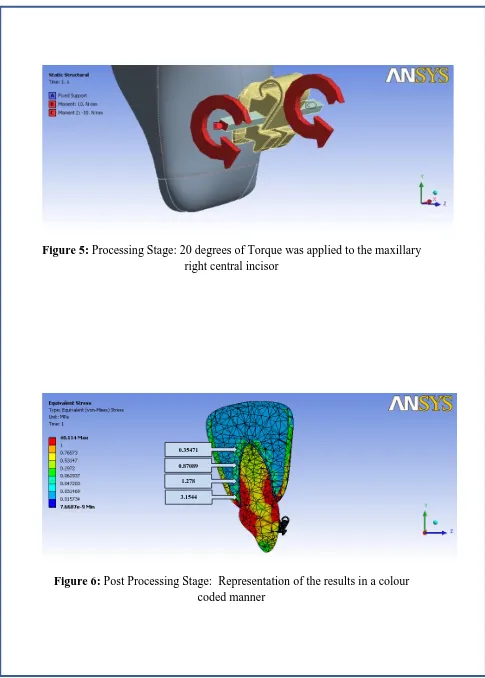
conventionally ligated Discovery. Torque of 20° was applied to the maxillary
right incisor with 0.46 X 0.64mm2 (0.018 X 0.025 in) and 0.48 X 0.64 mm2
(0.019 X 0.025 in) archwires. Three kinds of wire alloys were used: stainless
steel, titanium molybdenum, and nickel titanium. For the conventional Discovery
brackets, 2 types of ligation were modeled: elastic and stainless steel wire
ligatures. The torque angle/torque moment curves seemed to be dominated by the
characteristics of the wire. The change of wire dimension increased the torque
moments less than the change of wire alloy (125% increase for a 0.48X0.64mm2
instead of a 0.46X0.64mm2 stainless steel wire, and 220% for a 0.46 X 0.64 mm2
stainless steel instead of a nickel-titanium wire). The combined change of the wire
alloy and wire dimension resulted in a 600% increase for a 0.48 X 0.64 mm2
stainless steel instead of a 0.46 X 0.64 mm2 nickel-titanium wire. The play of the
0.46 X 0.64 mm2 wires was about 9.0°, and the play of the 0.48X0.64mm2 wires
was about 7.5°, with slightly more play for the Damon. Therefore, improving the
adaptation of torque movements to the biomechanical reactions of the
periodontium is best done by proper selection of both wire dimension and wire
25
of brackets with an active clip (eg, Speed), which had the least play and the
lowest torquing moments of all the wires.
Kojimaa et al (2010)61 calculated the long-term tooth movements in en-masse sliding mechanics. Long-term tooth movements in en-masse sliding
mechanics were simulated with the finite element method. Tipping of the anterior
teeth occurred immediately after application of retraction forces. The force system
then changed so that the teeth moved almost bodily, and friction occurred at the
bracket-wire interface. Irrespective of the amount of friction, the ratio of
movement distances between the posterior and anterior teeth was almost the same.
By increasing the applied force or decreasing the frictional coefficient, the teeth
moved rapidly, but the tipping angle of the anterior teeth increased because of the
elastic deflection of the archwire. Finite element simulation clarified the tooth
movement and the force system in en-masse sliding mechanics.
Xua et al (2011)116 determined the elastic modulus of the periodontal ligament (PDL). The study was carried out on eight human maxillary jaw
segments containing central incisors. Displacements were measured under load
using a electronic speckle pattern interferometry (ESPI). Subsequently, FEM
presenting the same individual geometry as the respective autopsy material were
developed to simulate tooth mobility numerically under the same force systems as
were used in the experiment. A bilinear material parameter set was assumed to
simulate tooth deflections. Thus, the force/deflection curves from the
measurements showed a significant nonlinear behavior of elastic stiffness of the
26
2) TORQUE AND SELF LIGATING BRACKETS IN ORTHODONTICS
Rauch (1959)88 stated that in order to attain our present-day goals of treatment, a definite technique for the application of torque force becomes
imperative. The orthodontist will experience little difficulty if he will keep in
mind the following fundamental principles: the crown of a tooth moves in the
direction of torque; the root of a tooth moves in the opposite direction of torque;
and, by the application of an auxiliary force derived from elastics or other
sources, this torque action can he altered in such a way as to cause either the root
or the crown of a tooth to move in whichever direction the operator may desire.
Germane et al (1989)35 studied the facial surface contours of 600 maxillary and mandibular teeth, including 50 of each type of tooth from central
incisors to first molars, were measured. The magnitude of the variation found was
so great as to suggest that differences between patients or differences in height of
bracket placement are greater than the differences between the standard torque
prescriptions now used in orthodontics. No single point, including the coronal
midpoint (LA point), was found to be constant among teeth of the same type.
Variation in facial surface contour tended to be greater in the posterior teeth than
in the anterior teeth. Future custom construction of brackets, adjusted to
individual facial contour differences, will also require information regarding
optimal tooth position in the head, including compensations necessary for
27
Creekmore et al (1993)20 stated that the frequently anticipated results of treatment are not achieved by using preadjusted appliances and straight wires.
This is due to inaccurate bracket placement, variations in tooth structure,
variations in the maxillary/mandibular relationships, tissue rebound, and
mechanical deficiencies of edgewise orthodontic appliances. Beyond the accuracy
or inaccuracy of bracket placement and the fact that brackets are placed away
from the center of resistance, orthodontic appliances have two additional
significant mechanical deficiencies; play between the arch wire and the arch wire
slot, and force diminution. These deficiencies cannot be eliminated from current
appliances, however, they can be minimized by using reasonably stiff arch wires
approximating the size of the arch wire slots. The amount of play plus the amount
of force diminution inherent in your appliance can be added to or subtracted from
the torque, tip, rotation, and height parameters for each bracket to deliver the teeth
to the desired positions. Therefore treatment goals can be achieved with
maximum efficiency.
Isaacson et al (1993)48 reported traditional edgewise orthodontic mechanics are significantly limited in their ability to provide incisor torque
control because of the limitations of bracket-to-bracket mechanics and the poorly
defined reciprocal actions inherently produced. The science of mechanics dictates
that all incisor torque control mechanisms must act through one of two basic
principles: the moment of a couple or the moment of a force. The torquing arch is
a modification of the traditional edgewise system and employs the moment of a
28
effects. Alternatively, the base arch uses the moment of a force to also rotate
incisors in a crown facial/root lingual direction. The base arch, however, includes
a large moment to rotate molars in a crown distal/root mesial direction, and
concurrent equilibrium forces to intrude incisors and extrude molars. Depending
on how they are employed, torquing arches and base arches may also rotate
molars in a faciolingual direction, enhance or diminish posterior anchorage, and
increase or conserve arch perimenter.
Odegaard et al (1994)79 demonstrated that the amount of play between bracket and wire in torsion for individual tooth movement is considerably larger
than the amount expected. It has also been shown that the initial portion of the
load/deflection curves are relatively flat for the smaller dimensions before a linear
relationship between moment and deflection is achieved, indicating a restraining
effect caused by the ligature. The resulting curves using wire "a" without ligature
illustrates this point. The linear portions of the curves show that the change in
effective rotational moment will change rapidly for small changes in the tooth
axial inclination, suggesting that reactivation of the wires should take place at
frequent intervals. For individual tooth torque, a more efficient method can be the
use of highly elastic wires in combination with brackets with variable torque.
Shivapuja et al (1994)95 reported the increased use of self-ligating bracket systems frequently raises the question of how they compare with conventional
ligation systems. An in vitro and clinical investigation was undertaken to evaluate
and compare these distinctly different groups, by using five different brackets.
29
(Ormco, GIendora, Calif.), and SPEED (Strite Industries Ltd., Cambridge,
Ontario) self-ligating bracket systems displayed a significantly lower level of
frictional resistance, dramatically less chairtime for arch wire removal and
insertion, and promoted improved infection control, when compared with
polyurethane elastomeric and stainless steel tie wire ligation for ceramic and
metal twin brackets.
Harradine (2003)42 reported that currently available self-ligating brackets offer the very valuable combination of extremely low friction and secure full
bracket engagement and, at last, they deliver most of the potential advantages of
this type of bracket. These developments offer the possibility of a significant
reduction in average treatment times and also in anchorage requirements,
particularly in cases requiring large tooth movements. Whilst further refinements
are desirable and further studies essential, current brackets are able to deliver
measurable benefit with good robustness and ease of use.
Harzer (2004)43 investigated slot deformation and the equivalent torque capacity of polycarbonate brackets with and without a metal slot in comparison
with those of a metal bracket. For this purpose, the expansion characteristics and,
in a further investigation, the labial crown torque of an upper central incisor, were
measured in a simulated intra-oral clinical situation, using the orthodontic
measuring and simulation system (OMSS). Three types of bracket with a 0.018
inch slot were tested: polycarbonate Brillant without a metal slot, Elegance with a
metal slot and the metal bracket, Mini-Mono. For testing purposes the brackets
30
archwires. In the activating experiments, significantly higher torque losses and
lower torquing moments were registered with both rectangular archwires with the
polycarbonate brackets than with the metal bracket. In the simulation tests,
significantly higher torquing moments were registered with the metal bracket than
with the polycarbonate brackets. On the basis of the present results, all three
brackets can be recommended for torquing. However, in view of the high torque
losses, the torques programmed in the straightwire technique must be seen as
questionable.
Cash et al (2004)13 evaluated the slots of five upper left central incisor brackets from 11 commercially available bracket systems of 0.022-inch (0.5588
mm) dimension. Results indicate that all bracket slots are oversized. Three
bracket systems slots (Twin Torque, Clarity, and Mini Mono) were within 5%
(61.08, 1.655, 1.75) of their stated dimensions with essentially parallel slot walls.
The Elegance Plastic slot was parallel sided but oversized by 12% (61.15). The
geometry of bracket slots was also variable. The Victory Series slot was slightly
divergent with the top oversized by 6% (61.035). The Nu-Edge slot was divergent
and slot top oversized by 14% (61.32). The Mxi Advant-Edge, Damon II SL, Elite
Mini Opti-MIM Roth, and MBT were all convergent, and the base of the Damon
slot was oversized by 17% (61.79). The Discovery bracket was convergent, and
the slot base was oversized by 24% (61.255), which was the largest recorded
variance. This bracket also had a 7% difference between the widths of the slot top
31
undersized archwires may directly and adversely affect three-dimensional tooth
positioning.
Pandis et al (2006)83 A randomized clinical trial done that the engagement mode of wire to bracket affects the buccolingual inclination of
maxillary incisors in extraction and non-extraction treatment with self-ligating
and conventional brackets. Difference in the buccolingual inclination of maxillary
incisors before and after treatment with the two appliances across the two
treatment groups (extraction and non-extraction). Angular measurements of the
Sella-Nasion and Nasion-A point to maxillary incisor axis was calculated. No
difference was found in the mean difference of the two angles measured for the
two bracket groups studied. Self-ligating brackets seem to be equally efficient in
delivering torque to maxillary incisors relative to conventional brackets in
extraction and non-extraction cases.
Pandis et al (2007)81 investigated the effect of intraoral aging on the force applied during engagement of a wire into the slot of active self-ligating brackets.
Two types of brackets were used: Speed and In Ovation-R. No difference was
found between as-received and used brackets with respect to force exerted by the
spring in 1 bracket group, whereas the other group showed extensive relaxation
after use; neither group had permanent deformation. The consistency of the initial
force levels varied significantly in each bracket group. Thus, the initial force
levels and the effect of intraoral conditions on the stiffness of the clip seem to
vary between products, with potential implications for the archwire engagement
32
Turnbull et al (2007)109 conducted a prospective clinical study, where they assessed the relative speed of archwire changes, comparing self-ligating
brackets with conventional elastomeric ligation methods, and further assessed this
in relation to the stage of orthodontic treatment represented by different wire sizes
and types. The main outcome measure was the time to remove or place
elastomeric ligatures or open/close self-ligating brackets for 2 matched groups of
fixed appliance patients: Damon2 self-ligating bracket (SDS Ormco, Orange,
Calif) and a conventional mini-twin bracket (Orthos, SDS Ormco). The Damon2
self-ligating system had a significantly shorter mean archwire ligation time for
both placing and removing wires compared with the conventional elastomeric
system. Ligation of an archwire was approximately twice as quick with the
self-ligating system. The type of bracket and the size of wire used are statistically
significant predictors for speed of ligation and chairside time. The self-ligating
system offered quicker and arguably more efficient wire removal and placement
for most orthodontic treatment stages.
Streva et al (2007)99 verified the torque precision of metallic brackets with MBT prescription using the canine brackets as the representative sample of
six commercial brands. Twenty maxillary and mandibular canine brackets of one
of the following commercial brands were selected: 3M Unitek, Abzil, American
Orthodontics, TP Orthodontics, Morelli and Ortho Organizers. The results showed
that for the maxillary canine brackets, only the Morelli torque (-3.33º) presented
statistically significant difference from the proposed values (-7º). For the
(-33
6.25º) presented statistically significant differences from the standards (-6º).
Comparing the brands, Morelli presented statistically significant differences in
comparison with all the other brands for maxillary canine brackets. For the
mandibular canine brackets, there was no statistically significant difference
between the brands. There are significant variations in torque values of some of
the brackets assessed, which would clinically compromise the buccolingual
positioning of the tooth at the end of orthodontic treatment.
Morina et al (2008)76 investigated the torque capacity of active and passive self ligating brackets compared with metallic, ceramic, and polycarbonate
edgewise brackets. Six types of orthodontic brackets were included in the study:
the self-ligating Speed and Damon2, the stainless steel (SS), Ultratrimm and
Discovery, the ceramic bracket, Fascination 2, and the polycarbonate bracket,
Brillant. All brackets had a 0.022-inch slot size and were torqued with 0.019 ×
0.025-inch SS archwires. For this purpose, the labial crown torque of an upper
central incisor was measured in a simulated intraoral clinical situation using the
orthodontic measurement and simulation system (OMSS). A torque of 20 degrees
was applied and the correction of the misalignement was simulated
experimentally with the OMSS. The ceramic bracket (Fascination 2) presented the
highest torquing moment (35 Nmm) and, together with a SS bracket, the lowest
torque loss (4.6 degrees). Self-ligating, polycarbonate, and selective metallic
brackets demonstrated almost a 7-fold decreased moment developed during
insertion of a 0.019 × 0.022- inch SS wire into a 0.022-inch slot and a 100 per
34
Badawi et al (2008)6 measured the difference in third-order moments that can be delivered by engaging 0.019 X 0.025-in stainless steel archwires to 2
active self-ligating brackets (In-Ovation, GAC, Bohemia, NY; Speed, Strite
Industries, Cambridge, Ontario, Canada) and 2 passive self-ligating brackets
(Damon2, Ormco, Orange, Calif; Smart Clip, 3M Unitek, Monrovia, Calif). A
bracket/wire assembly torsion device was developed. There was a significant
difference in the engagement angle between the 2 types of brackets; on average,
torque started to be expressed at 7.5° of torsion for the active self-ligating
brackets and at 15° of torsion for the passive self-ligating brackets. The torque
expression was higher for the active self-ligating brackets up to 35° of torsion.
Torsion of the wire past this point resulted in a linear increase of the measured
torque for the Damon2, the Smart Clip, and the In-Ovation brackets. The torque
was relatively constant past 35° of torsion for the Speed bracket. They concluded
that active self-ligating brackets are more effective in torque expression than
passive self-ligating brackets.
Nishio et al (2009)77 evaluated the resistance to deformation or fracture of esthetic brackets produced by archwire torsion. Six types of maxillary right
central incisor brackets were analyzed: traditional ceramic brackets (cer); ceramic
brackets reinforced with a stainless steel slot (cer/ss); ceramic brackets reinforced
with a gold slot (cer/gold); traditional polycarbonate brackets (poly);
polycarbonate brackets reinforced with a stainless steel slot (poly/ss); and
polycarbonate brackets reinforced with ceramic fillers and a stainless steel slot
35
to deformation or fracture, although gold slots and ceramic fillers are ineffective
for reinforcing esthetic brackets.
Pandis(2009)82 comparatively assessed the magnitude and direction of forces and moments generated from different bracket systems, during the initial
levelling and alignment stage of orthodontic treatment. Three types of brackets
were used: Orthos2 (Ormco), Damon2 (Ormco), and In-Ovation R (GAC). The
model was mounted on the Orthodontic Measurement and Simulation System
(OMSS) and six static measurements were taken at the initial crowded state per
bracket for the lateral incisor, canine, and first premolar. The lingually inclined,
crowded lateral incisor presented an extrusive and buccal movement and showed
the lowest force in the vertical direction, whereas the self-ligating group of
brackets generated the highest force in the buccolingual direction. The moments
applied by the three bracket systems followed the general trend shown for forces;
in the vertical axis, the self-ligating brackets exerted lower forces than their
conventional counterpart. This was modified in the buccolingual direction where,
in most instances, the self-ligating appliances applied higher moments compared
with the conventional bracket. In most cases, the magnitude of forces and
moments ranged between 30 – 70 cN and 2 – 6 N mm, respectively. However,
maximum forces and moments developed at the lateral incisor were almost four
times higher than the average.
Chung et al (2009)17 examined the influence of third-order torque on kinetic friction in sliding mechanics involving active and passive self-ligating
36
of brackets and tubes within a simulated posterior dental segment with 15, 10,
-5, 0, +-5, +10, and +15 of torque placed in the second-premolar bracket; a working
archwire was pulled through the slots. They concluded that third-order torque in
posterior dental segments can generate frictional resistance during anterior
retraction with the archwire sliding through self-ligating bracket slots. With small
torque angles, friction is less with passive than with active self-ligating brackets,
but bracket design is a factor. Frictional forces are substantial, regardless of
ligation if the wire-slot torque exceeds the third-order clearance.
Pandis et al (2010)80 compared the time required to complete the alignment of crowded maxillary anterior teeth (canine to canine) between Damon
MX (Ormco, Glendora, Calif) and In-Ovation R (GAC, Central Islip, NY)
self-ligating brackets. No difference in crowding alleviation was found between the 2
bracket systems. Higher irregularity index values were associated with the
increased probability of delayed resolving of crowding. Conclusions: The use of
passive or active self-ligating brackets does not seem to affect treatment duration
for alleviating initial crowding.
Chen et al (2010)16 conducted a systematic review to identify and review the orthodontic literature with regard to the efficiency, effectiveness, and stability
of treatment with self ligating brackets compared with conventional brackets.
Sixteen studies met the inclusion criteria, including 2 randomized controlled trials
with low risk of bias, 10 cohort studies with moderate risk of bias, and 4 cross
sectional studies with moderate to high risk of bias. Self-ligation appears to have a
37
studies. Analyses also showed a small, but statistically significant, difference in
mandibular incisor proclination. No other differences in treatment time and
occlusal characteristics after treatment were found between the 2 systems. No
studies on long-term stability of treatment were identified. They concluded that
despite claims about the advantages of self-ligating brackets, evidence is
generally lacking. Shortened chair time and slightly less incisor proclination
appear to be the only significant advantages of self-ligating systems over
conventional systems that are supported by the current evidence.
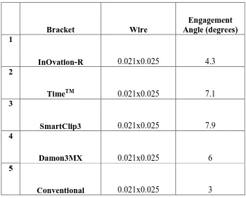
Archambault et al (2010)4 evaluated the quantitative effects on torque expression of varying the slot size of stainless steel orthodontic brackets and the
dimension of stainless steel wire, and to analyze the limitations of the
experimental methods used. In vitro studies measuring torque expression in
conventional and self ligating stainless steel brackets with a torque-measuring
device, with the use of straight stainless steel orthodontic wire without
second-order mechanics and without loops, coils, or auxiliary wires, were sought through
a systematic review process. On the basis of the selected studies, in a 0.018 inch
stainless steel bracket slot, the engagement angle ranges from 31 degrees with a
0.016 X 0.016 inch stainless steel archwire to 4.6 degrees with a 0.018 X 0.025
inch stainless steel archwire. In a 0.022 inch stainless steel bracket slot, the
engagement angle ranges from 18 degrees with a 0.018 X 0.025 inch stainless
steel archwire to 6 degrees with a 0.021 X 0.025 inch stainless steel archwire.
Active stainless steel self-ligating brackets demonstrate an engagement angle of
38
show an engagement angle of approximately 14 degrees with 0.019 X 0.025 inch
stainless steel wire in a 0.022 inch slot. They concluded that the engagement
angle depends on archwire dimension and edge shape, as well as on bracket slot
dimension, and is variable and larger than published theoretical values. Clinically
effective torque can be achieved in a 0.022 inch bracket slot with archwire torsion
of 15 to 31 degrees for active self-ligating brackets and of 23 to 35 degrees for
passive self-ligating brackets with a 0.019 X 0.025 inch stainless steel wire.
Fleming et al (2010)27 evaluated the clinical differences in relation to the use of self-ligating brackets in orthodontics. Randomized controlled trials (RCTs)
and controlled clinical trials (CCTs) investigating the influence of bracket type on
alignment efficiency, subjective pain experience, bond failure rate, arch
dimensional changes, rate of orthodontic space closure, periodontal outcomes, and
root resorption were selected. Concluded at this stage there is insufficient
high-quality evidence to support the use of self ligating fixed orthodontic appliances
over conventional appliance systems or vice versa.
Major et al (2010)64 stated that in all manufacturing processes there are tolerances; however, orthodontic bracket manufacturers seldom state the slot
dimensional tolerances. Their experiment developed a novel method of analyzing
slot profile dimensions using photographs of the slot. Five points are selected
along each wall, and lines are fitted to define a trapezoidal slot shape. This
investigation measures slot height at the slot’s top and bottom, angles between
walls, slot taper, and the linearity of each wall. Slot dimensions for 30 upper right
39
evaluated. Speed brackets have a slot height 2% smaller than the nominal
0.559mm size and have a slightly convergent taper. In-Ovation brackets have a
divergent taper at an average angle of 1.47 degrees. In-Ovation is closest to the
nominal value of slot height at the slot base and has the smallest manufacturing
tolerances. Damon Q brackets are the most rectangular in shape, with nearly
90-degree corners between the slot bottom and walls. Damon slot height is on
average 3% oversized.
Major et al (2011)65 investigated the third-order torque on different types of self-ligated brackets by analyzing the bracket’s elastic and plastic deformations
in conjunction with the expressed torque at varying angles of twist. An
orthodontic bracket was mounted to a load cell that measured forces and moments
in all directions. The wire was twisted in the bracket via a stepper motor,
controlled by custom software. At the maximum torquing angle of 63° with 0.019
X 0.025-in stainless steel wire, the total elastic and plastic deformation values
were 0.063, 0.033, and 0.137 mm for Damon Q (Ormco, Orange, Calif), In-
Ovation R (GAC, Bohemia, NY), and Speed (Strite Industries, Cambridge,
Ontario, Canada), respectively. The total plastic deformation values were 0.015,
0.006, and 0.086 mm, respectively, measured at 0_ of unloading. Conclusions:
In-Ovation R had the least deformation due to torquing of the 3 investigated bracket
types. Damon Q and Speed on average had approximately 2.5 and 14 times
40
Major et al (2011)66 conducted a study was to quantify torque expression in 3 self-ligation bracket systems (Damon Q, In-Ovation R, and Speed) during
loading and unloading. A stepper motor was used to rotate a wire in a fixed
bracket slot from –15º to 63º in 3º increments, and then back to –15º. The bracket
was mounted on top of a load cell that measured forces and moments in all
directions. Results showed that Damon’s and In-Ovation’s maximum average