FAYTH K. YOSHIMURA*
ANDKURT DIEM
Department of Biological Structure, University of Washington, Seattle, Washington 98195
Received 11 July 1994/Accepted 4 November 1994
We previously identified a protein-binding site (MLPal) that is located downstream of the enhancer element
in the long terminal repeat (LTR) of a mink cell focusing-forming (MCF) murine leukemia virus (F. K.
Yoshimura, K. Diem, H. Chen, and J. Tupper, J. Virol. 67:2298–2304, 1993). We determined that the MLPal
site regulates transcription specifically in T cells and affects the lymphomagenicity of the MCF isolate 13
murine leukemia virus with a single enhancer repeat in its LTR. In this report, we present evidence that two
different proteins, a T-cell-specific protein and a ubiquitous protein, bind the MLPal site in a sequence-specific
manner. By mutational analysis, we determined that the T-cell-specific and the ubiquitous proteins require
different nucleotides in the MLPal sequence for DNA binding. By competitive electrophoretic mobility shift
assays, we demonstrated that the T-cell-specific protein that binds MLPal is identical or similar to a protein
from nonactivable T cells that interacts with the binding site of the nuclear factor of activated T cells (NFAT).
Unlike the NFAT-binding site, however, the MLPal site does not bind proteins that are inducible by T-cell
activation. We observed that the MLPal sequence is conserved in the LTRs of other mammalian retroviruses
that cause T-cell diseases. Furthermore, the MLPal sequence is present in the transcriptional regulatory
regions of cellular genes that either are expressed specifically in T cells or are commonly rearranged by
provirus integration in thymic lymphomas. Thus, the MLPal-binding proteins may play a role in the
tran-scriptional regulation not only of the MCF virus LTR but also of cellular genes involved in T-cell development.
The long terminal repeat (LTR) of slowly transforming
ret-roviruses is a major determinant of the type of neoplastic
disease that these viruses produce (11, 14, 15, 17, 25, 26).
Studies of murine leukemia viruses (MLVs) differing in their
pathogenic characteristics have demonstrated a direct
correla-tion between the cell-type-specific transcripcorrela-tional activity of
the LTR and the cell type involved in disease (15, 27, 42).
The best-studied region of the LTR for transcriptional
reg-ulation is the enhancer element. Protein-binding sites within
the enhancer that contribute to T-cell-specific transcription
have been identified for murine retroviruses that cause thymic
lymphoma (4, 14, 15, 27, 41, 43, 45). It has been demonstrated
that these protein-binding sites also contribute to the disease
specificity of these MLVs (15, 42). Besides the enhancer
ele-ment, the region downstream of it also has been shown to be
important for specificity of retroviral pathogenesis and
tran-scriptional regulation (16, 17, 24, 26, 46). We recently observed
that deletion of the entire region between the promoter and
enhancer of the LTR of the mink cell focus-forming (MCF)
MLV significantly reduced the incidence of thymic lymphoma
and increased the latency of disease (46). Furthermore, we
observed that deletion of this region resulted in a marked
reduction of transcription as assessed by transient expression
assays (46). Within this LTR region, we identified a 25-bp
protein-binding site that contributes to T-cell-specific
tran-scription (52, 53). We refer to this protein-binding site as the
MCF virus LTR palindrome (MLPal) because it contains an
18-bp palindromic sequence.
Our studies of the mechanism by which the MLPal site
regulates transcription revealed that its activity depends on
interactions with the enhancer, which is located 14 bp
up-stream of the MLPal site. Although multimerized copies of
MLPal had no effect on the MCF virus promoter, the addition
of a single copy of MLPal increased transcription of one copy
of the viral enhancer repeat and promoter by 10-fold (52). We
further observed that the MLPal site partly overlaps a GATA
consensus sequence, which is a binding site for a multigene
family of transcription factors that regulate gene expression in
a cell-type-specific manner (34). Our preliminary analysis of
interactions between the MLPal- and GATA-binding sites
sug-gest that the GATA site inhibits transcriptional activation by
MLPal, most likely by interfering with protein binding to the
MLPal site (unpublished data).
In this report, we describe studies that examine protein
binding to the MLPal site. We have detected an
MLPal-bind-ing protein that is present only in T cells. By mutational
anal-ysis, we identified the bases in the MLPal sequence that are
involved in protein binding. We present evidence that the
MLPal site binds a nuclear protein that may also be part of the
multisubunit nuclear factor of activated T cells (NFAT) (8, 40).
We also discuss our detection of sequences with homology to
MLPal that are present in other mammalian retroviruses that
cause T-cell diseases and in the transcriptional regulatory
re-gions of several cellular genes.
MATERIALS AND METHODS
Cell lines.Murine T-lymphoma cell lines L691, Ti6, EL4, and BW5147.3 were grown in RPMI 1640 (GIBCO Laboratories, Grand Island, N.Y.) containing 10% fetal bovine serum (FBS) (GIBCO). The murine B-cell line S194 and human T-cell line Jurkat were grown in RPMI 1640, 10% FBS, and 531025
M b-mercaptoethanol. NIH 3T3 mouse and AH927 feline fibroblast cell lines were propagated in Dulbecco’s modified Eagle’s medium (GIBCO) plus 10% FBS. Jurkat cells were activated by the addition to the medium of 25 ng of 12-O-* Corresponding author. Mailing address: Department of Biological
Structure, SM-20, University of Washington, Seattle, WA 98195. Phone: (206) 685-1535. Fax: (206) 543-1524. Electronic mail address: [email protected].
994
on November 9, 2019 by guest
http://jvi.asm.org/
tetradecanoylphorbol-13-acetate (TPA; Sigma) per ml and 1.4mg of ionomycin (Calbiochem) per ml for 3.5 h.
Nuclear extract preparation.Cells were collected by centrifugation at 5003g for 10 min at 48C and rinsed once with phosphate-buffered saline (2 mM Na2HPO4, 2 mM KH2PO4, 150 mM NaCl); lysis buffer (LB; 10 mM
N-2-hydroxyethylpiperazine-N9-2-ethanesulfonic acid [HEPES; pH 8.0], 50 mM NaCl, 7 mMb-mercaptoethanol, 0.5 M sucrose, 1 mM EDTA, 0.25 mM EGTA, 0.5 mM spermidine, 0.15 mM spermine) was then added. Cells were lysed with LB containing 0.5% Nonidet P-40, with a 5- to 10-min incubation on ice. After this step, all buffers contained 0.5 mM phenylmethylsulfonyl fluoride, aprotinin (3mg/ml), leupeptin (1mg/ml), and pepstatin A (1mg/ml) as protease inhibitors. After centrifugation, nuclei were washed with extraction buffer (EB; identical to LB except that 10% glycerol is substituted for sucrose). Nuclei were resuspended in 5 to 10 ml of EB to which was added an equal volume of EB that contained 0.55 N NaCl dropwise with constant swirling. Nuclei were incubated on ice for 30 min with gentle shaking every 5 min. Treated nuclei were centrifuged at 10,800 3g for 20 min at 48C. The supernatant was decanted, and ammonium sulfate was slowly added to a final concentration of 70% saturation for protein precipitation. The ammonium sulfate precipitate was collected by centrifugation at 17,2003g for 20 min at 48C. Protein was resuspended in 500ml of EB and dialyzed against 100 ml of buffer B (EB without the spermine and spermidine) in the cold. Nuclear extracts were clarified by centrifugation at 15,0003g for 10 min before storage at2708C. Protein was measured by the Bradford method (5).
EMSA.The electrophoretic mobility shift assay (EMSA) is based on the method devised by Garner and Revzin (13). One nanogram of 32
P-labeled synthetic double-stranded oligonucleotides (approximately 50,000 Cerenkov cpm) was incubated with 2 to 4mg of nuclear extract and 1mg of poly(dI-dC) (Boehringer Mannheim) in 13EB buffer for 30 min on ice. Unlabeled double-stranded oligonucleotide competitors were preincubated with the nuclear extract for 10 min prior to addition of the labeled probe. Samples were electrophoresed through a 5% polyacrylamide gel in 0.33TBE buffer (13TBE buffer is 81 mM Tris base, 81 mM boric acid, and 1.8 mM EDTA) at 17 V/cm for 2 h at 48C. Gels were dried and exposed for various times to Kodak X-Omat film with a DuPont Lightning Plus intensifying screen at2708C.
Complementary oligonucleotide strands were synthesized with an Applied Biosystems 380B synthesizer by a Howard Hughes Medical Institute core facility at the University of Washington. Coding strand sequences of the oligonucleo-tides used in EMSAs were as follows: MLPal, ATAAAGCGAAACTAGCAG CAGTTTC; Pal-1, TACAGTCGCTGCACTGTA; Pal-2, GAAACAGCACCG ACAGAA; P sequence, CGAAAATTTCC; and NFAT, TGCCCAAAGAGGA AAATTTGTTTCATACAG. Nucleotides that are underlined in Pal-1 and Pal-2 sequences are identical to the nucleotides in the same positions in the MLPal sequence.
Preparation of DNA probes for EMSAs.Double-stranded oligonucleotides were32P labeled by annealing both strands and labeling with [a-32P]dATP and
[a-32P]dCTP with the Klenow fragment of Escherichia coli DNA polymerase I
(Boehringer Mannheim) at room temperature for 30 min. Specific activities ranged from 13107to 5 3107cpm/mg. Labeled probes were purified by
electrophoresis through a 4% polyacrylamide gel. After autoradiography, the DNA was excised from the gel and eluted by overnight incubation in 10 mM Tris-Cl (pH 7.5)–1 mM EDTA at 378C. The DNA was ethanol precipitated with yeast tRNA as the carrier before further use.
RESULTS
A T-cell-specific protein and a ubiquitous protein bind the
MLPal site.
Our previous studies of the MLPal
protein-bind-ing site (Fig. 1) involved the use of nuclear extracts isolated
only from T cells (52). To determine whether the MLPal site
binds a T-cell-specific or a ubiquitous protein, we compared
EMSA reactions with nuclear extracts isolated from different
cell types. For these EMSAs, we used a
32P-labeled
oligonu-cleotide probe that contained the MLPal sequence (see
Mate-rials and Methods). Nuclear extracts from four different
mu-rine T-cell lines, BW5147.3, L691, Ti6, and EL4, produced
protein-DNA complexes with similar mobilities (Fig. 2A, lanes
2 to 5). We detected two major bands, top and bottom, that
were generated by nuclear extracts from all of the T-cell lines
that we examined. Two additional bands with intermediate
mobilities were most noticeable with nuclear extracts from
BW5147.3 (lane 2) and EL4 (lane 5) cells. Bands lower in
mobility than the top band were also detectable with nuclear
extract from two T-cell lines (lanes 2 and 5). Nuclear extracts
from either a murine B-cell line, S194 (lane 6), or a feline
(AH927; lane 7) or murine (NIH 3T3; lane 8) fibroblast cell
line produced protein-DNA complexes with mobilities
corre-sponding to the bottom and/or intermediate bands that were
detectable with the T-cell extracts. The bottom band was
de-tectable with nuclear extracts from all cell types that we
ana-lyzed. Non-T cells lacked the top band that was generated by
the T-cell nuclear extracts. For S194 B cells (lane 6), we
de-tected a band that migrated slightly faster than the
T-cell-specific complex.
We observed that the signals corresponding to the top and
bottom bands increased in intensity when we added increasing
amounts of L691 T-cell nuclear extract to the reaction mixture
(Fig. 2B). This result indicated that binding of the nuclear
factors that produce the top and bottom bands to the MLPal
probe was not due to nonspecific protein binding to DNA. We
further address this point below.
Specificity of protein binding to the MLPal sequence.
To
[image:2.612.327.549.72.245.2]determine whether the protein-DNA complexes corresponding
to the top and bottom bands that were detectable with T-cell
FIG. 1. MCF13 LTR. Open boxes correspond to 69-bp direct repeats; the hatched box indicates the MLPal protein-binding site; CAT and TATA boxes are indicated; the horizontal arrow marks the start site of transcription. Numbering of nucleotides is relative to the start site of transcription. The nucleotide se-quence of the MLPal site is shown below; the palindromic sese-quence is under-lined.
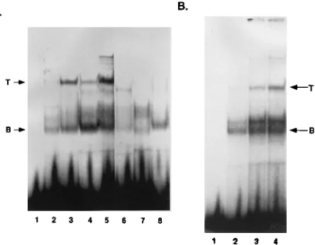
FIG. 2. (A) Cell line comparison of nuclear protein binding to the MLPal sequence. Two nanograms of32
P-labeled MLPal oligonucleotide probe (lane 1) was reacted with 4mg each of nuclear extracts isolated from various cell lines. T-cell lanes: 2, BW5147.3; 3, L691; 4, Ti6; 5, EL4. B-cell lane: 6, S194. Feline and murine fibroblast lanes: 7, AH927; 8, NIH 3T3. Arrows indicate the top (T) and bottom (B) bands that correspond to the two major protein-DNA complexes detectable with T-cell nuclear extracts. Binding reactions were performed in EB for 30 min at 48C. Polyacrylamide gel electrophoresis and processing of the gel were performed as described in Materials and Methods. (B) Titration of protein-DNA complexes generated by binding of T-cell nuclear protein to the MLPal sequence. Two nanograms of32P-labeled MLPal oligonucleotide probe (lane 1)
was reacted with increasing amounts of L691 T-cell nuclear extract (2 [lane 2], 4 [lane 3], and 6 [lane 4]mg) as described for panel A. T, top band; B, bottom band. Polyacrylamide gel electrophoresis was performed as described in Materials and Methods.
on November 9, 2019 by guest
http://jvi.asm.org/
nuclear extracts were generated by specific DNA binding, we
performed EMSA competition assays (Fig. 3). For this
deter-mination, we included in the EMSA binding reaction mixture
competing unlabeled double-stranded oligonucleotides that
contained either the wild-type MLPal sequence (lanes 2 and 3)
or two different sequences that correspond to mutations of
only the palindromic region of MLPal (Pal-1 [lanes 4 and 5]
and Pal-2 [lanes 6 and 7]) at 20- or 100-fold molar excess
compared with the labeled MLPal probe. The Pal-1
oligonu-cleotide contains base changes that maintain a palindromic
structure with the same GC content as the wild-type MLPal
sequence. Pal-2 has mutations of the palindromic sequence
that eliminate the twofold symmetry. In a separate experiment,
we also used an oligonucleotide that contained the sequence of
a cis-acting element that is located upstream of the promoter
of the interleukin-4 gene (P sequence [1]) at 10- or 50-fold
molar excess as a competitor (lanes 12 and 13). We observed
that the oligonucleotide with the MLPal sequence was an
ef-fective competitor in both experiments (lanes 2, 3, 10, and 11).
Competition for the top band was more efficient that
compe-tition for the bottom band at all molar ratios used.
Competi-tion for the bottom band is seen more easily in lane 3 than in
lane 11. We detected no comparable competition by
oligonu-cleotides that contained either the Pal-1 (lanes 4 and 5), Pal-2
(lanes 6 and 7), or P (lanes 12 and 13) sequence. For unknown
reasons, inclusion of the Pal-1 and Pal-2 oligonucleotides
in-creased the intensity of the bottom band compared with the
reaction with no competitor (lanes 4 to 7). Competition of the
bands migrating more slowly than the top band by Pal-1 and
Pal-2 (lanes 4 and 7, respectively) indicated that these bands
were generated by nonspecific DNA binding. From the results
of these competition assays, we concluded that the two major
protein-DNA complexes detectable with the wild-type MLPal
probe represented specific binding of T-cell nuclear proteins to
the MLPal sequence.
Mutational analysis of protein binding to the MLPal site.
To identify the bases within the MLPal site that participate in
protein binding, we mutated the wild-type sequence as shown
in Fig. 4A. Synthetic double-stranded oligonucleotides
con-taining each of the indicated mutations were
32P labeled and
used as probes in binding reactions with L691 T-cell nuclear
extract (Fig. 4B). We observed that mutations of nucleotides
(nt)
2
166 to
2
170 (Mut-1) resulted in the loss of the top and
bottom bands corresponding to the T-cell-specific and
ubiqui-tous protein-DNA complexes (lane 2). Mutations of nt
2
161 to
2
165 (Mut-2) eliminated the top but not the bottom band and
increased the intensity of the intermediate bands (lane 3).
Mutations of nt
2
153 to
2
160 in the middle of the palindrome
(Mut-3) did not completely eliminate but significantly reduced
the intensity of the top band (lane 4). These mutations did not
affect the bottom band. Mutations of nt
2
146 to
2
149 (Mut-4)
did not significantly affect the top band and produced a
mod-erate increase in the intensity of the bottom and intermediate
bands (lane 5). These data indicated that the sequence of the
first 10 bases of MLPal is essential for T-cell-specific
protein-binding and that the precise sequence of the last 6 bases of
MLPal that lie within the palindrome is not important. We also
concluded that the nucleotides involved in binding of the
ubiq-uitous protein differed from those essential for binding of the
T-cell-specific protein.
[image:3.612.74.278.71.219.2]The MLPal site binds a nuclear protein related to NFAT.
A
FIG. 3. Competition for T-cell nuclear protein binding to MLPal by beled oligonucleotides containing various sequences. Excess amounts of unla-beled double-stranded oligonucleotides were reacted with 4mg of L691 nuclear protein for 10 min prior to addition of 4 ng of32
P-labeled MLPal oligonucleotide in an EMSA binding reaction. Lanes 1 and 9, MLPal probe and nuclear protein with no competitor oligonucleotide. Unlabeled competitor oligonucleotides at indicated molar ratio excesses were in lanes 2 (203MLPal), 3 (1003MLPal), 4 (203Pal-1), 5 (1003Pal-1), 6 (203Pal-2), 7 (1003Pal-2), 10 (103MLPal), 11 (503MLPal), 12 (103P sequence), and 13 (503P sequence). Lane 8 is the MLPal probe with no protein. Sequences of oligonucleotides used for competi-tion are listed in Materials and Methods. T, top band; B, bottom band. Gel electrophoresis and processing were as described in the legend to Fig. 2A.
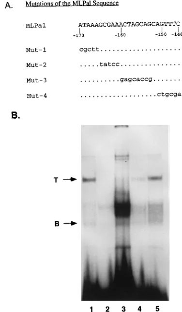
FIG. 4. (A) Sequences of oligonucleotides containing mutations of the ML-Pal sequence used for binding to T-cell nuclear extract in EMSA reactions. Mutated bases are identified by lowercase letters; dots correspond to identical bases. (B) Identification of bases in the MLPal sequence that are involved in the generation of the top and bottom protein-DNA complexes. Wild-type and mu-tant MLPal oligonucleotides in lanes 1 (wild type), 2 (Mut-1), 3 (Mut-2), 4 (Mut-3), and 5 (Mut-4) were32P labeled, reacted with 4mg of T-cell nuclear
extract, and electrophoresed as described in the legend to Fig. 2A. Arrows indicate top (T) and bottom (B) bands.
on November 9, 2019 by guest
http://jvi.asm.org/
[image:3.612.338.526.72.394.2]nucleotide sequence comparison of the MLPal site with known
cis-acting transcriptional regulatory elements revealed
homol-ogy with the binding site for NFAT (12, 40) (Fig. 5A). NFAT
is a multisubunit complex consisting of at least three identified
proteins (3, 18, 19, 28a, 29, 32, 33) and is required for the
trans-criptional activation of the interleukin-2 gene (8, 12, 40). We
observed that the sequence motif
AAA--G-GAAANT---GTTTC was shared by the MLPal and NFAT sites. An
addi-tional similarity that we detected between these two binding
sites is the location of methylated guanines that interfere with
protein binding (indicated by the filled circles in Fig. 5A)
(reference 18 and unpublished data).
To determine whether these two sites bind similar proteins,
we compared protein binding to the MLPal and NFAT sites by
EMSA analysis. When L691 T-cell nuclear extract was used in
the binding reaction, we detected a band for the NFAT probe
(Fig. 5B, lane 6) with mobility similar but not identical to that
of the MLPal top band (Fig. 5B, lane 5). The slight difference
in mobilities between the MLPal and NFAT bands was
repro-ducibly detectable. The L691 cells that were used for these
binding assays are T cells that are not activable and do not
produce the NFAT nuclear complex in response to agents that
mimic antigen activation of T cells (unpublished data). The
protein-DNA complex that was detectable with the NFAT
probe and L691 nuclear extract had a mobility different from
that of the NFAT complex detectable with nuclear extract
from activated Jurkat cells (Fig. 5B; compare lanes 4 and 6).
With the L691 nuclear extract, we also detected NFAT
com-plexes with mobilities similar to those of the intermediate and
bottom bands that were detectable with the MLPal probe.
We performed EMSA competition experiments to test the
possibility that the MLPal and the NFAT sites bind the same
protein in L691 nuclear extracts. When we used the MLPal
oligonucleotide as a probe, we detected effective competition
of the top band by the unlabeled double-stranded NFAT
oli-gonucleotide (Fig. 6A). Competition by the NFAT
oligonucle-otide (lanes 7 and 8) was comparable to that by the MLPal
oligonucleotide (lanes 3 and 4). The oligonucleotide
contain-ing the P sequence did not compete (lanes 5 and 6). When we
performed the converse experiment, we observed that the
un-labeled MLPal oligonucleotide effectively competed with the
NFAT band with mobility similar to that of the MLPal top
band (Fig. 6B, lanes 5 and 6). Competition by MLPal was
about as effective as that by the cognate NFAT competitor
oligonucleotide (lanes 3 and 4). We did not detect comparable
competition for the top NFAT complex by the Pal-1 (lanes 7
and 8) or Pal-2 (lanes 9 and 10) oligonucleotide. Effective
competition for the additional bands that were generated by
the NFAT probe was not observed for any of the
oligonucle-otides at the excess molar ratios used in this experiment.
FIG. 5. (A) Consensus sequence shared by the MLPal and NFAT protein-binding sites. The NFAT nucleotide sequence corresponds to nt2296 to2267 of the distal NFAT site of the murine interleukin-2 gene promoter (18). Identical bases are indicated by uppercase letters, and lowercase letters indicate differ-ences. Dashed lines correspond to gaps in the sequence. Filled circles identify the methylated guanines in the coding strand that interfere with protein binding (reference 18 and unpublished data). The consensus sequence derived for the MLPal and NFAT sites is shown. (B) Comparison of protein binding to MLPal and NFAT sequences for nuclear extracts isolated from activable and nonacti-vable T cells. A32
P-labeled MLPal or NFAT double-stranded oligonucleotide was reacted with 4mg of nuclear protein isolated from L691 cells (lanes 5 and 6) or Jurkat cells that were either treated with 25 ng of TPA per ml and 1.4mg of ionomycin per ml (lanes 2 and 4) or untreated (lanes 1 and 3). Binding conditions were as described in the legend to Fig. 2A. Lanes: 1, 2, and 5, MLPal probe; 3, 4, and 6, NFAT probe. The arrow labeled NFAT indicates the band in lane 4 that corresponds to the NFAT complex that is inducible in activated T cells. Arrows labeled T and B correspond to the top and bottom bands, respectively, in lane 5 that are detectable with the MLPal probe and L691 nuclear extract. NA and A designate nonactivated and activated Jurkat cells, respectively.
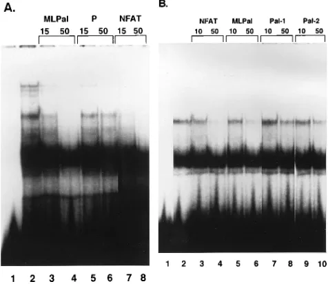
FIG. 6. (A) Competition by the NFAT oligonucleotide for T-cell nuclear protein binding to the MLPal sequence. Conditions for the binding reaction were as described in the legend to Fig. 2A. Unlabeled competitor oligonucleotides at either 15- or 50-fold excess molar ratio were preincubated with nuclear protein for 10 min before the addition of the32
P-labeled MLPal probe. Lanes: 1, MLPal labeled probe without protein; 2, MLPal labeled probe with 6mg of protein; 3, 153unlabeled MLPal; 4, 503unlabeled MLPal; 5, 153unlabeled P sequence; 6, 503unlabeled P sequence; 7, 153unlabeled NFAT; 8, 503unlabeled NFAT. (B) Competition by the MLPal oligonucleotide for L691 T-cell nuclear protein binding to the NFAT sequence. Unlabeled competitor oligonucleotides at either 10- or 50-fold excess molar ratio were preincubated with nuclear protein before the addition of the32
P-labeled NFAT probe. Lanes: 1, NFAT probe without protein; 2, NFAT probe with 6mg of protein; 3, 103NFAT competitor; 4, 503 NFAT competitor, 5, 103MLPal competitor; 6, 503MLPal competitor; 7, 103 Pal-1 competitor; 8, 503Pal-1 competitor; 9, 103Pal-2 competitor; 10, 503
Pal-2 competitor.
on November 9, 2019 by guest
http://jvi.asm.org/
[image:4.612.316.554.73.279.2]used to stimulate T cells to detect the NFAT nuclear complex
(3). When we compared EMSAs of nuclear extracts from
ac-tivated and nonacac-tivated Jurkat cells, we observed identical
band patterns with the MLPal probe (Fig. 5B, lanes 1 and 2).
In contrast, a comparison of the binding of these nuclear
ex-tracts to the NFAT probe resulted in our detection of the
NFAT nuclear complex only for activated Jurkat cells (Fig. 5B,
lanes 3 and 4). Our results suggested that a protein or proteins
that are inducible by the activation of T cells do not bind
MLPal. In contrast, MLPal binds nuclear proteins that are
constitutively expressed in mouse and human T cells.
Conservation of the MLPal sequences in the LTRs of other
MLVs and transcriptional regulatory regions of cellular genes.
We compared the nucleotide sequences located at a site
anal-ogous to MLPal in the LTRs of other retroviruses and detected
a strong conservation of the MLPal sequence in mammalian
viruses that induce T-cell diseases (Fig. 7A). For MLVs that
induce thymic lymphoma (radiation leukemia virus [20], MCF
virus isolate 247 [MCF247] [21], and Tikaut [6]), there were
one to three base pair differences. The analogous site in the
LTR of feline leukemia virus (44), which induces T-cell
lym-phoma, also has sequence similarity with MLPal. MLVs that
are either nononcogenic (Akv [47]) or induce diseases
involv-ing non-T cells (Friend [22]) had 7-of-25 base pair differences
for sites that were analogous to MLPal.
We searched the GenBank nucleotide database to
deter-mine whether we could detect the MLPal sequence in the
transcriptional regulatory regions of cellular genes. For this
search, we compared only nt
2
161 to
2
170 of the MLPal site,
which our mutational studies demonstrated were most critical
for T-cell-specific protein binding as described above (Fig. 4B).
We detected significant homology with these MLPal bases in
the transcriptional regulatory regions of eight cellular genes
that either are of significance to T cells or are proto-oncogenes
that are frequently rearranged in T-cell lymphomas (7, 9, 48)
(Fig. 7B). Five of these cellular genes, the T-cell receptor
(TCR) C
b
2, V
b
2, and
d
genes, CD8
b
-chain gene, and CD3
ε
-chain gene (6, 28, 31, 35–37), are normally expressed only in
T cells. The N-myc gene (10) is expressed preferentially in
kidney, intestine, and newborn brain (54), and the c-myc gene
(2) is expressed in all cell types (54). pim-1 (39) expression
occurs in hematolymphoid tissues and testis (30). The
up-stream region of N-myc (10) and the intron between the TCR
J
b
2 and C
b
2 (28) genes each possessed two sites with
homol-ogy to MLPal. A total of 11 sites with significant sequence
homology with MLPal were identified for the cellular genes
cited above. Six of the homologous sequences were in the
forward orientation, and five were reversed.
DISCUSSION
We previously identified a protein-binding site (MLPal) that
is located near the 3
9
end of the enhancer element in the LTR
of the MCF13 MLV. We demonstrated that the MLPal
se-quence is a cis-acting element that potentiates transcription
specifically in T cells (46, 52). In this report, we describe our
detection of a T-cell-specific protein that binds the MLPal site.
A sequence search uncovered homology between the MLPal
and NFAT sites, and EMSAs demonstrated that both
se-quences competed for binding by a T-cell nuclear protein. It is
not clear, however, which, if any, of the multiple proteins that
have been identified in the NFAT complex binds MLPal.
These NFAT-binding proteins include Fos, Jun, and NFATp
(3, 18, 19, 32, 33). Unlike NFAT, however, the MLPal
se-quence did not bind proteins that are inducible by T-cell
acti-vation. It has been shown that Fos and Jun are the inducible
proteins in the NFAT complex (3, 18). Additional reasons why
it is unlikely that Fos and Jun bind MLPal include the absence
of an AP-1 motif in MLPal and our observation that mutations
of the analogous bases that affect Fos and Jun binding to
NFAT (3) had no effect on protein binding to MLPal.
Fur-thermore, we have not been able to detect an effect on the
mobility or generation of the MLPal complex by the addition
of antibodies to Fos and Jun (unpublished data). Our current
data indicate that the same or a closely related nuclear protein
that is present in nonactivable T cells binds both the
MLPal-and NFAT-binding sites. However, in activated T cells,
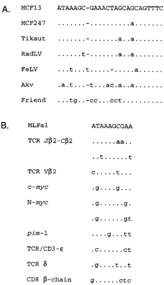
differ-FIG. 7. (A) Nucleotide sequence comparison of the MLPal sites in the LTRs of various mammalian retroviruses. Identical bases are indicated by dots, dashes represent missing bases, and lowercase letters correspond to differing bases. Sequences shown: MCF13 (51); MCF247 (21); Tikaut MLV (7); radiation-induced leukemia virus (RadLV) (20); feline leukemia virus (FeLV) (44); Akv 614 murine nonleukemogenic virus (47); Friend MLV clone 57 (22). (B) Nucle-otide sequences in transcriptional regulatory regions of cellular genes with ho-mology to the first 10 bases of the MLPal sequence. Identical bases are repre-sented by dots; differing bases are indicated by lowercase letters. TCR Jb2-Cb2, the Jb2-Cb2 intron of the murine TCRb-chain gene (28); TCR Vb2, 91 to 100 nt upstream of the transcriptional start site of the murine Vb2 gene (36); c-myc, 252 to 261 nt upstream of the P1 promoter of the murine c-myc proto-oncogene (2); N-myc, 723 to 732 and 681 to 690 nt upstream of the transcription initiation site of the murine N-myc proto-oncogene (10); pim-1, GC-rich promoter region of the murine pim-1 proto-oncogene (39); TCR Cb2, 39enhancer of the human TCR Cb2 gene (35); TCR/CD3-ε, 39enhancer of the human TCR/CD3-εgene (6); TCRd, enhancer of the human TCRdgene (37); CD8b-chain, 59-flanking region of the murine CD8b-chain gene (31).
on November 9, 2019 by guest
http://jvi.asm.org/
[image:5.612.350.519.67.361.2]ent proteins bind these two sites. Thus, we conclude that the
MLPal- and NFAT-binding sites bind similar but not identical
proteins. The MLPal-binding protein may be a novel member
of the family of NFAT-binding proteins that has recently been
identified, but one which is constitutively expressed in the
nucleus (28a, 32). Although there is a constitutive component
(NFATp) of the NFAT multisubunit complex, it is localized in
the cytoplasm (19, 29, 32, 33) and hence should be absent from
our T-cell nuclear extracts.
Our data from both EMSA and mutational analysis of the
MLPal sequence indicated that more than one protein binds
this site. We detected two major protein-DNA complexes that
corresponded to specific protein binding as determined by
competitive EMSAs. One band (bottom) appeared with
nu-clear extracts from various cell types. This observation
sug-gested that the protein that generates this complex is
ubiqui-tous. The second specific complex (top band) was detectable
only with T-cell nuclear extracts. Mutational analysis of the
MLPal sequence revealed that the ubiquitous protein requires
nt
2
166 to
2
170 for binding (Fig. 4A). In contrast, nt
2
153 to
2
170 of MLPal were found to be important for binding of the
T-cell protein, although nt
2
161 to
2
170 were more critical for
binding than nt
2
153 to
2
160. Single base mutations of these
nucleotides will be required to determine whether all or only a
few of them are essential for protein binding. We observed that
disruption of the palindrome by mutations of nt
2
146 to
2
151
had no effect on the generation of the top band, suggesting that
the specific sequence and not solely secondary structure is
required for binding of the protein from T cells.
The presence of sequences similar to MLPal in an analogous
site of the LTRs of several mammalian retroviruses that induce
T-cell diseases suggests that this protebinding site is
in-volved in viral pathogenesis. However, it is not an absolute
requirement since other lymphomagenic viruses do not have
this protein-binding site (25). In a previous study, we observed
that deletion of MLPal had a moderate effect on MCF13 MLV
pathogenicity only when the virus contained a single enhancer
element. Our detection of sequences with close homology to
the first 10 bases of MLPal, which are most stringently required
for generation of the T-cell-specific complex, in the
transcrip-tional regulatory regions of cellular genes is intriguing. Several
of these genes are those that encode proteins expressed
spe-cifically in T cells, such as the TCR C
b
2, V
b
2, and
d
, CD3
ε
-chain, and CD8
b
-chain proteins (6, 28, 31, 35–37).
More-over, it has been shown that the sites with homology to MLPal
in the TCR
d
gene enhancer and the intron between J
b
2 and
C
b
2 are bound by protein (28, 37). It is particularly intriguing
that the cellular genes c-myc, pim-1, and N-myc, which are
frequently rearranged in T-cell lymphomas (7, 9, 48), have
sequences with homology to MLPal in upstream regions of
their promoters. For c-myc, these homologous sequences are
found immediately upstream of the P1 promoter (2). It has
been observed that deregulation of the c-myc proto-oncogene
in T-cell tumors correlated with a shift in usage of the myc
promoter from P2 to P1 (38). We speculate that protein
bind-ing to the MLPal site may be required for the activation of the
P1 promoter. For pim-1, the MLPal sequence is also present in
the promoter region, where it partly overlaps an Oct-1-binding
site (39). Studies with transgenic mice have suggested a strong
cooperation between pim-1 and c-myc or N-myc in
lym-phomagenesis (49). The MLPal protein-binding site may
con-tribute to the coordinate expression of these proto-oncogenes.
Thus, the proteins that bind the MLPal site may have an
important role in both the normal regulation of cellular genes
and their deregulation in tumor cells. The identification and
isolation of these MLPal-binding proteins will allow us to make
this determination.
ACKNOWLEDGMENTS
We are grateful to Ted Young for critically reading the manuscript. This work was supported by Public Health Service grant CA44166 from the National Institutes of Health and a grant from the Royalty Research Fund of the University of Washington to F.K.Y.
REFERENCES
1. Abe, E., R. D. W. Malefyt, I. Matsuda, K. Arai, and N. Arai. 1992. An 11-base-pair DNA sequence motif apparently unique to the human inter-leukin 4 gene confers responsiveness to T-cell activation signals. Proc. Natl. Acad. Sci. USA 89:2864–2868.
2. Bernard, O., S. Cory, S. Gerondakis, E. Webb, and J. M. Adams. 1983. Sequence of the murine and human cellular myc oncogenes and two modes of myc transcription resulting from chromosome translocation in B lymphoid tumours. EMBO J. 2:2375–2383.
3. Boise, L. H., P. Bronislawa, X. Mao, C. H. June, C. Wang, T. Lindsten, R. Bravo, K. Kovary, J. M. Leiden, and C. B. Thompson.1993. The NFAT-1 DNA-binding complex in activated T cells contains Fra-1 and JunB. Mol. Cell. Biol. 13:1911–1919.
4. Boral, A. L., S. A. Okenquist, and J. Lenz. 1989. Identification of the SL3-3 virus enhancer core as a T-lymphoma cell-specific element. J. Virol. 63:76– 84.
5. Bradford, M. M. 1976. A rapid and sensitive method for the quantitation of microgram quantities of protein utilizing the principle of protein-dye bind-ing. Anal. Biochem. 72:248–254.
6. Clevers, H., N. Lonberg, S. Dunlap, E. Lacy, and C. Terhorst. 1989. An enhancer located in a CpG-island 39to the TCR/CD3-εgene confers T lymphocyte-specificity to its promoter. EMBO J. 8:2527–2535.
7. Corcoran, L. M., J. M. Adams, A. R. Dunn, and S. Cory. 1984. Murine T cell lymphomas in which the cellular myc oncogene has been activated by retro-viral insertion. Cell 37:113–122.
8. Crabtree, G. R. 1989. Contingent genetic regulatory events in T lymphocyte activation. Science 243:355–361.
9. Cuypers, H. T., G. Selten, W. Quint, M. Zijlstra, E. R. Maandag, W. Boelens, P. van Wezenbeek, C. Melief, and A. Berns.1984. Murine leukemia virus-induced T-cell lymphomagenesis: integration of proviruses in a distinct chro-mosomal region. Cell 37:141–150.
10. DePinho, R. A., E. Legouy, L. B. Feldman, N. E. Kohl, G. D. Yancopoulos, and F. W. Alt.1986. Structure and expression of the murine N-myc gene. Proc. Natl. Acad. Sci. USA 83:1827–1831.
11. DesGroseillers, L., E. Rassart, and P. Jolicoeur. 1983. Thymotropism of murine leukemia virus is conferred by its long terminal repeat. Proc. Natl. Acad. Sci. USA 80:4203–4207.
12. Durand, D. B., J. P. Shaw, M. R. Bush, R. E. Replogle, R. Belagaje, and G. R. Crabtree.1988. Characterization of antigen receptor response elements within the interleukin-2 enhancer. Mol. Cell. Biol. 8:1715–1724.
13. Garner, M. M., and A. Revzin. 1981. A gel electrophoresis method for quantifying the binding of proteins to specific DNA regions: application to components of the Escherichia coli lactose operon regulatory system. Nucleic Acids Res. 9:3047–3060.
14. Golemis, E., Y. Li, T. N. Fredrickson, J. W. Hartley, and N. Hopkins. 1989. Distinct segments within the enhancer region collaborate to specify the type of leukemia induced by nondefective Friend and Moloney viruses. J. Virol. 63:328–337.
15. Hallberg, B., J. Schmidt, A. Luz, F. S. Pedersen, and T. Grundstrom. 1991. SL3-3 enhancer factor 1 transcriptional activators are required for tumor formation by SL3-3 murine leukemia virus. J. Virol. 65:4177–4181. 16. Hanecak, R., P. K. Pattengale, and H. Fan. 1991. Deletion of a GC-rich
region flanking the enhancer element within the long terminal repeat se-quence alters the disease specificity of Moloney murine leukemia virus. J. Virol. 65:5357–5363.
17. Ishimoto, A., M. Takimoto, A. Adachi, M. Kakuyama, S. Kato, K. Kakimi, K. Fukuoka, T. Ogiu, and M. Matsuyama.1987. Sequences responsible for erythroid and lymphoid leukemia in the long terminal repeats of Friend mink cell focus-forming and Moloney murine leukemia viruses. J. Virol. 61:1861– 1866.
18. Jain, J., P. G. McCaffrey, V. E. Valge-Archer, and A. Rao. 1992. Nuclear factor of activated T cells contains Fos and Jun. Nature (London) 356:801– 804.
19. Jain, J., Z. Miner, and A. Rao. 1993. Analysis of the preexisting and nuclear forms of NF-AT (nuclear factor of activated T cells). J. Immunol. 151:837– 848.
20. Janowski, M., J. Merregaert, J. Boniver, and J. R. Maisin. 1985. Proviral genome of radiation leukemia virus: molecular cloning of biologically active proviral DNA and the nucleotide sequence of its long terminal repeat. J. Virol. 55:251–255.
21. Kelly, M., C. A. Holland, M. L. Lung, S. K. Chattopadhyay, D. R. Lowy, and
on November 9, 2019 by guest
http://jvi.asm.org/
25:7401–7408.
24. Laimins, L. A., P. Gruss, R. Pozzatti, and G. Khoury. 1984. Characterization of enhancer elements in the long terminal repeat of Moloney murine sar-coma virus. J. Virol. 49:183–189.
25. Lenz, J., D. Celander, R. L. Crowther, R. Patarca, D. W. Perkins, and W. A. Haseltine.1984. Determination of the leukemogenicity of a murine retrovi-rus by sequences within the long terminal repeat. Nature (London) 308:467– 470.
26. Li, Y., E. Golemis, J. W. Hartley, and N. Hopkins. 1987. Disease specificity of nondefective Friend and Moloney murine leukemia viruses is controlled by a small number of nucleotides. J. Virol. 61:693–700.
27. LoSardo, J., A. L. Boral, and J. Lenz. 1990. Relative importance of elements within the SL3-3 virus enhancer for T-cell specificity. J. Virol. 64:1756–1763. 28. Mauxion, F., M. G. Pray, and R. Sen. 1990. Multiple nuclear factors interact with sequences within the Jb2-Cb2 intron of the murine T cell receptor b-chain gene. J. Immunol. 145:1577–1582.
28a.McCaffrey, P. G., C. Luo, T. K. Kerppola, J. Jain, T. M. Badalian, A. M. Ho, E. Burgeon, W. S. Lane, J. L. Lambert, T. Curran, G. L. Verdine, A. Rao, and P. G. Hogan.1993. Isolation of the cyclosporin-sensitive T cell transcription factor NFATp. Science 262:750–754.
29. McCaffrey, P. G., B. A. Perrino, T. R. Soderling, and A. Rao. 1993. NF-ATp, a T lymphocyte DNA-binding protein that is a target for calcineurin immu-nosuppressive drugs. J. Biol. Chem. 268:3747–3752.
30. Meeker, T. C., J. Loeb, M. Ayres, and W. Sellers. 1990. The human pim-1 gene is selectively transcribed in different hemato-lymphoid cell lines in spite of a G1C-rich housekeeping promoter. Mol. Cell. Biol. 10:1680–1688. 31. Nakayama, K., Y. Shinkai, K. Okumura, and H. Nakauchi. 1989. Isolation
and characterization of the mouse CD8b-chain (Ly-3) genes. J. Immunol. 142:2540–2546.
32. Northrop, J. P., S. N. Ho, L. Chen, D. J. Thomas, L. A. Timmerman, G. P. Nolan, A. Admon, and G. R. Crabtree.1994. NF-AT components define a family of transcription factors targeted in T-cell activation. Nature (London) 369:497–502.
33. Northrop, J. P., K. S. Ullman, and G. R. Crabtree. 1993. Characterization of the nuclear and cytoplasmic components of the lymphoid-specific nuclear factor of activated T cells (NF-AT) complex. J. Biol. Chem. 268:2917–2923. 34. Orkin, S. H. 1992. GATA-binding transcription factors in hemaopoietic
cells. Blood 80:575–581.
35. Prosser, H. M., R. A. Lake, D. Wotton, and M. J. Owen. 1991. Identification and functional analysis of the transcriptional enhancer of the human T cell receptorbgene. Eur. J. Immunol. 21:161–166.
36. Ratanavongsiri, J., S. Igarashi, S. Mangal, P. Kilgannon, A. Fu, and A. Fotedar.1990. Transcription of the T cell receptorb-chain gene is controlled by multiple regulatory elements. J. Immunol. 144:1111–1119.
37. Redondo, J. M., S. Hata, C. Brocklehurst, and M. S. Krangel. 1990. A T cell-specific transcriptional enhancer within the human T cell receptord locus. Science 247:1225–1229.
38. Reicin, A., J. Q. Yang, K. B. Marcu, E. Fleissner, C. F. Koehne, and P. V. O’Donnell.1986. Deregulation of the c-myc oncogene in virus-induced
thy-with the 75-base-pair repeat of the Moloney murine leukemia virus en-hancer. Mol. Cell. Biol. 7:1101–1110.
42. Speck, N. A., B. Renjifo, E. Golemis, T. N. Fredrickson, J. W. Hartley, and N. Hopkins.1990. Mutation of the core or adjacent LVb elements of the Moloney murine leukemia virus enhancer alters disease specificity. Genes Dev. 4:233–242.
43. Speck, N. A., B. Renjifo, and N. Hopkins. 1990. Point mutations in the Moloney murine leukemia virus enhancer identify a lymphoid-specific viral core motif and 1,3-phorbol myristate acetate-inducible element. J. Virol. 64:543–550.
44. Stewart, M. A., M. Warnock, A. Wheeler, N. Wilkie, J. I. Mullins, D. E. Onions, and J. C. Neil.1986. Nucleotide sequences of a feline leukemia virus subgroup A envelope gene and long terminal repeat and evidence for the recombinational origin of subgroup B viruses. J. Virol. 61:2659–2669. 45. Thornell, A., B. Hallberg, and T. Grundstrom. 1988. Differential protein
binding in lymphocytes to a sequence in the enhancer of the mouse retro-virus SL3-3. Mol. Cell. Biol. 8:1625–1637.
46. Tupper, J. C., H. Chen, E. F. Hays, G. C. Bristol, and F. K. Yoshimura. 1992. Contributions to transcriptional activity and to viral leukemogenicity made by sequences within and downstream of the MCF13 murine leukemia virus enhancer. J. Virol. 66:7080–7088.
47. Van Beveran, C., E. Rands, S. K. Chattopadhyay, D. E. Lowy, and I. M. Verma.1982. Long terminal repeat of murine retroviral DNAs: sequence analysis, host-proviral junctions, and preintegration site. J. Virol. 41:542–556. 48. Van Lohuizen, M., M. Breuer, and A. Berns. 1989. N-myc is frequently
activated by proviral insertion in MuLV-induced T cell lymphomas. EMBO J. 8:133–136.
49. Van Lohuizen, M., S. Verbeek, P. Krimpenfort, J. Domen, C. Saris, T. Radaszkiewicz, and A. Berns.1989. Predisposition to lymphomagenesis in pim-1 transgenic mice: cooperation with c-myc and N-myc in murine leuke-mia virus-induced tumors. Cell 56:673–682.
50. Weiss, A., and J. D. Stobo. 1984. Requirement for the coexpression of T3 and T cell antigen receptor on a malignant human T cell line. J. Exp. Med. 160:1284–1299.
51. Yoshimura, F. K., B. Davison, and K. Chaffin. 1985. Murine leukemia virus long terminal repeat sequences can enhance gene activity in a cell-type-specific manner. Mol. Cell. Biol. 5:2832–2835.
52. Yoshimura, F. K., K. Diem, H. Chen, and J. Tupper. 1993. A protein-binding site with dyad symmetry in the long terminal repeat of the MCF13 murine leukemia virus that contributes to transcriptional activity in T lymphocytes. J. Virol. 67:2298–2304.
53. Yoshimura, F. K., J. Tupper, and K. Diem. 1989. Differential DNA binding of nuclear proteins to a long terminal repeat region of the MCF13 and AKV murine leukemia viruses. J. Virol. 63:4945–4948.
54. Zimmerman, K. A., G. D. Yancopoulos, R. G. Collum, R. K. Smith, N. E. Kohl, K. A. Denis, M. M. Nau, and O. N. Witte.1986. Differential expression of myc family genes during murine development. Nature (London) 319:780– 783.