Copyright © 1997, American Society for Microbiology
Cellular Recombination Pathways and Viral Terminal Repeat Hairpin
Structures Are Sufficient for Adeno-Associated Virus
Integration In Vivo and In Vitro
C. C. YANG,1X. XIAO,1X. ZHU,1† D. C. ANSARDI,1‡ N. D. EPSTEIN,2M. R. FREY,3
A. G. MATERA,3ANDR. J. SAMULSKI1*
Gene Therapy Center and Department of Pharmacology, University of North Carolina at Chapel Hill, Chapel Hill, North Carolina 27599-73521; National Heart, Lung, and Blood Institute,
National Institutes of Health, Bethesda, Maryland 208292; and Department of
Genetics, Case Western Reserve University, Cleveland, Ohio 441063 Received 3 July 1997/Accepted 10 September 1997
The human parvovirus adeno-associated virus (AAV) is unique in its ability to target viral integration to a specific site on chromosome 19 (ch-19). Recombinant AAV (rAAV) vectors retain the ability to integrate but have apparently lost this ability to target. In this report, we characterize the terminal-repeat-mediated integration for wild-type (wt), rAAV, and in vitro systems to gain a better understanding of these differences. Cell lines latent for either wt or rAAV were characterized by a variety of techniques, including PCR, Southern hybridization, and fluorescence in situ hybridization analysis. More than 40 AAV-rAAV integration junctions were cloned, sequenced, and then subjected to comparison and analysis. In both immortalized and normal diploid human cells, wt AAV targeted integration to ch-19. Integrated provirus structures consisted of head-to-tail tandem arrays with the majority of the junction sequences involving the AAV inverted terminal repeats (ITRs). No complete viral ITRs were directly observed. In some examples, the AAV p5 promoter sequence was found to be fused at the virus-cell junction. Data from dot blot analysis of PCR products were consistent with the occurrence of inversions of genomic and/or viral DNA sequences at the wt integration site. Unlike wt provirus junctions, rAAV provirus junctions mapped to a subset of non-ch-19 sequences. Southern analysis supported the integration of proviruses from two independent cell lines at the same locus on ch-2. In addition, provirus terminal repeat sequences existed in both the flip and flop orientations, with microhomology evident at the junctions. In all cases with the exception of the ITRs, the vector integrated intact. rAAV junction sequence data were consistent with the occurrence of genomic rearrangement by deletion and/or rearrange-ment-translocation at the integration locus. Finally, junctions formed in an in vitro system between several AAV substrates and the ch-19 target site were isolated and characterized. Linear AAV substrates typically utilized the end of the virus DNA substrate as the point of integration, whereas products derived from AAV terminal repeat hairpin structures in the presence or absence of Rep protein resembled AAV–ch-19 junctions generated in vivo. These results describing wt AAV, rAAV, and in vitro integration junctions suggest that the viral integration event itself is mediated by terminal repeat hairpin structures via nonviral cellular recombi-nation pathways, with specificity for ch-19 in vivo requiring additional viral components. These studies should have an important impact on the use of rAAV vectors in human gene therapy.
Adeno-associated virus (AAV) is a defective parvovirus that requires coinfection with a helper virus such as adenovirus (3) or herpes simplex virus (10) for efficient replication and pro-duction of progeny. In the absence of helper virus, AAV in-fection results in integration of the AAV genome into host genomic DNA. Subsequent superinfection of cells latently in-fected with AAV helper virus results in the rescue and repli-cation of the AAV genome, thereby completing the virus life cycle (4). AAV integration appears to have no adverse effect on cell growth or morphology in vitro (18). Furthermore, la-tently infected cells established in cell culture appear to be
stable and have maintained integrated viral DNA for.150 cell
passages (5, 40). The efficient integration of AAV into the host cell genome to establish viral latency in the absence of helper virus possesses a feature that so far is unique among DNA viruses of eukaryotes. Analysis of genomic DNA isolated from latently infected cells indicated that AAV targets integration toward q13.4-ter of chromosome 19 (ch-19) (24–26, 40). Se-quence analysis of several independent integration events showed that virus-host DNA junctions occurred within a 100-bp region (40). The chromosomal breakpoints isolated from wild-type (wt) latent cell lines differ from junctions es-tablished in a transient integration system, eses-tablished by Berns and colleagues, using an Epstein-Barr virus (EBV) shut-tle vector (15, 16) but are similar in that both require the same ch-19 locus. This targeted integration by wt AAV has been demonstrated in a variety of human cell lines, ranging from aneuploid 293, HeLa, Detroit 6, and IB3 cells to diploid WI-38, colon, and T cells (17, 20, 27, 33, 40).
The nonpathogenic nature of AAV, its site-specific targeting of integration in establishment of viral latency, and the ability of the virus to infect a wide range of cell lines and cell types suggest that AAV possesses many characteristics that make its
* Corresponding author. Mailing address: Gene Therapy Center, CB 7352, 7119 Thurston Bowles, University of North Carolina at Chapel Hill, Chapel Hill, NC 27599-7352. Phone: (919) 962-3285. Fax: (919) 966-0907. E-mail: [email protected].
† Present address: Department of Biological Sciences, Carnegie-Mellon University, Pittsburgh, PA 15213.
‡ Present address: Department of Microbiology, University of Ala-bama—Birmingham, Birmingham, AL 35294.
9231
on November 9, 2019 by guest
http://jvi.asm.org/
recombinant derivatives attractive for use as gene transduction vectors in gene therapy. At this time, however, the structure of the AAV provirus in its latent state within ch-19, the charac-teristics of the wt virus that are retained by recombinant AAV (rAAV) vectors (in particular, the site-specific targeting of integration), and the actual DNA substrates used for viral integration are less clear. Recent studies using an EBV–ch-19 episomal integration assay have now established the minimal host cis sequence required for targeting (15, 16, 30, 31), thus providing essential information required to understand the targeted integration of wt AAV. In this work, we characterized (i) the provirus structure of wt AAV from various latent cell lines through PCR analysis and sequence determination of junction breakpoints; (ii) the integration of rAAV in its estab-lishment of cellular latency through fluorescence in situ hy-bridization (FISH), Southern analysis, and bacterial cloning of provirus structures; and (iii) the in vitro integration of AAV DNA substrates into the ch-19 preintegration site. The results from the work described in this report indicate that head-to-tail structures are commonly observed for wt AAV proviruses with inversions of the genomic DNA and novel junctions in-volving the AAV p5 promoter region in the reverse orienta-tion. Furthermore, we describe the first isolation and charac-terization of provirus clones derived from independent latent cell lines that contain virus-cell junction sequences. rAAV was observed to integrate at loci on chromosomes 1, 2, 7, and 17, with two independent isolates targeting the same locus of ch-2. All of these provirus structures exhibited microhomology to the chromosome target sequence and resulted in deletions of the inverted terminal repeat (ITR) sequences. Finally, the in vitro integration of various AAV DNA substrates into the ch-19 integration site was characterized both in the presence and absence of AAV Rep proteins. Analysis of the resulting AAV–ch-19 integration products that were cloned and se-quenced revealed that while linear AAV substrates integrated into the target locus at the end of the DNA substrate, mole-cules that contained the AAV ITR hairpin structure generated junctions that were indistinguishable from wt AAV–ch-19 junctions characterized in vivo. The structures of junctions generated from wt, rAAV, and in vitro reactions in this study suggest that the AAV ITR hairpin is the required substrate for AAV integration and that host enzymes are sufficient for this integration event. While these results indicate that a common cellular pathway is utilized in the integration of rAAV and wt virus, the integration event itself does not appear to be a simple insertion of the viral genome into the host genome, and additional viral proteins (Rep) are required for targeting to ch-19. The understanding of the initial steps required for AAV and rAAV integration will have important implications for the use of this virus as a vector for human gene therapy.
MATERIALS AND METHODS
Cell lines and viruses.293, HeLa, Detroit 6, and WI-38 cell lines were main-tained on Dulbecco’s modified Eagle’s medium supplemented with 10% fetal calf serum, 100mg of penicillin per ml, and 100mg of streptomycin per ml in an environment maintained at 5% CO2. AAV/OR1-OR2 and rAAV (DD-Neo) viral stocks were prepared by using the AAV plasmids pSMOR, pBS35 (38, 40), and pDD-Neo (47) (see below) according to methods previously described. Latently infected cell lines were established as previously described (40, 47). Subconfluent-monolayer tissue culture cells were infected with AAV/OR1-OR2 or DD-Neo viral stocks at a multiplicity of infection of$10. In the case of DD-Neo, the infected cells were placed under G418 selection 48 h postinfection. Individual drug-resistant colonies were isolated and expanded after 2 weeks of selection.
DNA cloning and sequencing.The AAV hybrid viral vectors pSMOR and pBS35 used to generate AAV/OR1-OR2 viral stocks have been described pre-viously (40). The rAAV plasmid pDD-Neo used to generate recombinant viral vector stocks of DD-Neo was constructed by inserting the XhoI-SalI neomycin resistance fragment from pMC1neo poly A (Stratagene) into the SalI site of the
rAAV plasmid pDD-2 (47). High-molecular-weight chromosomal DNA was extracted as previously described (35). PCR amplification products derived from cell lines latently infected with AAV were TA cloned into the plasmid pCR1000 according to the protocol provided by the supplier (Invitrogen). The cosmid pHC-RJ was kindly provided by N. D. Epstein.
Cloning of complete (or portions of) rAAV proviruses containing provirus junction sequences from genomic DNA was performed according to the follow-ing procedure. Ten to twenty micrograms of genomic DNA was digested to completion with BglII, HindIII, or SspI and recircularized with DNA ligase. DNA ligations were performed in a total volume of 1 ml in the presence of 12 Weiss units of T4 DNA ligase under conditions outlined by the supplier (New England BioLabs). A Bio-Rad Gene Pulser was used as described in the manufacturer’s protocol for the transformation of electroporation-competent Escherichia coli (SURE strain; Stratagene) with the resulting recircularized DNA ligation prod-ucts.
The integration target plasmid pRE2 used in the in vitro integration experi-ments was constructed by cloning the 2.7-kb BamHI fragment containing the AAV–ch-19 integration site (40) into the BamHI site of pGEM-3Z (Promega). “No-end” AAV and rAAV DNA substrates were prepared, as previously de-scribed (41), using the PvuII fragment the AAV plasmid SSV9. Plasmid prepa-rations and other routine DNA manipulations were performed by standard procedures (35). DNA was sequenced at the University of North Carolina Au-tomated DNA Sequencing Facility on a model 373A DNA sequencer (Applied Biosystems) with a TaqDyedeoxy Terminator Cycle Sequencing Kit (Applied Biosystems).
PCR and DNA hybridization analysis.PCRs were carried out as described by the manufacturer (Perkin-Elmer Cetus) in a total reaction volume of 100ml. The AAV primers H (59-ACAAAGCTGTCAGAAATGCC-39), T (59-ATAAGTAG
CATGGCGGGTTA-39), and TR6 (59-CCTCAGTGAGCGAGCGAGCG-39)
and the ch-19 primer C (59-GCATAAGCCAGTAGAGCTCA-39) were synthe-sized at the University of North Carolina Nucleic Acids Core Facility. PCRs proceeded for 30 cycles in a Perkin-Elmer GeneAmp 9600 thermal cycler pro-grammed for 1 min at 94°C, 1 min at 58°C, and 1.5 min at 72°C. PCR products were analyzed on 1% agarose gels and then isolated from reaction mixtures by phenol extraction and ethanol precipitation by standard procedures (35).
Dot blot filters were prepared by spotting total genomic DNA (5mg) or PCR products (4ml of a 100-ml reaction volume) denatured in 0.4 N NaOH–0.6 M NaCl on Gene Screen Plus (New England Nuclear) nylon membranes soaked in the same solution. DNA panels containing high-molecular-weight chromosomal DNA were prepared by restriction enzyme digestion of total genomic DNA (10 to 20mg) followed by separation on a 0.8% agarose gel and transferred to Gene Screen Plus nylon membranes as recommended by the manufacturer. The mouse-human somatic cell hybrid panels representing each individual human chromosome was obtained from Oncor (Gaithersburg, Md.). Random primed 32P-labeled DNA probes were produced with a labeling kit and unlabeled probe DNA as described by the kit’s supplier (Boehringer Mannheim, Indianapolis, Ind.). Southern hybridizations (42) were performed by the following hybridiza-tion and wash procedures.32P-labeled DNA probes were hybridized to DNA panels at 65°C in 63SSC (13SSC is 0.15 M NaCl plus 0.015 M sodium citrate)–sodium dodecyl sulfate (SDS) overnight. Following hybridization, the membranes were washed for 30 min to 1 h in 23SSC–1% SDS at 65°C and then for 30 min in 0.23SSC–1% SDS at room temperature; then they were exposed to X-ray film with an intensifying screen.
In situ hybridization.Human metaphase spreads were prepared from cultured lymphoblasts for FISH by a standard methanol-acetic acid protocol. The cosmid clone pHC-RJ, containing the right-hand cellular junction preintegration region from the cell line DD16, was labeled by nick translation, incorporating biotin– 16-dUTP (Boehringer Mannheim). Approximately 70 ng of labeled DNA was then ethanol precipitated along with 2mg of human Cot 1 DNA (Gibco/BRL, Gaithersburg, Md.) and 9mg of salmon sperm DNA for use on each metaphase slide. The hybridization solution consisted of 50% formamide–23SSC–10% dextran sulfate at 37°C. After an overnight incubation at 37°C, washings were performed in 50% formamide–23SSC at 42°C and then in 13SSC at 60°C. Fluorescein isothiocyanate-conjugated avidin (Vector Laboratories, Burlingame, Calif.) was used for detection of hybridization signals. Counterstaining with 49,6-diamidino-2-phenylindole (DAPI) generated the G/Q banding pattern. A cooled charge-coupled device camera (Photometrics, Tucson, Ariz.) was utilized in combination with a standard epifluorescence microscope (Zeiss Axioplan). The 16-bit source images were stored as normalized 8-bit Gray scale data files by using the software program CCD Image Capture (Yale University, New Haven, Conn.). High-plane parallel bandpass filters maintained the proper image reg-istration. Image merging and pseudocoloring were accomplished with Gene Join (Yale University) on an Apple Macintosh computer. Finished color prints were produced with Adobe Photoshop 3.0.4 (Adobe Systems) and a Tektronix Phaser IISDX printer.
In vitro integration reactions.Cell extracts from untransfected 293 cells and 293 cells transfected with pHIV-Rep (2) were prepared as previously described (44) and dialyzed against 20 mM Tris-HCl–0.1 mM EDTA–50 mM NaCl–10% glycerol–1 mM dithiothreitol. Extracts were stored at280°C prior to use. In vitro integration reactions were performed in 10 mM Tris-HCl–30 mM HEPES, pH 7.5, containing 7 mM MgCl2, 0.1 mM dinucleoside triphosphates, 4 mM ATP, 40 mM creatine phosphate, 0.05 mM EDTA, 25 mM NaCl, 5% glycerol, 0.5 mM
on November 9, 2019 by guest
http://jvi.asm.org/
dithiothreitol, and 1mg of creatine phosphokinase. Typically, 0.5 to 1mg of DNA substrate, 0.5 to 1mg of DNA target, and approximately 200mg of 293 cell extract protein were added to a reaction mixture to a total reaction volume of 30ml. Reaction mixtures were incubated at 37°C for 4 to 6 h, after which they were incubated for 1 h at 37°C in the presence of 0.1 mg of proteinase K per ml. Following the proteinase K treatment, the resulting DNAs were subject to phenol extraction and ethanol precipitation. The DNA pellets were then resus-pended in 25ml of Tris-EDTA buffer, pH 7.0, containing RNase at 20mg/ml. PCR amplification of in vitro integration products was performed, as outlined previously, with 1ml of resuspended integration reaction mixture. TA cloning of PCR products into the vector pCR II was carried out, as described earlier in this report, by the supplier’s protocol (Invitrogen). Positive clones were screened for the presence of AAV–ch-19 inserts by PCR with the previously described prim-ers. Isolated clones were screened by digestion with EcoRI, which liberates the inserted DNA fragment from the cloning vector. Clones that contained inserts larger than 500 bp were selected for DNA sequence analysis.
RESULTS
Characterization of wt AAV–ch-19 integration in
immortal-ized and normal diploid cells. Genomic DNA was isolated
from several individual HeLa latent cell lines, a pool of WI-38 cells latently infected with AAV and the hybrid virus AAV/ OR1-OR2, and the initial latently AAV-infected cell line De-troit 5. The resulting DNAs were subject to PCR amplification with several AAV-specific and a ch-19-specific primer combi-nations. The two AAV-specific primers utilized (H and T) are unique to the Rep and Cap regions, respectively, and read out toward the AAV ITRs at either end of the AAV genome, while the ch-19-specific primer (C) used was selected to a 100-bp region within the ch-19 preintegration locus previously identi-fied (25, 40). The resulting PCR amplification products were subjected to dot blot analysis, and individual junctions derived from AAV–ch-19 integration events were cloned and se-quenced.
The dot blot analysis used to identify PCR products from cell lines latently infected with AAV is depicted in Fig. 1A. A probe for ch-19 junction-specific sequences was used to ana-lyze products of PCRs with the C primer alone as well as with T-C and H-C primer combinations, whereas a probe specific to the AAV ITR was used to analyze products of PCR with the T primer alone, the H primer alone, and the H-T primer com-bination. No detectable PCR product that hybridized with ei-ther probe was produced with any primer combination in un-infected HeLa cells. All cell lines shown, with the exception of C12, exhibited PCR products produced with the T-C primer combination that hybridized with the ch-19 junction-specific probe (Fig. 1A). Furthermore, with the exception of G12, PCR products generated with the H-C primer combination hybrid-ized to this same probe. To generate both head and tail junc-tions with the same cellular sequence, an inversion and dupli-cation of the genomic and/or viral DNA had to have taken place. No detectable PCR product hybridized to this probe in reactions containing only the single ch-19-specific primer C, suggesting that this inversion-duplication was directly related to wt AAV integration. With the AAV ITR-specific probe and the H-T primer combination, all cell lines exhibited ITR-pos-itive PCR products. Only one cell line, G11, demonstrated evidence for an AAV tail-to-tail product, while no head-to-head structures were detected. The positions of the wt AAV breakpoints in relation to the ch-19 sequence are shown in Fig. 1B. The summary of these results supports a common integra-tion structure for wt AAV which primarily involves head-to-tail tandems with cellular junctions containing rearrangements of genomic and/or viral DNA. These results are consistent with the original characterization of wt AAV integration in the Detroit 6 cells as previously described (23) and with a model for targeted AAV integration proposed by Berns and cowork-ers (30, 31). In addition to the ITR, the AAV p5 promoter (see
below) was the only other common viral sequence we observed being used in the generation of junctions.
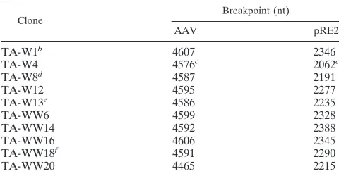
To extend the analysis of wt integration in normal diploid cells, we focused on junctions generated in WI-38 cells. A pooled infection was used to generate PCR junction products. The PCR products produced with the various primer combi-nations indicated in Fig. 1A were cloned into E. coli, and individual isolates were selected by colony hybridization to a ch-19-specific probe. Ten unique positive clones were then sequenced. The junction breakpoint locations within the AAV ITR and the ch-19 preintegration site are listed in Table 1. Breakpoints in the ch-19 integration site were observed within an approximately 270-bp region mapping to nucleotides (nt) 2062 to 2346 of the BamHI subclone containing the AAV ch-19 integration site described previously (40). The locations of these ch-19 breakpoints listed in Table 1 are shown in the map depicted in Fig. 1B. The cloned junctions contained breakpoints scattered throughout the putative hairpin struc-ture of the AAV ITR (Fig. 2A and 2D). Several of these junctions contained sufficient portions of the ITR to allow determination of the original orientation (flip or flop) of the hairpin. Six of these clones contained junctions with simple
FIG. 1. (A) Dot blot analysis of PCR amplification products generated from AAV-cell junctions. Genomic DNAs isolated from various cell lines that were latently infected with AAV are listed horizontally. PCR amplification was per-formed with various combinations of the AAV primers H, which hybridizes to AAV sequences at positions 400 to 381, and T, which hybridizes to positions 4498 to 4514, and/or the ch-19 cellular primer C, which hybridizes to the AAV preintegration site present on the cloned BamHI fragment found on ch-19. The various probes used are described in the text. (B) Locations (in nt) of chromo-somal breakpoints of cloned AAV–ch-19 junctions derived from WI-38 cell lines (black arrows), A9–ch-19 junctions (open arrows), and P5–ch-19 junctions (gray arrows). See the text for a more detailed description.
on November 9, 2019 by guest
http://jvi.asm.org/
[image:3.612.309.545.81.393.2]crossovers between viral (AAV ITR) and cellular (ch-19) se-quences. The breakpoint locations within the AAV ITRs of these clones are depicted in Fig. 2A.
The four remaining clones contained DNA sequences be-tween the breakpoint of the AAV terminal repeat and the ch-19 target sequence that could not be accounted for by a simple crossover between viral terminal repeat and cellular DNA sequences and are noted in Table 1. The lengths of these sequences inserted between contiguous viral and cellular se-quences ranged from 4 to 115 bp. These inserted sese-quences at the virus-cell junction matched DNA sequences present in the AAV genome. Two of these clones, TA-W1 and TA-WW18,
contained short stretches (#5 bp) of DNA that matched
se-quence further downstream within a complete AAV ITR. The presence of these sequences can also be accounted for by a simple deletion within the AAV ITR. The other two clones,
TA-W8 and TA-W13, contained significantly larger (.100 bp)
insertions at the virus-cell junctions. These insertions matched sequence immediately upstream of the P5 promoter in the AAV genome and were in an inverted orientation relative to the AAV ITR. One of them is a simple 113-nt insertion of nt 266 to 154 from AAV. The other contains a 118-nt insertion of nt 265 to 158 from AAV, with a rearrangement of DNA se-quence between nt 217 to 211.
No complete AAV ITRs were observed in any of the virus-host junctions derived from WI-38 latent cell lines (Table 1) that were cloned through PCR amplification with AAV prim-ers upstream of the start of the AAV ITR. Although it may be argued that the deletions in the AAV ITRs observed in these junctions are an artifact of the PCR amplification itself, it has been previously shown that it is possible to prepare complete and replication-competent AAV ITRs by PCR amplification (47). Furthermore, the junctions listed in Table 1 are similar to AAV-cell junctions characterized previously in aneuploid cell lines (23, 30, 40) in that incomplete ITRs and, in some cases, short stretches of nucleotides inserted at the virus-host junc-tion are observed. Moreover, AAV ITR-cell juncjunc-tions cloned from PCR amplification products (see below) derived from DD-Neo latent cell lines were identical to junctions that were cloned directly into E. coli. This result serves as a control and
indicates that the PCR reactions did not cause significant al-terations in the junctions that were characterized in this study.
To characterize junctions with breakpoints in the A9region
of the AAV ITR, a PCR amplification of virus-cell junctions that did not require polymerization through the entire AAV ITR was designed. Junctions from the HeLa latent cell lines G11, H3, and C11 were PCR amplified from genomic DNA by using the AAV primer TR6 and the ch-19 primer C. The AAV primer TR6 hybridizes to the A region of the AAV ITR and
matches sequence in the A9region (Fig. 2). Four independent
junctions (three from H3 and one from C11) that possessed
ITR breakpoints in the A9 region (nt 4655 to 4675) were
characterized. These junctions and their respective breakpoint locations within the AAV ITR and the ch-19 target region are listed in Table 2. AAV ITR breakpoints were located between nt 4655 and 4673, whereas ch-19 breakpoints were located between nt 1763 and 2119. The physical structure of the clone TA-H14 is depicted in Fig. 2B.
In addition to the expected A9-cell junctions, a second group
of novel junctions was produced by PCR amplification with the TR6-C primer pair. These junctions possessed AAV break-points that fell within the AAV P5 promoter region upstream of the start of the Rep open reading frame (nt 146 to 320) joined to ch-19 sequence, whose breakpoints fell between nt 1731 and 2294. The physical structure of the clone TA-H21 is also depicted in Fig. 2B. Nine junctions of this type (six from G11 and three from H3) were characterized and are listed in Table 2. Two of these junctions contained 11- to 12-bp inser-tions of DNA sequence at the virus-cell junction. These two groups of clones are consistent with virus-cell junctions
pos-sessing complete AAV ITRs with breakpoints within the A9
region or provirus structures involving a head-to-tail tandem structure with a virus-cell breakpoint in the P5 region of the AAV genome. However, because there is no information on sequence upstream of the TR6 primer, neither the integrity of
the ITR structure 59to the PCR primer nor the exact structure
of the provirus upstream of the A9region can be commented
on based on these results.
Besides the ITR- and P5-specific ch-19 junctions, the only other PCR product we obtained with any frequency involved viral head-to-tail junctions. Two unique clones containing head-to-tail junctions, obtained by using a combination of H and T primers, are shown in Fig. 2C. A physical map of the two unique junction sequences is depicted in Fig. 2C. Both of these junctions contain significant deletions within the AAV termi-nal repeat. The clone TA-ht5 crosses over from the A region of the tail terminal repeat into the D region of the head terminal repeat, while the clone TA-ht11 crosses over from the C region of the tail terminal repeat in the flop orientation into AAV sequence upstream of the P5 promoter at the head. While Southern blot analysis data supported the existence of frag-ments consistent with intact ITRs in these head-to-tail arrange-ments, none of the PCR products we isolated contained a complete terminal repeat. It is not clear, however, whether deletions and/or rearrangements contained in these head-to-tail clones were an artifact (47) or were produced during the integration process. The locations of all the unique AAV breakpoints within the putative terminal repeat hairpin of the AAV junction clones listed in Tables 1 and 2 are depicted in Fig. 2D.
Analysis of rAAV provirus structures.Numerous attempts
[image:4.612.49.289.81.206.2]to isolate an intact wt AAV provirus structure resulted in failure. While various clones could be isolated, they either were not stable in E. coli or rearranged as we propagated the bacteria as previously described (6) (data not shown). To char-acterize an intact AAV provirus structure, we focused on a
TABLE 1. AAV–ch-19 junctions from WI-38 latent cell linesa
Clone Breakpoint (nt)
AAV pRE2
TA-W1b 4607 2346
TA-W4 4576c 2062c
TA-W8d 4587 2191
TA-W12 4595 2277
TA-W13e 4586 2235
TA-WW6 4599 2328
TA-WW14 4592 2388
TA-WW16 4606 2345
TA-WW18f 4591 2290
TA-WW20 4465 2215
aJunctions were cloned as described in Materials and Methods. AAV and
pRE2 breakpoint locations are based on nucleotide map positions as described in reference 40.
bContains a 4-bp insertion (CACT) between the AAV and pRE2 breakpoints. cPreviously published in reference 40.
dContains an insertion of AAV nt 265 to 158 between the AAV and pRE2
breakpoints.
eContains an insertion of AAV nt 266 to 155 between the AAV and pRE2
breakpoints.
fA 5-bp insertion (TGGTC) between AAV and pRE2 breakpoints matches
sequence in B9of the AAV ITR. Inserted DNA sequences are listed reading from the AAV ITR out toward the cellular junction sequence.
on November 9, 2019 by guest
http://jvi.asm.org/
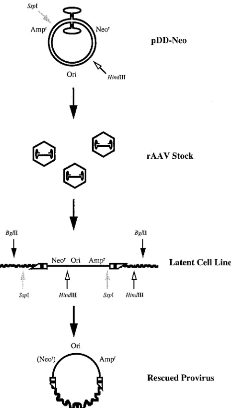
rAAV vector containing neomycin and ampicillin resistance genes and a bacterial origin of replication. The construct, DD-Neo, is shown in Fig. 3. Recombinant stocks of DD-Neo were generated from the unique vector plasmid pDD-Neo as
previ-ously described (47). The strategy for retrieving intact rAAV provirus, also outlined in Fig. 3, involved direct cloning of the vector from genomic DNA isolated from established latent cell lines (G418 resistant) into E. coli by using the bacterial origin
FIG. 2. (A) Structural maps of unrearranged AAV–ch-19 junctions derived by PCR amplification of genomic DNA from latently infected WI-38 cell lines. Nucleotide maps of the AAV ITR in its two possible orientations are shown at the top. (B) Structural maps of representative AAV–ch-19 junctions derived by PCR amplification of latently infected HeLa cells with AAV primer TR6 and ch-19 primer C. (C) Structural maps of head-to-tail structures derived by PCR amplification with AAV primers H and T of genomic DNA from HeLa cells latently infected with AAV as previously described. The AAV terminal repeat regions are indicated as previously described. (D) Physical location of the AAV junction breakpoints within the putative AAV terminal repeat hairpin structure. Asterisks indicate junctions derived by PCR amplification with the AAV primer TR6. Green arrows indicate junctions that contained insertions of nucleotides between ITR and cellular sequences. The nucleotides in the B-B9and C-C9regions are listed as N, since the purpose of this map is to indicate physically the portion of the ITR remaining at the junction, regardless of the ITR orientation (flip or flop).
on November 9, 2019 by guest
http://jvi.asm.org/
[image:5.612.93.509.73.597.2]of replication and an ampicillin selection marker contained on the vector. Detroit 6 cells were used for infection with recom-binant viral stocks of DD-Neo in the presence of the selective antibiotic geneticin G418. Individual drug-resistant colonies were selected, and cell lines that had undergone single or few vector integration events, as confirmed by Southern analysis, were chosen for provirus cloning. All of these clones were negative for rescue when assayed by wt AAV-adenovirus coin-fection.
Cloning of the rAAV provirus.Genomic DNA from six
DD-Neo latent cell lines established in Detroit 6 human cells was isolated and digested with BglII, a restriction enzyme that does not cut the DD-Neo vector. Southern hybridization analysis of
BglII-digested DNA from six latent cell lines with a probe
specific for the DD-Neo vector indicated the presence of unique BglII fragments larger than the 4-kb vector itself in five of the six cell lines (data not shown). One cell line, DD2, exhibited two DNA fragments that hybridized to the vector probe that were larger than pDD-Neo. Digestion of genomic DNA with HindIII or SspI, each of which cuts the vector once, revealed the presence of two DNA fragments that contained vector sequences, whereas the cell line DD2 showed four DNA fragments that hybridized to the vector probe (data not shown). These data supported single or relatively simple inte-gration patterns, which were further characterized by direct genomic cloning of the provirus.
BglII-digested genomic DNA was used for direct cloning of
plasmids containing vector-cell DNA junctions. The digested DNA was recircularized by T4 DNA ligase, and the ligation mixture was used to transform E. coli. Ampicillin-resistant trans-formants were grown in liquid medium, and the resulting plas-mid clones were isolated and characterized. Six plasplas-mid clones from five independent cell lines, chosen on the basis of size
(.4 kb) and the presence of restriction fragments
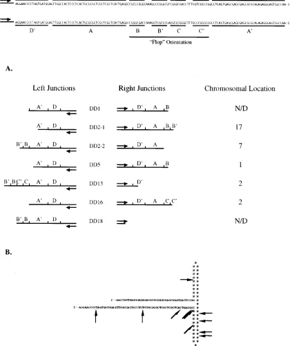
character-istic of the DD-Neo vector, were characterized. Two indepen-dent clones, DD2-1 and DD2-2, were isolated from the cell line DD2. Restriction enzyme mapping of the provirus clones re-vealed that the vector DD-Neo had integrated into the host genome intact in all cases, with breakpoints located in or near
the terminal repeats (Fig. 4A). One clone, derived from the latent cell line DD18, possessed a breakpoint in the vector about 200 bp downstream from the ampicillin resistance gene. DNA sequence analysis of the provirus clones indicated that none of the vector integrants contained a complete viral ter-minal repeat at the virus-cell junction (Fig. 4A). The break-points within the vector ITR at the crossover between vector
and host for both the left (59end, or head) and right (39end,
[image:6.612.50.290.88.252.2]or tail) junctions are shown in Fig. 4A. As with the cloned wt AAV junctions, several of these DD-Neo junctions contained sufficient portions of the terminal repeats to indicate the vector orientation of the terminal repeat (e.g., flip for the right junc-tion of DD16 and flop for the right juncjunc-tion of DD5). The locations of these cloned vector junction breakpoints within
TABLE 2. AAV junctions from HeLa cells amplified with primers TR6 and Ca
Clone Breakpoint (nt)
AAV pRE2
TA-H14 4673 1991
TA-H15 4674 1957
TA-H20 4668 2119
TA-C25 4655 1763
TA-G2b 269 1827
TA-G5 264 1930
TA-G6 294 1783
TA-G8 270 1744
TA-G9 281 2099
TA-G12c 262 2120
TA-H16 160 1838
TA-H21 288 2222
TA-H23 278 2307
aJunctions were cloned as described in Materials and Methods. AAV and
pRE2 breakpoint locations are based on nucleotide map positions as described in reference 40.
bContains an 11-bp insertion (TTAAATACCCA) between the AAV and
ch-19 breakpoints.
cContains a 12-bp insertion (TGGGTATTTAAG) between the AAV and
[image:6.612.311.543.245.655.2]ch-19 breakpoints.
FIG. 3. Strategy for the construction, establishment, and isolation of rAAV provirus DNA. pDD-Neo was used to generate recombinant virus stocks, which in turn were used to establish DD-Neo latent cell lines. Genomic DNAs isolated from individual drug-resistant colonies were digested with restriction enzymes that either cut outside the vector genome or cut once in the vector, recircularized with DNA ligase, and propagated in E. coli. The resulting rescued proviruses containing vector-cell junctions were sequenced and further characterized by Southern analysis and by FISH to human chromosomes.
on November 9, 2019 by guest
http://jvi.asm.org/
the putative AAV terminal repeat hairpin are depicted in Fig. 4B.
Attempts to clone a complete provirus from the latent cell line DD1 by the techniques previously described proved un-successful. Southern analysis of the genomic DNA from the latent cell line DD1 indicated that the provirus was contained
on a BglII fragment in excess of 30 kb (data not shown). To characterize the junctions for this provirus, genomic DNA was digested with either HindIII or SspI and was recircularized as described for BglII genomic DNA, and the resulting pool was used to transform E. coli. By using one of these enzymes that uniquely cuts the vector, the left and right junctions of the
FIG. 4. (A) DD-Neo rAAV terminal repeat sequences present at left- and right-hand vector-cell junctions of individual latent cell lines. Proviruses whose chromosomal locations have been mapped by hybridization to rodent-human somatic cell hybrid panels (see text for description) are indicated on the right. A nucleotide map of the AAV ITR is shown at the top in its two possible orientations (flip or flop). N/D, not determined. (B) Physical locations of the DD-Neo junction breakpoints within the putative AAV terminal repeat hairpin. The nucleotides in the B-B9and C-C9regions are listed as N, since the purpose of this map is to indicate physically the portion of the ITR remaining at the junction, regardless of the ITR orientation (flip or flop).
on November 9, 2019 by guest
http://jvi.asm.org/
[image:7.612.88.508.97.598.2]integrated provirus may be cloned out separately (Fig. 3). Ampicillin-resistant clones containing provirus junction se-quences were isolated from both ligation pools and character-ized. DNA sequence analysis revealed that a single type of clone was derived from each pool. Breakpoints in the ITR for the junctions from DD1 are also shown in Fig. 4A. As with the complete proviruses cloned from the other five latent cell lines, neither junction contained a complete AAV terminal repeat. However, the right junction sequence for DD1, derived from the cloning of HindIII digestion fragments, contains a 12-bp stretch of sequence, inserted at the vector-cell junction,
match-ing sequence present in the C9region of the AAV ITR. As with
the AAV–ch-19 junctions described earlier in this report, this result can be accounted for by either an insertion of this
frag-ment at the vector-cell junction or a deletion of the B9and C
sequences within the AAV ITR.
As with the cloning of PCR amplification products, the clon-ing and propagation of rAAV junctions may have altered the vector-cell junction sequences, given the potential for instabil-ity of AAV terminal repeat sequences in E. coli (8, 28, 37, 39). To address this question, vector-cell junction sequences de-rived from the provirus clones described in this work were compared to junctions cloned through PCR amplification of genomic DNA. Primers specific to cellular DNA sequences derived from junction clones were used with primers specific to the head and tail regions of the DD-Neo vectors. Agarose gel mobility and DNA sequence analysis revealed that the PCR products of junctions amplified from genomic DNA and the respective provirus clones were identical in both size and se-quence (data not shown). Furthermore, the single-copy provi-rus in DD16 was directly cloned in independent experiments, and the vector-cell junctions from both experiments were iden-tical in sequence. While it may be argued that both techniques may be subject to potential deletions, it is unlikely that two independent protocols would produce identical artifacts. These results together indicate that the junctions cloned in this work, either directly or from PCR amplification products, are authentic and not experimental artifacts.
Analysis of the first 500 bp of cellular DNA sequence ad-joining the vector-cell crossover of these cloned proviruses indicated that none of the sequences present at either the left or right junctions in any of the seven clones shared identity (data not shown). Furthermore, when compared to the ch-19 preintegration site for AAV, none of the sequence obtained from these vector-cell junctions matched the sequence of the
BamHI subclone (40) containing a 2.7-kb fragment of the
AAV preintegration site or the 4.1-kb EcoRI fragment AAVS1 (24), supporting non-ch-19-targeted integration (see Fig. 7 and data not shown).
Characterization of vector integration loci and chromosome
mapping.Using the DNA sequence obtained from the
provi-rus-cell junctions of the plasmid clone pDD16, PCR primers were designed to produce DNA probes for the left and right
junctions of this provirus. Random primed 32P-labeled
frag-ments of the left and right junctions were used to probe BglII-digested total genomic DNA from several latent cell lines. Figure 5A depicts a Southern blot of genomic DNA from several cell lines latently infected with DD-Neo and probed
with a32P-labeled DNA fragment specific to the right junction
of the provirus in the latent cell line DD16. In all cell lines, the probe hybridized to a DNA fragment corresponding to a mo-lecular weight of approximately 4 kb. In addition, the probe hybridized to a DNA fragment corresponding to a molecular weight of approximately 8.5 kb in the cell line DD16 and to a 10-kb BglII fragment present in the cell line DD13. The 8.5-kb and the 10-kb BglII fragments from the latent cell lines DD16
and DD13, respectively, comigrated with the provirus clones derived from those two cell lines (data not shown). The left-junction probe from DD16 hybridized to a 7-kb BglII fragment from all of the latent cell lines, as well as to an 8.5-kb fragment in the cell line DD16 and a 10-kb fragment in DD13 (Fig. 5B). These results support integration by two independent rAAV vectors into a cellular DNA sequence. Furthermore, Southern analysis of the preintegration site indicates that the left and right junctions of DD16 are not contiguous, since the left and right junction probes from DD16 hybridize to different geno-mic DNA fragments (Fig. 5A and B). These results suggest that rearrangement of the genomic DNA occurs during or after vector insertion, similar to the wt (data not shown). This observation is of extreme interest since an independent study has also mapped the integration of rAAV vectors into ch-2, suggesting that a preferred integration is being observed with these vectors (20).
Rodent-human somatic cell hybrid panels digested with
BamHI, each containing the total DNA from an individual
human chromosome, were used to determine the chromo-somal locations of the left and right junctions of the provirus cloned from the latent cell line DD16. Both probes hybridized to the panel corresponding to ch-2. Neither fragment
hybrid-FIG. 5. (A) Southern blot of genomic DNA isolated from individual cell lines latently infected with DD-Neo, as described in the text, and probed with cellular sequences from the right-hand junction derived from the cell line DD-16. Lane assignments are indicated horizontally, and molecular size markers (in kilobases) are noted vertically. Lanes: 1, DD5; 2, DD13; 3, Detroit 6; 4, DD16; 5, DD18. (B) Southern blot of genomic DNA isolated from individual latent cell lines as described in the text and probed with cellular sequences from the left-hand junction derived from the cell line DD-16. Lane assignments are the same as for panel A. (C) Southern blot of a somatic cell hybrid panel (ONCOR, Inc.) probed with cellular junction sequences derived from the cell line DD-16. Marker loca-tions and lane assignments are indicated horizontally.
on November 9, 2019 by guest
http://jvi.asm.org/
[image:8.612.311.545.69.373.2]ized to hamster or mouse DNA. The left-junction probe hybri-dized to a 10-kb BamHI fragment and exhibited some weaker cohybridization to a similarly sized fragment in ch-1 (Fig. 5B). The right-junction probe hybridized to a 7-kb BamHI frag-ment, with no cohybridization to any other panel (data not shown). A probe specific for the left junction from another clone of the cell line DD2 exhibited hybridization to a 1-kb
BamHI fragment present on ch-17, as well as sequences
present in the hamster (data not shown). The locations of the junction breakpoints within the vector ITR of the individual proviruses are shown in Fig. 4A, and their locations within the putative ITR hairpin are shown in Fig. 4B. The cellular se-quences at the vector-cell junctions of the provirus clones from the cell lines DD5, DD16, DD2-2, and DD2-1 were used as probes, and they have been mapped to chromosomes 1, 2, 7, and 17, respectively, by use of the same rodent-human somatic hybrid panels (Fig. 4A). There is a remote possibility, however, that these sequences have been translocated in Detroit 6, given their aneuploid nature or as a result of the integration event itself. Although this possibility cannot be ruled out at this time, FISH analysis of the latent cell lines supported the results obtained with the somatic cell hybrid panel (data not shown). In situ chromosome and DNA sequence analysis of the ch-2
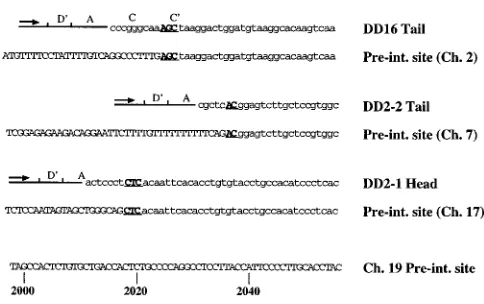
provirus preintegration site.The cosmid pHC-RJ, which
con-tains the right-junction preintegration region for the provirus contained in the cell line DD16, was isolated and used as a probe for in situ chromosome analysis to further map the location of this preintegration region in Detroit 6 cells. The hybridization signals localized exclusively to ch-2, as shown in Fig. 6A. DAPI (G/Q) banding and FLpter analysis (64 to 70%) of approximately 20 metaphase spreads showed that the pHC-RJ signals were located within the q24-q31 subregion. Several individual examples from independent metaphase spreads are shown in Fig. 6B. The minor differences in size of the individual chromosomes can be attributed to the different states of condensation during metaphase for chromosomes coming from different spreads. The localization of the hybrid-ization signals is diagrammed by the ideogram in Fig. 6C. The cosmid pHC-RJ was sequenced with primers derived from cellular sequences from the plasmid pDD16. The left-junction probe for DD16 did not hybridize to this cosmid, consistent with the results we observed for Southern analysis (Fig. 5A and 5B), and this is indicative of rearrangement (deletion and/or translocation) of genomic DNA at the provirus integration site.
The preintegration sequence surrounding the right (39, or
tail) vector-cell junction for DD16 is shown in Fig. 7, along with the preintegration sequences for the left junction of DD2-1, located on ch-17, and the right junction of DD2-2, located on ch-7. Comparison of terminal repeat and cellular preintegration sequences reveal 2 or 3 bp (depicted in boldface in Fig. 7) of microhomology that cannot be distinguished in viral and cellular DNA. rAAV provirus structures were similar to wt integrants in that both flip and flop orientations of the ITRs were observed, microhomology between the viral and cellular breakpoints existed, and vector integration resulted in rearrangement of cellular DNA. In addition, we saw an accu-mulation of similar breakpoints between rAAV and wt ITR sequences (compare Fig. 2C with Fig. 4B). Unlike the wt, rAAV vectors did not appear to integrate as tandems, and although we observed a common integration site on ch-2, tar-geting to ch-19 was completely lost.
In vitro integration.The in vitro integration of several AAV
and rAAV DNA substrates into the AAV ch-19 target site located on a circular plasmid (pRE2) was performed under conditions described earlier in Materials and Methods. Four different AAV DNA substrates were examined for their ability
to integrate in vitro into the ch-19 AAV integration sequence contained on the plasmid pRE2: no-end AAV DNA, described previously (34, 41); the linear XbaI fragment derived from AAV plasmid SSV9; the SSV9-derived linear SmaI fragment; and the SSV9-derived linear PvuII fragment. These AAV sub-strates are depicted in Fig. 8A.
In vitro integration of no-end AAV substrates. The DNA
sequences of 24 individual clones derived from the PCR am-plification of products of in vitro integration reactions between no-end DNA and pRE2 were obtained. Two primer combina-tions were used in the PCR amplification of the products from the in vitro integration reactions: primer pair H-C and primer pair T-C. These clones and the locations of the virus-cell breakpoints are listed in Table 3. Integration reactions carried out with extracts of 293 cells that had been transfected with pHIV-Rep are noted as such. Integration junctions were ob-tained both in the presence and absence of Rep in the cell extract.
Nineteen independent junction clones were isolated from the PCR amplification of products of in vitro integration re-actions between no-end AAV DNA and pRE2 with primers C and H. Of these junction clones, 11 contained breakpoints that were located in the AAV terminal repeat region (nt 4 to 184). The remaining junction clones contained breaks within the P5 promoter region of AAV (nt 192 to 352). None of these virus-cell junctions contained a complete, intact AAV terminal re-peat. A map of the physical locations of the AAV breakpoints of the 11 in vitro junction clones that were located in the AAV ITR is depicted in Fig. 8B. Breakpoints in the pRE2 target DNA for 15 of these clones mapped between nt 1903 and 2377. The pRE2 (ch-19) breakpoints from these junctions are de-picted in Fig. 8C. The four remaining clones contained rear-rangements of the substrate and/or target DNA sequences. Insertions of these sequences were observed in these clones between either an AAV breakpoint in the P5 region or the AAV terminal repeat and a pRE2 breakpoint mapping be-tween nt 2070 and 2395. The rearranged DNA sequences are noted in Table 3. Two of these clones, 34.4.5 and 15.5.9, con-tained insertions of AAV sequences from the capsid coding regions. The other clones contained more complex rearrange-ments, in which insertions of both AAV- and pRE2-associated sequences occurred in both direct and inverted orientations. All integration junctions that contained insertions or inver-sions of AAV and/or target sequences at the junction break-points were generated with 293 cell extracts in the absence of Rep protein.
In vitro integration junctions from five individual clones derived by PCR amplification with the T-C primer combina-tion were also characterized. All of the junccombina-tions cloned with the T primer contained AAV breakpoints within the AAV terminal repeat region (nt 4489 to 4675) and were located within a 10-bp region surrounding the Rep nicking site (nt 4566). Breakpoints in pRE2 of these clones were located be-tween nt 2042 and 2202. One of these junction clones con-tained an insertion of AAV sequences from nt 2358 to 2639, between the AAV ITR and pRE2 breakpoints. These clones are also summarized in Table 3, and the physical map locations of the AAV ITR and pRE2 breakpoints are also shown in Fig. 8B and C.
In vitro integration of linear AAV derivatives into pRE2.In
vitro integration junctions derived from linear, double-strand-ed AAV DNA substrates that contain portions of the complete AAV terminal repeats and pRE2 were PCR amplified with primers H and C and cloned in a manner similar to junctions derived from the in vitro integration of no-end DNA. These linear AAV substrates were derived from the AAV plasmid
on November 9, 2019 by guest
http://jvi.asm.org/
SSV9. The PvuII fragment contains the entire linear AAV genome with two complete intact AAV terminal repeats, while the SmaI fragment contains the AAV genome and half of each AAV terminal repeat. The XbaI fragment contains the AAV genome without AAV terminal repeat sequences. Integration junctions between these substrates and pRE2 were isolated by
PCR amplification and TA cloning as previously described for no-end DNA. The junctions that were characterized in these experiments are listed in Table 4.
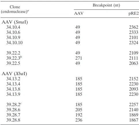
No junction clones were obtained from the linear PvuII fragment. Fifteen junctions between pRE2 and the SmaI and
XbaI linear AAV substrates were cloned and characterized. Six
FIG. 6. (A) FISH of metaphase human chromosome spreads. (B) Fluorescein isothiocyanate-conjugated avidin was used to detect hybridization signals by use of cellular junction sequences derived from the cell line DD16, while a counterstain with DAPI generated the G/Q banding pattern. Individual ch-2 from several metaphase spreads indicating hybridization to the long arm in the interval q24-q31. (C) Ideogram of ch-2 indicating locations of hybridization signals determined by using the previously described probe.
on November 9, 2019 by guest
http://jvi.asm.org/
[image:10.612.95.506.67.604.2]of the seven junctions obtained from the in vitro integration of the SmaI linear AAV substrate into pRE2 were joined at the
SmaI site terminus, whereas five of the eight junctions
ob-tained from the in vitro integration of the XbaI linear AAV substrates into pRE2 were joined at the XbaI site terminus. The remaining substrate-target junctions that were cloned con-tained deletions of DNA sequences from the end of the AAV substrate from the SmaI site (nt 46) or the XbaI site (nt 184). Breakpoints for these junction clones in the pRE2 target se-quence were located between nt 1867 and 2367.
DISCUSSION
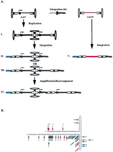
In this report, we have characterized the sequence structure of integrated wt AAV proviruses located in ch-19 of diploid and aneuploid cells and the structure, sequence, and location of the provirus of rAAV from latent tissue culture cells and described what we believe to be the first analysis of in vitro integration of various AAV substrates into a plasmid contain-ing the human ch-19 target site. From these results, the fol-lowing conclusions can be made. Establishment of wt AAV latency primarily involves head-to-tail viral structures, with the ITRs and p5 promoter sequences being involved in the cross-over (Fig. 9A). Virus integration typically results in duplication and inversion of virus and/or host DNA sequences at the target site. Breakpoints in the chromosome and ITR are clustered (Fig. 9B) and, although not sequence specific, show micro-homology between ITR and host DNA sequences at the cross-over point. Characterization of direct clones of single-copy rAAV proviruses indicates that the vector genome integrates intact, with no rearrangement of vector sequences except for the ITRs which form the crossover points (Fig. 9A). The struc-ture of the host genome is rearranged to some extent, and the vector has lost specificity for ch-19. As with wt, rAAV also demonstrates breakpoints in the ITR that are clustered (Fig. 9B). Analysis of in vitro junctions between various AAV sub-strates and the AAV ch-19 target site indicates that the termi-nal-repeat hairpin is the likely substrate required for integra-tion in the establishment of viral latency. Substrate-target DNA sequence rearrangements sometimes seen in vitro are consistent with the structures seen in wt AAV integration. The
observed junctions seen in all these systems indicate that wt, recombinant, and in vitro AAV integration uses a hairpin ITR for an integration substrate (Fig. 9B) and generates cellular junctions that are similar (exhibiting microhomology) and sug-gests that the integration event itself is mediated for the most part by cellular factors.
AAV-rAAV integration and its requirements.To date, the
cloning of a complete wt AAV provirus has proven to be elusive. Recently, Berns and coworkers have proposed a model describing the site-specific integration of AAV (30, 31). This model requires the Rep binding site (RBS) and the Rep nick-ing site (terminal resolution site) and involves multiple strand switching during novel DNA strand synthesis within a Rep-mediated complex formed between AAV and the AAV target site. The multiple-strand switches proposed in this model could account for the various provirus structures we observed. Fur-thermore, additional cellular machinery required for DNA replication and repair is necessary for completion of the inte-gration process. The results obtained in this study character-izing wt AAV, rAAV, and in vitro junctions indicate that this cellular machinery and the AAV ITR are sufficient for AAV or rAAV integration. At the same time, however, these results do not infer or imply any less of a role of the viral Rep protein in the targeted integration observed for wt AAV and how it may affect the formation of some of the various provirus structures observed during the establishment of viral latency.
If the hairpin structure is the correct substrate for tion and intact ITRs are not retained during or after integra-tion, then the presence of head-to-tail tandems in provirus structures would strongly support the occurrence of a replica-tion step prior to integrareplica-tion, as illustrated in Fig. 9A, step I. This tandem substrate could then participate in integration, forming the virus-cell breakpoints we observed (Fig. 9A, steps II and III), and retain sufficient sequences of the ITR for the rescue process (39) through the head-to-tail tandem. A similar model was proposed in 1993 to explain how rescue and repli-cation could ensure with provirus structures that contained rearranged ITR junctions (36). While this model is attractive, a primary concern with analysis of latent cell lines for struc-tural information on the mechanism of AAV integration is the possibility of subsequent events occurring before characteriza-tion can take place (2 to 4 weeks to derive clonal cell lines). This concern is echoed in the literature with the observation that high-passage-number, latently wt-AAV-infected cell lines produce free-monomer AAV genomes, suggesting that a dy-namic provirus state exists even after extended latency (11, 32). It is very possible that the complex viral structures we have characterized with wt AAV in this study are a result of vector amplification and rearrangement after integration (Fig. 9A, step IV). Transient expression of Rep would be sufficient to initiate a rescue step, which in turn could generate nicked substrates that would initiate replication in situ, generating substrates reminiscent of the onion skin model proposed for simian virus 40 in the early 1980s. The results support the existence of a complex provirus structure in latently infected cell lines, which is consistent with previous observations (5, 11, 27, 32, 46); this complex structure has been reproduced in an EBV episome containing the AAV ch-19 target sequence, in which the provirus frequently possesses a head-to-tail structure (15).
[image:11.612.49.291.64.216.2]An effort to characterize AAV integration without the con-cern of rearrangement during the extended time period re-quired to generate clonal cell lines has supported the use of the transient integration system established by Berns and col-leagues. Several classes of AAV–ch-19 junctions were ob-served in the study of wt AAV integration into an EBV shuttle
FIG. 7. DNA sequences of the vector-cell junctions for three independent provirus clones and their respective chromosomal preintegration loci. The ch-19 preintegration (Pre-int.) locus is shown at the bottom of the figure. DNA quences upstream of the integration site are in capital letters, while DNA se-quences that cannot be differentiated from vector or host DNA sese-quences are in boldface and underlined. The preintegration region of ch-19 displayed is from nt 1998 to 2056 of the BamHI subclone containing the AAV target site described previously (40), where the majority of AAV–ch-19 junctions were observed in that study.
on November 9, 2019 by guest
http://jvi.asm.org/
vector containing the ch-19 AAV target site (15, 16). In addi-tion to the juncaddi-tions between viral and ch-19 sequences formed within the AAV ITR, virus-host junctions were observed at or near the AAV P5 promoter, as well as between AAV se-quences and the EBV episome or unknown DNA sese-quences (15). In this report, we characterized several AAV–ch-19
junc-tions cloned from both diploid and aneuploid cell lines. In addition to the previously described junctions formed between the AAV ITR and the target site, we cloned several junctions that involved virus-cell crossovers within the AAV P5 pro-moter, junctions which parallel those observed previously in the EBV shuttle vector integration system. These results
sug-FIG. 8. (A) AAV DNA substrates for in vitro integration reactions derived from the AAV plasmid SSV9. The AAV ITR sequences are shaded. No-end DNA has been described previously (40). Locations of the XbaI, SmaI, and PvuII restriction sites are as indicated. (B) Breakpoint locations within the AAV ITR for in vitro integration junctions generated between pRE2 and no-end DNA. ITR breakpoints derived by PCR amplification of the head (59end; black arrows) and tail (39end; open arrows) junctions are as indicated. (C) pRE2 (ch-19) breakpoint locations from in vitro integration reactions. Black and open arrows indicate where ch-19 sequences are joined to head and tail ITR sequences, respectively. The gray arrows indicate ch-19 breakpoints joined to AAV sequences within the P5 promoter region.
on November 9, 2019 by guest
http://jvi.asm.org/
gest that the integration of wt AAV in the EBV shuttle vector retains many of the characteristics that have been described for the integration of wt AAV at ch-19. Since no other prominent viral sequences involved with cellular junctions were observed, it suggests that the RBS located in the P5 promoter is most likely responsible for the involvement of this DNA at the crossover points. It should also be noted that although the virus-cell junctions appear similar in both systems, 9 of over 40 AAV clones from the EBV shuttle vector contained the com-plete AAV genome, and only 2 of these exhibited the ability to rescue. This is in contrast to latently wt-AAV-infected cell lines, which exhibit significantly higher frequencies of viral rescue. This suggests that there may be as-yet-unidentified differences between the transient integration and stable latent cell lines.
Regardless, one significant question we addressed in this study is the potential difference in the establishment of viral
latency in aneuploid (D6 and HeLa) versus normal diploid
(WI-38) cells. Because of the finite lifetime of these cells (506
10 doublings), a mixed population of infected cells was char-acterized. However, the results (shown in Fig. 1A and 2D) are consistent with data from studies with HeLa cells and the latently AAV-infected cell line Detroit 5, which strongly sup-port the observation that there is no significant difference be-tween wt AAV infection of normal diploid cells and that of aneuploid cells.
Cloning of the rAAV provirus and its structural
character-ization.We have also cloned complete rAAV proviruses from
several individually established latent cell lines. Even though our screen was set up to characterize low-copy-number inte-grants, in all cases the provirus remained intact, without any of the complex structures observed with the wt virus; e.g., head-to-tail tandem arrays were not present in these cell lines, nor was there rearrangement of the rAAV genome. All of the observed vector-cell junctions were in or near the AAV ITR. Furthermore, none of the cloned rAAV proviruses was located at the ch-19 target site characterized for the wt virus, and none of the junction sequences shared significant homology with ch-19. It must be noted that the latently rAAV-infected cell lines were established in the absence of Rep protein, which has previously been suggested to be required for targeted integra-tion of wt AAV at ch-19 (7, 12, 45). Given the absence of Rep in the establishment of these cell lines, targeted integration of rAAV to ch-19 might not be expected. Although none of these proviruses was located in ch-19, Southern analysis of the par-ent cell lines indicated that two proviruses were located in the same chromosomal locus on ch-2. This is of interest since an independent observation of vector integration into ch-2 has been reported (20). It is not clear whether this represents a recombination hot spot in aneuploid cell lines or discriminat-ing potential of the vector in the absence of Rep.
The cellular junction sequences obtained from cloned
pro-TABLE 3. In vitro integration junctions from no-end DNA and pRE2
Clone Rep6a
Breakpoint (nt)b
AAV pRE2
Derived from PCR amplification with primers H and C
14.4.30 2 285 2200
14.4.38 2 270 2108
14.6.15 1 111 2285
14.6.21 1 120 2377
14.6.22 1 280 2135
14.6.40 1 120 2174
14.5.3.Z 2 111 2282
14.5.6.Z 2 140 2193
14.5.9.Zc 2 115 2395
14.6.2.Z 1 35 2194
14.6.5.Z 1 255 1903
14.6.7.Z 1 248 2021
14.6.15.Z 1 195 1972
14.6.17.Z 1 88 2056
14.6.19.Z 1 73 2200
34.4.5d 2 352 2070
34.4.6 2 71 2013
34.4.11e 2 245 2384
34.4.12f 2 153 2283
Derived from PCR amplification with primers T and C
15.5.7 2 4566 2042
15.5.8 2 4568 2199
15.5.9g 2 4567 2202
15.5.11 2 4564 2201
15.5.12 2 4572 2192
a1, Rep protein present during integration;2, Rep protein absent during
integration.
bNucleotide locations are based on map locations as described in reference 40. cContains an insertion of pRE2 nt 105 to 433 joined to pRE2 nt 523 to 466
between the indicated breakpoints.
dContains an insertion of AAV nt 3635 to 3027 between the indicated
break-points.
eContains an insertion of pRE2 nt 387 to 666 joined to AAV nt 4275 to 4424
and pRE2 nt 995 to 1180 between the indicated breakpoints.
fContains an insertion of pRE2 nt 4542 to 4421 between the indicated
break-points.
gContains an insertion of AAV nt 2358 to 2639 between the indicated
[image:13.612.50.292.91.428.2]break-points.
TABLE 4. In vitro integration junctions from linear AAV substrates and pRE2
Clone (endonuclease)a
Breakpoint (nt)
AAV pRE2
AAV (SmaI)
34.10.4 49 2362
34.10.6 49 2333
34.10.9 49 2101
34.10.10 49 2324
39.22.2 49 2109
39.22.3b 271 2111
39.22.5 49 2063
AAV (XbaI)
34.13.2 185 2152
34.13.4 185 2230
34.13.8 185 2093
34.13.9 185 2230
39.28.2c 185 2257
39.28.6 205 2140
39.28.7 192 1869
39.28.8 236 1867
aJunction clones were derived by PCR amplification with primers H and C. bContains an insertion of AAV nt 460 to 526 between the indicated
break-points.
cContains an insertion of pRE2 plasmid backbone nt 3879 to 3818 between
the indicated breakpoints.
on November 9, 2019 by guest
http://jvi.asm.org/
[image:13.612.307.548.467.685.2]FIG. 9. (A) Scheme outlining the integration of AAV-rAAV. Rep binding sites are indicated by the black boxes on the AAV genome (in the A stem of the ITR or in the P5 promoter, as indicated by the arrow) and at the chromosomal integration site. The pathway on the left describes the integration of wt AAV, in which head-to-tail multimers of AAV may be generated in replication prior to integration (I) or in an amplification-rearrangement step following integration (IV). The integration event may include deletion, duplication, and/or inversion of host DNA sequences, which can occur at either of these two steps. Potential provirus integration structures are also depicted, which include junctions in the P5 promoter (II) or the AAV ITR (III). The pathway on the right outlines the integration of rAAV, in which a single copy of the unrearranged vector is inserted in the genome (V). Rearrangement of the host genome occurs during the integration event. This must also be a pathway for wt AAV, based on results described in reference 15. See the text for further details. This figure is not meant to be exhaustive and may not be complete in detail. (B) Physical map of the locations of junction breakpoints within the putative AAV ITR for in vitro integration junctions (in black), along with junction breakpoints from wt AAV (in red, WI-38 junctions without rearrangements; in green, junctions with insertions at breakpoints) and rAAV (DD-Neo; in blue). Asterisks indicate junctions derived by PCR amplification with the AAV primer TR6. The nucleotides in the B-B9and C-C9regions are listed as N, since the purpose of this map is to indicate physically the portion of the ITR remaining at the junction, regardless of the ITR orientation (flip or flop). See the text for a more detailed description.