STATURE ESTIMATION FROM HAND LENGTH AND FOOT LENGTH IN
ADULTS – A REGIONAL STUDY IN CHENNAI, TAMILNADU.
Dissertation submitted to THE TAMILNADU Dr. M.G.R.MEDICAL
UNIVERSITY in partial fulfillment for the award of the degree of
DOCTOR OF MEDICINE IN FORENSIC MEDICINE
Branch XIV
INSTITUTE OF FORENSIC MEDICINE
MADRAS MEDICAL COLLEGE
CHENNAI - 600 003
March, 2008
CERTIFICATE
This is to certify that the dissertation entitled, “Stature
estimation from hand length and foot length in adults – A
regional
study
in
Chennai,
Tamilnadu”
submitted
by
Dr.K.S.M.Venkatachalam, in partial fulfillment for the award of the
degree of Doctor of Medicine in Forensic Medicine by the
Tamilnadu Dr. M.G.R. Medical University, Chennai is a bonafide
record of the work done by him in the Institute of Forensic Medicine,
Madras Medical College, during the academic year 2005 – 2008.
Dean,
Director and Professor,
Madras Medical College &
Institute of Forensic Medicine,
Government General Hospital,
Madras Medical College,
TABLE OF CONTENTS
Sl. No.
SECTIONS
Acknowledgement
Abbreviations
I
INTRODUCTIONII
STUDY OBJECTIVESIII
REVIEW OF LITERATUREIV
MATERIALS AND METHODSV
RESULTS AND DISCUSSIONVI
CONCLUSIONVII
SUMMARYVIII
BIBLIOGRAPHYIX
APPENDICES• Study Protocol
ACKNOWLEDGEMENT
At the outset, I thank the souls which the God has provided me for this study. I pray for the souls to rest in peace.
I am greatly obliged to the Dean, Dr. T.P. Kalanithi MD, Madras Medical College and Government general hospital for allowing me to conduct this study by utilizing the patients as my study subjects.
I immensely thank our Director and professor of Forensic Medicine Dr.
R. Vallinayagam MD. for his expert guidance in bringing out this dissertation
for my M.D. exam.
I thank professor of Forensic Medicine Dr. P. Ravisankar MD, DHA. Presently Dean, Govt. Medical College, Tiruchirapalli for his kind suggestion in executing this project.
I also thank Director and professor of Internal medicine Dr.
Thirumalaikolundu Subramanian MD., for his novel ideas in designing the
study protocol.
I am thankful to Dr. R. Selvakumar, MD., Reader in forensic medicine for his guidance to this study.
I am very much proud to acknowledge my humble gratitude to Dr.
N.Srinivasa Ragavan. MD., PGDMLE., Assistant professor of Forensic
My very special thanks to my father-in-law Mr. R. Arumugam, Proprietor Arbaa Industries, Kumbakonam, for making instruments for my study.
I am very grateful to Dr. A. John William Felix M.Sc., Dip. S.A.D.P.
PhD. selection grade lecturer cum statistician, Annamalai University, for his fine
statistical analysis and presentation.
I also thank Mr. J. John Jeyasekar B.Sc., MLIS, Dip.in French, Librarian of Forensic Science Laboratory, Chennai for helping me in collecting all the literature for my study.
I am greatly indebted to my cousin Mr. P. Manikandan M.Sc., MBA,
M.phil for his continuous help day and night, in data processing, DTP work and
finalizing this manuscript.
I thank Mr. T. Thirumoorthy B.com. DCA. Junior Assistant, Annanmalai University for making all the electronic typing work for my study.
I thank all my colleagues and students for helping in collecting data for my project.
Last but not least, I would like to pay my gratitude to my spouse and
ABBREVIATIONS
a.m. - ante meridian
C2 - 2nd Cervical Vertebra
cm. - Centimeter
C-H - Crown Heel
C-R - Crown Rump
Dr. - Doctor
F - Female / Femur
F.H. Plane - Frankfort Horizontal Plane Govt. - Government
H - Humerus
HSC - Higher Secondary Course
i.e. - That is.
L5 - 5th Lumbar Vertebra
LH - Left Hand
LHL - Left Hand Length
LF - Left Foot
LFL - Left Foot Length
M - Male
Max. - Maximum
M.F. - Multiplication Factor
Min. - Minimum
mm. - Mille meter
No. - Number
P - Probability value
r - Pearson correlation co-efficient
R - Multiple correlation co-efficient / Radius R2 - co-efficient of determination
RH - Right Hand
RF - Right Foot
RHL - Right Hand Length RFL - Right Foot Length S1 - 1st Segment of Sacrum.
S - Stature
Sl. - Serial
SE - Standard Error
SEE - Standard error of estimate
SPSS - Statistical Product and Service Solution (a software) SSLC - Secondary School Leaving Certificate
t - Student’s‘t’ test
Introduct
Introduct
Introduct
SECTION – I
INTRODUCTION
Identification is necessary in living persons, recently dead persons, decomposed bodies, mutilated bodies and skeleton. The police have to establish the identity of a person. If the victim’s identity is not known, it becomes difficult for the police to solve the crime.1
Visual identification becomes difficult or impossible in cases of fires, explosions, advanced decomposition, mutilation, aircraft accidents, earthquakes, mass disasters and other terrorist activities.1 Identification of individual by stature estimation has thrown a great degree of challenge to the forensic experts after the earthquake in Turkey in August 1999, the terror attack on the World Traders Centre in United States and the mass casualties reported during Tsunami in December 2005.
The main part of corpus delicti (i.e. the body of the offence; the essence of crime) is the establishment of the identity of the dead body.1 Identification of dead body and proof of corpus delicti is essential and integral part of any criminal and civil justice delivery system throughout the world.
mutilated or amputated limbs or parts of limbs has obvious significance in the field of forensic anthropology.
Studies on the estimation of stature, mostly of the long bones have been reported as indicated by the published work of Karl Pearson (1899), Dupuertuis and Hadden (1951), Trotter and Glesser (1952). The Indian perspective of the problem of stature estimation has been studied by Pan (1924), Nat (1931), Siddiqui and Shaw (1944), Athwale et al (1963), Patel et al (1964), Joshi et al (1964, 1965), Lal and Lala (1972), Kalte and Bausal (1974), Thakur and Rai (1987), Saxena (1984), Bhatnagar et al (1984), Jasuja (1987), Jasuja et al (1991, 1993, 1997).2
There are lot of international and regional level studies regarding the stature estimation and other aspects of identification from various body parts. A mere superficial perusal of the various studies regarding the stature estimation clearly indicates that there is wide and distinct variation from country to country, region to region and place to place. Hence this study of stature by hand and foot length specific to our region (Chennai, Tamil Nadu) assumes great deal of importance and interest.
Study objectives
Study objectives
Study objectives
SECTION – II
STUDY OBJECTIVES
1) To find the correlation of stature with hand length, foot length and its various combinations in adult individuals.
2) To find out sexual variations in determining this correlation.
3) Making multiple regression formulas and simple regression formulas for stature estimation of both sexes from
i) Right Hand Length, Left Hand Length, Right Foot Length and Left Foot Length – using all four selected variables.
ii) Right Hand Length and Right Foot Length
iii) Left Hand Length and Left Foot Length
iv) Right Hand Length and Left Foot Length
v) Left Hand Length and Right Foot Length
vi) Right Hand Length alone
vii) Left Hand Length alone
ix) Left Foot Length alone
Review of literature
Review of literature
Review of literature
SECTION – III
REVIEW OF LITERATURE
Stature
By stature we mean body height or body length of a person3. It is an important criterion for identification of a person. Identity of a person is incomplete without mentioning his/her height. 4
Growth pattern – Conception to adulthood
Starting from the intra-uterine life, stature increases up to 20-21 years of age of a person. After this age, though not of much significance, it slightly reduces with further increase of age.3 Both malnutrition and advancing years reduce stature. After the age of thirty, the natural processes of senile degeneration cause gradual decrease in stature by about 0.6 mm. per year on an average1. Morant (1950) has shown that after reaching its maximum, stature falls about 2.5 cm. for every twenty-five years and Trotter and Gleser (1953) suggest subtracting 0.06 cm. for each year in age over thirty. 5, 6
There are a number of formulas for estimating fetal age from measurement of crown-heel length (C-H length) or crown-rump length (C-R length). The useful ones are given below.
2. C-H length and fetal age Rule of Haase (1895): This is rough method of calculating the age of the fetus. The length of the fetus is measured from the crown to the heel in centimeters. During the first five months of pregnancy, the square root of the length in cm. gives the approximate age of the fetus in months. During the last five months, the length in cm. divided by five gives the age in months. 1
Method of calculating Fetal Age by various parameters
On an average, ideal length of a full-term baby at birth is 50 cm. It rises to 60 cm at 3 months, 70 cm at 9 months, 75 cm at one year, 85 cm at 2 years, 95 cm at 3 years and 100 cm at 4 years7. Thereafter, the child gains little over 5 cm every year until the age of 10 years. After this, increments in height vary according to the age at the onset of puberty. There is a marked acceleration of the growth during puberty.8
Method of
On an average, adolescent growth spurt occurs at 10 to 12 years in girls and 12 to 13 years in boys. Growth in stature generally extends up to 16 to 17 years in girls and 18 years in boys.
The maximum stature ( at least in American men ) was found in 1951 to be reached at the age of 23 year, a change from earlier estimates in which the peak was found to occur between 18 – 21 years of age.6
Adolescent boys and girls gain about 25 percent of the adult height and 50 percent of the adult weight during puberty. The age of onset and the rate of pubertal development show a wide variation. With the pubertal spurt, boys tend to become more muscular and girls show fat deposition in characteristic female distribution. Growth spurt occurs in three phases. Phase 1: There is a moderate increase in height velocity in the prepubescent phase.
Boy Girls
Growth curves comparison between Boys and Girls9
Age in Heig
Phase 2: In the pubescent phase, both height and weight increase rapidly.
Phase 3: In the post pubescent phase the velocity of growth decelerates, but
the weight gain continues even after the increase in the height stops due to epiphyseal fusion8.
Body Ratios7
Upper / lower segments ratio (as measured from the pubis) at birth is
1.7:1. With the greater increase in the length of the legs compared to the trunk, by the age of 10 to 12 years the ratio becomes approximately 1:1.
Stem stature index refers to the sitting height (crown-rump length) as
percentage of the total height or recumbent length. It is 70 at birth, 66 at 6 months, 64 at 1 year, 61 at 2 years, 58 at 3 years, 55 at 5 years, 52 at puberty and 53 to 54 at 20 years.
Span is the distance between tips of middle fingers when the arms are
outstretched. It is equal to height at 10 years. In earlier years, it is 1 to 2 cm less than the length / height. After 12 years, it is 1 to 2 cm. more than height.
Classification of Adult person by Stature10
According to Schmidt According to Martin
Male(cm) Female(cm)
Adult height
classification Male (cm) Female (cm)
169 - Median 165 154
- - Pygmies ≤129.9 ≤120.9
≤152.9 ≤141.9 Very short 130 – 149.9 121 – 139.9 153 - 162.9 142 – 150.9 Short 150 – 159.9 140 – 148.9 163 – 166.9 151 – 154.9 Lower medium 160 – 163.9 149 – 152.9 167 – 169.9 155 – 157.9 Medium 164 – 166.9 153 – 155.9 170 – 172.9 158 – 159.9 Upper medium 167 – 169.9 156 – 158.9 173 – 182.9 160 – 169.9 Tall 170 – 179.9 159 – 167.9 183 – 203.9 170 – 188.9 Very Tall 180 – 199.9 168 – 186.9
Stature variation3
Stature is more3
1. It is maximum between 20 – 25 years of age of a person.
2. It is more during morning hours of the day.
3. It is more in the recumbent position (1 – 3 cm)
4. It is more in dead bodies during the stage of primary relaxation (up to 1.5 cm in males and 2 cm. in females).6
Stature is less
1. After the age of 25 years, it decreases about 1 mm. per year.
2. During the evening hours, the stature of a person may be about 1.5 cm. less than what it is in the morning hours. This is due to decreased elasticity and increased tonicity of the vertebral muscles in the evening.
3. In standing posture, the stature is slightly less than what it is in the recumbent position.
4. In reduces drastically while the person takes heavy load on his head.11
5. In a dead body, the length reduces during the stage of rigor mortis.
Determination of stature
1. The length from the tip of the middle finger to the tip of the opposite middle finger, when arms are fully extended laterally in a horizontal position away from the body, closely equals the height
1,3,4,12,13,14,15,16,17,18
2. The length from the vertex to the symphysis pubis is roughly half of stature. After 14 years of age the symphysis pubis lies about halfway up the body. Before 14 years the trunk is longer than the lower limbs1,3,4,12,13,14,15,16,17,18,19,20,21.
From the vertex of the skull to the symphysis pubis is not always one half of the height, it some time exceeds by two inches.18
The pubic symphysis lies at the center of the body at 20-25 year till life16,17.
3. Twice the length of one arm, with 30 cm. added for two clavicles, and four cm. for the sternum, is equal to the height. 1,3,4,13,14,15,16,17,18,19,21
4. The length from the sternal notch to symphysis pubis multiplied by 3.3 gives the stature (i.e. 1/3 of stature) 1, 3,4,13,14,17,19.
5. The length of forearm measured from tip of olecranon process to tip of the middle finger is equal to 5 /19 of the stature (i.e. stature is 3.7 times of cubit length) 1,12,13,14,15,16,17,19.
6. i. The height of head measured by the vertical distance from the top of the head to the tip of the chin is about 1/7 of the total height. 1,14,17
7. The length of skull is about 1/8 of the total height. 1,15,16,17,20 .
The often quoted rule that the length of the body is the same as the width between the outstretched arms is quite inaccurate. The rule that “eight times the length of head in equal to the height of the body” is patently nonsense, as there is great variation in the ratio of head size to body size. 22
8. The length of vertebral column is 35/100 of the height1,13,14.
9. To the length of entire skeleton add two and half cm. to four cm for the thickness of the soft parts1,3,4,12,13,15,16,17,19,20,23,24.
10. Maximum foot length divided by 0.15 gives stature1,14.
11. From the base of the skull to the os coccyx is about 44 percent of the height.18
12. A useful rule of thumb is that the humerus is 20%, in tibia 22%, the femur 27%, and the spine is 35% of the individual’s height in life.1,22,25
13. Multiplication factor = Average height (stature) of the body / Average length of long bone or Body part.
Therefore, Stature = Average length of long bone or body part x M.F.
Multiplication factor for different bones are 26
Bones Multiplication factor
Femur 3.7
Tibia 4.5
Radius 6.5
The multiplication factor for estimation of stature from Clavicle worked out to be 11.1 from a study at East Punjab by Singh and Sohal.16
Factors Affecting Multiplication Factor4
The multiplication factors vary and depend on various factors, such as:
Sex – varies in male and female
Age – varies in adults and children
Bones – varies from one long bone to another long bone
Type of bone – varies for wet and dry bones
Race – varies from race to race
14. The following table for height estimation found very useful.23
Height Estimation
Osteological data showing ratios of principal measurements to total
height, expressed in percentages.
Age Birth 2 yrs. 4-6 yrs. 8-12 yrs. 15 yrs. 18-19 yrs. Adult
Height 100.00 100.00 100.00 100.00 100.00 100.00 100.00
Spine 36.84 31.48 33.71 29.76 30.74 30.83 34.15
Head
circumference 79.00 65.55 51.42 43.72 35.70 33.00 31.54
Humerus 18.50 17.40 18.85 19.30 19.25 19.00 19.54
Hand 16.30 11.48 11.71 11.86 10.55 11.11 11.23
Femur 22.00 22.94 26.00 26.51 27.40 26.38 27.51
Tibia 18.50 18.88 20.28 21.86 21.48 22.16 22.15
Foot 18.50 13.33 14.57 14.65 14.81 13.83 16.03
Transvers diameter
of pelivs 6.80 8.14 7.14 7.21 7.03 7.83 8.00
Anterio – posterior
diameter of pelivs 6.80 8.14 7.14 7.21 6.66 7.50 6.61
Measurement of long bones
Long bones must be measured in the proper manner which involves the use of a Hepburn-type osteometric board; measurements made by the use of tapes, calipers, string or other methods are suspect.5 To maintain a uniform standard, some authors have recommended use of right side bones only.3
The manner in which these bones should be measured was agreed at an international meeting of anthropologists in Geneva at the turn of the 20th century. “For the reconstruction of the stature with the aid of long bones, the maximum length shall be measured in all cases, except in those of the femur and tibia where the length measured in oblique position is taken”.23
Osteometric board1: This has a rectangular base with a ruler fixed along one of its long sides. An upright is fixed at one end of the board, and a second one slides along the board. The bone is placed with one of its ends against the fixed upright and the movable upright is brought up to the other end of the bone. The distance between the uprights is then shown on the ruler. Hepburn
Measurements of femur, tibia, humerus and radius are useful. Long bone lengths are measured as follows:
1. Femur : Head to medical condyle.
2. Tibia : Lateral condyle to tip of medial malleolus.
3. Fibula : Tip of head to tip of lateral malleous
4. Radius : Medial margin of head to tip of styloid process.
5. Ulna : Top of head to tip of styloid process.
Dimensions of dried bones for estimations of stature
Anthropometry
10Anthropometry – the measurement of man – provides scientific methods and techniques for taking various measurements and observations on the living man and the skeleton.
The origins of anthropometry are very ancient. The word anthropometry was first used in the 17th century by a German physician, J. Sigismund Elshwltz (1623-88) for his graduation thesis entitled “Authropometria”.
Broca in 1875 published a paper containing instruction regarding craniometry and craniology. He detailed methods of collecting and preserving weak and brittle bones. He also defined the measurement, landmarks to be used as well as the instrument required in taking them. His methods were widely used by anthropologist.
Anthropometry can be sub-divided into the following sections:
1. Osteometry:- measurements of skeleton
a. Craniometry:- measurements of skull
i. Odontometry:-measurements of teeth
b. Measurements of the post-cranial skeleton
2. Somatometry:- Measurements of the body i.e. the outermost measurements of the living or dead body.
a. Measurements of the total and post-cephalic body.
Instruments:
A number of instruments have been devised by anthropologists for taking accurate measurements on the living as well as on the skeleton.
Anthropometer Rod: It is the most used instrument for many of the anthropometric measurements on the living. It is used to take height measurement as well as transverse breadths of the body. It consisits of four segments which when joined together form a rigid rod of 200 cm.
Anthropometer Rod: Four Segments with Cross Bar.
Osteometry10:
The difference in the length of the living and skeletal long bones is because of drying up and the thickness of the cartilaginous joints. This varies between approximately 3.2 mm in radius to 7.1 mm in femur according to Martin and Saller (1957). The shrinkage is much greater in case of the newborns. The shrinkage of the upper and lower epiphysis is up to 25 percent of the total length of the bone. The shrinking is also greater in children. In adults the water content is replaced by minerals and the epiphysis become thinner.
The difference in the length of different long bones of the living and skeleton because of loss of water and drying up is as follows.
Femur - 2.3 – 2.6 mm.
Tibia - 1.7 mm.
Humerus - 1.3 mm.
Radius - 0.7 mm.
The cartilaginous joints also completely disappear in skeleton and the average thickness of the upper and lower joints taken together for various long bones are;
Femur - 4.5 mm.
Tibia - 4.5 mm.
Humerus - 2.8 mm.
In the skull all major dimensions are reduced by 1-2% by drying. In the vertebral column, total length is reduced by 2.7% by drying.1
Boyd and Trevor (1953) advise the subtraction of the following figures from the length of the bones if they are in a more recent condition with their cartilage intact: femur – subtract 7mm, tibia – subtract 5mm, humerus – subtract 5mm., radius – subtract 3mm.5
Somatometry:
Somatometric techniques: measurements of the body should be taken with the minimum number of clothes. The subject should stand erect barefoot on a level floor against the wall with his back and hips touching the wall, the feet should run parallel to each other and the heals must touch the wall. The shoulders should not be raised upwards. Arms should hang to the maximum and the palms of the hand should touch the thighs. Note that there is enough light on both the subject and the instrument. The head of the subject should rest without any strain in the eye-ear plane / Frankfurt horizontal plane.
Frankfurt plane: Line between lower border of orbit and the porion of external
auditory meatus.
Porion: The upper most and outer most point of bony external auditory
meatus.
for most of the body measurements 3.0- 5.0mm. and for stature and span 10.0mm. may be allowed.
1. Height vertex: Vertex to Sole (Floor)
2. Eye height: Inner corner of right eye to Sole (Floor)
3. Height tragus: Tragus of ear to Sole (Floor)
4. Shoulder height : Acromion to Sole (Floor)
Somatometric Measurement – Height Measurement of the Body in Standing Position10
Somatometric Landmarks Human Body – Lateral View10
1 2 3
4
5 6
7
8
5. Height Suprasternale: Suprasternal notch to Sole (Floor)
6. Nipple height: Right nipple to Sole (Floor)
7. Height Substernale: Substernal to Sole (Floor)
8. Height Symphysion: Symphysion to Sole (Floor)
Symphysion-Upper edge of the junction of the penis with abdomen.
9. Knee height: Upper most point of patella to Sole (Floor)
Methods of estimating stature from the skeleton11
The source materials for developing methods for stature estimation from the skeleton differ in character. They are of three basic types.
1) Collections of cadavers where cadaver lengths have been measured before and bone measurements taken after maceration. It had already been observed by the end of the nineteenth century that the length of the cadaver tended to be about 2.5 cm longer than the actual stature of the individual when living. This is explained by the relaxation of ligaments and vertebral disks as well as a flattening of the vertebral disks as well as a flattening of the vertebral curvature after rigor mortis. This means that the bone measurements are exact, but stature has to be subtracted because of the extension of the corpse as well as corrections because of reduction due to age.
2) Collections of living individuals where stature is known but the bone lengths have to be estimated from corresponding somatometric measurements on the body. In this case, it is possible to select the material so that reduction of stature due to age is eliminated. Thus, in these cases stature is known, but the bone lengths have to be estimated. The advantage of such a method is that large samples may be collected by measurement of individuals according to the same protocol, the disadvantage is the errors introduced when bone lengths are estimated.
is not influenced by older individuals whose statures have been reduced due to age.
Four different principles have been applied for stature estimation from the
skeleton.
1) The apparently most simple is based on crude ratios between the stature and the skeletal measurements, because for every individual the stature may be expressed by the skeletal measurement multiplied by a certain factor. So far, however, the actual standard errors connected with the use of crude factors have not been calculated. The errors made therefore remain unknown, which is a disadvantage of the method.
measurements are known according to one of three types of source materials. For this process least – squares simple regression is used. This is a mathematical method that minimizes the standard error of the stature estimate, and from that point of view this is the best choice for estimating stature.
3) The third principle aims at reproducing correctly the stature of the tallest and the shortest individuals, at the cost of a slightly higher standard error. This kind of method has, so far, only been developed for situations comparable to simple regression, estimates of stature based on the measurement of one bone at a time. It is related to least – squares regressions. The method, known as the reduced
major axis method, minimizes the sum of products of the deviation
between actual and estimated stature and corresponding actual and estimated bone measurements in the source material. This kind of equation may also well be used in order to estimate the bone measurement from the stature, whereas this is not even possible for simple regression, because another regression equation is needed.
4) The fourth principle is called the anatomical method. It is based on the sum of measurements of the skull height, the individual vertebrae of the vertebral column, femur, tibia, talus and calcaneus in articulated position, to which estimates of missing soft tissue are added, depending on the sum of the measurements.
No other identification procedure used by forensic anthropologists has undergone such a complicated course of development involving so many identifiable contributors as that concerned with the estimation of stature from more or less detached parts of the skeleton.28 When a full skeleton is available, then obviously direct measurement of the correctly assembled bones will give the original height within a few centimeters. When incomplete skeletal material is available, calculations have to be made on the basis of one or more bones. Where possible, all available bones should be used and a consensus results assessed, though the accuracy derived from different bones varies and the result from – say, a femur – is more reliable than that from an ulna. The descending order of usefulness is: femur, tibia, humerus , radius.6
History28
Thomos Dwight (1894) proposed two different methods of stature
reconstruction namely 1) anatomical method 2) mathematical method. In his words,
“The mathematical method rests on the proportion of certain bones to
the height….. There are persons with short legs and persons with long legs, and therefore no single rule of proportion can be true for both. This method therefore, is to be used when we can do no better.
If it is a matter of finding the closest possible estimate in a particular case, the anatomical method should yield it. On the other hand, for most purposes, application of the mathematical method will suffice.
Mathematical method
Sue to Topinard:
Jean Joseph Sue (1710-1792), an anatomy professor at the Louvre,
France published in 1755 four body measurements and the maximum length of many of the bones of fourteen cadavers, ranging in age from a six-week-old fetus to an adult of twenty-five years. The body measurements – stature, trunk length, upper-extremity length, and lower extremity length – provided perhaps the first clear documentation of two important facts concerning change in body proportions during growth, namely, (1) that the length of the trunk exceeds that of the lower extremities until about fourteen years of age, after which both lengths are equal (in other words, after fourteen the pubic symphysis is usually the center of body length) and (2) that the length of the upper extremities exceeds that of the lower extremities until about birth, after which the lower extremities are the longer. Sue said little about how the measurements were taken, but clearly indicated that the units of measure were the pied (foot),
pouce (thumb or inch), and ligne (line, 12 to the inch). His purpose in
publishing the measurements was to provide artists with a means of rendering the human body in correct proportions.
Matthieu Joseph Bonaventure Orfila (1787-1853), a professor of legal
example and reported the same selection of measurements for his own series of fifty one cadavers and twenty skeletons. He departed from Sue’s example only in using the metric units of measure. In order to determine the stature of a skeleton from the measurements of Sue and Orfila one needed to measure the length of one or more bones, say a femur and / or a humerus, then find in the tables comparable bone lengths and note the corresponding cadavers statures.
Soon the authors of other medicolegal textbooks picked up the Sue – Orfila measurements. In the United States one of the earliest to do so was
T.R.Beck (1823).
While this was going on in Britain, the French anthropologists were still active. Paul Broca (1824-1880), a medical anthropologist, founded the Societe
d anthropologie de Paris in 1859 and, among many other things, introduced
the osteometric board for measuring long-bone length more accurately. Then between 1885 and 1888 Paul Topinard (1830-1911), Broca’s successor as head of the Societe, published papers discrediting the procedures being used in Britain, giving a method of his own, and appealing for skeletal data collected according to recommendations he set forth.
By combining his own data with those of Orfila and Humphry, Topinard by 1888 had measurements on a series of 141 skeletons with which he showed that for the combined sexes the following average long bone stature (=100) ratios held: Maximum length of humerus Maximum Length of radius Maximum length of femur Maximum length of tibia
20.0 14.3 27.3 22.1
Using these ratios, he offered the following formula for stature estimation
R: 100:: L : x ; where R = the relationship of the particular long-bone to stature (=100), L = the length of the bone measured, and x = the stature sought. x = L/R x 100. Add 35 mm and you have the true stature, that of the living.
Rollet to Pearson
(1862-1973), was the student; the project was for his doctoral thesis. The latter
was published in 1889. Although Rollet had set out to follow Topinard’s recommendations about methods of measurement, size of series, age limitations, etc., he succeeded in completing only fifty male and fifty female cadavers instead of the desired 100 of each sex, and fifty one of those studied were over sixty years of age, the recommended upper age limit. Nevertheless, he offered his data in a variety of ways, including tables which enable one to see readily the bone length corresponding to a particular stature and
vice-versa.
Almost immediately (1893) Leonce Manouvrier (1850-1927),
Topinard’s successor as the leader of anthropology in Paris, having taken exception to the way in which Rollet had developed and organized his tables, published his own version which thereafter, owing to Manouvrier’s prestige, along was widely used. The following comparison of sample from both men’s tables shows the nature of the differences, namely in Rollet’s case, the average lengths of long bones from cadavers of the same length, and in Manouvrier’s case, the average lengths of cadavers with the same long-bone length.
Rollet, 1989
Lower extremity Upper extremity Male
Stature Femur Tibia Fibula Humerus Radius Ulna Cm 152 154 156 mm 415 421 426 mm 334 338 343 mm 329 333 … mm 298 302 … mm 223 … … mm 233 … … Manouvier, 1893
Lower extremity Upper extremity
Fibula Tibia Femur
Male
mm 318 323 328 mm 319 324 330 mm 392 398 404 cm 153.0 155.2 157.1 mm 295 298 … mm 213 … … mm 227 … …
Manouvier also took into account the fact that Rollet had measured the bones while fresh. He included with his tables therefore the recommendation that in using them to determine stature from dried bones, 2 mm be added to the dried – bone length for cartilage loss and that 2 cm be added to the corresponding statures in the tables to convert cadaver stature to living stature.
Manouvrier’s tables in turn were superseded, although not as quickly, by a new statistical procedure of the biometric school in England. It is tribute to Rollet, nevertheless, that his detailed skeletal data made possible this further advance. Karl Pearson (1857-1936), who was mainly responsible for this advance said at the time (1899) that “The only data available for the calculation of the correlation between stature and long bones occur in the measurements made by Dr.Rollet on 100 corpses in the dissercting room at Lyons”.
Pearson’s approach to stature estimation was based on the regression theory, which involves the calculation of standard deviations for the series of long bones and of coefficients of correlation between the different bones and stature.
preferred oblique length, Pearson went back to maximum length. And whereas both Topinard and Manouvrier had objected to the inclusion in the cadaver series of individuals over sixty years of age, Pearson saw no reason to omit any of Rollet’s aged subjects. Moreover, since Pearson’s main reason for entering this field was to continue the traditional British investigation of the statures of ancient races, he produced separate series of regression equations for both fresh and dried bones.
Stevenson to Trotter
Paul Stevenson (1890-1971) was the first to test the general
applicability of Pearson’s equations. An American trained at Washington University School of Medicine in St. Louis, he served from 1917 to 1937 as Associate Professor of Anatomy at Peking Union Medical College in China. During that time he followed Rollet’s procedure of measuring the statures and long-bone lengths of the cadavers in the dissecting room, eventually ending up with data on forty-eight male Chinese, Like Pearson, he used these data to derive regression equations for estimating stature. Surprisingly, his equations did not work successfully with European data, and neither did Pearson’s equations work successfully with Chinese data.
Leaving aside Breitinger’s (1937) contribution to this field because of its very different methodological approach, Manouvrier’s tables and Pearson’s equations were regarded throughout the early decades of the twentieth century as the only acceptable means to stature estimation.
that symposium on the deficiencies of the Rollet data upon which both Manouvrier’s tables and Pearson’s equations are based, and then said:
“Someone should work up the extensive records of cadaver stature and
bone lengths assembled at Western Reserve University and Washington University (St.Louis). We need not only better correlation data for whites, but special data for other races and a better idea of the probable error involved in individual determinations”.
As it happened, Wesley Dupertuis of Case Western Reserve University and Mildred Trotter of Washington University were present at that meeting and acted upon the suggestion within the next few years (Dupertuis and
Hadden, 1951; Trotter and Gleser, 1952).
Although Dupertuis and Hadden were the first to publish usage of their equations was almost immediately superseded by that of Trotter’s and Gleser’s equations. Three circumstances mainly were responsible for this 1) Trotter and Gleser had supplemented their cadaver data with the extensive data obtained by Trotter on the young American dead of World War II (Pacific Theater) for which statures measured in life were available 2) they had extended their racial coverage to the Mongoloid war dead; and 3) they had introduced with their equations a correction for age (Trotter and Gleser, 1951).
It is noteworthy, too, that besides producing specific equations for American whites and blacks, Dupertuis and Hadden, ignoring Stevenson’s experience, had produced general equations for use on any group.
Trotter and Gleser study
Terry collections Racial
Groups
World war II Males
Korean
War Males Males Females
Total
Whites 710 2817 255 63 3845
Blacks 80 385 360 177 1002
Mongoloids - 68 - - 68
Mexicans - 63 - - 63
Puerto Ricans
- 49 - - 49
Total 790 3382 615 240 5027
With adult limb bones
Pearson’s (1899) regression tables for calculating stature from dried long
bones. 6, 29
Male Female
S = 81.306 + 1.880 F
S = 70.651 + 2.894 H
S = 78.664 + 3.378 T
S = 85.925 + 3.271 R
S = 71.272 + 1.159 (F + T)
S = 71.441 + 1.220 F + 1.080 T
S = 66.855 + 1.730 (H + R)
S = 69.788 + 2.769 H + 0.195 R
S = 68.397 + 1.030 F + 1.557 H
S = 67.049 + 0.913 F + 0.600 T + 1.225 H – 0.187 R
S = 72.844 + 1.945 F
S = 71.475 + 2.754 H
S = 74.774 + 2.352 T
S = 81.224 + 3.343 R
S = 69.154 + 1.126 (F + T)
S = 69.561 + 1.117 F + 1.125 T
S = 69.911 + 1.628 (H + R)
S = 70.542 + 2.582 H + 0.281 R
S = 67.435 + 1.339 F + 1.027 H
S = 67.467 + 0.782 F + 1.120 T + 1.059 H – 0.711 R
Later in 1894 Thomos Dwight attempted to reconstruct stature from vertebrae by obtaining straight line length of the whole spine (atlas to sacral promontry ) and by relating this to body length derived coefficients for both the sexes. These coefficients had great utility for determining the stature of burned remains in which only the spinal column has survived intact.
Male Female
Length of spine Coefficient Length of spine Coefficient cm
Under 57 57 to 60 60 to 63 63 to 66 Over 66 2.93 2.84 2.78 2.79 2.65 cm Under 54
When one has determined the length of the spine in such a case, one locates in the above table the coefficient for the category into which this length falls and multiplies the two. However, Thomos Dwight concluded that “While I believe that a typical sternum has a fairly constant ratio to the height in each sex, sternal variations are so numerous that his method must be discarded”.28
Vertebral segments have been found to be very useful in stature estimation, especially in mutilated human remains; as with other regression formulae, these are population specific and should be adapted to local groups (Jason & Taylor 1995).30
Macdonnel (1901) reported on estimation of stature from other
measures than long bone lengths. He incorporated finger III length, cubit length (from elbow to tip of the middle finger) and foot length. The estimation formulae formulated by Macdonnel follows as under:29
Stature = 166.4572 + 7.7849 (Finger III – 11.5474) = 166.4572 + 2.6301 (Cubit – 45.0586) = 166.4571 + 4.0301 (Foot – 25.6877)
Depuertuis and Hadden’s formulae for stature from long bones 6, 19
Male (cm) Female (cm)
2.238 F + 69.089 2.317F + 61.412
2.392T + 81.688 2.533T + 72.572
2.970H + 73.570 3.144H + 64.977
3.650R + 80.405 3.876R + 73.502
1.255 (F + T) + 69.294 1.233 (F + T) + 65.213 1.728 (H + R) + 71.429 1.984 (H + R) + 55.729 1.422F + 1.062 T + 66.544 1.657F + 0.879T + 59.259 1.789 H + 1.841R + 66.400 2.164H + 1.525R + 60.344 1.928F + 0.568H + 64.505 2.009F + 0.566H + 57.600 1.422F + 0.931T + 0.083H + 0.480R +
56.006
1.544F + 0.764T + 0.126H + 0.295R + 57.495
The most useful and widely followed Trotter and Glesser equations modified in 1977 from original 1952 table by Krogman and Iscan (1986) are given below:6
White males SE Black males SE
3.08 H + 70.45 4.05 3.26 H + 62.10 4.43
3.78 R + 79.01 4.32 3.42 R + 81.56 4.30
3.70 U + 74.05 4.32 3.26 U + 79.29 4.42
2.38 F + 61.41 3.27 2.11 F + 70.35 3.94
2.52 T + 78.62 3.37 2.19 T + 86.02 3.78
2.68 F + 71.78 3.29 2.19 F + 85.65 4.08
1.30 (F + T) + 63.29 2.99 1.15 (F + T) + 71.04 3.53 1.42 F + 1.24 T + 59.88 2.99 0.66 F + 1.62 T + 76.13 3.49 0.93 H + 1.94 T + 69.30 3.26 0.90 H + 1.78 T + 71.29 3.49 0.27 H +1.32 F +1.16 T + 58.57 2.99 0.89 H+1.01 R+0.38 F+1.92 T+
74.56
3.38
White females SE Black females SE
3.36 H + 57.97 4.45 3.08 H + 64.67 4.25
4.74 R+ 54.93 4.24 3.67 R + 71.79 4.59
4.27 U + 57.76 4.30 3.31 U + 75.38 4.83
2.47 F + 54.10 3.72 2.28 F+ 59.76 3.41
2.90 T + 61.53 3.66 2.45 T + 72.65 3.70
2.93 F + 59.61 3.57 2.49 F + 70.90 3.80
1.39 (F + T) + 53.20 3.55 1.26 (Fr + T) + 59.72 3.28 1.48 F + 1.28 T + 53.07 3.55 1.53 F + 0.96 T + 58.54 3.23 1.35 H+ 1.95 T + 52.77 3.67 1.08 H+ 1.79 T 3.58 0.68 H+1.17 F + 1.15 T + 50.12 3.51 0.44 H+ 20 R + 1.46 F+ 0.86 T
+ 56.33
With adult limbs bones plus vertebrae28
In 1960 Fully and Pineau of France introduced an important variation of the prevailing mathematical method of stature estimation. They took into consideration the fact that the combined lengths of the femur and Tibia represent less than half of the combined lengths of all the skeletal parts contributing to stature and that of all the other parts the longest and most variable is the vertebral column.
Next, Fully and Pineau examined the correlation between stature and a combination of representative parts from the trunk and lower extremities. The combinations most favored were (I) femur length with length (height) of the five lumbar vertebrae (correlation with stature is 0.926), and (2) Tibia length with length (height) of the five lumbar vertebrae (correlation with stature is 0.908). For these two combinations they developed the following regression equations for use in estimating male stature.
1.09 (Femur + 5 lumbars) + 12.67 + 2.35 cm
2.32 (Tibia + 5 lumbars) + 48.63 + 2.54 cm
Considering their low standard errors, these equations may prove to yield more reliable stature estimates for some European populations than any of the Trotter and Gleser equations.
With partial adult limb bones
Gertrude Muller of Vienna focused her attention to the bones fragments
More recently (1970) Gentry Steele used the least squares method of regression correlation with bones fragments, replacing the radius with the femur. Applying the various bone segments Steele produced series of equation involving various combinations.
With subadult limb bones
In Finland Telkka et al. (1962) studied rapidly growing children stature with their subadult limb bones, by measuring on radiographs the diaphyseal lengths of the long limb bones of 3848 children under fifteen years of age.
Oliver (1969) constructed a prediction table for estimating sub-adult stature from length of the femoral diaphysis. Yet another study by Oliver and Pineau (1958) cited by Oliver provide regression equations for estimating fetal stature from bone length.29
Fetal stature (cm) = 7.92 H – 0.32 ±1.80
= 13.80 R - 2.85±1.80
= 8.73 U – 1.07 ± 1.59
= 6.29 F + 4.42 ± 1.82
= 7.85 F + 2.78 ± 1.65
Fetal bones 5, 23, 24
From measurements of the stature of 50 newly born children and the subsequent measurements of the dried bones without articular cartilages (i.e. diaphyses only) the following ratios have been calculated.
Stature of child = Femur X 6.71
Stature of child = Tibia X 7.63
Stature of child = Humerus X 7.60
Stature of child = Radius X 9.20
Stature of child = Clavicle X 11.30
Stature of child = Lower Jaw
( symphysis to condylar surface) X 10.00
Anatomical method as practiced by Thomos Dwight28
1. Making due allowance at the top end of the table for the eventual addition of the skull (step 8), embed the atlas and each succeeding vertebra down to the sacrum in a bed of clay with careful attention to the articulation of the posterior facets and thereby the reconstructions of the original curves.
2. Add the pelvis, articulating the posterior sacral facets with those of L5 in the same way as for the other vertebrae and taking care that the anterior superior spines of the ilia are on the same horizontal plane as the spines of the pubes.
3. Place the head of one of the femora in its acetabulum, making sure that the femoral head does not touch the rim of the acetabulum and that the plane of the inferior surfaces of both condyles is at a right angle to the long axis of the table.
4. Add the Tibia, leaving a space of 6 mm between it and the femur.
5. Add the talus, leaving a space of 3 mm between it and the T.
6. Add the calcaneus, leaving a space of 3 mm between it and the talus.
7. Allow 12 mm for the soft parts of the sole of the foot.
8. Add the skull to the atlas, leaving a space of 3 mm between their condyles.
9. Allow 6 mm for the thickness of the scalp.
As Practiced by Fully28
Fully’s interest in using the vertebral column in connection with the estimation of stature has already been mentioned. In 1956 when Fully described his anatomical method in print he called it a “new method”, because he was not aware of Dwights effort along this line made previously.
The essence of Fully’s simplifications are (1) putting together of the bone measurements instead of the bones themselves, and (2) the substitution of a single correction factor for all the spaces between the bones plus scalp and sole thicknesses. The measurements utilized are as follow:
Skull: Basion – bregma height taken with spreading caliper.
C2 to L5 inclusive: Maximum height of the corpus of each taken with
spreading caliper. The height of the dens is included in the height of C2 and thus takes care of the height of the atlas.
S1: Anterior height taken with sliding caliper.
Femur: Oblique length taken on standard osteometric board.
Tibia: Length without spine taken on Broca’s osterometric board.
Talus and calcaneus articulated: Distance between the superior part of the tibiotalar articular surface and the most inferior part of the bearing surface of the calcaneus taken on standard osteometric board.
For skeletal statures of 153.5 cm or less add 10.0 cm
For skeletal statures of 165.5 cm or more add 11.5 cm
An example of stature calculation by Fully’s anatomical method
Basion-bregma height - 14.0 cm Combined height C2-L5 - 51.7 cm Height of S1 - 3.0 cm Length of femur - 46.5 cm Length of Tibia - 36.6 cm Talocalcaneal height - 6.0 cm
--- Total skeletal height - 157.8 cm Correction - +10.5 cm
--- Estimated stature 168.3 cm
Fully’s anatomical method based on European skeletons. For other populations it needs to be tested for any correction factors.
These formulae are simpler to work out in the sense that a bone is to be multiplied by a factor to get the stature of the person. To mention some such formulae which are in use in our country.
1. Pan’s formulae (1929) for males and females of Bihar, Bengal and Orissa;
2. Nat’s formulae (1931) for male subjects of Uttar Pradesh and
3. Siddiqui and Shah’s formulae (1944) for the males of Punjab and neighboring areas.3
Multiplication factors for different bones for calculation of stature of
persons of different parts of India, based on the works of Pan (1924), Nat
(1931) and Siddiqui and Shah (1944).3
Multiplication factors to get the stature For Bengal, Bihar and Orissa
– Pan (1924)
For U.P Nat (1931)
For Punjab Siddiqui & Shah (1944) Bones
Male Female Male Male
Femur 3.82 3.8 3.7 3.6
Tibia 4.49 4.46 4.48 4.2
Fibula 4.46 4.43 4.48 4.4
Humerus 5.31 5.31 5.3 5.0
Radius 6.78 6.7 6.9 6.3
Ulna 6.0 6.0 6.3 6.0
Multiplication factors for hand and foot dimensions among different living
population groups of India10
Multiplication factor Population / Sex / Region
Hand length
Foot length
Author
A. North India
1. Himachal Pradesh
2. Delhi
Hindu Baniya (F) 9.11 6.64 Nath and Krishan, 1990 Punjabi (F) 9.12 6.67 Nath et al. 1990
Sikh (F) 9.18 - Kaur and Nath,1997
Jats (F) 8.66 6.59 Jain et al., 1998
Sikh (M) 9.05 6.60 Kaur, 1998
Sikh (F) 9.24 6.79 Kaur, 1998
Bengali Sudras (F) 9.14 6.67 Nath et al., 1988 Bengali Brahmins (F) 9.19 6.68 Nath et al., 1998 Punjabi (F) 8.99 6.79 Sethi, 1998 Jain (F) 9.07 6.83 Sethi, 1998
Jain (M) 9.04 6.62 Jain, 1999
Jain (F) 9.12 6.75 Jain 1999
Bengali Badiyas (F) 9.21 6.79 Nath et al., 1999 Bengali Kayasthas (F) 9.16 6.68 Nath et al., 1999 Yadavs (M) 8.62 6.63 Nath et al., 1999
3. Uttranchal
Brahmin (M) – Ranikhet 9.15 7.43 Tiwary, 1986 Raiput (M) – Ranikhet 9.14 7.46 Tiwary,1986 Rajput (M) – Dehradun 9.16 6.66 Garg, 1987 Raiput (F) – Dehradun 9.10 7.28 Garg, 1987 Brahmin (M), Garhwal 9.81 6.18 Anand, 1990 Brahmin (F) – Garhwal 9.08 6.79 Anand, 1990
Rajput (M) – Garhwal 9.08 6.42 Anand and Nath, 1990 Rajput (F)- Garhwal 9.39 7.13 Anand and Nath, 1990 Rajputs(M)–,Garhwal 9.25 6.87 Nath et al., 1987 Rajputs (F) –, Garhwal 9.37 6.73 Nath et al., 1998 Brahmins (M) –Garhwal 9.36 6.64 Nath et al., 1998 Brahmins (F) –, Garhwal 9.33 6.68 Nath et al., 1998
4. Rajasthan
Jat (M) Churu 9.04 6.60 Nath, 1997 Jat (F) Churu 9.10 6.68 Nath, 1997
B. West India 1.Maharashtra
Warli (M) 9.06 6.49 Jain et al., 1999
C. East India 1.Manipur
Mongoloid Type (M+F) 9.00 - Momonchand, 1992 Meiteis (M) 9.12 6.80 Devi Sunita, 2001 Meiteis (F) 9.09 6.88 Devi Sunita, 2001
2.West Bengal
Lodha (M) 9.05 6.45 Duggal and Nath, 1986 Lodha (F) 9.13 6.22 Duggal and Nath, 1986 Munda (M) 8.65 6.24 Duggal and Nath, 1986 Munda (F) 8.94 6.26 Duggal and Nath, 1986
Journal references on stature estimation 2, 31-39
Various Indian level and international level studies on stature estimation from dimensions of hands and feet 2, 31,32, cephalo – facial dimensions 33,34,35, vertebral column36, bones of forearm 37, fragments of tibia38, and calcaneus39 showed positive conclusions and open the way for further regional level researches.
Medico-legal and general importance of stature
1. Stature is one of the three primary characteristics of identification(the other two characters are age and sex) Stature is one of the great four of forensic anthropology (the other three characters are race, sex and age)
2. Generally adult males are taller than females.
3. To a certain extent assessment of race is possible by stature. Americans, Africans, Europeans are usually taller. Chinese, Japanese, Mongolians are usually shorter.
4. It is an important tool in assessing the fetal age.
5. To a certain extent, it is used to assess children’s age till puberty.
6. It is an important parameter along with weight in children’s growth monitoring.
7. Adults can be classified by stature according to Martin and Schmitz table given before.
8. Adult’s ideal weight calculation mainly based on stature40.
b. Ponderal Index = Height in cm. / cubic weight of body weight in kg.
c. Broca Index = Height in cm. - 100 = Ideal weight in kg for adults. d. According to one rule
(Height in cm. X Chest circumference in cm.) / 240, gives ideal weight in kg for adults.
9.
a. Some medical conditions with short stature as follows.
Dwarfism, Chronic malnutrition, Down’s syndrome, Turner’s syndrome, Hypopituitarism, Hypothyroidism
b. Some medical conditions with tall stature as follows.
Acromegaly, Gigantism, Klienfelter’s syndrome, Marfan’s syndrome, XXY male.
10. Stature is one of the important criteria in all personal profiles like, Bio-data, passport, identity card etc.,
11. In missing person complaint, stature is an important criterion.
12. During their first pregnancy females less than 140 cm. are considered as short statured primi-gravida – A high risk pregnancy.
13. In public road transport, persons above 130 cm. are charged full fare. 14. In postmortem examination, it is important to note the height and weight
of the body.21 for identification purpose.
Materials and methods
Materials and methods
Materials and methods
SECTION – IV
MATERIALS AND METHODS
The present study was conducted in the Institute of Forensic Medicine, Madras Medical College and also in the Institute of Internal Medicine, Government General Hospital, Chennai-600 003 in the year of 2007 over a period of 9 months from January to September.
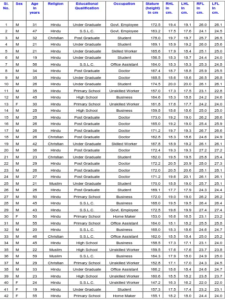
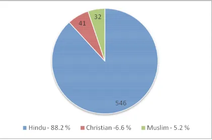
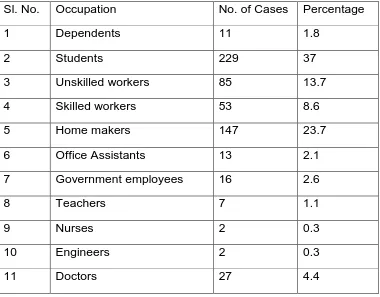
The study sample consists of 619 Healthy individuals comprising 311 males and 308 females in the age group of 18 to 59 years. In this study the samples included are:
i) The medical students of second MBBS attached to the Institute of Forensic Medicine, Madras Medical College, Chennai.
ii) Apparently healthy individuals attending the outpatient department of general medicine and their accompanying attenders.
iii) Patients admitted in the medical wards having no abnormality in the hand and foot and spine and their accompanying attenders.
iv) General healthy population.
The size of sample was decided after consultation with the statistician who advised to involve minimum of 300 subjects in each sex to get good results. Hence 311 male and 308 female subjects were selected for the study.
18 years. This is because all the centers of ossification in the foot, hand and long bones get completely fused by 18 years.
Cases above 60 years were excluded since stature, hand length and foot length significantly decreases due to osteoporotic changes in the bones and various factors which affect old age individuals.
The study sample consists of mostly right handed preponderance however some cases of left hand dominance are also included. All the measurements were taken in well lighted room. Due care was taken while taking measurements to avoid any diurnal variation. In this study all the recording were made in the morning hours between 8 a.m. to 11 a.m.
Before taking measurements every subject was asked to remove his foot wear and head wear. The measurements were always taken by one observer and recorded by another person in order to avoid inter observer error. The measurements were taken using standard anthropometric instruments in cm. to the nearest mm accuracy according to the technique given in manual biological anthropology.10
The subjects included in the study were healthy and free from any apparent symptomatic deformity. Some general information pertaining to Name, Age, Sex, Address, Religion, Educational Status, Economic Status, Occupational status etc. were also obtained.
Inclusion Criteria:
Willing healthy adult individuals of both sexes from Madras Medical College and Government General Hospital, Chennai between 18 to 59 years of age.
Exclusion Criteria:
1. Subjects less than 18 years
2. Subjects more than 59 years
3. Subjects with spinal deformities
4. Subjects with injuries to hand
5. Subjects with injuries to foot
6. Subjects with any major systemic diseases
7. Subjects with endocrine disorders
8. Pregnant women and lactating mothers
9. Unwilling individuals
Before venturing into the study, Ethical clearance was obtained from the Ethical committee headed by the Chairman (Dean, MMC). Prior permissions were obtained from the heads of the respective institutes for recording measurements from the subjects.
understand well. Each subject’s standing height, Right hand length, Left hand length, Right foot length and Left foot length were measured using standard anthropometric instruments by applying standard techniques. The data collected were subjected to statistical analysis by using SPSS (Statistical product and service solution) statistical software and regression formulas were derived with various combinations to reach the best estimate possible.
Landmarks and techniques involved in taking anthropometric
measurements10
Stature:
It can be measured in two ways, one in standing position i.e. height-Vertex and the other in lying position i.e. horizontal body length.
1. Height-vertex or Standing height:
It is vertical distance between the point vertex and the floor when the subject stands in anatomical position with palms touching the thighs with head in Frankfurt horizontal plane
Anatomical position: The anatomical position of a man is erect posture
with feet parallel to each other and arms hanging by the sides with palms facing forward.
Vertex: It is highest point on the head when head is held in Frankfurt
2. Horizontal body length or Supine length:
It measures the projective distance between vertex and plantare on vertical axis.
Plantare: It is the lowest point on the sole. It normally lies on the lateral
side.
Supine length usually measured in babies, dead bodies, in living subjects in emergency situation. This measurement is taken by laying the subject on a table. The subject should lie with his back in horizontal position on the table with his soles touching a vertical wall. The head should be in such a position that eye-ear plane is at right angle to the surface of the table. Supine length usually greater than standing height by 1.5cm – 2cm. It is not desirable to take measurement in the lying position unless there is urgent need. Measurements are usually taken in standing position all over the world.
Out of the two methods of taking height of a person, the standing height method was chosen for taking stature of a person, because it is most accepted method worldwide.
Instrument:
downwards, perpendicular to the vertical calibrated wooden rod is used to note the vertex point.
Technique:
The subject is made to stand in an erect posture and measurement is taken without any wear on head and foot. The subject should stand up on the platform against the vertical calibrated wooden rod, feet axis parallel or slightly divergent with head balanced on neck in F.H. plane. Hands should hang down. The movement of projected horizontal sliding wooden bar is controlled by the right hand, and moved from above downwards along mid saggital plane of the subject to just touch the vertex point. No pressure should be exerted since this is a contact measurement. Then it is fixed by the given screw at that level. The subject is then asked to step down and measurement noted in cm to the nearest mm accuracy.
Hand length:
There are four various methods by which the hand length can be measured like;
1. Length of hand: It measures the straight distance between the points interstylion and dactylion III.
Interstylion: It is the mid point of the line joining the two stylia (i.e.
Dactylion III: It is the distal most point of the third finger (i.e. the tip
of the middle finger) of the hand
2. Projective hand length: It is the vertical projective distance between stylion and dactylion, when the subject stands with arms and hands hanging down along the side of the body.
Stylion: It is the deepest point on the styloid process of radius while
the arm hanging by the side of the subject.
Dactylion: It is the lowest point on the anterior margin of the middle
finger with the arm hanging on the side of the subject.
3. Total hand length: It is straight distance from dactylion and mid-point of the most distal flexing crease of the wrist, while the hand is extended along the long axis of the forearm.
4. Approximate hand length: It is straight distance between stylion and dactylion.
Note: Dactylion generally denotes tip of the middle finger and stylion
generally denotes tip of the radial styloid process. Dactylion-II means the tip of the second finger. Stylion ulnare means the tip of the ulnare styloid process.
In this study, the total hand length method is applied to take hand length measurement to get more accurate results.
A specially designed instrument (depth gauge- 12” size) used commonly for measuring the depth of the holes is used for taking hand length measurements. It consist of a metal caliber which has measurements from 0 - 30 cm. with nearest mm accuracy. A movable metallic piece, which has flat surface at right angle to the caliber, attached with fixation screw and it can be moved along the long axis of the scale by releasing the fixation screw. It is used to touch the dactylion gently.
Technique:
The subject is asked to sit on a stool and to extend his hand with all the fingers together in correspondence with long axis of the forearm, on a table with dactylion protruding away from the edge of the table. The observer stands along the subject, place the instrument on the surface of the palm with the free end (zero point – 0.0cm) of the instrument fixed on the mid-point of the most distal flexing crease of wrist and the movable metallic piece is slided to touch the tip of the middle finger. Then it is fixed by tightening the fixation screw and it is taken out of the hand. Reading is noted. The same technique is applied for measuring both sides.
Foot length:
There are two methods by which the foot length can be measured like