1
Dissertation on
A STUDY ON PREVALENCE OF CARDIOVASCULAR
AUTONOMIC NEUROPATHY IN TYPE 2 DIABETES
AND UTILITY OF CORRECTED QT INTERVAL FOR
ITS DIAGNOSIS
Submitted to
THE TAMILNADU DR. M.G.R. MEDICAL UNIVERISTY
CHENNAI – 600 032
In partial fulfillment of the regulations For the Award of the Degree of
M.D. (GENERAL MEDICINE) BRANCH -1
MADRAS MEDICAL COLLEGE
CHENNAI – 600 003
2
CERTIFICATE
This is to certify that the dissertation titled “PREVALENCE OF CARDIOVASCULAR AUTONOMIC NEUROPATHY IN TYPE 2 DIABETES AND UTILITY OF CORRECTED QT INTERVAL FOR ITS DIAGNOSIS” is the bonafide original work of
Dr.G.VIGNESH in partial fulfillment of the regulation for M.D. Branch–I (General Medicine) Examination of the Tamilnadu Dr. M.G.R Medical University to be held in MARCH 2009. The Period of study was from May 2007 to August 2008.
Prof. R.SUKUMAR, M.D Professor Of Medicine,
Madras Medical College & Research Institute, Govt. General Hospital,
Chennai – 600 003.
Prof. C.RAJENDIRAN, M.D Director,
Institute of Internal Medicine,
Madras Medical College and Research Institute, Govt. General Hospital,
Chennai -600 003.
Prof.T.P.KALANITI, M.D Dean
3
DECLARATION
I, Dr G.VIGNESH, solemnly declare that dissertation titled “PREVALENCE OF CARDIOVASCULAR AUTONOMIC NEUROPATHY IN TYPE 2 DIABETES AND UTILITY OF CORRECTED QT INTERVAL FOR ITS DIAGNOSIS” is a bonafide work done by me at Madras Medical College and Govt. General Hospital from May 2007 to August 2008 under the guidance and supervision of my unit chief PROF.R.SUKUMAR,MD, Professor of medicine.
This dissertation is submitted to Tamilnadu Dr. M.G.R Medical University, towards partial fulfillment of regulation for the award of M.D. Degree (Branch – I) in General Medicine.
Place : Chennai.
4
SPECIAL ACKNOWLEDGEMENT
I would like to thank my beloved Dean Prof. T.P.Kalaniti, M.D.,
5
ACKNOWLEDGEMENT
I would like to express my sincere gratitude to my beloved professor and director, Institute of Internal Medicine
Prof. C.RAJENDIRAN, M.D., for his guidance and encouragement .
With extreme gratitude, I express my indebtedness to my beloved chief Prof. R.SUKUMAR, M.D., for his motivation, advice and valuable criticism which enabled me to complete this work.
I am extremely thankful to my Assistant Professors Dr.KANI SHEIK MOHAMMED, M.D., Dr.R.S.A.ALEXANDER, MD and Dr.S.DEEPA M.D., for their guidance and encouragement.
I would always remember with extreme sense of thankfulness for the co-operation and criticism shown by my post graduate colleagues.
6
CONTENT
SL.NO TITLE PAGE NO
1 INTRODUCTION
2 AIMS AND OBJECTIVES 3 REVIEW OF LITERATURE 4 MATERIALS AND METHODS 5 OBSERVATION AND RESULTS 6 DISCUSSION
7 CONCLUSION BIBLIOGRAPHY ANNEXURE
7
ABBREVIATION
ANS - Autonomic Nervous System
CAN - Cardiovascular Autonomic Neuropathy DAN - Diabetic Autonomic Neuropathy
DCCT - Diabetes Control and Complications Trial DPP - Dipeptidyl Peptidase
ECG - Electrocardiogram ED - Erectile Dysfunction E:I - Expiration-to-Inspiration GI - Gastrointestinal
HDL – High Density Lipoprotein HRV - Heart Rate Variability LDL – Low Density Lipoprotein
LVDD – Left Ventricle Diastolic Dysfunction MI - Myocardial Infarction
PSA - Power Spectral Analysis
QSART - Quantitative Sudomotor Axon Reflex Test TGL – Triglyceride
8
INTRODUCTION
Diabetes Mellitus is a heterogenous group of metabolic disorders characterized by hyperglycemia resulting from defects in insulin secretion, insulin action or both1. Type 2 Diabetes is one of the major health problems all over the world.1 WHO recently reported that total diabetics in India in 2000 was 31.7 million and this number likely to increase to 79.4 million by 20302.
Autonomic dysfunction is common in diabetics but symptomatic autonomic neuropathy is not that common. Presence of Cardiac Autonomic Neuropathy(CAN) is responsible for silent myocardial infarction and sudden death in diabetics. Hence recognizing cardiac dysautonomia early, which is asymptomatic will helpful to delay or arrest its progression.
9 function tests are now widely used for the assessment of autonomic function. These tests are non-invasive and do not require sophisticated equipment. All that required is an electrocardiogram machine, Heart rate monitor and sphygmomanometer. This highlights the importance of simple non-invasive tools like ECG and sphygmomanometer in diagnosing asymptomatic cardiac autonomic neuropathy.
Type 2 diabetic patients with abnormal CVR tests may have increased mortality, and those combined with postural hypotension have higher mortality than those without 4.
In 1980, for the first time, an association of prolonged QTc interval with cardiac autonomic neuropathy was given, thereby opening the possibility of a rapid objective method for detecting cardiac autonomic neuropathy. Further studies demonstrated an association of prolonged QTc interval with cardiac dysautonomia in diabetes mellitus5.
This study is performed to estimate the Prevalence of Cardiovascular Autonomic Neuropathy with relation to duration of diabetes in our GGH and to check the utility of corrected QT interval in diagnosing it.
10
AIMS AND OBJECTIVES
1. To evaluate the prevalence of Cardiovascular Autonomic Neuropathy in Type 2 Diabetes patients in our hospital.
2. To correlate the prevalence of Cardiovascular Autonomic Neuropathy with duration of diabetes.
11
REVIEW OF LITERATURE
The Diabetic pandemic is rapidly spreading and mostly affects developing countries like India. Indeed, India presently has the largest number of diabetic patients in the world and has been infamously dubbed as the ‘Diabetic capital of the world’. Although by definition, diabetes is characterized by elevated glucose concentrations, the impact of diabetes on both the health of individuals and on the health care systems, resides almost entirely in the long term complications of diabetes affecting almost every system in the body including eyes, kidneys, heart, feet and nerves.2
Neuropathy occurs more or less with the same frequency in both type 1 and type 2 diabetes, suggesting a common etiological mechanism based on chronic hyperglycemia .2
12 affect daily activities of individuals with diabetes and may invoke potentially life-threatening outcomes.4,5n
Autonomic nervous system
The autonomic nervous system conveys sensory impulses from the blood vessels, the heart and all of the organs in the chest, abdomen and pelvis through nerves to other parts of the brain (mainly the medulla, pons and hypothalamus). These impulses often do not reach our consciousness, but elicit largely automatic or reflex responses through the efferent autonomic nerves, thereby eliciting appropriate reactions of the heart, the vascular system and all the organs of the body to variations in environmental temperature, posture, food intake, stressful experiences and other changes to which all individuals are exposed. There are two major components of the autonomic nervous system, the sympathetic and the parasympathetic systems. The afferent nerves sub-serving both systems convey impulses from sensory organs, muscles, the circulatory system and all the organs of the body to the controlling centers in the medulla, pons and hypothalamus.
13 (ganglia) along the side of spine where the messages are relayed to other nerve bodies (or neurons) that travel to a large extent with the blood vessels to all parts of the body. Through these nervous pathways, the autonomic nerves convey stimuli resulting in largely unconscious reflex, bodily adjustments such as in the size of the pupil, the digestive functions of the stomach and intestines, the rate and depth of respiration and dilatation or constriction of the blood vessels.
The function of Autonomic nervous system in its regulation of the internal organ is highly independent. When the autonomic nervous are interrupted, these organ continue to function, but they are no longer as effective in maintaining homeostasis and adapting to the demands of changing internal condition and external stress.
Autonomic dysfunction
14
PATHOGENESIS OF DAN
Autonomic dysfunction that accompanies diabetic neuropathy is the one that has extensively evaluated.6,7,8 Several different factors have been implicated in thispathogenic process3. Hyperglycemic activation of the polyol pathway leading to accumulation of sorbitol and potential changes in the NAD:NADH ratio may cause direct neuronal damage and/or decreasednerve blood flow. Activation of protein kinaseC induces vasoconstriction and reduces neuronal blood flow. Increased oxidative stress, with increased free radical production, causes vascular endothelium damage and reduces nitric oxidebioavailability. Alternately, excess nitric oxide production may result in formation of peroxynitrite and damage endotheliumand neurons, a process referred to as nitrosative stress. In a subpopulation of individuals with neuropathy, immune mechanisms may also be involved. Reduction in neurotrophic growth factors, deficiency of essential fatty acids, and formation of advanced glycosylation end products (localized in endoneurial blood vessels) also result in reduced endoneurialblood flow and nerve hypoxia with altered nerve function.The result of this multifactorial process may be activation of polyADP ribosylation depletion of ATP, resulting in cellnecrosis and activation of genes involved in neuronal damage.
15 communicantes and loss of lateral horn cells in the spinal cord. They believe the later changes to be secondary.
Clinical features of Autonomic Dysfunction can be divided as follows10
(i) Cardiovascular symptoms: Postural hypotension, vertigo, syncope, dizziness, blurring of vision, neck pain, weakness.
(ii) Upper GI symptoms: Dysphagia, nausea, emesis, early satiety, bloating.
(iii) Lower GI symptoms: Nocturnal Diarrhea, constipation involuntary loss of stools and incomplete evacuation.
(iv) Urological symptoms: Dysuria, involuntary dribbling of urine, prolonged dribbling, incomplete bladder emptying, Erectile dysfunction(ED).
(v) Others: Hypohydrosis, Gustatory sweating.
Physiological basis for testing Abnormalities of Autonomic Nervous System
16 McLeod and Tuck11 state that in changing from recumbent to standing position a fall of more than 30mmHg systolic and 15 mmHg diastolic is abnormal; others give figures of 20 and 10mmHg. This excessive drop in Blood pressure indicates inadequate sympathetic vasoconstrictor activity. In response to the induced drop in blood pressure, the pulse rate normally increases. The failure of rise in heart rate in response to drop in blood pressure withstanding is a good indication of vagal nerve dysfunction12. In addition the pulse after rising initially in response to upright posture slows after about 15 beats to reach a stable rate by the 30th beat. The ratio of R-R interval in the ECG, corresponding to the 30th and the 15th beats is an even more sensitive measure of the integrity of Vagal inhibition the sinus node. A ratio in nonelderly of less than 1.05 is usually abnormal, indicating loss of vagal tone12.
17 dizziness) to autonomic dysfunction. In Valsalva maneuver, the subject exhales into a manometer or against a closed glottis for 15 seconds. Creating a markedly positive intra- thoracic pressure.
The response to performance of the Valsalva maneuver has four phases3 and in healthy individuals can be observed as follows:
• PhaseI: Transient rise in blood pressure and a fall in heartratedue to compression of the aorta and propulsion of blood into the peripheral circulation. Hemodynamic changes are mostlysecondary to mechanical factors.
• Phase II: Early fall in blood pressurewith a subsequent recoveryof blood pressure later in the phase.The blood pressure changes are accompanied by an increase inheart rate. There is a fallin cardiac output due to impairedvenous return causing compensatorycardiac acceleration, increasedmuscle sympathetic activity,and peripheral resistance.
• Phase III: Blood pressure falls and heart rate increases with cessationof expiration.
18 The response is mediated through alternating activation of parasympathetic and sympathetic nerve fibers. Failure of the heart rate to rise during positive intra thoracic pressure indicates sympathetic dysfunction and failure of the heart rate to slow during blood pressure overshoot indicates parasympathetic dysfunction. In patients with autonomic failure, the fall in BP is not aborted during the last few seconds of increased intra thoracic pressure and there is no over shoot of BP when breath is released 12.
Vasomotor reactions
Measurement of the skin temperature is a useful index of vasomotor function. The integrity of sympathetic reflex arc which includes baroreceptors of the aorta and carotid sinus, their afferent pathways, the vasomotor centers and the sympathetic and parasympathetic outflow, can be tested in a general way by the cold pressor test, grip test, mental arithmetic test and Valsalva maneuver.
19
Sudomotor function
Test for integrity of efferent sympathetic pathway. In sympathetic or galvanic skin- resistance test, a set of electrodes placed on the skin measures the resistance to passage of weak current in skin, the change in electric potential is the result of a ionic current within sweat glands. The response depends on sympathetic response to sweat glands (Gutrecht)13. A more quantitative and reproductive method is examination of postganglionic sudomotor function termed QSART (quantitative sudomotor axon reflex test)14. The QSART involves iontophoresis of a cholinergic agonist to measure axon reflex-medicated sudomotor responses quantitatively to evaluate postganglionic sudomotor function. Four sites are used and studied simultaneously with the patient supine. Thetest, typically done by recording from the forearm and three lower-extremity skin sites, has high sensitivity, specificity,and reproducibility, with a coefficient of variation of 20%if performed by trained personnel. The test is not generallyavailable and requires the purchase of expensive specializedequipment.
Assessing Pupillary function
20
Cardiac autonomic neuropathy (CAN)
CAN probably contribute to the poor prognosis of CHD and CHF in diabetes. The majority of patients with CAN come to clinical attention with complaints of postural hypotension, resting tachycardia, exercise intolerance (or) prior myocardial ischemia (or) infarction. The risk for CAN depends on the duration of diabetes and the degree of glycemic control and leads to parallel the development of other end organ disease related to diabetes such as retinopathy, nephropathy and vasculopathy. Symptoms and signs of CAN often occur relatively late in the natural history of this complication. 1
Simple bedside tests for cardiac autonomic neuropathy diagnosis16:
Heart rate variability test Blood pressure test
Score Deep breathing
Valsalva ratio
Response to standing
Response to handgrip
Response to standing
0 >15 >1.20 >15 >15 ≤ 10
1 11-15 1.11-1.20 12-15 11-15 11-29
21 For grading of cardiovascular autonomic function, results are classified into normal, borderline and abnormal (scores 0,1,2 respectively). An overall score of ‘0’ or ‘1’ was considered normal, score 2,3,4 were considered borderlines and score ≥5 were judged was abnormal autonomic function16.
Prevalence of Cardiac autonomic neuropathy (CAN) 17
The reported prevalence of CAN varies with the population studied and methods used. Regarding of these variations CAN appears to be common in diabetes. A summary of 15 reports on CAN suggests that the prevalence is in between 2.6 and 90% in diabetic population. The average incidence is about 30%.
Pathology of Autonomic neuropathy in Diabetes9
22 There were inflammatory changes in autonomic ganglia in all cases and in (or) around bundles of unmyelinated nerve fibers in many. These findings suggest that there may be several different pathogenetic mechanisms involved in the development of autonomic neuropathy in diabetes. 9
Diabetic patients have a high rate of Coronary Artery disease, which may be asymptomatic owing to autonomic neuropathy. Silent ischemia is significantly more frequent in patients with autonomic neuropathy than in those without autonomic neuropathy (38% versus 5%).18
It is clear however that a reduced appreciation of ischemic pain can impair timely recognition of myocardial ischemia (or) infarction and there by delay in initiating appropriate therapy. 18
23 the tendency for development of ventricular arrhythmia and cardiovascular events after infarction.18
Autonomic manifestations
Abnormal autonomic function tests are often found in patients with clinical features of sensory neuropathy, and almost any aspect of autonomic function can be affected. However, symptoms of autonomic neuropathy are not always present and are commonest in patients with poorly controlled type I diabetes. In all of the autonomic syndromes described below, symptoms may be chronic or strikingly intermittent. 19
Most longitudinal studies point to a common sequence of autonomic involvement in patients with diabetic autonomic neuropathy. In the cardiovascular system, the earliest abnormality detected by systematic and sensitive testing is a decrease in heart- rate variability during deep breathing, followed by alterations in the heart rate on standing and then the Valsalva maneuver. Postural hypotension is a relatively late event. This progression suggests that parasympathetic (vagal) fibers are damaged first, followed by the sympathetic outflow. Abnormal autonomic function tests are much more common than significant autonomic symptoms. 19
24 this is related to the neuropathy itself or to coexistent macrovascular disease or other complications. 19
Resting Tachycardia
Whereas abnormalities in HRV are early findings of CAN, resting tachycardia and a fixed heart rate are characteristic late findings in diabetic patients with vagal impairment5. Resting heart rates of 90 to 100 bpm and occasional heart rate increments up to 130 bpm occur. The highest resting heart rates have been found in patients with parasympathetic damage, occurring earlier in the course of CAN than sympathetic nerve function; in those with evidence for combined vagal and sympathetic involvement, the rate returns toward normal but remains elevated. A fixed heart rate that is unresponsive to moderate exercise, stress, or sleep indicates almost complete cardiac denervation5. Thus, heart rate may not provide a reliable diagnostic criterion of CAN in the absence of other causes unless it is increased by more than 100 bpm.
Exercise Intolerance
25 Presently, there is inadequate evidence to recommend routine screening of asymptomatic diabetic patients with an exercise ECG test. Emerging data support the utility of stress imaging testing in identifying diabetic patients with preclinical coronary artery disease, particularly patients with high-risk features, and comorbidities such as long-standing disease, CAN, multiple chronic renal failures, resting ECG abnormalities, and peripheral artery disease.
Abnormal sweating
This is one of the commonest, but frequently neglected, symptoms of diabetic autonomic neuropathy. Gustatory sweating, precipitated by eating cheese and other foods, is characteristic. Sweating is often profuse over the face and sometimes upper chest, corresponding to the area supplied by the superior cervical ganglion. Some patients find this socially debilitating.
Other abnormalities of sweating include the dry neuropathic foot, which allows cracking of the skin and the entry of infection and episodic nocturnal sweating that cannot be attributed to hypoglycemia.19
Cardiovascular changes
26
Postural hypotension
This is due to the impairment of the increase in cardiac output and of the sympathetically mediated vasoconstrictor response in the lower limbs, which normally follow standing upright. In diabetic neuropathy, this is a common manifestation of sympathetic denervation. It can be exacerbated by administration of insulin (which acts as a vasodilator at pharmacological concentrations) and of diuretics, antihypertensives and tricyclic antidepressants (which also have vasodilator activity). On standing, blood pressure is normally maintained, may rise slightly or may fall, but the systolic drop does not usually exceed 10 mmHg; a significant drop is generally taken as > 30 mmHg, although the magnitude of the systolic fall on standing does not correlate tightly with symptoms of dizziness.19 Postural hypotension is often worse on getting out of bed in the morning, but often varies substantially with time. The diagnosis is made simply by measuring the blood pressure with the patient lying and then standing for up to 2 min (some patients show a delayed fall).19
Autonomic Cardiopathy5
27 the severity of CAN with reduced peak diastolic filling rate and with an augmented atrial contribution to diastolic filling as assessed by Doppler echocardiography. It is difficult to judge, however, whether CAN is an independent contributor to these abnormalities, because other factors such as interstitial myocardial fibrosis and microangiopathic or metabolic changes (discussed in the pathogenesis of diabetic heart muscle disease) may also be responsible for LV dysfunction. CAN is associated with LV diastolic dysfunction (LVDD) at rest, both in patients with long-term type 225 or type 1 diabetes24. LVDD may progress to heart failure, mainly with preserved LV systolic function (diastolic heart failure), which is also related to high morbidity and mortality rates.26,27 The pathophysiology of LVDD includes delayed relaxation, impaired LV filling, and/or increased stiffness28. Diabetes mellitus can produce functional, biochemical, and morphological myocardial abnormalities independent of coronary atherosclerosis and hypertension, contributing to heart failure with normal LV systolic function29.
Silent myocardial ischemia/cardiac denervation syndrome
28 individual’s sensitivity to regional ischemia by interrupting pain transmission 30. The presence of CAN does not exclude painful myocardial infarction(MI) among individuals with diabetes 31. Chest pain in any location in a patient with diabetes should be considered to be of myocardial origin until proven otherwise; but, of equal importance, unexplained fatigue, confusion, tiredness, edema,hemoptysis, nausea and vomiting, diaphoresis, arrhythmias, cough, or dyspnea should alert the clinician to the possibility ofsilent MI 32.
Association of cerebrovascular disease and CAN
The frequency of ischemic cerebrovascular events is increased in individuals with type 2 diabetes. The impact of autonomicdysfunction on the risk of the development of strokes was examined by Toyry et al. Abnormalities of parasympathetic and sympathetic autonomic function were found to be independentpredictors of stroke in this cohort 33.
Cardiac Radionuclide Imaging 3
29 using MIBG and single–photon emission computed tomography is more sensitive in detecting CAN than indirect autonomic reflex testing, because MIBG uptake is reduced in patients with normal autonomic tests3. The MIBG uptake defects are localized predominantly in the LV posterior and inferior segments. In advanced CAN, completely absent MIBG uptake may be observed.
Cardiovascular autonomic neuropathy and increased cardiovascular morbidity and mortality in diabetes mellitus possible mechanisms17
1. Impaired angina recognition, Silent ischemia and infarction.
2. Decreased threshold for ischemia
- Increased resting heart rate and blunted chronotropic response to exercise.
- Impaired coronary vasomotor regulation.
3. Prolonged QT interval
- Increased lethal arrhythmias and sudden death (with or without myocardial ischemia)
4. Abnormal diastolic and systolic function
30 5. Increased peri-operative risk
- Increased need for hemodynamic support
- Reduced hypoxia-induced respiratory drive.
6. Alteration of normal circadian variation of sympatho-vagal activity
- Lack of normal nighttime decrease in blood pressure
- Loss of night time protection against myocardial infarction
7. Increased prevalence of other cardiovascular risk factors and other Complications
Increased microvascular complication
Increased rate of progression of glomerulopathy
Increased prevalence of hypertension and dyslipidemias17
PHARMACOLOGIC MANAGEMENT FOR CONTROL DIABETES
Sulfonylureas
31 sodium-adenosine triphosphate (Na-ATP) channel and cause an increase in intracellular calcium results in insulin exocytosis.
Some experts point to a possible risk of increased myocardial damage in patients with known CAD who use sulfonylureas at the time of an ischemic event. Prevention of protective ischemic preconditioning of the heart by inhibition of the potassium (K)-ATP channel is the putative mechanism. The UKPDS data do not support this concern. The authors agree and use sulfonylureas in appropriate patients with CAD. 3
Repaglinide
This newer insulin secretagogue binds to a different receptor site than do the sulfonylureas on the K-ATP channel. The half-life of this agent is 3.7h, which makes it effective for postprandial rather than preprandial hyperglycemia, for use in the elderly and for diabetic patients with chronic renal failure.
Metformin
32 inititation of therapy. Significant decrease in LDL cholesterol and triglycerides occur. The incidence of lactic acidosis with metformin is 9 per 100,000 person-years. Contraindications to its use include an elevated creatinine level (>1.4 in women, >1.5 in men), congestive heart failure, severe pulmonary disease, or any hypoxic state.
Thiazolidinediones
Thiazolidinediones promote insulin-stimulated glucose transport in muscles and adipocytes through a mechanism of action involving peroxisome proliferators activated receptor-gamma (PPAR-γ) ligands.
Binding to the nuclear receptor promotes differentiation of adipocytes and increased expression of glucose transporter. Thiazolidinediones has been shown to be effective both as monotherapy and in combination with insulin,sulfonylureas, and metformin.
33 and HDL cholesterol concentrations between 12 and 19 percent, with changes in serum triglycerides similar to those seen with placebo.
Pioglitazone, the newest thiazolidinedione, has been approved for use as monotherapy and in combination with metformin, sulfonylureas, and insulin. Weight gain(20%), ankle edema and fluid overload are the common side effects. Hepatotoxicity can occur. Contraindicated in Volume overload states, Hepatitis, Ischemic heart disease and macular edema.
Alpha-Glucosidase Inhibitors
Acarbose and miglitol work in the intestine to reversibly inhibit brush border alpha-glucosidases, resulting in a delay in carbohydrate absorption. These agents are poorly absorbed from the gastrointestinal tract. These drugs cause a 30 percent decrease in postprandial glucose in contrast to a 10 percent decrease in fasting glucose levels. They are adjuncts to other oral agents and rarely are potent enough to be used as mono therapy.
GLP Analogues and Di peptidylpeptidase inhibitors1
34 distal intestine. GIP and GLP-1 is rapidly inactivated by the enzyme dipeptidyl peptidase-IV (DPP – IV). Newer therapeutic candidates for the treatment of type 2 diabetes are those drugs that enhance glucose dependent insulin secretion called Incretin mimetics. The use of DPP-IV inhibitors to improve the duration of endogenous GLP-1 ativity to target post-prandial glucose excursions along with effects on fasting glucose levels. They do not demonstrate any hypoglycemic effects. Sitagliptin is the first approved drug which can be used as monotherapy or combination therapy. Reduced hypoglycemic effect and reduced weight gain are the advantages over Sulphonylureas.
Insulin
The natural history of type 2 diabetes is one of progressive beta-cell failure. Therefore, after approximately 10 years of the use of oral hypoglycemic agents, insulin may be required in combination with oral agents as the sole therapy. Although endogenous hyperinsulinemia is clearly associated with atherogenesis, there is no compelling evidence of increased risk of cardiovascular disease or increased mortality from exogenous insulin therapy.
Treatment for Autonomic Dysfunction3
35 if those patients at high risk or those shownto be in early stages are not treated until advanced symptomatology is present, little has been achieved.
Unfortunately, information presented at the fifth Regenstrief conference on the intensive management of type 2 diabetes indicatedthat physicians may feel that screening is not of value because treatment options for identified complications are limited34.Such a view does not take into account the clinical research advances that have been made in the treatment of diabetes. Tests that provide evidence of further health consequences may bringpatients to medical attention before other signs of diabetic end-organ injury emerge, making proactive treatment, particularly the establishment of intensive diabetes care, possible. The results of autonomic function testing can contribute to good patient management in the following ways.
To assist in the establishment (or reestablishment) of tight glycemic control
36 to be effective for patientswith type 2 diabetes, although clinical studies are underwayin the latter.
In its earliest stages, there has been some clinical demonstration that autonomic dysfunction may be influenced within a few daysto a few weeks with effective treatment36,37. Delay in instituting appropriate interventions can only increase the likelihood of developing advanced neuropathies. Stabilization of the neuropathies(generally considered to be any delays in further progression)through tight glycemic control seems possible, whereas reversal of the condition may be less likely36. Again, the results from the DCCT show that intensive glycemic treatment can prevent the development of abnormal heart rate variation and slow the deterioration of autonomic dysfunction over time 38.
37
To facilitate the decision to initiate treatment for cardiovascular autonomic dysfunction3
Several different factors have been implicated in the potential metabolic-vascular pathogenic process of diabetic neuropathy (e.g., activation of the polyol pathway, increased oxidativestress, reduction in neurotrophic growth factors, deficiency of essential fatty acids, and formation of advanced glycosylation end products). Thus, timely identification ofautonomic dysfunction in diabetic patients may expedite end-organprophylaxis such as the use of ACE inhibitors and Aspirin and the use of pharmacological and non-pharmacological interventions to improve blood pressure and lipid control. Improved nutritionand reduced alcohol and tobacco consumption are additional options available to patients with diabetes who are identified with autonomic nerve dysfunction. Interventions to modulate reduced heart rate variation currently being studied in clinical trials are based on theories of the pathogenesis of CAN.
38 individuals with CAN but may mitigate the effectsof statins and niacin in treating or preventing macrovasculardisease.
Studies using ACE inhibitors as a means to improve heart rate variation have resulted in conflicting results. Whereas quinapril significantly increased parasympathetic activity after 3 months of treatment, cardiovascular autonomic function did not change significantly after 12 months of treatment with trandolapril.
Non-pharmacological measures like adequate intake of salt and fluids, in the range of 10 to 20g, of salt / day and 2-3 litres of fluid / day. Sleeping with the head end elevated will minimize the effects of supine nocturnal hypertension. Patients are advised to sit with legs dangling over the edge of the bed for several minutes before attempting to stand. Other maneuvers like leg–crossing, contraction of leg muscles for 30 seconds. Such measures compress leg vein and increase systemic resistance. Compression stocking may be helpful. Anemia should be corrected.
39 inhibiting gastrointestinal peptides prevent the vasodilation and hypotension effects.
The use of cardioselective (e.g., atenolol) or lipophilic (e.g., propranolol) ß-blockers may also modulate the effects of autonomic dysfunction.32 By opposing the sympathetic stimulus,they may restore the parasympathetic-sympathetic balance. Recently, the administration of metoprolol to ramipril-treated diabetic patients with abnormal albuminuria has been shown toimprove autonomic dysfunction40.
Because the pathogenesis of CAN is most likely a multifactorial process, a combination of therapies directed simultaneously at different parts of the pathogenicpathway may be needed. In addition, the goal of these interventions should be directed at the prevention of further deterioration of cardiovascular autonomic dysfunction rather than expectingto realize improved function.
To emphasize the importance of adherence to diet and exercise interventions
40 importance of lifestyle interventions and screening at or soon after diagnosis.
Motivation to adhere and remain compliant with non-pharmacologicalinterventions is difficult. Current research suggests that preventive measures (glycemic control, diet, and exercise) introduced to the general diabetic population are difficult to sustain and consequently less than effective. This is due, in part, to thelong-term commitment that must be made to the practice of preventive measures. Although individuals with diabetes are faced with the immediate pressures of disease management on a day-to-day basis, it is the long-term risks of micro- and macrovascular complications that pose the most serious risks42. The ability to determine early stages of autonomic dysfunction could intensify the salience of measures such as diet and exercise that directly affect efforts to establish tight glycemic control and delay the development of autonomic dysfunction. Colloquial patient management strategies could be introduced to a now potentiallymotivated patient.
41 Clinicians working together with the patient can develop an appropriate exercise programthat will yield a plan for reaping maximum benefits.
QTc – Interval and prolongation
QT interval taken from the onset of QRS complex to end of the ‘T’ wave and corrected for heart rate give the corrected QT interval (QTc). A QTc interval of more than 440 millisecond is considered prolonged 44. It reflects the total duration of ventricular myocardial depolarization and repolarization in the ECG. It is usually corrected for heart rate by Bazett formula, where QTc = QT √RR interval. The Bazett formula gives a slight over correction of QT interval at higher heart rates. Hodges et al57 also gave a formula to get corrected QT as follows:
QTc = QT + 1.75 (rate-60) and is gaining more acceptance. QTc prolongation in ischemic heart disease carries an increased risk (2-5 times) of sudden cardiac death.
The exact mechanism of QTc prolongation is not clearly defined. It is generally agreed that parasympathetics have little influence on QTc
42 ECG can be used to diagnose CAN with reasonable sensitivity, specificity and positive predictive value.
43
MATERIALS AND METHODS
STUDY POPULATION
A total of 150 patients satisfying all the inclusion and exclusion criteria were enrolled for the study from the population of Type 2 Diabetes patients who attended the out patient clinics and Inpatients of Institute of Internal Medicine, Government General Hospital . Written consent was obtained from all the patients participating in the study after clearly explaining the study procedure.
The patients were grouped into three according to the duration of diabetes. Autonomic neuropathy testing by simple bet side tests was done in op department and Medical ward using ECG monitor, Pulseoxymeter and BP apparatus for the same 150 patients. Each groups were compared with one another with the available statistical data.
STUDY DURATION
This study was conducted for a period from May 2007 to August 2008.
STUDY DESIGN
44 with duration of Diabetes. To investigate the relationship between cardiac autonomic dysfunction and corrected QT interval.
METHODS
Detailed clinical history was taken from each patients and a completes review of their case notes performed. A complete clinical examination of the cardiovascular system was done for each patient.
Tests for autonomic functions
On the day of testing patients were instructed not to ingest caffeine containing products. All recordings the done 5-8 hours post prandially. Blood pressure was recorded manually using standard sphygmomanometer. The heart rate variation was calculation using standard Heart rate monitor, Pulseoxymeter and continous ECG recording. A baseline ECG was taken with a Standard ECG machine for calculation of QTc interval.
The simple bedside tests for assessing the autonomic nervous system were described by Ewing and Clarke47. All patients were subjected to a battery of five tests as described below:
Heart rate response to valsalva maneuver
45 sphygmomanometer, to maintain a pressure of 40 mm Hg for 10 seconds The ratio of the maximum heart rate during blowing to the minimum during the compensatory bradycardia after stopping is calculated. The maneuver was repeated three times with one minute interval in between and results were expressed as
Valsalva ratio = Max heart rate ÷ Minimum heart rate.
The mean of the three-valsalva ratios was taken as the final value.
Heart rate variation during deep breathing
The subject was asked to breathe deeply at six breaths / min (Five seconds “in” and five seconds “out”) for one minute. The average heart rate difference (maximum minus minimum during the respiratory cycle) is calculated while the patient breaths deeply for 1 min. The results were expressed as the mean of the difference between maximum and minimum heart rates for the six measured cycles in beats / min.
Immediate heart rate response to standing
46
Blood pressure response to standing
This test measured the subject’s blood pressure with a sphygmomanometer while he was lying quietly. Then he was made to stand up and the blood pressure again after one minute. The postural fall in blood pressure was taken as the difference between the systolic pressure lying and the systolic pressure standing. The test was in repeated three times and the mean systolic blood pressure was calculated.
Blood pressure response to sustained hand grip
The blood pressure of the patient was taken three times before the maneuver. A modified sphygmomanometer was used to sustain handgrip. The patient was asked to grip the inflatable rubber and apply maximum voluntary pressure possible. A reading from the attached mercury manometer was taken during maximum voluntary contraction. There after, the patient was asked to maintain 30% of maximum voluntary contraction for as long as possible up to five minutes. Blood pressure was measured at one minute intervals during the handgrip. The result was expressed as the difference between the highest diastolic blood pressure during the handgrip exercise and the mean of the three diastolic blood pressure readings before the handgrip began.
47 H/o light headedness
H/o weakness or tiredness
H/o vertigo
H/o anxiety
H/o palpitations
H/o sweating abnormality
H/o diarrhea
H/o constipation
48
INTERPRETATION OF THE TEST WAS BASED ON THE WORKS OF EWING AND CLARKE16.
Heart rate variability test Blood pressure test Score Deep
breathing
Valsalva ratio
Response to standing
Response to handgrip
Response to standing
0 >15 >1.20 >15 >15 ≤ 10
1 11-15 1.11-1.20 12-15 11-15 11-29
2 ≤ 10 ≤1.10 <12 ≤ 10 >30
For grading of cardiovascular autonomic function, results are classified into normal, borderline and abnormal (scores 0,1,2 respectively). An overall score of ‘0’ or ‘1’ was considered normal, score 2,3,4 were considered borderlines and score ≥5 were judged was abnormal autonomic function56.
QT interval was taken from the onset of QRS complex to the end of T wave. QT was then corrected for heart rate using the Bazette’s formule44.
QTc interval = QT / √(R – R). A QTc interval more than 440
49
INCLUSION CRITERIA
1) Type 2 diabetes already on treatment and newly diagnosed patients.
EXCLUSION CRITERIA
1) Age above 60 years
2) Documented ischaemic heart disease
3) Documented valvular or congenital heart disease
4) Hypertension
5) COPD
6) Uraemia
7) Parkinsonism
STATISTICAL ANALYSIS
51
OBSERVATION AND RESULTS
POPULATION CHARACTERISTICS
The study group consists of 150 patients with Type 2 diabetes. The following tables lists the information about the age variation and sex distribution of the study group. The patients in the groups <5yrs, 5-10 yrs
[image:51.612.131.508.378.539.2]and >10 yrs were taken as group A, B and C respectively.
TABLE – 1 AGE VARIATION AMONG STUDY GROUPS
Duration of DM N Mean age SD
<5 years 50 50.08 4.251
5-10 years 50 52.76 3.217
>10 years 50 55.16 2.881
52
TABLE – 2 SEX DISTRIBUTION AMONG STUDY GROUPS
<5 years 5-10 years >10 years Duration of DM
n % n % n %
Male 27 54 29 58 28 56
SEX Female 23 46 21 42 22 44
Total 50 100 50 100 50 100
53
[image:53.612.123.516.225.521.2]CARDIOVASCULAR AUTONOMIC DYSFUNCTION IN THE STUDY GROUP
TABLE -3 FREQUENCY DISTRIBUTION OF NORMAL (0-1),
BORDERLINE (2-4), ABNORMAL (≥5)
Group
<5YRS 5-10YRS >10YRS CAN
n % n % n %
Significance
0-1 36 72 24 48 13 26
2-4 10 20 14 28 8 16 Score
≥5 4 8 12 24 29 58
Total 50 100.0 50 100.0 50 100.0
P<0.001
Significant
54
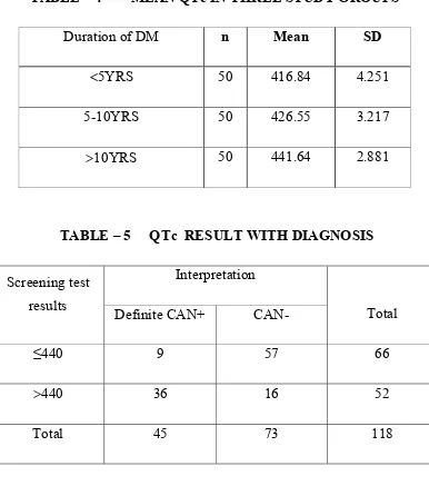
TABLE – 4 MEAN QTc IN THREE STUDY GROUPS
Duration of DM n Mean SD
<5YRS 50 416.84 4.251
5-10YRS 50 426.55 3.217
>10YRS 50 441.64 2.881
TABLE – 5 QTc RESULT WITH DIAGNOSIS
Interpretation Screening test
results Definite CAN+ CAN- Total
≤440 9 57 66
>440 36 16 52
Total 45 73 118
Sensitivity = 36 / 45 x 100 = 80% Specificity = 57 / 73 x 100 = 78.08%
55
TABLE - 6
CORRELATION BETWEEN AUTONOMIC NEUROPATHY (AN) AND QTc PROLONGATION IN TOTAL DIABETIC PATIENTS
QTc in msec
Definite
CAN %
Borderline
CAN %
No
CAN % p Value
≤440 9 20 22 68.8 58 79.5
>440 36 80 10 31.2 15 20.5
<0.001
significant
56
DISTRIBUTION OF SYMPTOMS OF CAN
57
58
DISCUSSION
The results of this study on native Indian population illustrate the fact that cardiac autonomic dysfunction is common in Diabetic patients and its prevalence increases with duration of diabetes. Similar results have been reported in previous studies conducted in India and other countries48-51. This study demonstrated significant abnormalities in autonomic function using basic cardiovascular autonomic function tests, which have well been validated.
59 Mohan et al51 studied the prevalence of CAN in 336 patients with NIDDM in south India. There was an increase in prevalence of CAN with duration of diabetes. In 0-5 years group the prevalence of autonomic dysfunction was 28.2%. In our study, the prevalence of autonomic dysfunctin in 0-5yrs group is 28% of which 8% had definite CAN and 20% had evidence of borderline/early CAN.
Toyry J P et al48 studied the clinical significance of autonomic neuropathy in NIDDM. A total of 133 patients with newly diagnosed NIDDM (70 men) and 144 control subjects (62 men) were examined at baseline and after 5 and 10 years of follow-up. The frequency of autonomic dysfunction at baseline, at 5 yrs and 10yrs was 4.9%, 19.6% and 65% respectively. In our study, the prevalence of definite CAN at <5yrs, 5-10yrs and >10yrs is 8%, 24% and 58% respectively which is comparable to the above study. Also, the prevalence of cardiovascular autonomic neuropathy increases with increase in duration of diabetes (p<0.001).
Pappachan J M et al52 studied the prevalence the CAN among 100 type 1 and type 2 diabetes mellitus, in south India assessed by the five autonomic function tests by Eving’s methodology. The prevalence of CAN was 60% which is comparable to the results obtained in this study.
60 Hence the autonomic tests that assess the parasympathetic system which include Heart Rate Variability(HRV) to deep breath, valsalva, lying to standing becomes abnormal before the blood pressure tests. In our study, the patients with borderline CAN almost everyone have abnormalities in Heart Rate Variability(HRV) with only few have blood pressure abnormalities. This again emphasis the importance of Heart Rate Variability testing as screening for early autonomic dysfunction in diabetes.
The symptoms of Autonomic dysfunction was present only in 19 out of 45(42.22%) patients with definite CAN. Almost 58% of patients with definite CAN were asymptomatic. This clearly shows the importance of screening for autonomic dysfunction even in asymptomatic diabetics. Patients with severe cardiac autonomic dysfunction have high risk of developing silent myocardial infarction and sudden death53.
Also, in our study, 14 out of 73(19.1%) patients without CAN had symptoms suggestive of autonomic dysfunction. Clinical symptoms of autonomic neuropathy generally do not occuruntil long after the onset of diabetes. Whereas symptoms suggestive of autonomic dysfunction may be common they may frequently bedue to other causes rather than to true a autonomic neuropathy3.
61 compared with 80.9 and 93.3% for patients with normal CVR test. He concluded that Type 2 diabetic patients with abnormal CVR tests may have increased mortality, and those combined with postural hypotension have higher mortality than those without. Abnormal CVR tests may be important predictors of mortality in Type 2 diabetes mellitus. Subclinical autonomic dysfunction can, however, occur withina year of diagnosis in type 2 diabetes patients and within twoyears in type 1diabetes patients.3 Massinet al54 demonstrated that early puberty is a critical periodfor the development of CAN and suggested that all type 1 diabetic patients should be screened for CAN beginning at the first stageof puberty.
Vinik et al3 proposed the measurement of HRV at the time of diagnosis of type 2 diabetes and within 5 yearsafter diagnosis of type 1 diabetes (unless an individual has symptoms suggestive of autonomic dysfunction earlier) serves to establish a baseline, with which 1-year interval tests can be compared. Regular HRV testing provides early detection and thereby promotes timely diagnostic and therapeutic interventions. The knowledge of early autonomic dysfunction can encourage patient and physicianto improve metabolic control and to use therapies such as ACEinhibitors and ß-blockers, proven to be effective for patients with CAN.
62 present in groups representing <5yrs, 5-10yrs and >10yrs duration respectively. Identifying these individuals at risk is only the first step in managingpatients and ultimately affecting outcomes. After identification, effective management must be provided. Proactive measures arerequired, because if those patients at high risk or those shownto be in early stages are not treated until advanced symptomatologyis present, little has been achieved. Early observations by researchers that near-normal glycemic control seems to be the most effective way to delay the onsetof CAN and arrest its progression. Hence it is important in emphasizing tight glycemic control for individuals with autonomic dysfunction with reeducation of the patient with regard to need for regular monitoring and hypoglycemia. Thus, timely identification ofautonomic dysfunction in diabetic patients may expedite end-organ prophylaxis such as the use of ACE inhibitors and aspirin and the use of pharmacological and non-pharmacological interventions to improve blood pressure and lipid control. Improved nutrition and reduced alcohol and tobacco consumption are additional options available to patients with diabetes who are identified with autonomic nerve dysfunction.3 Early identification of CAN permits timely initiationof therapy with the antioxidant alpha-lipoic acid (thioctic acid) and Vitamin E, which appears to slow or reverse progression of neuropathiesin some studies.55
63 and specificity of 80% and 78.08%. respectively. There was significant correlation (p<0.001) between CAN and QTc prolongation in this study group.
Pappachan J M et al52 studied the utility of prolongation of corrected QT interval (QTc) in the ECG to diagnose CAN in patients with diabetes. They calculated the sensitivity and specificity of QTc prolongation for the diagnosis of CAN were 77% and 62.5% in type 1 and 76.5% and 75% in type 2, respectively. They concluded that QTc interval in ECG can be used to diagnose CAN with reasonable sensitivity and specificity. This value of sensitivity and specificity correlates with our study.
64 There were certain limitations in this study:
65
CONCLUSION
The following are the conclusions from this study:
1. The Prevalence of Cardiovascular Autonomic Neuropathy is high in type 2 diabetics in our hospital.
2. The prevalence of CAN will increase with increase in the duration of diabetes. About half of the patients with type 2 diabetes have autonomic dysfunction after ten years.
66
BIBILIOGRAPHY
1. Nihal T, Vasan S, Bhatt R: A practical approach to
Diabetes Mellitus, 2007
2. Jayaram B M et al: Type 2 Diabetes Mellitus and its
complications: A preventive program, 2008
3. Vinik A I, Maser E R, Mitchell B D, Freeman R:Diabetic
autonomic neuropathy. Diabetes Care 2003; 26:1553-1579
4. Chen H S et al. Abnormal cardiovascular reflex tests are
predictors of mortality in Type 2 diabetes. Diabetic
Medicine 2001; 18(4):268-273
5. Aaron I. Vinik and Dan Ziegler: Diabetic Cardiovascular
Autonomic Neuropathy. Circulation 2007;115;387-397
6. Low .P.A, Walsh JC et al. The sympathetic nervous system
in diabetic neuropathy, a clinical and pathological study.
Brain 1975, 98: 341– 56.
7. Maser RE, Mitchell BD, et al. The associated between
cardio vascular autonomic neuropathy and mortality in
individuals with diabetes: a Meta – analysis. Diabeties
67
8. Burgos LG, Ebert TJ et al. Increased intra operative cardio
vascular morbidity in diabetes with autonomic
neuropathy. Anaesthesiology – 1989; 70: 591– 97.
9. Duchen LW, Anjorin A et al. Pathology of autonomic
neuropathy in DM. Ann. intern. Med. 1980; 92: 301.
10. Becker, Klaus, Gorlach et al. Characterisation and natural
course of cardiac autonomic neuron dysfunction 1997;
11(6): 751–757.
11. Mc leod JG, Tuck RR; Disorders of the autonomic nervous
system. Party I. Pathophysiology and clinical features.
Part II investigation and treatment An Neurl. 21: 419,
519–1973.
12. Adam and victor’s principles of neurology, McGraw – Hill
8th Edition
13. Gutrecht JA. Sympathetic skin response J. Clin. Neuro
physiol. 11: 519–1994
14. Low PA, Clinical Autonomic Disorders 2nd ed.
Philadelphia, Lippicott –Raven, 1997.
15. Clarke BF, Ewing DJ, Campbell IW: Diabetic autonomic
68
16. F Bellavere, G Bosello, D Fedele, C Cardon e, and M Ferri
Br Med J (Clin Res Ed). 1983 July 2; 287(6384): 61.
17. Braunwald’s Heart disease, 7th edition ELSEVIER
Saunders
18. Aoron I Vinik et al Semi Neurol. 2003; 23: 365-372
19. Textbook of Diabetes, John C Pickup & Gareth Williams,
3rd edition Blackwell Science
20. Vinik AI, Erbas T: Neuropathy. In Handbook of Exercise
in Diabetes. Ruderman N, Devlin JT, Schneider SH,
Kriska A, Eds. Alexandria, VA, American Diabetes
Association, 2002; 463–496.
21. Roy TM, Peterson HR, Snider HL, Cyrus J, et al.:
Autonomic influence on cardiovascular performance in
diabetic subjects. Am J Med 87:382–388, 1989.
22. Vinik AI, Erbas T: Neuropathy. In Handbook of Exercise
in Diabetes. Ruderman N, Devlin JT, Schneider SH,
Kriska A, Eds. Alexandria, VA, American Diabetes
69
23. Hayat SA, Patel B, Khattar RS, Malik RA. Diabetic
cardiomyopathy: mechanisms, diagnosis and treatment.
Clin Sci (Lond). 2004;107: 539–557
24. Didangelos TP, Arsos GA, Karamitsos DT, Athyros VG,
Karatzas ND. Left ventricular systolic and diastolic
function in normotensive type 1diabetic patients with or
without autonomic neuropathy: a radionuclide
ventriculography study. Diabetes Care. 2003;26:1955–
1960.
25. Mustonen J, Uusitupa M, Lansimies E, Vainio P, Laakso
M, Pyorala K. Autonomic nervous function and its
relationship to cardiac performance in middle-aged
diabetic patients without clinically evident cardiovascular
disease. J Intern Med. 1992;232:65–72.
26. Lenzen MJ, Scholte op Reimer WJ, Boersma E,
Vantrimpont PJ, Follath F, Swedberg K, Cleland J,
Komajda M. Differences between patients with a
preserved and a depressed left ventricular function: a
report from the EuroHeart Failure Survey. Eur Heart J.
70
27. Ansari M, Alexander M, Tutar A, Massie BM. Incident
cases of heart failure in a community cohort:
importance and outcomes of patients with preserved
systolic function. Am Heart J. 2003;146:115–120.
28. Mandinov L, Eberli FR, Seiler C, Hess OM. Diastolic
heart failure. Cardiovasc Res. 2000;45:813– 825.
29. Piccini JP, Klein L, Gheorghiade M, Bonow RO. New
insights into diastolic heart failure: role of diabetes
mellitus. Am J Med. 2004; 116(suppl 5A):64S–75S.
30. Ambepityia G, Kopelman PG, Ingram D, Swash M, Mills
P: Exertional myocardial ischemia in diabetes: J Am Coll
Cardiol 1990; 15:72–77.
31. Campbell IW, Ewing DJ, Clarke BF: Painful myocardial
infarction in severe diabetic autonomic neuropathy. Acta
Diabetol Lat 1978; 15:210–214
32. Vinik AI, Erbas T: Recognizing and treating diabetic
autonomic neuropathy. Cleve Clin J Med 2001;
68:928-944.
33. Toyry JP, Niskanen LK, Lansimies EA, Partanen KPL,
71
development of stroke in patients with
non-insulin-dependent diabetes mellitus. Stroke 1996; 27:1316–1318.
34. Clark CM, Vinicor F: Introduction: Risks and benefits of
intensive management in non-insulin-dependent diabetes
mellitus: the fifth Regenstrief conference. Ann Intern Med
1996; 124:81–85
35. DCCT Research Group: The effect of intensive diabetes
therapy on measures of autonomic nervous system
function in the Diabetes Control and Complications Trial
(DCCT). Diabetologia 1998; 41:416–423.
36. Ziegler D: Diabetic cardiovascular autonomic neuropathy:
prognosis, diagnosis and treatment. Diabetes Metab Rev
1994; 10:339–383.
37. Fraser DM, Campbell IW, Ewing DJ, Murray A, Neilson
JM, Clarke BF: Peripheral and autonomic nerve function
in newly diagnosed diabetes mellitus. Diabetes 1977;
26:546–550.
38. DCCT Research Group: The effect of intensive diabetes
therapy on measures of autonomic nervous system
function in the Diabetes Control and Complications Trial
72
39. Malik RA, Williamson S, Abbott C, Carrington AL, Iqbal
J, Schady W, et al.: Effect of
angiotensin-converting-enzyme (ACE) inhibitor trandolapril on human diabetic
neuropathy: randomised double-blind controlled trial.
Lancet 352:1978–1981, 1998
40. Ebbehoj E, Poulsen PL, Hansen KW, Knudsen ST,
Molgaard H, Mogensen CE: Effects on heart rate
variability of metoprolol supplementary to on going
ACE-inhibitor treatment in type I diabetic patients with
abnormal albuminuria. Diabetologia 2002; 45:965–975.
41. Singleton JR, Smith AG, Bromberg MB: Painful sensory
polyneuropathy associated with impaired glucose
tolerance. Muscle Nerve 2001; 24:1225–1228.
42. Sochett E, Daneman D: Early diabetes-related
complications in children and adolescents with type 1
diabetes: implications for screening and intervention.
Endocrinol Metab Clin North Am 1999; 28:865–882.
43. Howorka K, Pumprla J, et al.: Effects of physical training
on heart rate variability in diabetic patients with various
degrees of cardiovascular autonomic neuropathy.
73
44. Wagner GS. Marriott’s Practical electro cardiograph, 10th
ed. Philadelphia: Lippincott William and Wilkinn Co.
2001. Vol. I: 53 – 56.
45. Kahn JK, Sisson JC, Vinik AI. QT interval prolongation
and sudden cardiac death in diabetic autonomic
neuropathy. Jr of clinical Endocrinology and metabolism
1987; 64 (4): 751-4.
46. Bellavere F, Ferri M, Quarini L et al. Prolongation of QT
period in diabetic autonomic neuropathy. A possible role
in sudden cardiac death. Br HJ 1988; 59: 379-83.
47. D J Ewing and B F Clarke. Diagnosis and management of
diabetic autonomic neuropathy. Br Med J (Clin Res Ed).
1982; 285(6346): 916–918.
48. Toyry J P et al. Occurance, Predictors and Clinical
significance of Autonomic neuropathy in NIDDM. Ten
year follow-up from the diagnosis.Diabetes. 1996 Mar ;45
(3):308-15.
49. Doran A and Andrew J B et al. Diabetic Autonomic
Neuropathy. The clinical interpretation of improved
technology. Diabetes Technology and Therapeutics 2001;
74
50. Ratzmann K P et al. Prevalence of Peripheral and
Autonomic Neuropathy in newly diagnosed Type 2
Diabetes mellits. J Diabet Complications, 1991; 5: 1-5.
51. Mohan V et al. Autonomic Neuropathy in NIDDM and
fibrocalculus pancreatic diabetes in south India, Diabet
Med 1996; 13: 1038-43.
52. Pappachan J M et al. Cardiac autonomic neuropathy in
diabetes mellitus: prevalence, risk factors and utility of
corrected QT interval in the ECG for its diagnosis
Postgraduate Medical Journal 2008; 84:205-210.
53. Paul V et al. Predictive Value of Cardiac Autonomic
Neuropathy in Diabetic Patients With or Without Silent
Myocardial Ischemia Diabetes Care 2001; 24:339-343.
54. Massin MM, Derkenne B, Tallsund M, Rocour-Brumioul
D, Ernould C, Lebrethon MC, Bourguignon JP: Cardiac
autonomic dysfunction in diabetic children. Diabetes Care
1999; 22:1845–1850.
55. Ziegler D, Reljanovic M, Mehnert H, Gries FA:
Alpha-lipoic acid in the treatment of diabetic polyneuropathy in
Germany: current evidence from clinical trials. Exp Clin
75
56. Mathur et al: QTc Prolongation in Diabetes Mellitus – An
Indicator of Cardiac Autonomic Neuropathy. Journal,
Indian Academy of Clinical Medicine 2006; 7(2): 130-2
57. Hodges M, Salerno Q, Erlien D. Bazett's QT correction
reviewed. Evidence that linear QT correction for
heart rate is better. J Am Coll Cardiol 1983;1:694.
76
PROFORMA
• Name : • Age • Sex
• Smoking / Alcohol Status: • Duration of Diabetes:
• History Suggestive of Autonomic Neuropathy : • Examination:
Vital Signs: HR: BP: • Examination of cardio vascular system:
[
TESTS FOR CARDIO VASCULAR AUTONOMIC FUNCTION
Test 1 Test 2 Test 3 Mean
I Valsalva Ratio
ii Deep Breathing Test iii Supine to standing iv BP Response to standing v BP Response to
sustained Hand grip
• Examination of RS,GIT &CNS:
• ECG: QTc • Blood Sugar: Urea