A
A CLIN
THE TA
MADRA
NICAL S
AMIL NA
With
for
AS
MED
A D
STUDY
THE
Su
ADU Dr.
h fulfillm
r the aw
M.S..
(O
B
DICAL
CO
CHE
M
Dissertat
ON TE
OPTIC
ubmitted
. M.G.R.
CHENN
ment of
ward of t
PHTHAL
BRANCH
OLLEGE
A
NNAI
‐
6
MARCH
‐
2
tion on
EMPOR
C DISC
d to
MEDIC
NAI
the regu
the degr
LMOLOG
H
‐
III
AND
RES
00
003
CERTIFICATE
This is to certify that Dr.Thirumangai.S., Post Graduate student in M.S
Ophthalmology, at Regional Institute of Ophthalmology and Government
Ophthalmic hospital attached to Madras Medical College, Chennai, carried
out this dissertation on “A CLINICAL STUDY ON TEMPORAL
PALLOR OF THE OPTIC DISC” under my direct guidance and
supervision during the period from May 2006 to March 2009.
This dissertation is submitted to the TamilNadu Dr.MGR Medical
University, Chennai in partial fulfillment of award of M.S. Degree in
Ophthalmology
Prof. Dr. K.Namitha Bhuvaneswari Prof. Dr. M. Radhakrishnan Chief, Strabismology,Neuroophthalmology, Director & Superintendent, Paediatric ophthalmology clinic. RIO-GOH
RIO - GOH, Chennai-8 Chennai-8.
Prof.Dr. T. P. Kalanithi, M.D.,
Dean,
Madras Medical College, Chennai
DECLARATION
I, Dr.Thirumangai.S., solemnly declare that the dissertation titled
“A CLINICAL STUDY ON TEMPORAL PALLOR OF THE OPTIC DISC” has been prepared by me. This is submitted to The Tamil Nadu Dr. M.G.R. Medical
University, Chennai, in partial fulfillment of the requirement for the award
of M.S. Ophthalmology, degree Examination to be held in March 2009.
Place: Chennai
Date:
ACKNOWLEDGEMENT
I feel extremely grateful to GOD for granting me the wisdom to
complete this dissertation.
First and foremost I would like to offer my sincere gratitude to the
patients , without whose cooperation this dissertation would not have been
possible.
I am thankful to PROF. Dr. T.P. Kalanithi, M.D., DEAN, Madras
Medical College for allowing me to pursue my dissertation at the Regional
Institute of Ophthalmology.
I convey my sincere thanks to Prof. Dr. M. Radhakrishnan, M.S.,
D.O., Director and Superintendent, Regional Institute of
Ophthalmology for his support.
I express my gratitude to Prof. Dr. K.Namitha Bhuvaneswari,
M.S.,D.O., chief,Department of Paediatric ophthalmology and Strabismus
for her support and guidance.
I am extremely thankful to my unit Assistant Professors, Dr.
Zaibunissa, M.S. D.O., Dr. B.Pramila, M.S. , Dr. T.G.Umamaheswari
CONTENTS
S.NO PART -I PAGE
NO
I INTRODUCTION 1
II REVIEW OF LITERATURE 2
1 ANATOMY OF THE VISUAL PATHWAY 2
2 NORMAL APPEARANCE OF THE OPTIC DISC PHYSIOLOGICAL TEMPORAL PALLOR
4
3 ACQUIRED OR PATHOLOGICAL TEMPORAL 6
4 PATHOLOGY OF OPTIC ATROPHY/DISC PALLOR 10
5 DISCREPANCIES BETWEEN OPTIC DISC APPEARANCE AND VISUAL FUNCTION
13
6 ETIOLOGY OF TEMPORAL PALLOR 14
PART II
1 AIM OF THE STUDY 46
2 MATERIALS AND METHODS 48
3 ANALYSIS AND DISCUSSION 49
4 RESULTS 56
5 CONCLUSION 59
PART III
1 BIBLIOGRAPHY
2 PROFORMA
3 KEY TO MASTER CHART
4 MASTER CHART
INTRODUCTION
Temporal pallor of the disc is a very frequent fundus finding in a
number of patients attending Eye hospitals. This has to be differentiated
from the relative pallor of the temporal part of disc in normal people. A
variety of disease processes present with temporal pallor . The etiological
cause of this entity is of diagnostic challenge.
In this study , the criteria for etiological diagnosis , the exact
pathology , the various causes , clinical features, management and their
course and prognosis are discussed.
II Review of literature
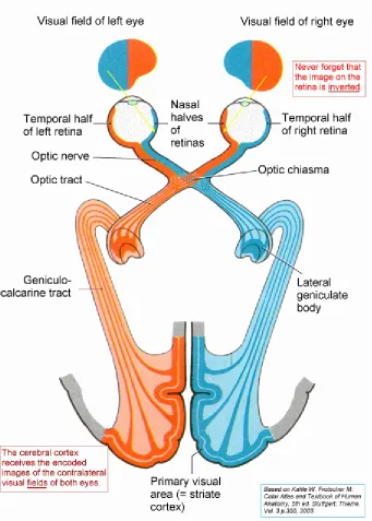
2.1 Anatomy of the visual pathway
The visual system as a whole is developmentally, morphologically
and functionally an outgrowth from the central nervous system. The visual
sensory system comprises of retina, optic nerve, optic chiasma, optic tract,
lateral geniculate bodies, optic radiation and the visual cortex.
The rods and cones of the neural epithelium of the retina form the end
organ of the visual afferent tracts. The first conducting nerve cell of the first
order is the bipolar cell of the inner nuclear layer of the retina with its axon
in the inner reticular layer. The neurons of the second order are the ganglion
cells in the retina, the processes of which pass into the nerve fiber layer and
along the optic nerve to the lateral geniculate body. The optic nerve contains
approximately 1.2 million nerve fibers and is divided into intraocular,
intraorbital, intra canalicular and intracranial till it reaches the optic chiasma.
In general, it may be said that the fibers from the peripheral parts enter
the periphery of the optic nerve, while those from parts of retina near the
optic disc enter the centre area of the nerve. The fibers from the macular
Figure 1‐VISUAL PATHWAY
[image:9.612.148.489.116.593.2]towards the cortex.This is called papillomacular bundle which become
centrally situated in the posterior part of the optic nerve. The retina is
divided into nasal and temporal halves by an imaginary vertical line passing
through the fovea . Fibers from the temporal half of retina enter the chiasma
and pass into the optic tract of the same side and synapse in layers 2,3,5 of
the LGB. Fibres from nasal half of retina enter the chiasma, decussate and
pass into the optic tract of the opposite side and synapse in layers 1,4,6 of
LGB. Each LGB, thereby receives visual information from both retinas. The
corresponding third order neurons originate in the LGB and then pass by the
optic radiations to the corresponding occipital lobes. Lesions of the optic
tract or occipital cortex will cause blindness of ipsilateral temporal retina
and contralateral nasal retina (Hemianopia).
The visual fibers in the optic radiations, run behind the motor fibers
in the internal capsule .Thereafter they separate considerably, the lower
fibers of retina run forwards into the temporal lobe (Meyer’s loop) before
they turn backwards to the lower portion of visual cortex. The dorsal fibers
run backwards in a more direct course to the upper part of the visual cortex.
They pass close to the posterior cornu of the lateral ventricle, so that when
The occipital cortex in and about the calcarine fissure differs from the
cortex elsewhere in the possession of the white line, the line of Gennari. This
primary visual sensory area is the cortical projection of the corresponding
halves of both retinae. The parts above and below the calcarine fissure
represent the upper and lower corresponding quadrants of both retinae
respectively and the posterior part of the occipital lobe represents the
macula.
2. 2 Normal appearance of the disc:
Physiologic temporal pallor :
An absolute prerequisite to recognition of the
abnormal disc is familiarity with normal disc colour. The temporal side of
the normal disc usually is paler than nasal side. This may be due to the
following causes:
i) The size of the physiologic cup – when it extends almost to the
temporal edge of the disc, the temporal side looks pale.
ii) Thin translucent nature of the temporal nerve fibre
iv) Sometimes a temporal crescent of sclera exposed by retraction of
RPE may enhance whitish appearance of disc.
v) In eyes with axial myopia, the normal temporal pallor is
accentuated because of the oblique entry of optic nerve, the
contents of the disc are displaced nasally. The physiologic cup is
shallow and its extension to temporal margin produces relative
temporal pallor.
vi) In infants, efforts to keep lids separated while doing fundus
examination, may produce inadvertent pressure on the eye and
may account for apparent temporal pallor.
vii) Other variable factors like the amount of light used, the colour
,temperature of the light source and the age of lens may give an
illusionary picture of whiteness of disc.
2.3) Acquired or pathologic temporal pallor of the optic disc
When the optic nerve degenerates, its blood supply
is reduced and the smaller vessels are no longer visible. In addition to
reduction in blood supply, formation of glial tissue has been said to occur
with optic atrophy. These two factors have been presumed to account for
the pallor that is associated with optic nerve atrophy.
Pallor of the optic disc may be diffuse or confined to one sector.
Kestenbaum introduced a “Capillary number test” is which the small
vessels at the margins of the disc which are usually 9-10 in number were
decreased in patients with diffuse optic atrophy.
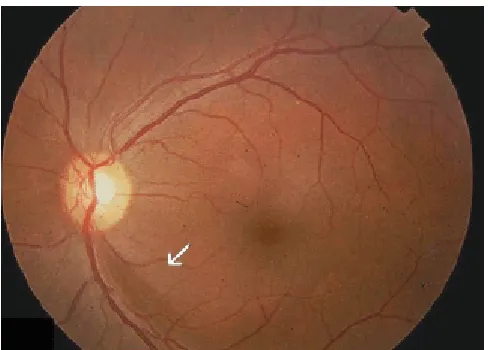
Acquired temporal pallor is most commonly associated with
segmental optic atrophy. The white area which generally extends from the
temporal edge of disc to the central vessels, may appear totally devoid of
capillaries. Margins of this white area tend to blend gradually with the
reddish yellow colour of the surrounding disc tissue. Sharply demarcated
wedge shaped temporal pallor is the consequence of discrete papillomacular
Figure 2‐TEMPORAL PALLOR OF THE OPTIC NERVE HEAD
[image:14.612.127.501.199.477.2]Superior, inferior or nasal sector shaped pallor seldom appears as sharply
circumscribed as temporal pallor.
Acquired temporal pallor is usually caused by optic neuropathies that
selectivity affect central vision and field, sparing the peripheral field. Such
optic neuropathies include toxic and nutritional optic neuropathies, dominant
hereditary optic atrophy and optic neuritis. when superior or inferior disc
pallor is present, an ischaemic etiology is more likely.
The evaluation of optic disc color, though looks simple has many
pitfalls. More objective evaluation of the pale disc can be obtained by
detailed observation of its configuration and neural tissues, its veins, arteries
and capillaries and the peripapillary retinal fiber layer that surrounds it.
Evaluation of peripapillary retinal nerve fiber layer in the diagnosis
of optic atrophy
Focal destruction of nerve fiber bundles is one of the common
pathologic denominators of disease that affects the inner retinal layers, optic
disc, retrobulbar optic nerve or a combination of these structures. When
bright red free light is used in ophthalmoscopy or in fundus camera, normal
linear striations that overlie the retinal vessels are seen which are the
Figure 3‐Localised nerve fibre layer defect
[image:16.612.196.440.195.370.2]Early focal loss of axons is represented by the development of dark
slits or wedges in the peripapillary retinal nerve fiber layer. The slit defects
are most easily identified in the superior and inferior arcuate regions where
the nerve fiber is particularly thick. When multiple nerve fiber bundle
defects are present, they impart a “raked appearance” to the nerve fiber
layer. With gross loss of axons, the defects coalesce producing a large
wedge pattern. Within the wedge, the entire retina takes on a flat granular
appearance with no striations being appreciated. The vessels in this area,
having lost their surrounding nerve fiber layer covering, appear darker than
normal. A prominent light reflex usually emanates from the side of vessels.
As per the zone affected, one can interpret the possibilities, for eg, in
temporal pallor due to toxic optic neuropathy, the papillomacular bundle will
show dark granular appearance without the usual linear striations. In
diseases like multiple sclerosis, the typical nerve fiber bundle defect can be
detected even prior to the disc pallor and aids in the detection of multisystem
involvement.
Diffuse thinning of nerve fiber layer around the optic disc is difficult
to recognize in the early stages. In more advanced stages, signs of atrophy
highlights on large and small retinal blood vessels, apparent reduced caliber
of blood vessels and pallor of optic disc with decreased visibility of the disc
capillaries(Figure 3)
Retinal vascular changes associated with optic atrophy:
In most cases of optic atrophy the retinal arteries are narrowed but not
all cases are associated with retinal vascular changes. In eyes with optic
atrophy from damage to the retro laminar optic nerve, the retinal vessels are
often unaffected. Eyes with retinal vascular changes associated with optic
atrophy presumably have suffered an additional insult directly affecting the
retinal vasculature.
In addition to the fundus appearance, several tests like visual acuity,
fields, color vision help in deciding the pathological nature of temporal
2.4) Pathology of Optic Atrophy:
Since the axons in the optic nerve arise almost entirely from the
ganglion cells located in the retina, damage to such axons may occur at
several locations.
a) The retinal nerve fiber layer or optic disc
b) From disease within or surrounding the different portions of the optic
nerve
c) From intracranial diseases of the optic chiasma, optic tracts or LGB
d) From diseases of the retrogeniculate pathways that produce
transsynaptic degeneration. Focal disruption anywhere along an axon
causes degeneration of the entire axon and its cell body, the retinal
ganglion cells. When large number of axons undergo such
degeneration, gross shrinkage of the optic nerve occurs. i.e optic
F
F
igure 4‐NO
igure 5‐OP
ORMAL NE
PTIC NERV
ERVE FIBRE
VE ATROPH
E LAYER
HY
Wallerian ascending degeneration
When a visual axon is severed, its distal segment being separated from
its nutrient ganglion cell body quickly disintegrates and disappears and its
investing myelin sheaths undergoes a slower breakdown into simple lipids,
this process is called Wallerian degeneration or Anterograde degeneration .
The nerve fibres undergo wallerian degeneration at a rate inversely
proportional to their thickness. Larger axons degenerate sooner and smaller
ones latter. They break up into fragments including the myelin sheaths at
constrictions. A series of ellipsoids form ,each surrounding a fragment of
axons. Myelin is broken down into simpler lipids and engulfed by
phagocytic microglia. Leucocytes are rarely present in Wallerian
degeneration. An essential feature of Wallerian degeneration is the swelling
and degeneration of terminal buttons of axons within the LGB. Wallerian
degeneration and the optic pallor it produces can only be observed clinically
when retinal axons are damaged within the eye .
Retrograde (Descending) Degeneration of Axons:
Although ascending degeneration begins and completes within 7 days,
this time, orthograde axonal transport continues in a normal manner. After
about 3 – 4 weeks, the entire remaining structure degenerates rapidly, so that
after 6 - 8 weeks, after severe optic nerve injury, no affected ganglion cells
remain viable. A most fascinating feature of retrograde degeneration of optic
nerve is that the time course of this degeneration is apparently independent
of the distance of injury from the ganglion cell body.
Regeneration:
Regeneration of axon in the optic nerve is quite limited but some
degree of remyelination does occur after injury. This has been proved in
multiple sclerosis and other immune related disorders.
Remyelination plays a larger role in the recovery from acute and
chronic compressive lesions of CNS as long as there has not been severe
irreversible axonal dama
2.4.1 Pathogenesis of acquired disc pallor:
Optic atrophy is consistently associated with reduction in blood
supply and formation of glial tissue. These two factors account for the disc
Experimental studies of animals suggested that factors that give the
pink colour of the disc are its thickness and the cytoarchitecture of nerve
fiber bundles passing between glial columns containing capillaries. They
postulated that light entering the disc rim is normally conducted along the
transparent nerve fiber bundles.The light diffuses among the adjacent
columns of glial tissue and capillaries and acquire the pink color of the
capillaries and the disc appears pink. In atrophic disc rim, the axonal
bundles are destroyed and the remaining astrocytes are arranged at right
angles to the entering light and little light passes into the disc substance to
traverse the capillaries that are still present and surrounded by layers of
astrocytes. Since light is reflected from opaque glial cells and does not pass
through capillaries it remains white and the optic disc appears pale. In some
areas, loss of tissue also allows light to pass directly to the opaque scleral
lamina and this adds to the pallor. Other explanations for optic disc pallor
include absence of capillaries and astroglial proliferation.
Discrepancies between the optic disc appearance and visual
function:
No judgment of pallor is meaningful until and unless the pallor is
color vision as well as quantitative perimetry, examination of pupils and
electro physiological studies. An optic disc may appear pale but other
examinations may not reveal any abnormality. In cases, it is most likely to
be physiologic pallor. Conversely, an optic disc may appear normal despite
optic nerve dysfunction. In most of these cases, careful evaluation of the
disc, peripapillary retinal nerve fiber layer will provide evidence of retinal
nerve fiber atrophy to produce pallor of disc.
2.6
Etiology of Temporal Pallor
:
Several scenario could cause temporal pallor and the commonly
encountered ones are
1) Nutritional and deficiency optic neuropathy
2) Toxic neuropathy including tobacco, alcohol and toxins
3) Retrobulbar, compressive and infiltrative optic neuropathy
4) Hereditary optic neuropathies like
i) Leber’s optic atrophy
ii) Dominant optic atrophy
iv) Sequelae of optic neuritis – demyelinating diseases
v) Chiasmal compression syndromes.
vi) Macular diseases
Since the causes are divergent , each entity in some detail is discussed
in sequence.
2.6.1 Nutritional optic neuropathy
Toxic and nutritional optic neuropathies are often described together
because they present with more less same symptoms.
Diagnostic criteria proposed by Lussel are :
1. Gradual bilateral painless progressive visual loss
2. Dyschromatopsia
3. Central or centrocoecal visual field defects
4. Optic disc appearance normal initially
5. Absence of metamorphopsia and hallucination
Others :
1) Later developing temporal disc pallor
2) Atrophy of the papillomacular bundle layer
With the exception of vit B12, no specific nutrient has been proved to
cause amblyopia in humans.
Vitamin B12 deficiency
This vitamin is found in milk, egg, meat and cheese and must be
ingested. Vitamin B12 deficiency which takes years to develop generally
arises in three different settings.
1) The first and most common is pernicious anaemia. This auto immune
condition results in gastric atrophy, reduced intrinsic factor
production by gastric parietal cells and B12 malabsorption. It affects
middle aged individuals and can cause megaloblastic anaemia. An
associated neurologic condition, sub acute combined degeneration of
the spinal cord, bilateral optic neuropathy can develop. In this setting,
patients develop both peripheral neuropathy and myelopathy. The
second setting is in patients with a previous history of partial or
absorb B12. The third setting, simple lack of B12 in the diet as might
occur with a strict vegan is the rarest.
Clinical picture:
Patients present with slowly progressive vision loss with
dyschromatopsia and centrocaecal scotomas. The optic nerve may be normal
or may demonstrate pallor and nerve fiber defects . The defects could be
temporal pallor, nerve fiber drop out in papillomacular bundle and rake like
defects in the nerve fiber layer. Vision loss may precede the recognition of
anaemia or other neurologic symptoms. Visual evoked response
abnormalities have been found in patients without visual symptoms.
The diagnosis is confirmed by low serum B12 levels or elevated
methyl malonic acid or homocystine , both of which require B12 for their
metabolism.
Treatment is with parenteral hydroxy cobalamin and with prompt
therapy, vision loss may be reversible.
Optic neuropathy has been shown in primate studies to be result of
demyelination involving the papillomacular bundle. The pathogenesis
(especially in cigarette smokers ) or improper fatty acid synthesis leading to
myelin dysfunction. More recently Rizzo proposed that adenosine
triphosphate deficiency may be a final common pathway for B12 optic
neuropathy, leber’s optic neuropathy and tobacco alcohol amblyopia.
Other vitamin deficiencies
Deficiencies in thiamine (B1), Pyridoxine (B6), folic acid, niacin
and riboflavin (B2) have all been suggested causes of optic neuropathy
Thiamine
Deficiency of thiamine (B1) typically results in Beriberi or wernickes
encephalopathy. There is experimental,biochemical and clinical data that
suggest that thiamin deficiency may cause an optic neuropathy. However the
experiments did not confirm conclusively that thiamine deficiency could be
the sole cause of optic neuropathy. Ketogenic and high protein, low
carbohydrate diets may also put a patient at risk for developing thiamine
deficiency. Though there is good biochemical evidence that many of the
patients with nutritional optic neuropathy were thiamine deficient, yet there
is no basis to conclude that thiamine deficiency is the primary cause of an
In case of pyridoxine, there has been no causal relationship
established between the deficiency state and optic neuropathy. However
certain drugs like Isoniazid, cycloserine bind with vit B6.Other drugs
interacting with B6 are chloramphenicol, hydrallazine, Pencillamine. It has
been postulated that the optic neuropathy associated with these drugs are due
to the pyridoxine deficiency rather than the drug themselves.
Niacin:
Deficiency of niacin causes pellagra, characterized by diarrhoea
,dermatitis and dementia. It may also have associated optic neuropathy
characterized by central or centrocaecal scotoma , reduced visual acuity and
temporal pallor of disc. Visual function improves if treated earlier.
Riboflavin vit B2
Riboflavin deficiency manifests less dramatically than Beriberi or
pellagra. However retrobulbar optic neuropathy has been associated with
burning feet syndrome among the malnourished prisoners of war in the far
east which is specifically due to riboflavin deficiency.
Certain drugs like thallium may cause optic neuropathy indirectly by
interfering with vit B2 metabolism.
Folic Acid
It plays a possible role in tobacco alcohol ambylopia.
Though there is no definitive evidence that such deficiencies play a
primary etiological role in nutritional amblyopia, it remains possible that
there are some patients who develop loss of vision from damage to optic
nerves consequent to dietary vitamin deficiencies.
Nutritional amblyopia may be primary when it is due to poor dietary
intake or secondary when it is associated with chronic alcoholism or
smoking or malabsorption syndrome.
TROPICAL OPTIC NEUROPATHY:
There are optic neuropathies endemic to tropical regions for which a
nutritional agent has been invoked. This epidemic was reported from
Jamaica. The clinical features of the patients were numbness and cramps in
hands and feet, bilateral visual loss, hearing loss, muscle wasting and pain,
There is an otherwise unexplained bilateral optic neuropathy called
“west Indian Amblyopia” The history is typically bilateral, painless,
progressive visual decline in a well nourished adult. The field defects were
central or centrocaecal scotomas but annular scotomas and peripheral
constrictions have been reported. Optic atrophy develops. Deafness was also
present. No treatment including various vitamin regimens is effective in
reversing the visual or non visual manifestations of this disorder. Although a
toxic basis (bush tea) has been postulated, these is no good evidence for
either a toxic or nutritional etiology.A similar disorder has been described in
Nigerians. The features include optic neuropathy, ataxia, peripheral
neuropathy, hearing loss and an unusual feature was peripheral field
constriction without central defects. The role of poor nutrition and ingestion
of exogenous cyanide from an excess of cassava in diet was unclear. The
evidence that cyanide plays a role in causing any optic neuropathy is
discussed further in section on tobacco optic neuropathy.
2.6.2 TOBACCO-ALCOHOL AMBLYOPIA:
The most commonly recognized nutritional or toxic optic neuropathy
may actually be a combination of both a nutritional deficiency state and a
The affected entity are heavy smokers, generally malnourished and
they are usually pipe smokers or cigar smokers using shag and strong
tobacco mixtures. Cigarette smokers are rarely affected Tobacco chewing
and passive smoking in tobacco factories can also cause the disease.
Tobacco amblyopia is found mostly in middle aged or elderly males.
The disorder is painless and characterized by slowly progressive bilateral
dyschromatopsia and visual loss. The characteristic field defect is
centrocaecal Scotoma . The optic discs are initially normal with pallor being
a late feature. In alcohol amblyopia there may be central or centrocaecal
scotoma especially for red with dense nuclei of scotoma in the centre and
not in the centrocaecal area(Figure 6).
ETIOPATHOGENSIS
The mechanism by which tobacco damages the optic nerve has not
been determined. One possibility relates to concurrent malnutrition. Vitamin
B12 deficiency and impaired vit B12 absorption was found in 40% of
patients. But in most cases of tobacco amblyopia, blood vit B12 levels were
within normal limits.
ROLE OF CYANIDE:
Cyanide is present in tobacco smoke and this led to the suspicion that
tobacco amblyopia is a limited form of cyanide poisoning. The cyanide
theory was reviewed and concluded that there is a defect in sulfur
metabolism and of cyanide detoxification in patients with visual loss
associated with tobacco smoking. The role of cyanide in tobacco amblyopia
is yet to be proved. As with other nutritional amblyopias, the etiology of
tobacco amblyopia in many cases is multifactorial.
Patients with tobacco amblyopia slowly improve if they stop smoking
or if they are treated with 1M injection of hydroxycobolamin 1000
micrograms, the dose being repeated five times at intervals of 4 days and
Figure 6‐Centrocaecal scotoma of tobacco‐alcohol amblyopia
[image:34.612.112.548.158.384.2]Note :
Bilateral centrocaecal scotoma may at times mimic the bitemporal
depression of chiasmal interference .The differentiating points are:
i) The VA is diminished
ii) The field defects extend across the vertical meridian .
iii) Especially demonstrable with red objects
iv) There is no peripheral hemianopic depression.
v) As the defects progress they appear more centrocaecal and less
hemianopic.
Drug Induced retro bulbar toxic optic neuropathies
Insidious and slowly progressive bilateral loss of function in the
central fields, with resultant diminished acuity, dyschromatopsia and central
scotoma should alert the possibility of intrinsic optic nerve disease related
to adverse effect of pharmaceuticals. Grant lists 40 such substances but the
evidence for a statistical association between most substances and optic
1. Amantidine hydro chloride
2. Amiadarone
3. Arsenicals
4. Asperdium
5. Carbon disulfide
6. Carbon tetrachloride
7. Chloro dinitro benzene
8. Chlorpromazine
9. Chlorpropamide
10.Cobalt chloride
11.Digitalis
12.Dinitrotoluene
13.Disulfiram
14.Ethambutol
15.Ethylene glycol
16.Hexachlorophene
17.Hydroxy quinolines
18.INH
19.Lead
21.Quinine
22.Streptomycin
23.Styrene
24.Thallium
25.Toluene
26.Trichloroethylene
27.Tricresyl phosphate
28.Vincristine
29.Amoproxan
Ethambutol:
It causes a dose related optic neuropathy. The toxicity is thought to
result from its chelating properties. A typical presentation includes either
central field defects with loss of acuity and dyschromatopsia or well
preserved central visual function with peripheral field loss. Doses less than
15mg /kg/day are safe but in patients taking between 15-25mg/kg/day visual
symptoms develop over a period of months. Dyschyromatopsia and loss of
contrast sensitivity are the early signs. Some recovery usually occurs with
discontinuation of the drug but abnormal usual fields and contrast sensitivity
INH:
It can also cause anterior and retrobulbar toxic optic neuropathy
which can occur with or without pyridoxine deficiency induced sensory
neuropathy.
AMIODARONE:
An optic neuropathy with disc swelling very similar to anterior
ischaemic optic neuropathy has been reported in patients taking amiodarone.
Visual loss may be insidious, with bilateral simultaneous involvement of
eyes and is less severe than AION.
HALOGENATED HYDROXY QUINOLINES:
These drugs have been implicated in the development of myelopathy
and optic neuropathy patients develop optic atrophy and severe vision loss
2.6.3 RETROBULBAR COMPRESSIVE OPTIC NEUROPATHIES:
Compressive optic neuropathies can be identified by their typical
presentation with slowly progressive visual loss and optic nerve atrophy.
SUMMARY OF THE IMPORTANT CAUSES OF OPTIC NERVE
COMPRESSION:
INTRAORBITAL:
Primary orbital tumors
Cavernous hemangioma
Shwannoma
Secondary orbital tumors
Sinus tumor
Metastatic tumors
Enlarged extra ocular muscles
Intra orbital / Intracanalicular / Intracranial
Optic nerve neoplasmas (Primary)
Malignant optic nerve glioma
Optic nerve sheath meningioma
Optic nerve neoplasms(metastatic)
Carcinomatous meningitis
Optic nerve metastasis
Lymphoma/leukemia/myeloma
Non tumour sinus causes
Mucocele
Paraclinoid tumours
Vascular causes
Aneurysms
Ectatic carotid arteries
Chiasmal lesions
Pituitary adenoma
Suprasellar meningioma
Other causes
Fibrous dysplasia, systemic diseases,thyroid disease,orbital hemorrhage.
Signs and symptoms:
Patients usually present with reduced visual acuity,colour vision and
visual field defects typical of optic neuropathy.The optic nerve may be
normal,pale and swollen.Compression will only result in optic nerve head
swelling when the lesion is intraorbital and optic atrophy results in
intracanalicular or intracranial lesions. The other symptoms are gaze evoked
amaurosis, pain on eye movements, orbital fullness, proptosis and ptosis.
The non ophthalmic manifestations are headache, facial numbness, anosmia,
endocrine abnormalities.
2.6.4 HEREDITARY OPTIC NEUROPATHIES
There are 3 types of hereditary optic neuropathies.
1. Those that occur primarily without associated neurologic or systemic
signs
3. Those in which the optic neuropathy is secondary to the overall
disease process
Monosymptomaic Hereditary Optic Neuropathy
2.6.4 A LEBER’S OPTIC NEUROPATHY:
Since the disorder was described by Leber is 1871, the clinical profile
of LHON has been well established, although it is highly variable in
members of the same pedigree.
Age: Usually presents in second or third decade.
Sex: Men are affected more than women.
Mode of Inheritance: It became clear from studies that the disease was due
to maternally inherited mitochondrial DNA.
CLINICAL FEATURES:
It causes an acute or sub acute failure of vision in one or both
eyes.The average interval between first and second eye involvement is
several months. There is painless vision loss, headache and uthoff’s
symptom may be associated. The patient presents with signs of an acute
is unusual. Field defects show central or centrocaecal scotoma. RAPD may
be present.
DISC APPEARANCE:
Leber commented on tortuosity of vessels and white striated opacity
in the peripapillary region .Findings also include
¾ Circumpapillary telangiectatic microangiopathy
¾ Pseudoedema (Swelling of nerve fiber layer around the disc)
¾ Absence of true edema or staining of the disc on FFA.
However, the disc may have a normal appearance is some cases.
LHON patients subsequently develop optic atrophy. After the onset of optic
atrophy, it is difficult to identify patients with LHON because
pseudopapilloedema and telangiectasia usually resolve. Initially the nerve
fiber loss is limited to the papillomacular bundle with temporal pallor of the
disc. Later the entire nerve fiber layer disappears with diffuse disc atrophy.
Ophthalmoscopic changes appear to be present prior to any subjective or
objective evidence of visual dysfunction and Farnsworth Munsell 100 Hue
test appears to be the earliest indication of optic nerve dysfunction. Even
findings and whether all of these will develop the leber’s disease is to be
seen with time.
OTHER ASSOCIATED DISEASES :
Although rare, concomitant neurologic disease, cardiac conduction
defects, arrthymias and skeletal deformities have been associated with
LHON.
MITOCHONDRIAL GENETICS:
Although mitochondrial inheritance became the most plausible
explanation, these mutations do not explain the variable occurrence of the
disease within the same pedigree, and there was wide spectrum of
phenotypic expression. Possible theories put forward for the variable
phenotypic expression are 1) heteroplasmy (coexistence of mutant & normal
mt DNA), 2) second genetic factor possibly on x chromosome 3)
environmental factors like cyanide poisoning (cigarette smoking), alcohol
and environmental toxins. These factors may possibly alter genetically
programmed cell death.
There is no known effective treatment or prophylaxis for the visual
loss that occurs with LHON. Steroids, cyanide antagonists, cyanacobalamine
have been tried with variable results. Surgical dissection of thickened
arachnoid tissue has been done in selective cases with disappointing results.
B. DOMINANT OPTIC ATROPHY
Two forms were described. Congenital or infantile form with
nystagmus and juvenile form.
Patients typically present before the end of first decade of life with
insidious onset of bilateral vision loss .They have central or centrocaecal
defects with intact peripheral isopters. 40% of patients retain acuity of
20/60 or better. Visual loss usually does not progress once the patient
reaches the second decade of life. Colour vision loss typically occurs along
the blue–yellow or tritanopic axis. The associated optic disc pallor often
takes on a striking wedge like appearance with temporal excavation of the
neuroretinal rim, although the whole disc is sometimes pale. Because of the
highly variable degree of visual dysfunction, seemingly asymptomatic adult
and child relatives can often be identified if visual acuity or colour loss or
optic atrophy is present. Recently pedigree analysis has identified the defect
identification of symptomatic and asymptomatic carriers. A much less
common focus on 18q12.2-12.3 has also been identified.
PATHOLOGY:
Studies have demonstrated diffuse loss of retinal ganglion cells,
suggesting primary ganglion cell death as the mechanism of disease.
TREATMENT:
There is no effective treatment at this time. Patients should also be
screened for sensory neural hearing loss as this occurs with increased
frequency in this group of patient.
C.RECESSIVE OPTIC ATROPHY:
The recessive form is more severe than the dominant form. Patient
present in early childhood (age 2 to 4) with more profound visual acuity
loss and searching nystagmus. There is often a history of parental
consanguinity. There is usually diffuse disc pallor and attenuation of retinal
vessels. Complicated, recessive optic atrophy when associated with spino
cerebellar degeneration, cerebellar ataxia, pyramidal tract dysfunction and
OPTIC ATROPHY ASSOCIATED WITH NEUROLOGIC AND
METASTATIC DISEASES;
Optic atrophy and progressive visual dysfunction frequently
accompanies a variety of other inherited, degenerative neurologic
conditions. Visual loss progresses along with the systemic neurologic
syndrome but vision loss usually remains moderate. The disorders frequently
involved are ataxias like spinocerebellar degenerations, Fredrich’s ataxia,
charcot maric tooth disease ,olivoponto cerebellar atrophy,
Mucopolysaccharidoses, lysosomal storage disorders, Tay-sachs, Niemann
pick A, krabbe’s disease, Pelizaeus-Merizbacher disease,
Adrenoleucodystrophy, Meta chromatic leukodystrophy, generalized
gangliosidosis.
Progressive optic atrophy is also recognized in patients with
Congenital deafness. In lysosomal storagedisorders,optic atrophy results
from ganglion cell damage due to these accumulated materials.
2.6.5 TEMPORAL PALLOR AS SEQUELAE OF OPTIC AND
RETROBULBAR NEURITIS:
i) Local Conditions : Uveitis and Retinitis
Meningitis
Intra Orbital infections
Nasal sinusitis, Venoms
ii) Demyelinating disease : Multiple sclerosis, Acute
disseminated encephalomyelitis
Myelitis, Devic’s neuro myelitis
optica , diffuse periaxial
encephalitis
iii) Infective encephalitis : Viral, bacterial, rickettsial, Protozoal
iv) Systemic Conditions : Diabetes, anaemia, lymphomatous
disease , collagen disease ,
endocrine disturbance , malignant
disease , Toxic and allergic
conditions, avitaminosis.
v) Giant cell arteritis
vi) Neurological syndromes : Cranial polyneuritis
vii) Specific infections : Tuberculosis, Syphilis, mycoses
Clinical features of active optic Neuritis:
The vision loss is rapid in onset, progressive involving central vision
more than peripheral. The loss may be mild to no PL. Reduced color vision
invariably accompanies the vision loss .Red objects may be described as
either pinker or browner in patients with dyschromatopsia , characteristic
pain precedes the vision loss. Globe tenderness on pain on eye movements
is typical. This pain characteristically disappears after 3-5 days. Patient
complains of phosphenes or flashes of light. The presence of phosphenes
should also raise the possibility of concomitant retinal disease as in neuro
retinitis .Pupillary constrictions to light is illsustained and there is afferent
pupillary defect on swinging flash light test. The characteristic field defect is
central scotoma especially for red. Other possible defects are paracentral,
arcuate, altitudinal and contraction of peripheral fields. Fundus examination
shows nothing in case of retro bulbar neuritis but in papillitis there are
disc edema, flame shaped haemorrhages, exudates, sheathing, cells in
posterior vitreous. Though the disease is mostly unilateral (71%) it is
but occurring with an interval of 3 months and less (12%) or more than
three months.
The sequelae of optic Neuritis:
Vision:Visual acuity returns to normal or near normal mostly. In spite
of the recovery of visual acuity to even 6/6 ,patients still feel that the quality
of vision is not as good as the fellow eye and describe their vision in the
affected eye as fuzzy.
Even after recovery people may experience uthoff’s phenomenon
i.e., blurring of vision with exercise, hot bath or emotional stress. This is
common in patients with multiple sclerosis but also noted in optic neuritis of
other origin as well as in leber’s optic atrophy .
The possible explanations are:
1) Axonal conduction delay in demyelinating disease with heat.
2) Humoral substances released during exercise or heat causing a
conduction delay.
Pulfrich phenomenon: Due to conduction delay ,simple to and fro
movements of a ball on a string in one plane appears as elliptical
and especially in parking. This is to some extent alleviated when the
normal eye is given sunglasses so that some filtering effect occurs,
neutralizing this effect.
Color vision:There is always some defects in color vision for
(Red)which can either be easily made out even by the gross pseudo
isochromatic charts or only by 100 Hue test in subtle defects.
Brightness sensitivity: Is reduced on the side affected by optic
neuritis previously.
Pupil:Afferent pupillary defect is present in gross lesions. In subtle
defects, the best method of detecting the defect is by edge pupil cycle time.
In this method patient in seated before slit lamp and beam of moderate
intensity of 0.5 mm thick horizontally is shone perpendicular to the iris to
edge of the pupil; pupil constricts immediately and then dilates again,
constricts and dilates. These oscillations occur indefinitely as long as the
light is kept on. Now the time taken for 100 oscillations is counted with a
stop watch and the time taken for a single oscillation is detected. This is
called pupil cycle time (normal is 720-940 milliseconds. Normal difference
is cycle time between 2 eyes 0-70millisecond). This time is significantly
latency time in that it can detect and quantitate subclinical defects in optic
nerve conduction time. It is an objective and quantitative method by which
each eye can be tested individually. It is a fast, simple and reliable clinical
test of optic nerve function.
Fundus examination: 50-80% of patients with optic neuritis
develop some degree of disc pallor, whether initially the disc was normal or
with swelling of the disc. The pallor is typically temporal but may also be
generalized in some cases. Nerve fiber layer defects occur invariably either
of papillomacular bundle or arcuate bundle or diffusely.
Visually evoked responses: Typically shows increase in the latent
period but the amplitude may be normal or slightly reduced.
2.6.6 CHIASMAL COMPRESSION AND TEMPORAL PALLOR.
As a rule pressure in the chiasmal region produce bilateral simple
optic atrophy with white optic discs with sharp margins. Involvement of
Figure 7‐PITUITARY TUMOUR
[image:53.612.108.391.71.254.2] [image:53.612.116.542.419.651.2]loss of any type associated with endocrine dysfunction.In cases of
bitemporal hemianopia, the pallor is rarely accentuated in the nasal half as
all the macular fibres as well as those supplying the area between the disc
and the macula pass through the temporal part of disc .So initially the pallor
may be restricted to the temporal part of the discs alone but as the
compression is unrelieved, the entire disc becomes pale. The most
common causes are compressive sellar masses and therefore the diagnosis
and management heavily depends on neuroimaging . This lesions produce
visual acuity and field defects by interfering with the optic nerves, chiasma
, optic tract or by obstructing the third ventricle and causing chronic
atrophic papilloedema . So an awareness of temporal pallor which may
occur due to chiasmal compression may aid in the early diagnosis of
tumors compressing chiasma(Figure 7&8) .
2.6.7 MACULAR DISEASES CAUSING TEMPORAL PALLOR.
If carefully examined, the vast majority of macular diseases can be
diagnosed easily. However there are several macular conditions that are not
well visualised and therefore optic neuropathy is suspected. They are
overlooked or the clinical suspicion is not high enough and the correct
diagnosis is never considered.
These conditions are:
1) central serous maculopathy
2) cystoid macular edema
3) Diabetic macular ischaemia.
4) Acute macular neuroretinopathy
5) cone dystrophy
6) Toxic maculopathies
7) Idiopathic blind spot enlargement syndrome
CLINICAL DISTINCTION BETWEEN OPTIC NEUROPATHY AND
MACULOPATHY .
SL.NO SIGN OPTIC
NEUROPATHY
MACULOPATHY
1 Reduced acuity Common Common
2 Dyschromatopsia Severe Mild
3 Amsler grid abnormality Missing portions or
gray spots
Distorted or bent
lines
4 Afferent pupillary defect Common Rare
5 Visual field defects Central, arcuate,
nasal, altitudinal
Central scotoma and
midperipheral defects
in photoreceptor
disease
6 Ophthalmoscopy Swollen,pale or
normal optic nerve
Occasionally pale
optic nerve , macular
abnormality (pigment
atrophy,edema)
Despite extensive evaluation, it may be impossible to localize the
cause of the patient’s visual loss . In this setting it is important to consider
the possibility of nonorganic or functional vision loss. When doubt exists
about the cause of the visual loss, one should consider screening the patient
for the treatable causes of optic nerve dysfunction including mass lesions ,
infections and nutritional processes.
SL.NO TESTS OPTIC
NEUROPATHY
MACULOPATHY
1 Electroretinography Normal Normal or
abnormal
(Especially focal
ERG)
2 Ocular coherence
tomography
Normal Abnormal
3 Visual evoked
response
Large latency
delay
Small latency
AIM OF STUDY
The aim of the study is to analyse
• the various causes
• clinical course
• associated findings of patients presenting with temporal pallor
of the optic nerve head
INCLUSION CRITERIA
Patients with temporal pallor of optic disc whose vision showed no
improvement with glasses or pin hole.
EXCLUSION CRITERIA
Patients with refractive errors,corneal diseases, cataract and glaucoma were
MATERIALS AND METHODS
This study was conducted in 50 patients presenting with temporal
pallor of the optic nerve head in the Regional institute of ophthalmology,
Govt ophthalmic hospital, Chennai during the period of March 2007 to
October 2008.
A detailed history of the nature of complaints, history pertaining to
smoking, alcohol, diet and long term intake of drugs especially ethambutol
was obtained.In all cases visual acuity,detailed anterior segment examination
with slitlamp biomicroscopy with special reference to pupillary
reflex,posterior segment evaluation with both direct and indirect
ophthalmoscope,colour vision and fields were done.X ray skull,CT brain and
ANALYSIS AND DISCUSSION
Total number of patients under study-50
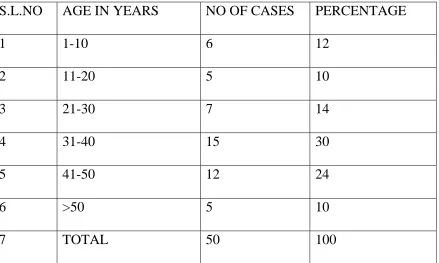
[image:62.612.109.548.239.502.2]AGE INCIDENCE
Table 1
S.L.NO AGE IN YEARS NO OF CASES PERCENTAGE
1 1-10 6 12
2 11-20 5 10
3 21-30 7 14
4 31-40 15 30
5 41-50 12 24
6 >50 5 10
7 TOTAL 50 100
SEX INCIDENCE
Table 2
S.L.NO SEX NO OF CASES PERCENTAGE
1 Male 35 70
2 Female 15 30
3 Total 50 100
The sex incidence showed a preponderance in the male population.
[image:63.612.104.546.433.665.2]PRESENTATION OF CASES
Table 3
Figure 9‐Age of presentation
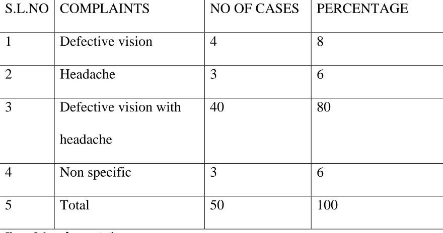
S.L.NO COMPLAINTS NO OF CASES PERCENTAGE
1 Defective vision 4 8
2 Headache 3 6
3 Defective vision with
headache
40 80
4 Non specific 3 6
[image:63.612.103.543.434.666.2]Fi
igure 10‐Sex d
distribution
1 to 10 11 to 20 21 to 30 31 to 40 41 to 50 >50
Male Female
0 0 0 0
In my study , 80 % of the patients presented with defective vision and
headache. 8% of the patients presented with only defective vision. 6% of
patients presented with only headache. 6% of patients presented with
nonspecific symptoms like deviation of the eyes.
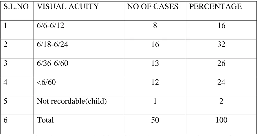
[image:65.612.102.546.293.528.2]VISUAL ACUITY
Table 4
S.L.NO VISUAL ACUITY NO OF CASES PERCENTAGE
1 6/6-6/12 8 16
2 6/18-6/24 16 32
3 6/36-6/60 13 26
4 <6/60 12 24
5 Not recordable(child) 1 2
6 Total 50 100
The visual acuity was tested with snellen’s chart. In my study, 32% of
patients had visual acuity of 6/18-6/24. 26% of the patients had visual acuity
of 6/36-6/60. 24% of patients had visual acuity less than 6/60.Though the
Fi
Fi
g
o
igure 11‐Prese
igure 12‐Visu
good vision
out the caus
entation of sym
al acuity
n, it may st
se of tempo
mptoms
till be nece
oral pallor
essary to do
. o necessary Defect Heada Defect headac Non sp
6/6‐6/1 6/18‐6/ 6/36‐6/ <6/60 Not rec
y investiga
tive vision
che
tive vision with che pecific 12 /24 /60 cordable(child)
ations to fin
h
)
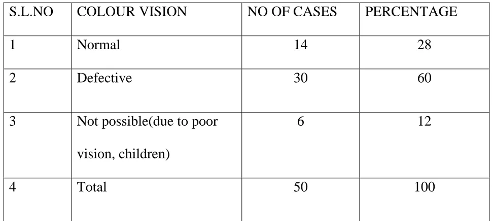
ESTIMATION OF COLOUR VISION
Table 5
S.L.NO COLOUR VISION NO OF CASES PERCENTAGE
1 Normal 14 28
2 Defective 30 60
3 Not possible(due to poor
vision, children)
6 12
4 Total 50 100
Estimation of colour vision was done with pseudo isochromatic chart. It was
defective in 60% of the patients. It was not possible in patients with poor
vision and in children in 12% of patients.
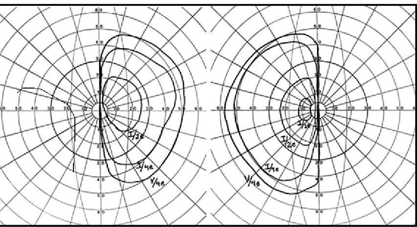
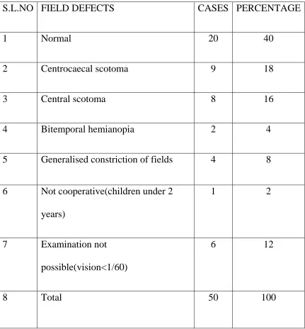
FIELDS
Table 6
S.L.NO FIELD DEFECTS CASES PERCENTAGE
1 Normal 20 40
2 Centrocaecal scotoma 9 18
3 Central scotoma 8 16
4 Bitemporal hemianopia 2 4
5 Generalised constriction of fields 4 8
6 Not cooperative(children under 2
years)
1 2
7 Examination not
possible(vision<1/60)
6 12
8 Total 50 100
Fi
Fi
igure 13‐Estim
igure 14‐Fields
mation of colo
s
our vision
Normal
Defective
Not possib vision, child
Normal
Centrocaec
Central sco
Bitempora
Generalise of fields Not cooper under 2 ye
le(due to poor dren)
cal scotoma
otoma
l hemianopia
d constriction
rative(children ars)
r
Examination of fields was done with Bjerrum’s screen using coloured
objects. The fields of the patients who had optic nerve pathology were tested
with red targets and those who had macular pathology were tested with blue
yellow targets. Patients having tobacco-alcohol amblyopia presented with
central and centrocaecal scotoma.Chiasmal compressive lesions presented
with bitemporal hemianopia.Patients with retrobulbar neuritis had
contraction of the visual fields.Automated perimetry confirmed the findings
of the kinetic perimetry.
[image:70.612.105.546.424.678.2]X RAY FINDINGS
Table 7
S.L.NO FINDINGS NO OF CASES PERCENTAGE
1 Normal 45 90
2 Enlargement of sella 2 4
3 Fracture wall of orbit 2 4
4 Not taken(child) 1 2
Fiigure 15‐X‐ray findings
Norma Enlarge Fractur Not tak al
ement of sella re wall of orbit ken(child)
The X-ray findings were normal in 90% of the patients.The enlargement of
sella due to pituitary adenoma was found in 4% of the patients. Two out of
six patients with traumatic optic neuropathy had fracture in the lateral wall
of the orbit.
MANAGEMENT
After appropriate investigations, in 2 patients the findings were
suggestive of pituitary tumour and in 3 patients the findings were suggestive
of suprasellar meningioma and they were referred to neurosurgery for
further management. In the remaining 45 cases, 10 doses of vitamin B1,
B6,B12 injections were given on alternate days and neurovitamin
supplements were given. All of them were asked to come for regular follow
up for one year. Three patients who were on ATT had ethambutol induced
toxic amblyopia and were advised to stop ethambutol and to continue the
other ATT drugs. Nine patients who had tobacco-alcohol amblyopia were
advised to stop taking tobacco and alcohol.
RESULTS
50 cases of temporal pallor of optic disc were taken for study.
In my study,
• 68% of the patients(38 patients) presented between third and fifth
decades.
• 70% of the patients(35 patients) were males and 30%(15 patients)
were females.
• 80% of the patients(40 patients) presented with defective vision and
headache and the visual acuity was not improving with pinhole.
• In 82% of the patients(41 patients), the visual acuity ranged between
6/18 - <6/60.
• In 60% of the patients(30 patients), the colour vision was defective.
• 46% of the patients(23 patients) had field defects. Patients with
tobacco-alcohol amblyopia and ethambutol induced toxic amblyopia
had central and centrocaecal scotomas(12 patients). Patients with
with retrobulbar neuritis had generalized constriction of the visual
fields(5 patients).
• X-ray findings showed enlargement of sella turcica due to pituitary
tumour in 4% of the cases. Out of 6 patients who had traumatic optic
neuropathy, the X-ray findings of 2 patients showed fracture in the
lateral wall of orbit.
• In 14 % of the patients(7 patients), CT scan findings were suggestive
of pituitary tumors, suprasellar meningioma,fracture of the wall of the
orbit.
• 18% of the patients(9 patients) who were diagnosed to have tobacco-
alcohol amblyopia were advised to stop taking tobacco/alcohol and
had slight improvement in vision.
• 6% of the patients(3patients) who were on ATT were advised to stop
ethambutol.
In our follow up of the patients for a period of 6 months, patients with
tobacco-alcohol and ethambutol induced toxic amblyopia had visual
improvement of two lines after stopping the agents and with neurovitamin
Out of the 50 patients, the causes of temporal pallor were,
Incidental finding 8
Idiopathic 6
Tobacco amblyopia 9
Post traumatic sequelae 6
On ATT(ethambutol) 3
RB Neuritis sequelae 5
Chiasmal compression 2
Suprasellar meningioma 3
Post meningitic sequelae 2
Post lactational 1
Anemia 2
ARMD 2
Figure 16‐CT brain axial view showing pituitary tumour
Figure 17‐CT brain coronal view showing suprasellar meningioma
[image:76.612.108.413.94.332.2] [image:76.612.108.429.378.671.2]CONCLUSION
Temporal pallor of the optic disc is one of the common ophthalmic
finding in our practice. It could be physiological or pathological.
• Most of the patients with temporal pallor presented between third and
fifth decades.
• 2/3 rd of the patients were males who gave history of prolonged
intake of tobacco and alcohol.
• Tobacco alcohol amblyopia form the important cause.The other
important causes were post traumatic, post neuritic,post lactational,
post meningitic, ethambutol induced, compressive lesions and
macular diseases.
• It is necessary to emphasise that patients with temporal pallor who
have defective visual acuity not improving with glasses, defective
colour vision have to be thoroughly investigated to detect the cause of
temporal pallor. The commonly encountered field defects were
central, centrocaecal scotomos, bitemporal hemianopia and
contraction of visual fields which aided in the diagnosis of the
• Inspite of careful history,clinical examination and detailed
investigations the cause of temporal pallor was not detected in 6
patients.
• Regarding the management and prognosis, those presenting early with
remediable cause had improvement in visual acuity,(tobacco
,alcohol,nutritional,post neuritic,drugs).Those presenting late like
chiasmal compression ,suprasellar meningioma,post traumatic did not
BIBLIOGRAPHY
Text books:
1. Walsh and Hoyt’s clinical ophthalmology - 4 th edition
2. System of Ophthalmology - Duke Elder : Vol XII : 1971;
3. Clinical Ophthalmology. Thomas D. Duane : 1986.
4. Grants Toxicology of the eye - 2 nd edition.
5. The visual fields. David -O- Harrington, 5thedition.
6. Parson’s Diseases of the eye. 20th edition.
Journals:
American Journal of Ophthalmology:
1. Baron G.J., Tapper L. and Iorine G. Ocular toxicity from Ethambutal.
77 : 256; 1974.
2. Caroll F.D. optic neuritis; 35 - 75 - 82 ; 1952
3. Caroll F.D.Jamaican Optic neuropathy in immigrants to U.S. 71 :
216-65; 1971
5. Sanderson P.A. Optic neuropathy presumably caused by Vincristine
therapy : 81 : 146 – 150 ; 1976
6. Stambolian. D;Optic neuropathy associated with vit B12 deficiency.
83;465 – 468; 1977.
7. Sykowski P. Digitoxin intoxication resulting in optic neuropathy 32;
572; 1949.
8. Sykowski : P. Streptomycin causing retrobulbar optic neuritis 34;
1446 : 1951.
9. Henkind. P. Chaarles. N.C. and Plarson J. Histopathology of
ischaemic optic neuropathy. 69; 78-90;1970.
10.Landers MB III. Bradbury M.J. Retinal vascular changes in retrograde
optic atrophy : 86 : 177 – 81 ; 1978.
11.Manor R.S Schleinn N. Yassin Y. Narrow band (540nm) green light
stereoscopic photography of the surface details of peripapillary retina
91 774-780; 1981.
12.Quigley HA and Andersons D.R. The histologic Basis of optic disc
13.Radius R.L. and Anderson D.R. Retinal ganglion cell degeneration in
experimental optic atrophy 86; 673-79;1978
14.Suydacker . D; The normal optic disc Ophthalmoscopic and
pathological studies 58; 958-964;1964.
15.Jonston P.B. Gaster. R.N. and Tripathi R.C. A clinico pathologic
study of autosomal dominant optic atrophy. 88; 868-875 1979.
16.Gudas P. Jr. Optic nerve myeloma : 71 ;1085-1089;1971.
17.Brude RM & Gallin P.F. Visual parameters associated with recovered
retrobulbar optic neuritis 79; 1034-37;1975.
18.Glaser I.S. Savino; The photostress recovery test in clinical
assessment of visual function 83; 193-201; 1978
19.Griffin J.F. and wray S.H. Acquired colour vision defects in
retrobulbar neuritis’ 86; 193-201 ; 1978
20.Miller S.D. and Thompson H.S., Pupil cycle time in optic neuritis
85;635-642; 1978 b.
21.Wilson W.B.VER differentiation of ischaemic optic neuritis from
Archives of ophthalmology :-
22. Baghdassarian S.A. Optic neuropathy due to lead poisoning. 80 :
721-723-1968.
23.Caroll F.D. Nutritional amblyopia 76 ; 406-411 ; 1966.
24.Foulds W.S. The optic neuropathy in pernicious anaemia 82 ; 427-432
; 1969 C;
25.Lerman S. Centrocaecal scotoma as the presenting sign in pernicious
anaemia ; 65 ; 381-385 ; 1961.
26.Lessel S. Experimental cyanide optic neuropathy ; 86 194-204 ; 1971.
27.Rodger F.C. Nutritional amblyopia A. statistical report of the course
and progress in 238 cases 47 ; 570-583 1952.
28.Victor M.Tobacco Alcohol Amblyopia 70 ; 313-318 ; 1963.
29.Victor M. Tobacco – Alcohol Amblyopia Further comments on its
pathology 74 ; 649-657 1965.
30.Victor M. Deficiency amblyopia in the alcoholic patient d64 ; 1-33;
31.Schwatz B; Pallor of the optic disc : Quantitative photographic
evaluation 89 : 278-279 ; 1973.
32.Trobe I.D. Glaser I.S; optic atrophy; Differential diagnosis by fundus
observation alone 98 ; 1040-45 ; 1980 a.
33.Constantine E.F. Lebers disease ; recovery 53; 608-609 ; 1955.
34.Nikoskelainen E. sogg R.L. The early phase in lebers hereditary optic
atrophy ; 95 969-978 1977.
35.Caldwell J.B.H. Dominant juvenile optic atrophy ; A study of two
families and review of the hereditary disease in childhood 85; 133-147
1971.
36.Kline L.B. “Glaser J.S. Dominant optic atrophy; The clinical profile
97 ; 1680-86 1979.
37.Manchester P.J. Dominant hereditary optic atrophy with bitemporal
field defects 60 ; 479-84 1958.
38.Frisen L. and Hoyt. W.F. Insidious atrophy of retinal nerve fibres in
39.Hirose – J; A simultaneous recording of electroretinogram and VER ;
95 – 1205 – 1208 ; 1977.
40.Nutritional amblyopia : Caroll F.D. 76 ; 406-411; 1966.
PROFORMA
1. Serial No
2. OP/IP No
3. Name of the patient
4. Age
5. Sex
6. Address
7. Occupation
8. Complaints Duration
9. History taking:-