Copyright © 1998, American Society for Microbiology. All Rights Reserved.
Mta Has Properties of an RNA Export Protein and Increases
Cytoplasmic Accumulation of Epstein-Barr Virus
Replication Gene mRNA
O. JOHN SEMMES,
1,2LIN CHEN,
1ROBERT T. SARISKY,
1ZHIGANG GAO,
1LING ZHONG,
1AND
S. DIANE HAYWARD
1,3*
Molecular Virology Laboratories, Department of Pharmacology and Molecular Sciences,
1and Department of
Oncology,
3Johns Hopkins School of Medicine, Baltimore, Maryland 21205, and Department of
Microbiology, University of Virginia Medical School, Charlottesville, Virginia 22908
2Received 7 July 1998/Accepted 9 September 1998
The Epstein-Barr virus (EBV) Zta and Mta regulatory proteins were previously found to be required for
efficient replication of oriLyt in cotransfection-replication assays, but the contribution of Mta to the replication
process was unknown. We now demonstrate that Mta regulates replication gene expression. Using the
poly-merase processivity factor BMRF1 as an example, we found that in transfected cells, total BMRF1 mRNA levels
were unaffected by Mta but that the amounts of cytoplasmic BMRF1 RNA and protein were greatly increased
in the presence of Mta. Mta also increased cytoplasmic accumulation of the BALF2, BALF5, BSLF1, and
BBLF4 replication gene mRNAs but did not affect cytoplasmic levels of BBLF2/3 mRNA. Thus, five of the six
core replication genes require Mta for efficient accumulation of cytoplasmic RNA. The contribution of Mta to
posttranscriptional RNA processing was examined. Examination of Mta localization in transfected cells by
indirect immunofluorescence revealed that Mta colocalized with the splicing factor SC35. We also found that
Mta has RNA binding activity. Glutathione S-transferase–Mta bound to BMRF1 and BMLF1 transcripts but
not to a control cellular gene RNA. Mta contains a consensus leucine-rich nuclear export signal. Such signal
sequences are characteristic of proteins that undergo nuclear export. Examination of Mta localization in a
heterokaryon assay provided evidence that Mta shuttles between the nucleus and the cytoplasm. Our
exper-iments indicate that Mta functions in RNA processing and transport and mediates cytoplasmic accumulation
of a number of EBV early mRNAs.
Epstein-Barr virus (EBV) encodes three transactivators, Zta
(BZLF1, ZEBRA), Rta (BRLF1), and Mta (BMLF1), that
together regulate EBV lytic cycle gene expression. Zta is a
bZIP family transcriptional activator that is represented only in
the gamma 1 class of herpesviruses (4). The Rta transcriptional
activator has homologs encoded within both gamma 1 and
gamma 2 herpesvirus genomes (52, 66), while homologs of the
Mta protein are encoded by alpha-, beta-, and
gammaherpes-viruses (13, 32, 50, 53, 57, 62, 79, 82, 90, 91, 93, 94). The most
extensively studied Mta homolog is herpes simplex virus
(HSV) IE63 (ICP27). HSV mutants that lack a functional IE63
gene overexpress immediate early and early viral genes and are
deficient in late gene expression (42, 45, 63–65, 80, 81). Further
analysis has shown that IE63 represses expression of genes
containing introns by inhibiting cellular pre-mRNA splicing.
IE63 associates with and reorganizes proteins associated with
small nuclear ribonucleoprotein particles (snRNPs). This
ac-tivity contributes to, but is not sufficient for, splicing inhibition
(27, 59, 61, 69, 70). HSV IE63 also has a posttranscriptional
stimulatory activity (71). IE63 expression leads to enhanced
binding of cleavage stimulation factor (CstF) to the
polyade-nylation signal of HSV genes (43, 44), and IE63 has recently
been shown to shuttle between the nucleus and cytoplasm,
indicating a role in facilitating RNA transport (60, 68, 83).
EBV Mta is a phosphoprotein (12, 92) that migrates in
denaturing polyacrylamide gels as a series of polypeptides with
a major species of 60 kDa (11, 92). Mta has been less well
characterized than HSV IE63 but is also recognized to have a
posttranscriptional mechanism of action. In transient
expres-sion assays, Mta was initially recognized to stimulate reporter
gene expression from a variety of heterologous promoters (40,
54). Activity in these assays was subsequently shown to be
reporter gene dependent, indicating a posttranscriptional
mechanism (8, 12, 37). Mta has also been implicated as a
contributor to replication via the lytic origin of replication,
oriLyt. In an initial evaluation of the requirements for oriLyt
replication in a cotransfection replication assay, both Zta and
Mta were required to obtain replication in transfected Vero
cells in addition to the six core replication genes, BMRF1
(polymerase-associated factor), BALF2 (single-stranded DNA
binding protein), BALF5 (DNA polymerase), BSLF1
(pri-mase), BBLF4 (helicase), and BBLF2/3 (primase accessory
protein) (19). In this replication assay, HSV IE63 could
par-tially substitute for Mta, suggesting that Mta may have been
contributing to oriLyt replication indirectly by augmenting
rep-lication gene expression as has been described for IE63 (87).
However, IE63 may also contribute to HSV DNA replication
in other ways. IE63 has been found to locate within replication
compartments in infected cells (95). We have now examined
the effects of Mta on expression of EBV replication genes and
show that five of the six core replication genes require Mta for
cytoplasmic accumulation of their mRNA transcripts. Further,
an examination of the mechanism of action of Mta revealed
that Mta associates with splicing factors, binds specific RNAs,
and shuttles between the nucleus and cytoplasm. Thus, Mta
functions analogously to HSV IE63 in facilitating RNA
pro-cessing and export of viral transcripts.
* Corresponding author. Mailing address: Department of
Pharma-cology and Molecular Sciences, Johns Hopkins School of Medicine,
725 N. Wolfe St., Baltimore, MD 21205-2185. Phone: (410) 955-2548.
Fax: (410) 955-8685. E-mail: [email protected].
9526
on November 9, 2019 by guest
http://jvi.asm.org/
MATERIALS AND METHODS
Plasmids. Expression plasmids in which the six core replication genes (BMRF1, BSLF1, BBLF4, BBLF2/3, BALF2, and BALF5) were expressed from heterologous promoters but contain natural 39untranslated sequences have been described elsewhere (20), as have the set in which the replication gene open reading frames (ORFs) were inserted into an SG5-based vector (72). The cDNA version of BBLF2/3, pEF76A, has been described elsewhere (19). To generate SG5-Flag-Mta vector pDH304, PCR amplification of the genomic region con-taining the BSLF2 and BMLF1 ORFs was performed with pTS6 as the template (11) and the primers 592195 (GTCAAGATCTATGGTTCCTTCTCAGAGA) and 391438 (TCAGAGATCTTTATTGATTTAATCCAGG). The PCR product was cleaved with BglII and ligated into the BglII-cut SG5-Flag vector pJH253. Glutathione S-transferase (GST)–Mta was generated by using the template pTS6 and the PCR primers 591439 (TCAGGGATCCGAGAGCCACATTCTGGAA) and 391438. The PCR product was cleaved with BamHI and BglII and ligated into the BglII site of pGH254 such that the BMLF1 ORF was expressed as a fusion with GST. pMBP-Tax was constructed by inserting sequences correspond-ing to the complete Tax cDNA into the BamHI site of pMAL-cR1 (New England Biolabs). SG5-CBF1(pJH156) contains the CBF1 coding sequence ligated as a BglII/BamHI fragment into the BglII site of SG5 (Stratagene, La Jolla, Calif.).
Cotransfection-replication assay.The assay was performed in Vero cells as previously described (20), using the oriLyt plasmid pSL77 and SG5-based ex-pression vectors for the six core replication genes, Mta, Rta, and Zta (72).
Western blotting.293T cells were seeded at 106in 100-mm-diameter dishes the day before transfection. Cells were transfected with 20mg of DNA, using calcium phosphate-BES [N,N9-bis(2-hydroxyethyl)-2-aminoethanesulfonic acid]-buffered saline (10). Cells were harvested 48 h after transfection, washed, and lysed in sample buffer (25 mM Tris, 2% sodium dodecyl sulfate [SDS], 10% glycerol, 5% b-mercaptoethanol, 0.02% bromophenol blue). Cell lysates were electropho-resed through an SDS–10% polyacrylamide gel, and the separated proteins transferred to nitrocellulose (Bio-Rad, Hercules, Calif.). After blocking in TBS-T (100 mM Tris [pH 7.5], 100 mM NaCl, 1% Tween 20) plus 5% nonfat dry milk for 1 h, the filter was incubated with anti-Flag antibody (1:1,000; Eastman Kodak Co., New Haven, Conn.) to detect Flag-Mta.
BMRF1 protein expression was examined in Vero cells grown in six-well cluster dishes and transfected with a total of 2mg of plasmid DNA per well. BMRF1 was detected with anti-BMRF1 monoclonal antibody (MAb; 1:2,000; Advanced Biotechnologies Inc., Columbia, Md.). The actin protein control was detected with antiactin MAb (1:3,000; Sigma, St. Louis, Mo.). Bacterially ex-pressed GST fusion proteins were detected with anti-GST MAb (1:500; Santa Cruz Biotechnology Inc., Santa Cruz, Calif.). Protein bands were visualized using chemiluminescence (Amersham Life Sciences, Arlington Heights, Ill.).
Immunofluorescence.Expression and colocalization assays were performed with Vero cells grown on glass tissue culture chamber slides (Nunc) and trans-fected with 2mg of DNA by the calcium phosphate-BES procedure. Fixing and staining were performed as previously described (88). Anti-Flag antibody (East-man Kodak) was diluted 1:1,500, and anti-SC35 culture supernatant (a gift from X. D. Fu and T. Maniatis) (23) was diluted 1:50.
The heterokaryon assay used HeLa cells grown on glass coverslips and trans-fected with Flag-Mta by the calcium phosphate method. The cells were seeded at approximately 30% confluent. After removal of the calcium-DNA precipitates by washing, the cells were allowed to grow for 24 h. Subsequently, Cos-TdRev cells were seeded onto the same dishes when approximately 50% confluent. This cell line stably expresses RevM10, a mutant of human immunodeficiency virus (HIV) Rev which is unable to export from the nucleus. The strong nucleolar staining of RevM10 was used to identify recipient cells. The combined cell populations were allowed to grow for 8 h to resume normal cell shape. Protein synthesis was inhibited by addition of cycloheximide (100 mg/ml) 30 min prior to heterokaryon formation to allow for clearance of residual cytoplasmic protein. Culture me-dium was removed, and fusion was mediated by incubation in 50% polyethylene glycol in phosphate-buffered saline (PBS) for 90 s. The polyethylene glycol was removed with four washes of PBS, and the fused cells were incubated for an additional hour in complete medium containing cycloheximide. The cells were then washed three times with PBS and fixed by incubation in 4% paraformalde-hyde at room temperature for 12 min. The fixed cells were then permeabilized with a 2-min incubation in 100% methanol at room temperature. Flag-tagged Mta was identified with anti-Flag MAb, and anti-Rev rabbit polyclonal antibody was used to recognize RevM10. The secondary antibodies were tetramethyl rhodamine isothiocyanate (TRITC)-conjugated goat anti-mouse and fluorescein isothiocyanate (FITC)-conjugated anti-rabbit. The immunostained coverslips were attached to glass slides with VectaShield (Vector Laboratories, Inc., Bur-lingame, Calif.), and the samples were analyzed on a Nikon microscope.
RNA analyses.Total RNA was isolated by the guanidinium thiocyanate-phe-nol method. Approximately 107HeLa cells were lysed by addition of 1 ml of denaturing solution (4 M guanidinium thiocyanate, 25 mM sodium acetate [pH 7.0], 100 mM 2-mercaptoethanol, 0.5% Sarkosyl) directly to the culture dish following removal of the medium. The homogenate was transferred to a 5-ml polypropylene tube, 0.1 ml of 2 M sodium acetate (pH 3.9) was added and mixed, and then 1 ml of water-saturated phenol and 0.2 ml of chloroform-isoamyl alcohol (49:1) were added. After 15 min on ice, the phases were separated by centrifugation at 10,0003g. The upper aqueous phase containing the RNA was
subjected to repeated precipitation (three times) in isopropanol. The RNA was then washed in ethanol and stored at220°C.
For preparation of cytoplasmic RNA, culture medium was removed, and the cells were scraped off in 1 ml of ice-cold PBS and collected by centrifugation (1,000 rpm for 5 min). The cell pellet was resuspended in 375ml of ice-cold lysis buffer (50 mM Tris-Cl [pH 8.0], 100 mM NaCl, 5 mM MgCl2, 0.5% Nonidet P-40, 1,000 U of RNAs per ml, 1 mM dithiothreitol) and incubated on ice for 5 min. Nuclei and cell debris were removed by centrifugation (15,000 rpm) in a micro-centrifuge for 2 min at 4°C. The supernatant was removed to a clean microcen-trifuge tube containing 4ml of 20% SDS and mixed. Proteinase K was added at 200mg/ml and allowed to incubate at 37°C for 15 min. The supernatant was extracted twice with 400ml of phenol-chloroform-isoamyl alcohol (25:24:1) and once with chloroform-isoamyl alcohol (24:1). RNA was precipitated from the extracted aqueous phase by addition of 40ml of 3 M sodium acetate (pH 5.2)–1 ml of ethanol and centrifugation at 15,000 rpm for 15 min at 4°C. The RNA pellet was rinsed with ethanol, air dried, and resuspended in water.
Total BMRF1 mRNA was detected by primer extension analysis. The BMRF1-specific probe, CCTTGGTCTAAAGCGGAG (positions 79937 to 79918 on the EBV genome [4]) was designed to yield a 128-base extension product. The oligonucleotide was end-labeled by standard methods (3). Isolated RNA (20mg) was mixed with 105cpm of labeled oligonucleotide and precipitated. The pre-cipitate was dissolved in 30ml of hybridization buffer [80% formamide, 40 mM piperazine-N,N9-bis(2-ethanesulfonic acid) (PIPES; pH 6.4), 400 mM NaCl, 1 mM EDTA (pH 8.0)] and incubated at 30°C overnight. RNA and oligonucleotide were precipitated by addition of 170ml of 0.3 M sodium acetate and 500ml of ethanol. The pellet was washed once with ethanol and air dried. Reverse tran-scription was done with avian myeloblastosis virus reverse transcriptase at 42°C for 90 min. The reaction was halted by addition of 1ml of 0.5 M EDTA and digested with 1mg of RNase A at 37°C for 30 min. The mixture was extracted with phenol-chloroform-isoamyl alcohol (25:24:1) and precipitated by addition of 100ml of 2.5 M sodium acetate and 300ml of ethanol. The pellet was washed once with ethanol and air dried. The extended products were analyzed on an 8% acrylamide-urea gel.
Total and cytoplasmic RNAs were also detected by Northern blotting. RNA (30mg) was electrophoresed through a 1% formaldehyde-agarose gel and trans-ferred onto a nylon membrane (Schleicher & Schuell Inc., Keene, N.H.). The membrane was incubated at 65°C with individual32P-labeled DNA probes which were generated by using a Random Primed DNA labeling kit (Boehringer Mann-heim). The DNAs used as probes were as follows: BBLF2/3, 0.8-kb BglI fragment from pRTS25; BALF2, 1-kb XbaI/HindIII/BglII fragment from pRTS12; BMRF1, 0.8-kb EcoRI/HindIII fragment from pMH2; BALF5, 3.0-kb Xba/ HindIII fragment from pRTS13; BSLF1, 2.6-kb Xba/HindIII fragment from pRTS11; and BBLF4, 2.4-kb Xba/HindIII fragment from pRTS28.
RNA-protein interactions.RNA probes for Northwestern analysis were syn-thesized by in vitro transcription in the presence of [32P]UTP. The BMRF1 probes were transcribed from pGEM74 linearized with EcoRI, resulting in an 847-base RNA product corresponding to BMRF1/2 message from positions 82081 to 82920 plus vector sequences. The BMLF1 probe was transcribed from pGEM74 linearized with HindIII, resulting in a 853-base RNA product corre-sponding to BMLF1 message from 82920 to 82081 plus vector sequences. All other RNA probes were synthesized by in vitro transcription of PCR-amplified genomic sequences containing a complete T7 site in the 59oligomer primer. The A1 probe primers were GGATCCTAATACGACTCACTATAGGGAGGTCT CTCTCCGGGCACT (82800) and TACAGTGAGGTTACACAGGTG (82662), resulting in a 138-base RNA product. The A2 probe primers were GGATCCT AATACGACTCACTATAGGGA (82924) and ATGGCCCTGACAAGTCGG CTG (82804), resulting in a 132-base RNA probe which also contained some vector sequences. The A2a probe primers were GGATCCTAATACGACTCA CTATAGGGA (82924) and AAAAGGGAGCTTAGCGTG (82870), resulting in a 63-base RNA product of which 10 nucleotides were vector sequences. The A2b probe primers were GGATCCTAATACGACTCACTATAGGGAGGGA CAGAGGCCGTGGAG (82870) and ATGGCCCTGACAAGTCGGCTG (82801), resulting in a 69-base RNA product. All RNA probes were purified by spin-gel exclusion chromatography to.98% purity as determined by percent incorporated radioactivity. The overall integrity of each probe was examined by 6% acrylamide-urea gel electrophoresis.
Glutathione-agarose purified GST-Mta was separated by polyacrylamide gel electrophoresis (PAGE) on an SDS–12% polyacrylamide gel. The proteins were transferred onto Immobilon-P (Millipore) by semidry transfer using an Immu-noblot Transfer Cell (Bio-Rad). The filter-bound proteins were denatured by immersion in 6 M guanidine-HCl in binding buffer (50 mM Tris-HCl [pH 7.5], 50 mM NaCl, 1 mM EDTA) for 10 min at room temperature. The denatured protein was then renatured by stepwise dilution of the guanidine-HCl with binding buffer. Dilutions (50%) were separated by 10-min incubations, with the fifth change being binding buffer. Nonspecific binding was reduced by incubation in prehybridization buffer (2.5% nonfat dry milk–1 mM dithiothreitol in binding buffer) for 60 min at room temperature. The filters were then washed twice in binding buffer for 5 min at room temperature. Labeled RNA probes were added at 106cpm/ml/filter in a total volume of 5 ml of hybridization buffer [10 mg each of calf thymus DNA and poly(I)-poly(C) per ml in binding buffer]. The protein and probes were incubated at room temperature for 1 h. Unbound probe was
on November 9, 2019 by guest
http://jvi.asm.org/
removed with four 10-min washes at room temperature. The filters were allowed to air dry, then wrapped in plastic wrap, and visualized by autoradiography.
RESULTS
The requirement for Mta for oriLyt replication in
trans-fected cells is vector dependent.
In the original oriLyt
cotrans-fection replication assays, the six core EBV replication
pro-teins were expressed from vectors that provided a strong
heterologous promoter but no other regulatory sequences and
the individual genes retained their natural 3
9
untranslated
se-quences (19). In this setting, oriLyt replication required Zta
and Mta in addition to the core replication proteins, while the
Rta transactivator was not essential. Zta acts as an oriLyt
origin binding protein (2, 19, 72–74) and facilitates tethering of
the core replication complex (24). The role of Mta in this
replication assay was not defined.
The ORFs for the six core replication genes and the Zta,
Rta, and Mta transactivators were recloned into the SG5
vec-tor such that all 5
9
and 3
9
regulatory sequences would be vector
derived. SG5 provides the rabbit
b
-globin intron that facilitates
splicing of expressed transcripts along with an efficient
polyad-enylation signal. A cotransfection replication assay using the
recloned replication genes was performed with Vero cells (Fig.
1). The recloned genes produced a more robust replication
signal than was obtained in assays using the original expression
constructions. As previously observed, oriLyt replication did
not require the presence of Rta and was dependent on Zta and
on the viral DNA polymerase. However, the use of the
re-cloned replication genes rendered oriLyt replication Mta
in-dependent.
Mta increases expression of BMRF1 protein.
Mta is a
post-transcriptional transactivator, and it seemed likely that the
original requirement for Mta lay at the level of replication gene
expression. To assess whether this is the case, we determined
the level of protein expression of the BMRF1 protein
(poly-merase accessory factor) in Vero cells transfected with the
original BMRF1 expression vector, pMH2, or the recloned
SG5-BMRF1 vector, pRTS14. The BMRF1 protein was
unde-tectable in cells transfected with pMH2 but became deunde-tectable
upon cotransfection of pMH2 with Mta (Fig. 2). Cells
trans-fected with the SG5-based vector pRTS14 expressed
detect-able BMRF1 in the absence of Mta, and the amount increased
upon cotransfection with Mta (Fig. 2). Thus, a requirement for
Mta in the cotransfection replication assays correlates with the
need for Mta to obtain detectable levels of protein expression.
Mta increases cytoplasmic transport of replication gene
mRNA.
To determine whether the increased accumulation of
BMRF1 protein in the presence of Mta correlated with
changes in accumulation of BMRF1 transcripts, we
deter-mined the levels of BMRF1 mRNA expressed from pMH2 in
the presence and absence of Mta. Cotransfection of Mta had
no effect on the amount of total BMRF1 RNA in transfected
cells, as measured by either Northern or S1 analysis (Fig. 3A
and B). In contrast, the level of cytoplasmic BMRF1 mRNA
dramatically increased upon cotransfection of Mta (Fig. 3C).
Expression of the other five core replication genes was
evalu-ated similarly in cells transfected with the original replication
gene constructions (19) in which expression is directed from a
strong heterologous promoter but the genes retain their
natu-ral 3
9
untranslated sequences. These assays revealed that Mta
was also required for efficient cytoplasmic accumulation of the
transcripts for BALF2 (single-stranded DNA binding protein),
BALF5 (DNA polymerase), BSLF1 (primase), and BBLF4
(helicase). However, Mta had no effect on the levels of
cyto-FIG. 1. oriLyt replication can occur in transfected cells in the absence of [image:3.612.372.485.67.115.2]Mta. A cotransfection-replication assay was performed with Vero cells which were transfected with recloned expression plasmids for the six core EBV repli-cation genes plus the three lytic transactivators Zta, Rta, and Mta. When strong splicing and polyadenylation signal sequences for replication gene expression are provided by the vector, oriLyt replication is independent of Mta. oriLyt replica-tion remains dependent on Zta and the EBV DNA polymerase.1ve, positive; Rep’d, replicated.
FIG. 2. Mta increases BMRF1 protein expression. Western analysis was used to determine BMRF1 protein levels in Vero cells transfected with BMRF1 expression vectors that provide a heterologous strong promoter and use either natural 39downstream sequences (pMH2) or vector-provided splicing and 39 processing signals (pRTS14). BMRF1 protein was detected with anti-BMRF1 MAb, and the actin control was detected on a duplicate membrane with antiactin MAb. In the presence of Mta (1), the amount of BMRF1 protein was increased in cells transfected with either vector. However, expression from pMH2 was dependent on Mta, and no BMRF1 protein was detected in the absence of Mta (2).
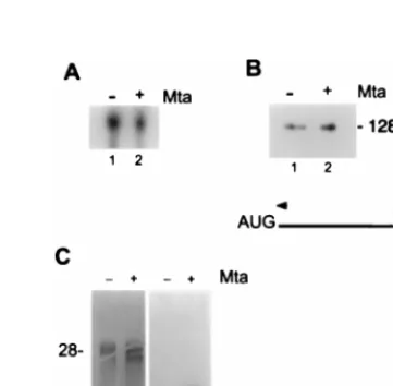
FIG. 3. Mta increases cytoplasmic accumulation of BMRF1 mRNA. HeLa cells were transfected with the BMRF1 expression plasmid (pMH2) and cotrans-fected with either the Mta expression plasmid (pRTS16) or vector control. (A) Total RNA was isolated from both control (lane 1) and Mta-expressing (lane 2) cells and subjected to Northern analysis for BMRF1 message expression. As shown, there was no discernible difference in total BMRF1 message between cells cotransfected with Mta or control vector DNA. (B) Total RNA was isolated from both control (lane 1) and Mta-expressing (lane 2) cells and subjected to primer extension analysis for BMRF1 message expression. Again, there was no discernible difference in total BMRF1 message between cells cotransfected with Mta or control DNA. The band migrating at 128 bases (128b) is marked. (C) Cytoplasmic RNA was isolated from control (lanes 1 and 3) and Mta-expressing (lanes 2 and 4) cells and subjected to Northern analysis for BMRF1 message expression. Lanes 1 and 2, methylene blue stain for rRNA; lanes 3 and 4, autoradiogram of cytoplasmic RNA reactive with the BMRF1-specific probe. BMRF1 message was undetectable in the absence of Mta (lane 3) but was abundant in Mta-expressing cells (lane 4).
on November 9, 2019 by guest
http://jvi.asm.org/
[image:3.612.103.235.69.153.2] [image:3.612.335.516.411.588.2]plasmic BBLF2/3 mRNA (Fig. 4). BBLF2/3
(primase-associ-ated factor) is encoded by two ORFs, BBLF2 and BBLF3, and
the mRNA, unlike that of the other five core replication genes,
is spliced (19). To determine whether the BBLF2/3 intron was
a necessary component of Mta independence, a cDNA version
of BBLF2/3 was tested. Cytoplasmic accumulation of
tran-scripts of the BBLF2/3 cDNA lacking the internal intron
re-mained Mta independent (Fig. 4), suggesting that signals in
addition to the presence of the intron itself influence the
re-quirement for Mta.
Mta binds directly to RNA.
The HSV IE63 homolog of Mta
has been shown to bind to RNA (7, 33, 46). An RNA binding
assay was performed with in vitro-transcribed,
32P-labeled
mRNAs transcripts and GST-Mta along with control GST and
maltose binding protein (MBP)-Tax proteins (Fig. 5). The
GST-Mta vector expressed the BMLF1 ORF fused to GST.
Only a small amount of the intact fusion protein was detected
in silver-stained gels. Most of the protein migrated as a triple
band of degradation products of 55 to 60 kDa. These three
protein bands contained the amino terminus of the fusion
protein, as demonstrated by their interaction with anti-GST
antibody (Fig. 5B) and on the basis of size, would represent the
amino-terminal half of the BMLF1 polypeptide.
The BMRF1 mRNA is believed to be coterminal with that
for BMRF2 and to use the polyadenylation signal located right
at the 3
9
end of the BMRF2 ORF (58). We generated an
847-base RNA probe that crossed the 3
9
100 bases of the
BMRF2 ORF and extended across the region between the
BMRF2 and BMLF1 ORFs to include sequences that might
potentially be present in a primary unprocessed transcript for
BMRF1 and BMRF2 (Fig. 5C). This RNA bound to the
GST-Mta fusion protein. The adjacent convergent mRNA is that for
BSLF2/BMLF1 (Mta). The polyadenylation site for this
tran-script has been mapped to the sequence coincident with the
terminus of the BMLF1 ORF (67). An equivalent 853-base
RNA that covered the 3
9
174 bases of the BMLF1 ORF and
the region between the BMLF1 and BMRF2 ORFs was also
generated to represent a primary Mta transcript. This RNA
also bound to GST-Mta (Fig. 5C). There was specificity to the
RNA binding in that a similarly sized RNA generated across
the ORF for the cellular DNA binding protein CBF1/RBPJk
(28) did not interact with GST-Mta. The BMRF1 and BMLF1
RNAs also did not interact with control GST protein or
MBP-Tax (Fig. 5).
The progressive degradation of the GST-Mta fusion protein
provided information on the approximate location of the
RNA-binding domain within Mta. All degradation products
greater than 55 kDa (GST plus 25 kDa of Mta polypeptide)
bound specifically to RNA. However, degradation products
containing less than 25 kDa of Mta had no RNA binding
activity. An arginine-rich region of Mta that is reminiscent of
domains in characterized RNA binding proteins is located
between amino acids (aa) 125 and 204. This domain would be
present in the RNA binding GST-Mta polypeptides.
[image:4.612.73.266.68.141.2]In an effort to further define the RNA region required for
interaction with Mta, two smaller nonoverlapping segments of
the original BMLF1 test RNA were synthesized as probes. The
A1 RNA (137 bases) covered sequences that might be present
in a primary BMLF1 transcript but would not be present in the
mature polyadenylated mRNA. This RNA did not bind to
Mta (Fig. 6A). The A2 RNA, which did bind to
GST-Mta, was 132 bases long and covered sequences within the
BMLF1 ORF, beginning at a position 54 bp upstream of the
translational termination signal. The difference in binding
abil-ity of the A1 and A2 probes was not a reflection of differences
in size or RNA integrity, as illustrated in Fig. 6C. When the A2
region was again subdivided into RNAs covering 51 and 69
nucleotides of the Mta gene, both RNAs bound to GST-Mta
(Fig. 6B). It is possible that there are multiple contacts
be-tween Mta and target RNAs, but this point needs further
evaluation. The predicted secondary structure of the A2 probe
FIG. 4. Mta expression increased cytoplasmic accumulation of five EBVrep-lication gene mRNAs. HeLa cells were transfected with the original expression vector for either BMRF1, BALF2, BSLF1, BBLF4, BALF5, or BBLF2/3 as indicated and cotransfected with either control (2) or Mta-expressing (1) plas-mid. Cytoplasmic RNA was isolated and subjected to Northern analysis for expression of the indicated message. Both genomic (BBLF2/3) and cDNA (BBLF2/3 cDNA) versions of BBLF2/3 were tested. Mta had no effect on BBLF2/3 message, which was found to be constitutively expressed in the cyto-plasm in the absence of Mta. In contrast, the other replication mRNAs were undetectable in the cytoplasmic fraction in the absence of Mta (2) and accu-mulated in the presence of Mta (1).
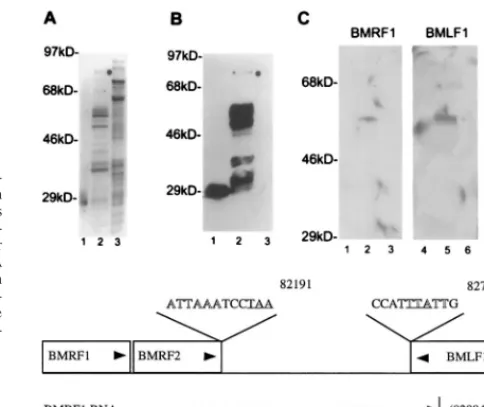
FIG. 5. Mta binds target mRNAs. Purified GST-Mta, control GST, and crude control MBP-Tax were separated by SDS-PAGE and transferred to Im-mobilon-P filters. The membrane-bound proteins were denatured and renatured as described in the text and incubated with the indicated RNA probes. (A) Silver stain of purified GST (lane 1), purified GST-Mta (lane 2), and crude MBP-Tax (lane 3). The full-length GST-Mta is indicated by a dot. The gel shows numerous background proteins that are available for nonspecific probe binding. (B) A membrane blot containing the proteins shown in panel A was immunoprobed with anti-GST MAb and visualized by chemiluminescence. The purified GST-Mta was partially degraded. Note that the degradation was largely progressive from the carboxyl terminus since most degradation products tested positive for the amino-terminal GST fusion partner (lane 2). (C) A membrane blot of the proteins shown in panel A was incubated with BMRF1 and BMLF1 RNA probes and a control probe transcribed from the cellular CBF1 cDNA. GST-Mta, but not GST or MBP-Tax, bound to the BMRF1 and BMLF1 RNA probes (compare lanes 2 and 5 to lanes 1, 3, 4, and 6). The control CBF1 RNA did not bind to GST-Mta (lane 8). (D) Map locations of the BMRF1 and BMLF1 probes relative to the BMRF1, BMRF2, and BMLF1 ORFs. The map coordinates are those for the B95-8 genome (4). Arrowheads indicate the direction of transcription of the genes and probe RNAs. The polyadenylation signals for these genes are shown in outlined type, and the translation termination codons for BMRF2 and BMLF1 are underlined.
on November 9, 2019 by guest
http://jvi.asm.org/
[image:4.612.307.549.69.273.2]is presented in Fig. 6D along with its relative location within
the BMLF1 ORF.
Intranuclear localization of Mta.
RNA binding proteins,
including the HSV IE63 protein, have been found to be
present in splicing bodies within the nucleus (70). The ability of
Mta to colocalize with splicing factors was examined in an
immunofluorescence assay. Cells transfected with Flag-Mta
were stained with anti-Flag antibody and FITC-conjugated
sec-ondary antibody to detect Mta and antibody recognizing the
SC35 splicing factor plus TRITC-conjugated secondary
anti-body to detect endogenous SC35. In transfected cells,
Flag-Mta gave a diffuse nuclear staining with underlying strongly
staining nuclear speckles. These speckles were found to
colo-calize with SC35-containing bodies (Fig. 7).
Mta shuttles between the nucleus and the cytoplasm.
HIV
type 1 Rev serves as the mechanistic prototype for proteins
that regulate cytoplasmic transport of viral transcripts. Rev
recognizes a specific RNA sequence, the Rev-responsive
ele-ment, via an arginine-rich region and mediates export of
un-spliced and incompletely un-spliced viral RNA (17, 18). The
abil-ity to shuttle between the nucleus and cytoplasm is believed to
be an essential component of Rev function. Rev
nucleocyto-plasmic shuttling is facilitated by a signal for nuclear entry
(nuclear localization signal) and a signal for nuclear export
(nuclear export signal [NES]). It is the NES which appears to
distinguish shuttling from nonshuttling nuclear proteins, and a
NES has been found in several other shuttling proteins,
includ-ing HSV IE63 (49, 51, 56, 68, 75). Inspection of the Mta amino
acid sequence revealed the presence of a motif between aa 226
and 237 that can be aligned with the NES sequences of Rev,
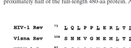
Rev-like proteins, and HSV IE63 (Fig. 8).
To determine whether Mta is capable of nucleocytoplasmic
shuttling, we performed a heterokaryon analysis. HeLa cells
transfected with Flag-Mta were fused to Cos-TdRev cells,
which stably express HIV RevM10, a Rev mutant that is unable
to export from the nucleus. The fused cells were incubated in
medium containing cycloheximide to block de novo protein
synthesis and then fixed and stained for Mta and for RevM10.
In heterokaryons, the donor cell nucleus expressing transfected
Flag-Mta and the recipient nucleus expressing RevM10 are
located within a single cytoplasm. Proteins that are capable of
shuttling will move from the nucleus of the transfected cell into
the cytoplasm. From the cytoplasm, the shuttling protein can
move back into either of the two available nuclei, resulting in
both donor and recipient nuclei staining for the transfected
protein. In this assay, Mta demonstrated the ability to shuttle
from the originally transfected cells into the RevM10-staining
nuclei of the recipient cells (Fig. 9).
DISCUSSION
Induction of the EBV lytic cycle by activators such as
anti-immunoglobulin antibody initiates transcription of the BZLF1
and BRLF1 genes, which encode the Zta and Rta
transcrip-tional activators (21, 47, 78, 85). Transcription across the
Mta-encoding BSLF2/BMLF1 gene follows, with the Mta promoter
responding to both the Zta and Rta activators (8, 36). Mta is
recognized to stimulate reporter gene expression in a
posttran-scriptional manner, but beyond that little is known about the
contribution of Mta to the regulation of EBV gene expression.
We have shown that Mta is necessary for efficient cytoplasmic
accumulation of mRNA for five of the EBV replication genes,
BMRF1, BALF2, BALF5, BSLF1, and BBLF4. Thus, Mta
contributes to EBV early gene expression. However, not all
early genes are Mta dependent. Abundant cytoplasmic mRNA
was detected for the BBLF2/3 replication gene in the absence
of Mta, and the level was not affected by the addition of Mta,
suggesting that some of the EBV early gene transcripts can be
processed efficiently by cellular factors. The BBLF2/3
tran-scripts, unlike the other five core replication genes, is spliced,
but removal of the intron in BBLF2/3 did not alter its behavior.
It is possible that the partial 5
9
and 3
9
splicing signals bounding
the intron continue to be recognized by cellular factors.
How-ever, the precise requirements for Mta independence and Mta
dependence remain to be determined.
[image:5.612.59.282.69.293.2]In an indirect immunofluorescence assay, Mta showed a
mixture of diffuse nuclear staining and strongly staining
nu-clear speckles. These speckles colocalized with the nunu-clear
speckles characteristic of the splicing factor SC35, indicating
that Mta is present at sites adjacent to nascent mRNA
synthe-sis and processing. HIV Rev and HSV IE63 also show an
association with SC35 in the nucleus (35, 70). The association
with SC35 together with the observation that using a vector
such as SG5 that introduces the
b
-globin intron into expressed
transcripts alleviates the dependence on Mta for BMRF1
pro-tein expression suggests that Mta may also function in part
through communication with splicing machinery. Although the
SG5-BMRF1 vector, pRTS16, was not dependent on Mta for
expression of BMRF1, protein expression was still increased
upon cotransfection with Mta. This result would also be
com-patible with Mta functioning to facilitate RNA processing. The
IE63 (ICP27) protein that is the HSV homolog of Mta has
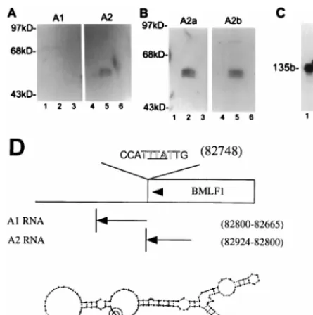
FIG. 6. Mta binds a 135-base sequence within BMLF1, as determined byNorthwestern analysis of the binding of nonoverlapping BMLF1-specific RNA probes to GST-Mta. (A) Protein blots identical to those in Fig. 5A were probed with different RNAs corresponding to sequences within the BMLF1 39region. The A1 probe encompasses the first base of the naturally occurring poly(A) signal and continues in the 39direction for 139 bases. The A2 probe comprises 130 bases upstream of the start of A1 and is entirely within the BMLF1 ORF. A2 retains the ability to bind Mta, whereas A1 shows no interaction. (B) Subdivision of the A2 probe failed to identify a single interactive region. The A2a probe is the 59 half of A2, and the A2b probe is the 39 half of A2; both probes bound GST-Mta. (C) All probes were analyzed on 8% urea-polyacrylamide gels for integrity. Full-length probes of comparable specific activity were generated. A known 135-base (135b) control RNA (lane 1) was used as a size marker for the A1 (lane 2) and A2 (lane 3) probes. (D) Schematic showing the probes, their relative genomic positions, and the predicted secondary structures of the Mta binding A2 RNA. The polyadenylation signal (outlined type), translational ter-mination codon (underlined), and direction of transcription (arrowheads) are indicated.
on November 9, 2019 by guest
http://jvi.asm.org/
been demonstrated to cause a redistribution of the snRNPs
that are components of the splicing complex and to colocalize
with the redistributed snRNPs (59). This redistribution is
be-lieved to contribute to the repression of host cell splicing that
occurs in HSV-infected cells. However, experiments
per-formed with temperature-sensitive mutants in IE63 indicated
that the repression of host cell splicing is more complex and
redistribution of snRNPs alone is insufficient to bring about
splicing inhibition (70). There is no evidence that Mta exerts a
comparable negative effect on cellular splicing, although the
question has not been rigorously examined. In our
experi-ments, we saw no repression of expression of the BBLF2/3
replication gene upon cotransfection of Mta. Perhaps the
pres-ence of spliced lytic cycle genes in EBV is not compatible with
a global splicing repression activity. The EBV lytic cycle
im-mediate-early Zta and Rta RNAs are spliced, as are several
early and late genes, including Mta itself, BHRF1, encoding
the bcl-2 homolog, and BLLF1, encoding the membrane
gly-coprotein gp350/220.
Mta was shown to bind to RNAs from the BMRF1 and
BMLF1 ORFs. The BMLF1 transcripts that showed specific
binding were derived entirely from sequences within the
BMLF1 ORF and included the 119 nucleotides of EBV
se-quence from positions 82920 to 82801 in the EBV genome.
The leftward-transcribed BMLF1 ORF terminates at position
82745. The cis-acting response elements in RNA bound by the
HIV Rev and human T-cell leukemia virus type 1 (HTLV-1)
Rex proteins have been extensively characterized. These
ele-ments are relatively large, 233 and 254 nucleotides,
respec-tively, and form stable stem-loop secondary structures.
Dele-tions that result in loss of structure also result in loss of
function. Mutational studies also provided evidence for the
presence of additional sequence specific subregions that may
be required for protein-RNA recognition (1). The HTLV-1
Rex-responsive element is located immediately downstream of
the polyadenylation signal for the env gene, and the HIV
Rev-responsive element is located within the env intron (41). The
simpler retroviruses that do not encode Rev-like proteins are
apparently able to utilize the cellular machinery for
posttran-scriptional processing of their RNAs. This function is mediated
via specific sequence and structural elements within the
tran-scripts. A constitutive RNA transport element that is required
for cytoplasmic transport of Mason-Pfizer monkey virus
intron-containing RNAs has been described elsewhere (6). This
ele-ment consists of 153 nucleotides located in the 3
9
untranslated
region of the env RNA, and the constitutive transport element
can substitute for the Rev–Rev-responsive element
combina-tion (15). Based on computer modeling, this element also
forms a stable stem-loop structure (16). Computer modeling
predicts that the BMLF1 RNA that bound GST-Mta is capable
of stem-loop structure formation, but the predicted structure
has not been validated experimentally. The location of an
RNA binding element within the BMLF1 ORF may be related
to the unusual placement of the BMLF1 polyadenylation
sig-nal, which is located immediately adjacent to the translational
termination signal for the BMLF1 ORF. Curiously, several
other EBV genes also have this unusually compact spacing
between the end of the ORF and the polyadenylation signal.
Examples include BORF2 (which encodes the large subunit of
ribonucleotide reductase), BLRF2 (a late gene), BLLF1 (the
gp350/220 membrane antigen gene), BBLF4 (the helicase
gene), and BMRF2 (a late gene).
[image:6.612.72.526.74.262.2]RNA binding by the Rev-like proteins and HSV IE63 is
mediated by an arginine-rich region that also serves as the
nuclear localization signal (29, 39, 68). The GST-Mta protein
used in our binding experiments yielded specific breakdown
products that based on size and interaction with anti-GST
antibody represented N-terminal polypeptides comprising
ap-proximately half of the full-length 480-aa protein. An
arginine-FIG. 7. Mta colocalizes with the SC35 splicing factor. Flag-tagged Mta was transfected into Vero cells. The Mta-expressing cells were fixed and immunostained with anti-Flag (rabbit polyclonal) and anti-SG35 (mouse monoclonal) primary antibodies. Secondary antibodies were FITC-conjugated anti-rabbit and TRITC-conjugated anti-mouse. Mta expression was diffusely nuclear, with a concentration in subnuclear regions which overlap with SC35 speckles.FIG. 8. A potential NES within EBV Mta. A sequence within Mta was compared with known functional NES sequences (5, 31, 37, 49, 68, 89).
on November 9, 2019 by guest
http://jvi.asm.org/
[image:6.612.321.535.627.705.2]rich region is present in this N-terminal region, between aa 125
and 204 of Mta, and it is likely that this domain mediates RNA
binding. The BMLF1 ORF together with BSLF2 encodes Mta.
The binding of Mta to its own message implies that Mta
reg-ulates its own synthesis. The same observation has been made
for the HSV IE63 (ICP27) protein (68). BMRF1 and BMRF2
have coterminal RNA transcripts, and the region of the
BMRF1 message that bound to Mta would also be present in
the BMRF2 transcript. It therefore seems likely that BMRF2,
a late gene, is also regulated by Mta and hence that Mta can
regulate both early and late classes of EBV mRNAs.
Mammalian viruses differ from the host cell by utilizing
intronless transcripts to encode many of their gene products.
Transcripts that are unspliced or incompletely spliced are
typ-ically retained in the nucleus. Thus, the unspliced viral
mes-sages need a mechanism to avoid nuclear retention. Viruses
circumvent this problem by encoding proteins that bind to
specific RNA sequences and transport unspliced and
incom-pletely spliced viral RNA into the cytoplasm (1, 9, 25, 26, 30,
41, 68, 77). These proteins contain a leucine-rich NES that
enables rapid nuclear export (5, 17, 34, 38, 48). The NES has
recently been shown to interact with a protein, CRM1 or
ex-portin 1, which is related to the karyopherin
b
family of nuclear
import proteins (22, 55, 76, 84). Exportin 1 interacts with the
NES-containing protein, and RanGTP to form a complex that
is transported through the nuclear pore (86). The combined
presence of a NES and a nuclear localization signal allows
RNA transport proteins to shuttle between the nucleus and the
cytoplasm. NES elements, in addition to the retrovirus
Rev-like proteins, have been identified in the adenovirus E4 34-kDa
protein (14) and most recently in the HSV IE63 (ICP27)
pro-tein (68). Mta has a leucine-rich region located between aa 215
and 250 that contains a consensus NES. The heterokaryon
assay has been a valuable tool for establishing that export
proteins shuttle between the nucleus and cytoplasm, and this
assay was exploited to determine whether Mta functioned in
this manner. Mta was readily detected in both the donor and
the acceptor nuclei of heterokaryons, indicating that Mta has
the ability to move from the nucleus of the donor cell into the
cytoplasm and from the cytoplasm into the nucleus of the
recipient cell. Thus, our experiments establish that Mta has the
properties of an RNA export protein. Mta binds to specific
EBV RNAs, shuttles between the nucleus and the cytoplasm,
and is required for efficient cytoplasmic accumulation of EBV
replication gene RNAs. EBV joins the growing list of viruses
that circumvent the problem of nuclear retention of RNAs by
encoding proteins that facilitate RNA processing and mediate
efficient cytoplasmic accumulation of their own transcripts.
ACKNOWLEDGMENTS
We thank M.-L. Hammarskjold for the TdRev cell line, X. D. Fu
and T. Maniatis for the SC35 antibody, Jon Finan for technical
assis-tance, and Feng Chang for manuscript preparation.
This work was funded by National Institutes of Health grant RO1
CA30356. R.T.S. was supported by training grant T32 CA09243.
S.D.H. is the recipient of American Cancer Society award FRA429.
REFERENCES
1. Ahmed, Y. F., S. M. Hanly, M. H. Malim, B. R. Cullen, and W. C. Greene. 1990. Structure-function analyses of the HTLV-I Rex and HIV-1 Rev RNA response elements: insights into the mechanism of Rex and Rev action. Genes Dev. 4:1014–1022.
2. Askovic, S., and R. Baumann. 1997. Activation domain requirements for disruption of Epstein-Barr virus latency by ZEBRA. J. Virol. 71:6547–6554. 3. Ausubel, F. M., R. Brent, R. E. Kingston, D. M. David, J. G. Seidman, J. A. Smith, and K. Struhl (ed.).1994. Current protocols in molecular biology, vol. 1. John Wiley & Sons, Inc., New York, N.Y.
4. Baer, R., A. T. Bankier, M. D. Biggin, P. L. Deininger, P. J. Farrell, T. J. Gibson, G. Hatfull, G. S. Hudson, S. C. Satchwell, C. Seguin, P. S. Tuffnell, and B. G. Barrell.1984. Organization of the B95-8 Epstein-Barr virus ge-nome. Nature 310:207–211.
5. Bogerd, A. M., R. A. Fridell, R. E. Benson, J. Hua, and B. R. Cullen. 1996. Protein sequence requirements for function of the human T-cell leukemia virus type 1 Rex nuclear export signal delineated by a novel in vivo random-ization-selection assay. Mol. Cell. Biol. 16:4207–4214.
6. Bray, M., S. Prasad, J. W. Dubay, E. Hunter, K.-T Jeang, D. Rekosh, and M.-L. Hammarskjold.1994. A small element from the Mason-Pfizer monkey virus genome makes human immunodeficiency virus type 1 expression and replication Rev-independent. Proc. Natl. Acad. Sci. USA 91:1256–1260. 7. Brown, C. R., M. S. Nakamura, J. D. Mosca, G. S. Hayward, S. E. Straus,
and L. P. Perera.1995. Herpes simplex virus trans-regulatory protein ICP27 stabilizes and binds to 39ends of labile mRNA. J. Virol. 69:7187–7195. 8. Buisson, M., E. Manet, M.-C. Trescol-Biemont, H. Gruffat, B. Durand, and
A. Sergeant.1989. The Epstein-Barr Virus (EBV) early protein EB2 is a posttranscriptional activator expressed under the control of EBV transcrip-tion factors EB1 and R. J. Virol. 63:5276–5284.
9. Chang, D. D., and P. A. Sharp. 1989. Regulation by HIV Rev depends upon recognition of splice sites. Cell 59:789–795.
10. Chen, C., and H. Okayama. 1987. High-efficiency transformation of mam-malian cells by plasmid DNA. Mol. Cell. Biol. 7:2745–2752.
11. Cho, M.-S., K.-T Jeang, and S. D. Hayward. 1985. Localization of the coding region for an Epstein-Barr virus early antigen and inducible expression of this 60-kilodalton nuclear protein in transfected fibroblasts cell lines. J. Vi-rol. 56:852–859.
12. Cook, I. D., F. Shanahan, and P. J. Farrell. 1994. Epstein-Barr virus SM protein. Virology 205:217–227.
[image:7.612.142.457.69.208.2]13. Defechereux, P., S. Debrus, L. Baudoux, B. Rentier, and J. Piette. 1997. Varicella-zoster virus open reading frame 4 encodes an immediate-early FIG. 9. Mta shuttles between the nucleus and the cytoplasm. In the heterokaryon assay, Flag-Mta was transfected into HeLa (the heterokaryon donor) cells. The Cos cell line TdRev, which is a stable producer of a Rev mutant incapable of nucleocytoplasmic shuttling, was used as the heterokaryon recipient. After heterokaryon formation, the cells were fixed and immunostained with anti-Flag (mouse monoclonal) and anti-Rev (rabbit polyclonal) primary antibodies. The secondary antibodies were TRITC-conjugated anti-mouse and FITC-conjugated anti-rabbit. The arrowheads point to two Mta-expressing cells in each panel. Note the even distribution of Mta between both donor (bottom arrow) and recipient (top arrow) cells.
on November 9, 2019 by guest
http://jvi.asm.org/
protein with posttranscriptional regulatory properties. J. Virol. 71:7073– 7079.
14. Dobblestein, M., J. Roth, W. T. Kimberly, A. J. Levine, and T. Shenk. 1997. Nuclear export of the E1B 55-kDa and E4 34-kDa adenoviral oncoproteins mediated by a rev-like signal sequence. EMBO J. 16:4276–4284. 15. Ernst, R. K., M. Bray, D. Rekosh, and M.-L. Hammarskjold. 1997. A
struc-tured retroviral RNA element that mediates nucleocytoplasmic export of intron-containing RNA. Mol. Cell. Biol. 17:135–144.
16. Ernst, R. K., M. Bray, D. Rekosh, and M. L. Hammarskjold. 1997. Second-ary structure and mutational analysis of the Mason-Pfizer monkey virus RNA constitutive transport element. RNA 3:210–222.
17. Fischer, U., J. Huber, W. C. Boelens, I. W. Mattaj, and R. Luhrmann. 1995. The HIV-1 Rev activation domain is a nuclear export signal that accesses an export pathway used by specific cellular RNAs. Cell 82:475–483. 18. Fischer, U., S. Meyer, M. Teufel, C. Heckel, R. Luhrmann, and G.
Raut-mann.1994. Evidence that HIV-1 Rev directly promotes the nuclear export of unspliced RNA. EMBO J. 13:4105–4112.
19. Fixman, E. D., G. S. Hayward, and S. D. Hayward. 1995. Replication of Epstein-Barr virus oriLyt: lack of a dedicated virally encoded origin-binding protein and dependence on Zta in cotransfection assays. J. Virol. 69:2998– 3006.
20. Fixman, E. D., G. S. Hayward, and S. D. Hayward. 1992. trans-acting re-quirements for replication of Epstein-Barr virus ori-Lyt. J. Virol. 66:5030– 5039.
21. Flemington, E. K., A. E. Goldfeld, and S. H. Speck. 1991. Efficient transcrip-tion of the Epstein-Barr virus immediate-early BZLF1 and BRLF1 genes requires protein synthesis. J. Virol. 65:7073–7077.
22. Fornerod, M., M. Ohno, M. Yoshida, and I. W. Mattaj. 1997. CRM1 is an export receptor for leucine-rich nuclear export signals. Cell 90:1051–1060. 23. Fu, X. D., and T. Maniatis. 1990. Factor required for mammalian
spliceo-some assembly is localized to discrete regions in the nucleus. Nature 343: 437–441.
24. Gao, Z., A. Krithivas, J. E. Finan, O. J. Semmes, S. Zhou, Y. Wang, and S. D. Hayward. 1998. The Epstein-Barr virus proteins lytic transactivator Zta interacts with the helicase-primase replication. J. Virol. 72:8559–8567. 25. Hadzopoulou-Cladaras, M. 1989. The Rev (Trs/Art) protein of human
im-munodeficiency virus type 1 affects viral mRNA and protein expression via a cis-acting sequence in the env region. J. Virol. 63:1265–1274.
26. Hammarskjold, M.-L., J. Heimer, B. Hammarskjold, I. Sangwan, L. Albert, and D. Rekosh.1989. Regulation of human immunodeficiency virus env expression by the rev gene product. J. Virol. 63:1959–1966.
27. Hardy, W. R., and R. M. Sandri-Goldin. 1994. Herpes simplex virus inhibits host cell splicing, and regulatory protein ICP27 is required for this effect. J. Virol. 68:7790–7799.
28. Henkel, T., P. D. Ling, S. D. Hayward, and M. G. Peterson. 1994. Mediation of Epstein-Barr virus EBNA2 transactivation by recombination signal-bind-ing protein Jk. Science 265:92–95.
29. Hibbard, M. K., and R. M. Sandri-Goldin. 1995. Arginine-rich regions suc-ceeding the nuclear localization region of the herpes simplex virus type 1 regulatory protein ICP27 are required for efficient nuclear localization and late gene expression. J. Virol. 69:4656–4667.
30. Hidaka, M., J. Inoue, M. Yoshida, and M. Seiki. 1998. Post-transcriptional regulator (rex) of HTLV-1 initiates expression of viral structural proteins but suppresses expression of regulatory proteins. EMBO J. 7:519–523. 31. Hope, T. J. 1997. Viral RNA export. Chem. Biol. 4:335–344.
32. Inchauspe, G., and J. M. Ostrove. 1989. Differential regulation by varicella-zoster virus (VZV) and herpes simplex virus type-1 trans-activating genes. Virology 173:710–714.
33. Ingram A., A. Phelan, J. Dunlop, and J. B. Clements. 1996. Immediate early protein IE63 of herpes simplex virus type 1 binds RNA directly. J. Gen. Virol. 77:1847–1851.
34. Kalland, K. H., A. M. Szilvay, K. A. Brokstad, W. Saetrevik, and G. Hau-kenes.1994. The human immunodeficiency virus type 1 Rev protein shuttles between the cytoplasm and nuclear compartments. Mol. Cell. Biol. 14:7436– 7444.
35. Kalland, K. H., A. M. Szilvay, E. Langhohff, and G. Haukenes. 1994. Sub-cellular distribution of human immunodeficiency virus type 1 Rev and colo-calization of Rev with RNA splicing factors in a speckled pattern in the nucleoplasm. J. Virol. 68:1475–1485.
36. Kenney, S., E. Holley-Guthrie, E. C. Mar, and M. Smith. 1989. The Epstein-Barr virus BMLF1 promoter contains an enhancer element that is responsive to the BZLF1 and BRLF1 transactivators. J. Virol. 63:3878–3883. 37. Kenney, S., J. Kamine, E. Holley-Guthrie, E. C. Mar, J. C. Lin, D.
Marko-vitz, and J. Pagano. 1989. The Epstein-Barr virus immediate-early gene product, BMLF1, acts in trans by posttranscriptional mechanism which is reporter gene dependent. J. Virol. 63:3870–3877.
38. Kim, F. J., A. A. Beeche, J. J. Hunter, D. J. Chin, and T. J. Hope. 1996. Characterization of the nuclear export signal of human T-cell lymphotropic virus type 1 Rex reveals that nuclear export is mediated by position-variable hydrophobic interactions. Mol. Cell. Biol. 16:5147–5155.
39. Kjems, J., M. Brown, D. D. Chang, and P. A. Sharp. 1991. Structural analysis of the interaction between the human immunodeficiency virus Rev protein
and the Rev response element. Proc. Natl. Acad. Sci. USA 88:683–687. 40. Lieberman, P. M., P. O’Hare, G. S. Hayward, and S. D. Hayward. 1986.
Promiscuous trans-activation of gene expression by an Epstein-Barr virus-encoded early nuclear protein. J. Virol. 60:140–148.
41. Malim, M. H., J. Hauber, S. Y. Le, J. V. Maizel, and B. R. Cullen. 1989. The HIV-1 rev trans-activator acts through a structured target sequence to acti-vate nuclear export of unspliced viral mRNA. Nature 338:254–257. 42. McCarthy, A. M., L. McMahan, and P. A. Schaffer. 1989. Herpes simplex
virus type 1 ICP27 deletion mutants exhibit altered patterns of transcription and are DNA deficient. J. Virol. 63:18–27.
43. McGregor, F., A. Phelan, J. Dunlop, and J. B. Clements. 1996. Regulation of herpes simplex virus poly(A) site usage and the action of immediate-early protein IE63 in the early-late switch. J. Virol. 70:1931–1940.
44. McLauchlan, J., A. Phelan, C. Loney, R. M. Sandri-Goldin, and J. B. Clem-ents.1992. Herpes simplex virus IE63 acts at the posttranscriptional level to stimulate viral mRNA 39processing. J. Virol. 66:6939–6945.
45. McMahan, L., and P. A. Schaffer. 1990. The repressing and enhancing functions of the herpes simplex virus regulatory protein ICP27 map to C-terminal regions and are required to modulate viral gene expression very early in infection. J. Virol. 64:3471–3485.
46. Mears, W. E., and S. A. Rice. 1996. The RGG box motif of the herpes simplex virus ICP27 protein mediates an RNA-binding activity and deter-mines in vivo methylation. J. Virol. 70:7445–7453.
47. Mellinghoff, I., M. Daibata, R. E. Humphreys, C. Mulder, K. Takada, and T. Sairenji.1991. Early events in Epstein-Barr virus genome expression after activation: regulation by second messengers of B cell activation. Virology 185:922–928.
48. Meyer, B. E., and M. H. Malim. 1994. The HIV-1 Rev trans-activator shut-tles between the nucleus and the cytoplasm. Genes Dev. 8:1538–1547. 49. Meyer, B. E., J. L. Meinkoth, and M. H. Malim. 1996. Nuclear transport of
human immunodeficiency virus type 1, visna virus, and equine infectious anemia virus Rev proteins: identification of a family of transferable nuclear export signals. J. Virol. 70:2350–2359.
50. Moriuchi, H., M. Moriuchi, H. A. Smith, and J. I. Cohen. 1994. Varicella-zoster virus open reading frame 4 protein is functionally distinct from and does not complement its herpes simplex virus type 1 homolog, ICP27. J. Vi-rol. 68:1987–1992.
51. Murphy R., and S. R. Wente. 1996. An RNA-export mediator with an essential nuclear export signal. Nature 383:357–360.
52. Nicholas, J., K. R. Cameron, H. Coleman, C. Newman, and R. W. Honess. 1992. Analysis of nucleotide sequence of the rightmost 43 kbp of herpesvirus saimiri (HSV) L-DNA: general conservation of genetic organization be-tween HVS and Epstein-Barr virus. Virology 188:296–310.
53. Nicholas, J., U. A. Gompels, M. A. Craxton, and R. W. Honess. 1988. Conservation of sequence and function between the product of the 52-kilodalton immediate-early gene of herpesvirus saimiri and the BMLF1-encoded transcriptional effector (EB2) of Epstein-Barr virus. J. Virol. 62: 3250–3257.
54. Oguro, M. O., N. Shimizu, Y. Ono, and K. Takada. 1987. Both the rightward and the leftward open reading frames within the BamHI M DNA fragment of Epstein-Barr virus act as trans-activators of gene expression. J. Virol. 61:3310–3313.
55. Ossareh-Nazari, B., F. Bachelerie, and C. Dargemont. 1997. Evidence for a role of CRM1 in signal-mediated nuclear protein export. Science 278:141– 144.
56. Pasquinelli, A. E., M. A. Powers, E. Lund, D. Forbes, and J. E. Dahlberg. 1997. Inhibition of mRNA export in vertebrate cells by nuclear export signal conjugates. Proc. Natl. Acad. Sci. USA 94:14393–14399.
57. Perera, L. P., S. Kaushal, P. R. Kinchington, J. D. Mosca, G. S. Hayward, and S. E. Strauss.1994. Varicella-zoster virus open reading frame 4 encodes a transcriptional activator that is functionally distinct from that of herpes simplex virus homology ICP27. J. Virol. 68:2468–2477.
58. Pfitzner, A. J., J. L. Strominger, and S. H. Speck. 1987. Characterization of a cDNA clone corresponding to a transcript from the Epstein-Barr virus BamHI M fragment: evidence for overlapping mRNAs. J. Virol. 61:2943– 2946.
59. Phelan, A., M. Carmo-Fonscca, J. McLauchlaw, A. I. Lamond, and J. B. Clements.1993. A herpes simplex virus type 1 immediate-early gene product, IE63, regulates small nuclear ribonucleoprotein distribution. Proc. Natl. Acad. Sci. USA 90:9056–9060.
60. Phelan, A., and J. B. Clements. 1997. Herpes simplex virus type 1 immediate early protein IE63 shuttles between nuclear compartments and the cyto-plasm. J. Gen. Virol. 78:3327–3331.
61. Phelan, A., J. Dunlop, and J. B. Clements. 1996. Herpes simplex virus type 1 protein IE63 affects the nuclear export of virus intron-containing tran-scripts. J. Virol. 70:5255–5265.
62. Ren, D., L. F. Lee, and P. M. Coussens. 1994. Identification and character-ization of Marek’s disease virus genes homologus to ICP27 and glycoprotein K of herpes simplex virus-1. Virology 204:242–250.
63. Rice, S. A., and D. M. Knipe. 1990. Genetic evidence for two distinct trans-activation functions of the herpes simplex virus alpha protein ICP27. J. Virol. 64:1704–1715.
on November 9, 2019 by guest
http://jvi.asm.org/
64. Rice, S. A., and V. Lam. 1994. Amino acid substitution mutations in the herpes simplex virus ICP27 protein define an essential gene regulation func-tion. J. Virol. 68:823–833.
65. Rice, S. A., V. Lam, and D. M. Knipe. 1993. The acidic amino-terminal region of herpes simplex virus type 1 alpha protein ICP27 is required for an essen-tial lytic function. J. Virol. 67:1778–1787.
66. Russo, J. J., R. A. Bohenzky, M.-C. Chein, J. Chen, M. Yan, D. Maddalena, J. P. Parry, D. Peruzzi, I. S. Edelman, Y. Chang, and P. S. Moore.1996. Nucleotide sequence of the Kaposi sarcoma-associated herpesvirus (HHV8). Proc. Natl. Acad. Sci. USA 93:14862–14867.
67. Sample, J., G. Lancz, and M. Nonoyama. 1986. Mapping of genes in BamHI fragment M of Epstein-Barr virus DNA that may determine the fate of viral infection. J. Virol. 57:145–154.
68. Sandri-Goldin, R. M. 1998. ICP27 mediates HSV RNA export by shuttling through a leucine-rich nuclear export signal and binding viral intronless RNAs through an RGG motif. Genes Dev. 12:868–879.
69. Sandri-Goldin, R. M., and M. K. Hibbard. 1996. The herpes simplex virus type 1 regulatory protein ICP27 coimmunoprecipitates with Sm anti-serum, and the C terminus appears to be required for this interaction. J. Virol. 70:108–118.
70. Sandri-Goldin, R. M., M. K. Hibbard, and M. A. Hardwicke. 1995. The C-terminal repressor region of herpes simplex virus type 1 ICP27 is required for the redistribution of small nuclear ribonucleoprotein particles and splic-ing factor SC35; however, these alterations are not sufficient to inhibit host cell splicing. J. Virol. 69:6063–6076.
71. Sandri-Goldin, R. M., and G. E. Mendoza. 1992. A herpesvirus regulatory protein appears to act post-transcriptionally by affecting mRNA processing. Genes Dev. 6:848–863.
72. Sarisky, R. T., Z. Gao, P. M. Lieberman, E. D. Fixman, G. S. Hayward, and S. D. Hayward.1996. A replication function associated with the activation domain of the Epstein-Barr virus Zta transactivator. J. Virol. 70:8340–8347. 73. Schepers, A., D. Pich, and W. Hammerschmidt. 1996. Activation of oriLyt, the lytic origin of DNA replication of Espstein-Barr virus, BZLF1. Virology 220:367–376.
74. Schepers, A., D. Pich, and W. Hammerschmidt. 1993. A transcription factor with homology to the AP-1 family links RNA transcription and DNA repli-cation in the lytic cycle of Epstein-Barr virus. EMBO J. 12:3921–3929. 75. Schoborg, R. V., and J. E. Clements. 1996. Definition of the RRE binding
and activation domains of the caprine arthritis encephalitis virus Rev pro-tein. Virology 226:113–121.
76. Seedorf, M., and P. A. Silver. 1997. Importin/karyopherin protein family members required for mRNA export from the nucleus. Proc. Natl. Acad. Sci. USA 94:8590–8595.
77. Seiki, M., J. Inoue, M. Hidaka, and M. Yoshida. 1988. Two cis-acting ele-ments responsible for posttranscriptional trans-regulation of gene expression of human T-cell leukemia virus type 1. Proc. Natl. Acad. Sci. USA 85:7124– 7128.
78. Sinclair, A. J., M. Brimmel, F. Shanahan, and P. J. Farrell. 1991. Pathways of activation of the Epstein-Barr virus productive cycle. J. Virol. 65:2237– 2244.
79. Singh, M., C. Fraefel, L. J. Bello, W. C. Lawrence, and M. Schwyzer. 1996.
Identification and characterization of BICP27, an early protein of bovine herpesvirus 1 which may stimulate mRNA 39 processing. J. Gen. Virol. 77:615–625.
80. Smith, I. L., M. A. Hardwicke, and R. M. Sandri-Goldin. 1992. Evidence that the herpes simplex virus immediate early protein ICP27 acts post-transcrip-tionally during infection to regulate gene expression. Virology 186:74–86. 81. Smith, I. L., R. E. Sekulovich, M. A. Hardwicke, and R. M. Sandri-Goldin.
1991. Mutations in the activation region of herpes simplex virus regulatory protein ICP27 can be trans dominant. J. Virol. 65:3656–3666.
82. Smith, R. H., Y. Zhao, and D. J. O’Callaghan. 1993. The equine herpesvirus 1 (EHV-1) UL3 gene, an ICP27 homolog, is necessary for full activation of gene expression directed by an EHV-1 late promoter. J. Virol. 67:1105–1109. 83. Soliman, T. M., R. M. Sandri-Goldin, and S. J. Silverstein. 1997. Shuttling of the herpes simplex virus type 1 regulatory protein ICP27 between the nucleus and cytoplasm mediates the expression of late proteins. J. Virol. 71:9188–9197.
84. Stade, K., C. S. Ford, C. Guthrie, and K. Weis. 1997. Exportin 1 (Crm1p) is an essential nuclear export factor. Cell 90:1041–1050.
85. Takada, K., and Y. Ono. 1989. Synchronous and sequential activation of latently infected Epstein-Barr virus genomes. J. Virol. 63:445–449. 86. Ullman, K. S., M. A. Powers, and D. J. Forbes. 1997. Nuclear export
recep-tors: from importin to exportin. Cell 90:967–970. (Comment.)
87. Uprichard, S. L., and D. M. Knipe. 1996. Herpes simplex ICP27 mutant viruses exhibit reduced expression of specific DNA replication genes. J. Vi-rol. 70:1969–1980.
88. Wang, Y., J. E. Finan, J. M. Middeldorp, and S. D. Hayward. 1997. P32/TAP, a cellular protein that interacts with EBNA-1 of Epstein-Barr virus. Virology 236:18–29.
89. Wen, W., J. L. Meinkoth, R. Y. Tsien, and S. S. Taylor. 1995. Identification of a signal for rapid export of proteins from the nucleus. Cell 82:463–473. 90. Whitehouse, A., M. Cooper, and D. M. Meredith. 1998. The immediate-early
gene product encoded by open reading frame 57 of herpesvirus saimiri modulates gene expression at a posttranscriptional level. J. Virol. 72:857– 861.
91. Winkler, M., S. A. Rice, and T. Stamminger. 1994. UL69 of human cyto-megalovirus, an open reading frame with homology to ICP27 of herpes simplex virus, encodes a transactivator of gene expression. J. Virol. 68:3943– 3954.
92. Wong, K. M., and A. J. Levine. 1989. Characterization of proteins encoded by the Epstein-Barr virus transactivator gene BMLF1. Virology 168:101–111. 93. Zhao, Y., V. R. Holden, R. N. Harty, and D. J. O’Callaghan. 1992. Identifi-cation and transcriptional analyses of the UL3 and UL4 genes of equine herpesvirus 1, homologs of the ICP27 and glycoprotein K genes of herpes simplex virus. J. Virol. 66:5363–5372.
94. Zhao, Y., V. R. Holden, R. H. Smith, and D. J. O’Callaghan. 1995. Regula-tory function of the equine herpesvirus 1 ICP27 gene product. J. Virol. 69:2786–2793.
95. Zhong, L., and G. S. Hayward. 1997. Assembly of complete, functionally active herpes simplex virus DNA replication compartments and recruitment of associated viral and cellular proteins in transient cotransfection assays. J. Virol. 3146:–3160.
on November 9, 2019 by guest
http://jvi.asm.org/