0022-538X/98/$04.0010
Copyright © 1998, American Society for Microbiology. All Rights Reserved.
Rapid Selection in Modified BHK-21 Cells of a Foot-and-Mouth
Disease Virus Variant Showing Alterations in Cell Tropism
CRISTINA ESCARMI´S,
1ELISA C. CARRILLO,
2MARCELA FERRER,
3JUAN F. GARCI´A ARRIAZA,
1NORA LOPEZ,
3CECILIA TAMI,
2NURIA VERDAGUER,
4ESTEBAN DOMINGO,
1*
AND
MARIA T. FRANZE-FERNA
´ NDEZ
3*
Centro de Biologı´a Molecular “Severo Ochoa” (CSIC-UAM), Universidad Auto´noma de Madrid, 28049 Madrid, Spain
1;
Instituto de Biotecnologı´a, Centro de Investigacio´n en Ciencias Veterinarias, INTA 1708 Moro´n, Buenos Aires,
2and Centro de Virologı´a Animal (CONICET), Serrano 669, 1414 Buenos Aires,
3Argentina; and
Centre de Investigacio´ i Desenvolupament (CSIC), Jordi Girona 6, 08028 Barcelona, Spain
4Received 26 June 1998/Accepted 9 September 1998
With persistent foot-and-mouth disease virus (FMDV) in BHK-21 cells, there is coevolution of the cells and
the resident virus; the virulence of the virus for the parental BHK-21 cells is gradually increased, and the cells
become partially resistant to FMDV. Here we report that variants of FMDV C
3Arg/85 were selected in a single
infection of partially resistant BHK-21 cells (termed BHK-Rb cells). Indirect immunofluorescence showed that
the BHK-Rb cell population was heterogeneous with regard to susceptibility to C
3Arg/85 infection. Infection of
BHK-Rb cells with C
3Arg/85 resulted in an early phase of partial cytopathology which was followed at 6 to 10
days postinfection by the shedding of mutant FMDVs, termed C
3-Rb. The selected C
3-Rb variants showed
increased virulence for BHK-21 cells, were able to overcome the resistance of modified BHK-21 cells to
infec-tion, and had acquired the ability to bind heparin and to infect wild-type Chinese hamster ovary (CHO) cells.
A comparison of the genomic sequences of the parental and modified viruses revealed only two amino acid
differences, located at the surface of the particle, at the fivefold axis of the viral capsid (Asp-93Ala in VP3 and
either Gly-1103Arg or His-1083Arg in VP1). The same phenotypic and genotypic modifications occurred in
a highly reproducible manner; they were seen in a number of independent infections of BHK-Rb cells with viral
preparation C
3Arg/85 or with clones derived from it. Neither amino acid substitutions in other structural or
nonstructural proteins nor nucleotide substitutions in regulatory regions were found. These results prove that
infection of partially permissive cells can promote the rapid selection of virus variants that show alterations
in cell tropism and are highly virulent for the same cells.
Foot-and-mouth disease virus (FMDV) is an important
an-imal pathogen of the genus aphthovirus of the Picornaviridae
family (48). FMDV usually causes an acute, systemic infection
in cloven-hooved animals and cytolytic infections in cell
cul-ture. However, FMDV can also establish persistent infections
in ruminants (3, 29, 36, 50, 53, 55, 56) and in cell cultures (9, 12,
21). Persistence in cell cultures was established by infecting
cloned BHK-21 cells (a population derived from a single cell)
with an FMDV of serotype C (clone C-S8c1) that was plaque
purified three times (9). The carrier cultures were obtained by
growing the cells that survived the cytolytic infection (at a
frequency of about 10
23) (9, 37). At the critical step of
initia-tion of persistence, the prevailing event ensuring cell survival
and viral replication was the rapid variation of the cells, which
became partially resistant to FMDV (37). The FMDV then
became increasingly virulent for BHK-21 cells (12, 37). In a
virulence assay that measures the minimal amount of virus
needed to kill 10
4BHK-21 cells in 24 h under standard
con-ditions, the virus shed by persistently infected cells at passage
100, termed R100, was 1,000-fold more virulent than the
pa-rental C-S8c1 (51).
The evolution of BHK-21 cells during FMDV persistence
could be characterized because treatment of the carrier cells
with ribavirin [(1-
b
-
D-ribofuranosyl)-1-H-1,2,4-triazole-3-car-boxamide] eliminated the virus from the cells (8). Cured cells,
in which virus could not be detected by reverse transcription
(RT)-PCR (37), were partially resistant to infection by several
isolates of FMDV of serotypes C, O, and A but produced
nor-mal yields of other RNA viruses, such as vesicular stomatitis
virus, encephalomyocarditis virus, and Semliki Forest virus (9,
12, 37). The degree of resistance to FMDV of carrier cells
increased with the passage number of the persistently infected
culture (11, 12). The evolving cell populations were
heteroge-neous with regard to susceptibility to FMDV, as revealed by
the analysis of 248 stable cell clones isolated from carrier cells
at passages 17, 19, 62, and 74 (11). At least six distinct cell
phenotypes were identified, the most-resistant clones
produc-ing 10
4times less FMDV C-S8c1 progeny than the standard
BHK-21 cells (11).
In the present study we have examined the ability of cloned
FMDV C
3Arg/85 to overcome the resistance of modified
BHK-21 cells. C
3Arg/85 is a standard South American type C
FMDV of which several isolates and cloned preparations have
been previously characterized genetically (39, 43) and
antigeni-cally (38, 54). The results show a surprising adaptability of
FMDV to overcome the cellular barrier in a single infection of
a monolayer of modified BHK-21 cells. The selected progeny
virus displayed increased virulence for BHK-21 cells and
mod-ified BHK-21 cells along with a number of additional
pheno-typic alterations. Two amino acid substitutions located at the
surface of the viral capsid are associated with the dramatic
phenotypic changes.
* Corresponding author. Mailing address for Esteban Domingo:
Centro de Biologı´a Molecular “Severo Ochoa,” Universidad
Auto´-noma de Madrid, Cantoblanco, 28049 Madrid, Spain. Phone:
34-91-3978485. Fax: 34-91-3974799. E-mail: [email protected].
Mail-ing address for Maria T. Franze-Ferna´ndez: Centro de Virologı´a
Animal, Serrano 669, 1414 Buenos Aires, Argentina. Phone and fax:
54-1-825-1863. E-mail: [email protected].
10171
on November 9, 2019 by guest
http://jvi.asm.org/
MATERIALS AND METHODS
Cells, viruses, and infections.The origin of BHK-21 cells and the procedures used for infection of BHK-21 and Chinese hamster ovary (CHO) cell monolayers with FMDV in liquid and in semisolid media have been previously described (2, 12, 16, 52). BHK-Rb cells derive from a subline of BHK-21 cells persistently infected with FMDV C-S8c1. They were cured of FMDV by ribavirin treatment (8) at cell passage 70 after the initiation of persistence. BHK-Rb cells correspond to the population termed C1Rc1-p70 Rbv in de la Torre et al. (11).
BHK-Rb cells were used between passages 9 and 15 after curing with ribavirin. Cell clone BHK-R74-A4 was obtained by growth of a single cell from passage 74 of the persistently infected cell population C1-BHK-Rcip74, as described by de la
Torre et al. (11). Cell populations BHK-Rb and BHK-R74-A4 were routinely monitored by RT-PCR amplification with primers corresponding to different regions of FMDV RNA, with negative results. Wild-type CHO cells and the two glycosaminoglycan-deficient CHO mutants pgs D-677 and pgs A-745 were kindly provided by J. D. Esko (27, 35).
The FMDV C3Arg/85 isolate used was passaged twice in BHK-21 cells and
plaque purified in BHK-21 cells, and then the cloned virus was passaged three times in BHK-21 cells to produce the viral preparation (working stock) for the experiments described here.
Generation of FMDV C3-Rb.Subconfluent BHK-Rb cell monolayers were infected either with C3Arg/85 or with plaque-purified viral clones derived from
the C3Arg/85 working stock and incubated with Dulbecco modified Eagle
me-dium (DMEM) containing 2% fetal calf serum until the cell monolayer became partially lysed at 2 to 3 days postinfection. At this time, the medium was replaced by DMEM containing 10% fetal calf serum, and the incubation was continued, with the medium changed every 24 h. Virus rescued from the supernatants at 8 to 10 days postinfection was generically designated C3-Rb.
Virulence assay.The virulence of FMDV for BHK-21 cells and for modified BHK-21 cells is defined as the minimum number of PFU required to kill a designated number of cells in a given time (both parameters are indicated for each experiment) under standard infection conditions (51).
Indirect immunofluorescence assays.Cells were grown on coverslips, washed extensively with phosphate-buffered saline, placed on ice, rinsed, fixed in acetone (10 min at220°C), air dried, and stored at220°C. Immunofluorescence was performed as previously described (28), with a 1:50 dilution of anti-FMDV C3
polyclonal antiserum raised in guinea pigs used as the primary antibody. The secondary antibody was a 1:100 dilution of a fluorescein-conjugated anti-guinea pig immunoglobulin G (Sigma).
Heparin-Sepharose binding of FMDV.Heparin-Sepharose CL-6B and control Sepharose CL-6B beads (ligand density,;2 mg of porcine heparin/ml of drained gel; Pharmacia Biotech) were equilibrated with a combination of DMEM, 25 mM HEPES, and 0.1% fetal calf serum (binding buffer) and resuspended as a 10% (vol/vol) slurry; 200ml of the slurry was mixed with 300ml of the virus diluted in binding buffer (105to 106PFU). The virus-heparin-Sepharose and
virus-Sepharose mixtures were incubated for 1 h at room temperature with gentle stirring and centrifuged at 1,0003g for 2 min. Virus recovered in the supernatants was quantitated by plaque assay. In all cases, 100% of the input PFU was recovered from the supernatant of the Sepharose CL-6B control beads, indicating stability of the virus during the procedure.
Isolation of viral RNA, cDNA synthesis, PCR amplification, nucleotide se-quencing, and determination of the size of the poly(C) tract.Viral RNA from the supernatants of infected cultures was obtained by treatment with proteinase K and sodium dodecyl sulfate, followed by phenol extraction, as previously de-scribed (26). To determine the length of the poly(C) tract, the supernatants of infected cells were treated with 2mg of DNase per ml and 4.5mg of pancreatic RNase per ml in 50 mM Tris (pH 7.5)–10 mM MgCl2for 50 min at 37°C prior
to treatment with proteinase K as described above. Amplification of FMDV RNA fragments by RT-PCR was carried out as described by Escarmı´s et al. (26) either with the sets of primers that have been previously described (2, 25, 26, 51) or with those designed for efficient RT-PCR amplification of the C3Arg/85
genome. The sequences of the oligonucleotide primers used will be provided upon request. DNA fragments were purified from unincorporated deoxynucleo-side triphosphates and primers by treatment with shrimp alkaline phosphatase and exonuclease I (Amersham). Nucleotide sequencing was performed on over-lapping DNA fragments with either the thermosequenase kit from Amersham or the femtomole DNA cycle sequencing kit from Promega (26). A few sequences were determined with an automatic sequencer (model no. ABI373). To our knowledge, these results provide the first entire genomic sequence of an FMDV of subtype C3. To determine the size of the polyribocytidylate [poly(C)] tract of
the FMDV RNA, we treated about 10 ng with 5 U of RNase T1in TE (10 mM
Tris-HCl [pH 8.0], 1 mM EDTA) in a volume of 5ml for 30 min at 37°C. Then, 1ml of a 103kinase buffer (500 mM Tris-HCl [pH 7.5], 100 mM MgCl2, 100 mM
dithiothreitol, 8 U of RNasin), 45mCi of [g-32P]ATP (150mCi/ml; 6,000 Ci/
mmol), and 2.5 U of T4 polynucleotide kinase were added, and the mixture (final volume, 10ml) was incubated 30 min at 37°C. The size of the poly(C) tract was determined by electrophoresis in a 6% polyacrylamide–7 M urea gel, with a nucleotide sequence ladder used as a size marker (25).
Extraction and quantification of intracellular FMDV RNA.To quantify rela-tive amounts of intracellular FMDV RNA, total cellular RNA was extracted by the procedure of Chomczynski and Sacchi (7). Aliquots of RNA corresponding
to 105, 104, and 103cells were spotted onto nitrocellulose filters and hybridized
onto a probe covering the entire P1 region of FMDV C3Arg/85. The probe was
labelled with [32P]phosphate by using the oligolabelling kit (Pharmacia).
Auto-radiographs were quantitated by the Fotoanalyst image analysis system (Foto-dyne, Inc.).
Positioning of amino acid replacements on the three-dimensional structure of FMDV.The crystallographic coordinates of C-S8c1 (pdb entry, 1fmd [34]) have been used as a reference to model the amino acid replacements found in FMDV C3-Rb. The substituted residues have been modeled with the program TURBO
(47) by placing the side-chain atoms in one of their standard conformations. The structure has been optimized by removing the close contacts by using the same program.
Nucleotide sequence accession numbers. The nucleotide sequences deter-mined in the present study were deposited in GenBank under accession no. AJ007347 and AJ007572.
RESULTS
Rapid evolution of FMDV C
3Arg/85 in BHK-Rb cells.
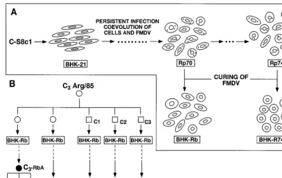
BHK-Rb cells, obtained by eliminating FMDV from persistently
infected C
1-BHK-Rc1p70 cells (11) (Fig. 1A), were partially
resistant to infection by FMDV C
3Arg/85. While parental
BHK-21 cells infected with C
3Arg/85 at a multiplicity of
infec-tion of 5 to 10 PFU/cell developed a complete cytopathic effect
at 16 to 20 h postinfection, parallel infections in BHK-Rb cells
did not show signs of cytopathology until 2 to 3 days after
infection. At that time, partial cell lysis was observed. Five
independent infections of BHK-Rb cells with either the initial
clonal preparation of C
3Arg/85 or three clonal derivatives of
the same preparation (-c1, -c2, and -c3) (Fig. 1B) were carried
out. The 20 to 50% of cells surviving at 2 to 3 days
postinfec-tion did not reach confluence in subsequent days and shed
virus that was increasingly virulent for the host cells. This
finding was revealed by a virulence assay that measures the
minimal amount of virus needed to kill BHK-Rb cells under
standard infection conditions (51) (Table 1). While 10
4to 10
5PFU of virus shed at 6 to 10 days postinfection was sufficient to
kill the BHK-Rb cells in each of five independent experiments,
a 100-fold-higher amount of the parental C
3Arg/85 either did
not affect or only partially killed the BHK-Rb cells (Table 1).
The C
3-Rb viruses shed by the BHK-Rb-infected cells at 8 or
10 days postinfection were termed C
3-RbA, C
3-RbB, C
3-RbC,
C
3-RbD, and C
3-RbE (Fig. 1B). In addition to increased
vir-ulence for BHK-Rb cells, all these viruses displayed a
small-plaque phenotype compared with that of the parental virus
in BHK-21 cells and acquired the ability to form plaques on
BHK-Rb cell monolayers. The parental working stock of
FMDV C
3Arg/85 and its three clonal derivatives did not form
visible plaques on BHK-Rb cells but formed normal size
plaques on BHK-21 cell monolayers. The three subclones
C
3Rb-Ac14, -c15, and -c16 and the parental C
3-RbA
popula-tion (Fig. 1B) showed identical phenotypes regarding virulence
for modified and parental BHK-21 cells and plaque
morphol-ogy (Table 2).
Growth characteristics of FMDV C
3Arg/85 and C
3-Rb in
BHK-21, BHK-Rb, and BHK-R74-A4 cells.
The infection of
BHK-21, BHK-Rb, and BHK-R74-A4 cells by C
3Arg/85 and its
C
3-Rb derivatives was studied by immunofluorescence and by
a comparative analysis of virus yields and levels of
intracellu-lar viral RNA. The limited permissivity of BHK-Rb cells to
FMDV C
3Arg/85 was due at least in part to the heterogeneity
of cells with regard to susceptibility to this virus, as indicated by
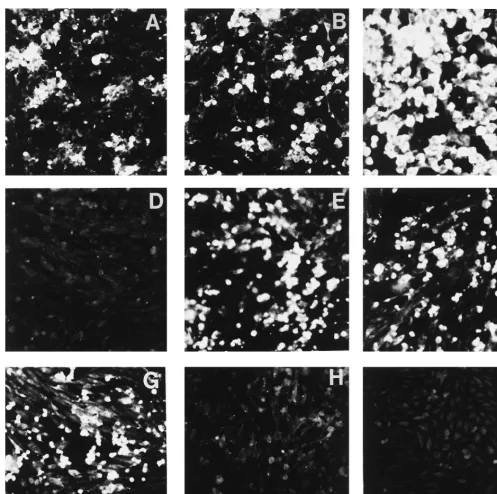
immunofluorescence analysis. Clusters of brightly fluorescent
BHK-Rb cells amounted to only half of the cell population,
even at 24 h postinfection (Fig. 2A and B). In contrast, in
infections of BHK-Rb cells with FMDV C
3-Rb, viral antigens
were detected in the majority of cells at 5 h postinfection (Fig.
2C). These results are in agreement with the higher yield of
virus in infections with the C
3-Rb variants in comparison to
10172
ESCARMI´S ET AL.
J. V
IROL.
on November 9, 2019 by guest
http://jvi.asm.org/
that in infections with the parental C
3Arg/85 and with the
observation of complete killing of BHK-Rb cells at 10 h after
infection with C
3-Rb variant FMDVs (Fig. 3B and E).
The ability of C
3-Rb viruses to overcome the BHK-Rb cells’
resistance to infection was further tested with BHK-R74-A4
cells, one of the stable cell clones isolated from an
FMDV-C-S8c1 carrier culture that was very resistant to infection by
FMDV C-S8c1 (11) (Fig. 1A). Single-step growth curves
re-vealed that while BHK-R74-A4 cells produce 100-fold-lower
yields of FMDV C
3Arg/85 than the standard BHK-21 cells do,
both cell types yield similar amounts of C
3-Rb virus (Fig. 3A
and C). The resistance of BHK-R74-A4 cells to C
3Arg/85 and
their permissivity to C
3-Rb viruses were confirmed by
compar-ing the levels of intracellular FMDV RNA in infections with
either virus (Fig. 3F). In addition, C
3-Rb viruses, but not the
parental C
3Arg/85, were able to kill BHK-R74-A4 cells,
al-FIG. 1. Diagram of the origin of the cells and viruses used in the present study. (A) Persistent infection of BHK-21 cells with FMDV C-S8c1 (9). At passage 70, the persistently infected culture was cured of FMDV by ribavirin treatment (8), yielding the uncloned population BHK-Rb. At passage 74, many cell clones free of FMDV were obtained by terminal dilution (11). One clone that is highly resistant to FMDV is BHK-R74-A4. (B) The C3Arg/85 used is a plaque-purified preparation
derived from the natural isolate C3Arg/85 (39, 43) as described in Materials and Methods. Either this preparation (open circles) or one of three plaque-purified
subpopulations (-c1, -c2, or -c3 [empty squares]) was used to infect BHK-Rb cells. Viruses rescued from the supernatant of infected BHK-Rb cells at 8 or 10 days postinfection are designated C3-RbA (8 days postinfection), -B, -C, -D, and -E (10 days postinfection) (filled circles). The letters correspond to experiments A to E
listed in Table 1. c14, c15, and c16 (filled squares) are clonal subpopulations obtained by plating population C3-RbA onto a BHK-Rb cell monolayer, isolating virus
[image:3.612.95.503.72.329.2]from three single plaques and then passaging the virus twice in BHK-Rb cells. Further details about experimental procedures are given in Materials and Methods.
TABLE 1. Generation of FMDV variants in BHK-Rb cells
Expta FMDVb Infecting
FMDV
Virus shed by BHK-Rb cells at indicated day postinfectionb
2 6 10
A
C
3Arg/85
.10
6d10
610
410
4B
C
3Arg/85
.10
6d10
610
410
4C
C
3Arg/85c1
.10
6cND
e.10
6d10
4D
C
3Arg/85c2
.10
6dND
10
410
4E
C
3Arg/85c3
.10
6cND
.10
6d5
3
10
4 aBHK-Rb cells were infected with the FMDVs indicated in the second column at a multiplicity of infection of 5 PFU/cell (experiment A) or 20 PFU/cell (experiments B to E), as described in Materials and Methods. The origins of the infecting FMDV C3Arg/85 and its clonal derivatives -c1, -c2, and -c3 are depictedschematically in Fig. 1 and described in Materials and Methods. bThe infecting FMDV C
3Arg/85 and virus shed by the infected BHK-Rb cells
at the indicated days postinfection were tested for ability to kill BHK-Rb cells in an adaptation of a previously described virulence assay (51). Numbers represent the minimum amount of PFU required to kill 73104BHK-Rb cells in 48 h.
c106PFU of virus caused no lysis of the cells.
d106PFU of virus caused partial lysis of the cells.
[image:3.612.307.548.532.621.2]eND, not determined.
TABLE 2. Properties of FMDV C
3Arg/85 and variants
selected in BHK-Rb cells
FMDVa No. of PFU required to kill indicated cells
b Diameter
of plaque (mm)c
BHK-21 BHK-Rb BHK-R74-A4
C
3Arg/85
10
6.10
6d.10
6e2–6
C
3-RbA
10
310
4ND
f#
2
C
3-RbAc14
10
310
410
6#
2
C
3-RbAc15
10
310
410
6#
2
C
3-RbAc16
10
310
410
6#
2
aThe origin of the FMDVs employed is described in Materials and Methods, Fig. 1, and Table 1.bVirulence for the indicated cells was measured as previously described (51) but with modifications depending upon cell number and time of infection for the three types of cells tested. Numbers represent the minimum number of PFU required to kill 53104BHK-21 cells in 24 h, 73104BHK-Rb cells in 48 h, or
105BHK-R74-A4 cells in 54 h.
cThe sizes of the plaques were measured at 30 h after plating onto a BHK-21 cell monolayer as previously described (16). For C3Arg/85,#5% of the plaques
were 2 to 3 mm in diameter, and$95% ranged from 3 to 6 mm in diameter. d106PFU of C
3Arg/85 caused a partial cytopathic effect.
e106PFU of C
3Arg/85 did not affect cells.
fND, not determined.
on November 9, 2019 by guest
http://jvi.asm.org/
[image:3.612.50.291.539.627.2]though their virulence was lower for these cells than for
BHK-Rb or BHK-21 cells (Table 2).
In the parental, unmodified BHK-21 cells, both C
3-Rb
vi-ruses and the parental C
3Arg/85 replicated equally well as
indicated by the equivalent amounts of virus yields and by the
levels of intracellular FMDV RNA at early times postinfection
(Fig. 3A and D). C
3-Rb viruses, however, produced complete
cell killing at around 9 h postinfection, in contrast to the 16 to
20 h required in infections with C
3Arg/85. It should be noted
that, in agreement with the latter observation, C
3-Rb viruses
are 1,000-fold more virulent than the parental C
3Arg/85 for
BHK-21 cells (Table 2). This finding indicates that the
hyper-virulence for BHK-21 cells of FMDV C
3-Rb is related to
fac-tors other than an increase in virus production.
Replication of C
3-Rb viruses in CHO cells.
CHO cells under
standard conditions cannot be infected by natural FMDV
iso-FIG. 2. Indirect immunofluorescence analysis of BHK-Rb and CHO cells infected with different FMDVs. Cells were infected at a multiplicity of infection of 10 PFU/cell. After virus adsorption (1 h, 37°C) the cell monolayers were extensively washed with DMEM and overlaid with DMEM-fetal calf serum (2% for BHK-Rb cells and 5% for CHO cells). At the time postinfection indicated for each experiment (panels A to I), the presence of FMDV antigens was revealed by indirect immuno-fluorescence analysis performed as indicated in Materials and Methods. (A to C) BHK-Rb cells were infected with either FMDV C3Arg/85 for 5 h (A) or 24 h (B) orFMDV C3-Rbc15 for 5 h (C). (D to G) Wild-type CHO cells infected for 24 h with C3Arg/85 (D), C3-Rbc15 (E), C3-RbB (F), and C3-RbE (G). (H and I) Mutant CHO
pgs D-677 (H) or CHO pgs A-745 (I) infected for 24 h with C3-Rbc15. Results indistinguishable from those shown with C3Arg/85 (panels A, B, and D) were obtained
with C3Arg/85c1, -c2, and -c3. Likewise, results indistinguishable from those shown with C3-Rbc15, C3-RbB, and C3-RbE (panels E to I) were obtained with the other
C3-Rb viruses selected in BHK-Rb cells (data not shown). The origins of the cells and the FMDVs are described in Materials and Methods, Fig. 1, and Table 1.
10174
ESCARMI´S ET AL.
J. V
IROL.
on November 9, 2019 by guest
http://jvi.asm.org/
[image:4.612.52.550.66.561.2]lates (2, 32, 41, 49). However, adaptation of FMDV to cell
culture often leads to populations that can infect CHO cells via
interaction with the surface glycosaminoglycan heparan sulfate
(2, 32, 41, 49). Thus, it was of interest to determine whether
FMDV C
3-Rb had acquired the ability to infect CHO cells
and to bind heparin. Infection of wild-type CHO cells with
C
3Arg/85 or with its clonal derivatives -c1, -c2, and -c3 did not
produce virus progeny (Table 3). Furthermore,
immunofluo-rescence assays indicated an absence of detectable FMDV
antigen in the cells, suggesting an early block in infection (Fig.
2D). In contrast, C
3-Rb viruses selected in five independent
experiments were able to replicate in wild-type CHO cells as
indicated by virus production and by immunofluorescence
analysis (Table 3 and Fig. 2E to G). Mutant CHO cells pgs
D-677 and pgs A-745, which are deficient in
glycosaminogly-cans (27, 35), were negative for FMDV antigens by
immuno-fluorescence assays (Fig. 2H and I) and did not yield C
3-Rb
progeny (results not shown).
Since infection of CHO cells by C
3-Rb viruses required the
presence of cell surface heparan sulfate, the abilities of the
dif-ferent virus populations to bind heparin were analyzed. The
results (Table 3) show that all variants tested acquired the
abil-ity to bind to heparin, although the extent of binding was lower
than that reported for FMDV C1 with a long history of
pas-sages in cell culture (2).
Few genetic changes are associated with the phenotypic
mod-ifications of C
3-Rb viruses.
To define the molecular basis of
the phenotypic modifications undergone by FMDV C
3Arg/85
in its replication in BHK-Rb cells, the entire genomic
nucleo-tide sequence of the working stock of FMDV C
3Arg/85 and
that of the virulent clone C
3-RbAc15 were determined and
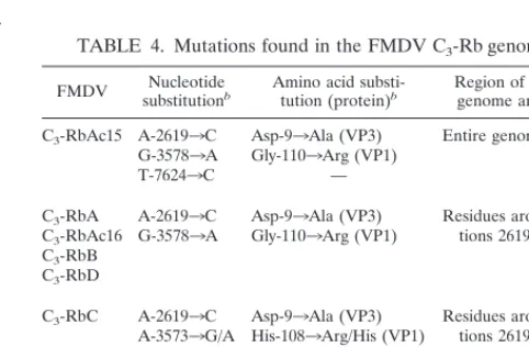
compared. Only two amino acid substitutions were identified in
the viral proteins as follows (Table 4): Asp-9
3
Ala in VP3 and
Gly-110
3
Arg in VP1. In addition, one silent mutation was
identified at position 7624. To determine whether the two
ami-no acid substitutions were consistently found in selected C
3Arg/85
FIG. 3. Determination of virus production (A to C) and intracellular viral RNA (D to F) upon infection of BHK-21 and modified BHK-21 cells with different FMDVs. BHK-21 cells (A and D) or BHK-Rb cells (B and E) or BHK-R74-A4 cells (C and F) were infected with C3Arg/85 (E), C3-Rbc15 (‚), or C3-Rbc16 (Œ) at
a multiplicity of infection of 10 PFU/cell. At the indicated times postinfection, samples from the culture medium were withdrawn and titrated (panels A to C). The time at which a complete cytopathic effect was observed is indicated by dotted arrows (infections with C3-Rbc15 or C3-Rbc16) or by a complete arrow (infection with C3Arg/85). For
other infections the cytopathic effect observed at the indicated times postinfection was not complete (Table 2). The viral yields from infections with other C3-Rb viruses
were very similar to those shown for C3-Rbc15 and -c16. In parallel infections, total cellular RNA was extracted at the indicated times (panels D to F). FMDV RNA was
extracted and quantitated by dot blot hybridization as described in Materials and Methods. In panels D to F, numbers on the ordinate represent the accumulated amount of FMDV RNA expressed as the percentage of the maximum value in each experiment. Infections were with C3Arg/85 (empty columns), C3-Rbc15 (columns with stripes), or
C3-Rbc16 (filled columns). Procedures for infection with FMDV, titrations with plaque assays, and quantification of FMDV RNA are detailed in Materials and Methods.
on November 9, 2019 by guest
http://jvi.asm.org/
variants, the genomic regions corresponding to amino acids 9
of VP3 and 110 of VP1 were sequenced for FMDVs C
3-RbA,
C
3-RbAc16, C
3-RbB, C
3-RbC, C
3-RbD, and C
3-RbE. All C
3-Rb viruses included an Asp-9
3
Ala substitution in VP3 and
either a His-108
3
Arg substitution or a Gly-110
3
Arg
substi-tution in VP1. In particular, C
3-RbC was a mixed population
containing both His and Arg at position 108 of VP1 and Gly
and Arg at position 110 of VP1, with only Ala at position 9 of
VP3. C
3-RbE included an Asp-9
3
Ala substitution in VP3 and
only a His-108
3
Arg substitution in VP1. The sequencing of
the entire P1-coding region of C
3-RbE revealed no other
mu-tations (Table 4). An analysis of 22 molecular clones from
population C
3-RbC showed that each clone included Arg at
either position 108 or position 110 in VP1. None of the clones
obtained had sequences with Arg in both positions 108 and 110
of VP1 (44). The size of the poly(C) tract was determined for
FMDV C
3Arg/85c1, C
3Arg/85c2, C
3Arg/85c3, C
3-RbA, C
3-RbAc15, C
3-RbAc16, C
3-RbB, C
3-RbC, C
3-RbD, and C
3-RbE.
No significant variations were seen between the C
3-Rb viruses
and the parental C
3Arg/85; the length of the poly(C) tract in all
the genomes analyzed ranged from 204 to 214 C residues.
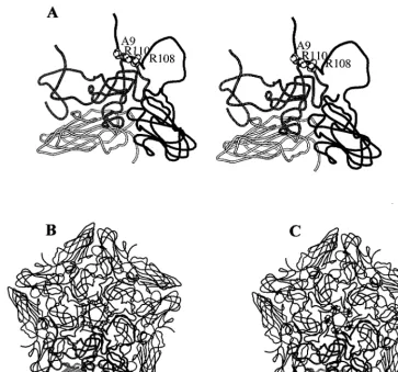
The three amino acid substitutions found in C
3-Rb viruses
cluster at the virion fivefold axis (Fig. 4) and represent a net
increase of positive charges on the virion surface. We conclude
that the same two capsid replacements at position 9 of VP3
and at position 108 or position 110 of VP1 were repeatedly
selected during infection of BHK-Rb cells by populations of
FMDV C
3Arg/85 and that these substitutions were associated
with the phenotypic modifications of C
3-Rb viruses.
DISCUSSION
The modified BHK-Rb cells were selected during the course
of a persistent infection of cloned BHK-21 cells with FMDV
clone C-S8c1 (9, 11, 12). The system selects for variant cells
which are partially resistant to the virus and for viruses which
are hypervirulent for the cells. This coevolution favors the
survival of both the host cells and the resident virus (11, 12,
37). Coevolution of cells and viruses has been described for a
number of other viral systems (1, 4–6, 40, 45, 46) and thus
cannot be regarded as a rare phenomenon. BHK-Rb cell
pop-ulations are heterogeneous in that they are composed of cell
types that differ in degree of cellular transformation and of
resistance to infection by FMDV (11, 12). Upon infection of
this heterogeneous cell population with FMDV C
3Arg/85, an
initial round of partial cell lysis took place on the second to
third day postinfection. The more-resistant cells persisted for
several additional days, supporting viral replication, as
sug-gested by their continuing viability and the shedding of virus
into the culture medium. The cell-to-cell variation in FMDV
susceptibility within the BHK-Rb cell population was
previ-ously documented by cell cloning and phenotypic testing (11).
The cellular heterogeneity has been confirmed and extended in
the present study by indirect immunofluorescence analysis of
infections with C
3Arg/85 (Fig. 2). The negative
immunofluo-rescence of about 50% of the BHK-Rb cells upon infection
with C
3Arg/85 suggests a block in an early step of the infectious
cycle of FMDV in this cell subpopulation (receptor
recogni-tion, entry, uncoating, or early protein or RNA synthesis). In
the more-resistant BHK-Rb cell subpopulations, variant
vi-ruses depicting minimal genetic changes but increased
viru-lence for their host cells were selected. The same amino acid
substitutions were selected in independent infections by clonal
C
3Arg/85 preparations, and therefore were the result of
inde-pendent mutational events (Fig. 1B and Table 4).
[image:6.612.50.290.97.248.2]The adaptation of RNA viruses is based on the continuous
generation of mutant genomes, resulting in extremely
hetero-geneous and dynamic mutant swarms, termed viral
quasispe-cies (14, 17, 18, 23, 24, 30). Average mutation rates for a
num-ber of RNA viruses have been estimated at 10
23to 10
25substitutions per nucleotide and round of copying (reviewed in
references 17 and 22). Thus, for an RNA genome of about 8
kb, such as FMDV, with a mutation rate of 10
24substitutions
TABLE 3. Replication in wild-type CHO cells and binding
to heparin-Sepharose of parental and variant
FMDV C3Arg/85 populations
FMDVa
PFU/PFU ratio
Replication in wild-type
CHO cellsb Binding to heparin-Sepharosec
C3Arg/85
0.9
1
C3Arg/85c1
,0.1
1.5
C3Arg/85c2
,0.1
1
C3Arg/85c3
1
1.6
C3-RbA
5
3
10
316
C3-RbAc15
1
3
10
310
C3-RbAc16
8
3
10
313
C3-RbB
6
3
10
310
C3-RbC
1
3
10
418
C3-RbD
1
3
10
311
C3-RbE
3
3
10
320
aThe origins of the FMDVs are described in Materials and Methods, Fig. 1, and Table 1.
bWild-type CHO cells were infected with virus at a multiplicity of infection of 10 PFU/cell. After virus adsorption (1 h, 37°C), cell monolayers were extensively washed with DMEM–1% fetal calf serum and maintained with DMEM–5% fetal calf serum. Results are expressed as the ratio of PFU/ml in supernatants of the infected cells at 36 h postinfection divided by the PFU/ml in the supernatants at 1 h postinfection (around 103PFU/ml). In a parallel experiment, replication of
C3Arg/85 in BHK-21 cells gave a ratio of 43104PFU at 20 h postinfection.
[image:6.612.308.549.486.644.2]cBinding experiments were performed as indicated in Materials and Methods. Numbers represent the ratio of PFU in the supernatant of the Sepharose beads after centrifugation divided by the PFU in the corresponding supernatant of the heparin-Sepharose beads after centrifugation (2). Results are the means of at least two independent titrations.
TABLE 4. Mutations found in the FMDV C3-Rb genomes
aFMDV substitutionNucleotideb Amino acid substi-tution (protein)b Region of the viralgenome analyzedb
C3-RbAc15 A-26193C Asp-93Ala (VP3) Entire genome
G-35783A Gly-1103Arg (VP1)
T-76243C —
C3-RbA A-26193C Asp-93Ala (VP3) Residues around
posi-tions 2619 and 3578 C3-RbAc16 G-35783A Gly-1103Arg (VP1)
C3-RbB
C3-RbD
C3-RbC A-26193C Asp-93Ala (VP3) Residues around
posi-tions 2619 and 3578 A-35733G/A His-1083Arg/His (VP1)
G-35783A/G Gly-1103Arg/Gly (VP1)
C3-RbE A-26193C Asp-93Ala (VP3) Capsid-coding region
(P1) A-35733G His-1083Arg (VP1)
aThe origin of each FMDV analyzed is described in Materials and Methods, Fig. 1, and Table 1. Mutations are defined by comparing the relevant sequences with those of the parental FMDV C3Arg/85. All parental C3Arg/85 preparations
(Fig. 1B) showed the wild-type sequence.
bNucleotide numbering is from the 59end of the genomic RNA of FMDV C3Arg/85, as described in reference 26. Amino acids are numbered
indepen-dently for each capsid protein. —, silent mutation. In C3-RbC, nucleotides and
amino acids separated by lines indicate mixed populations according to the bands in sequencing gels. The locations of the replaced amino acids in the three-dimensional structure of the FMDV capsid are shown in Fig. 4.
10176
ESCARMI´S ET AL.
J. V
IROL.
on November 9, 2019 by guest
http://jvi.asm.org/
per nucleotide copied, every progeny RNA molecule produced
during a replication process might contain an average of about
one mutation. The number of mutations per genome could
theoretically follow a Poisson distribution assuming that the
occurrence of mutations along the genome is random and
assuming their neutral character (15, 20). Obviously, mutations
may not occur at random, and many of them will be deleterious
and therefore eliminated by negative, or purifying, selection
(17). The evolution of a viral quasispecies will be influenced
not only by mutational input but also by the number of rounds
of genome copying and by the population size of the
replicat-ing genomes (17, 20, 23, 24, 30). The quasispecies structure and
dynamics predict that variants with one mutation or a few
mutations will be common in mutant distributions and thus will
be selected whenever they give the virus a selective advantage
in the environment at hand (14). This scenario provides a
plausible interpretation for the observations described in the
present study. Furthermore, repetitive, or deterministic, to use
a more fundamental term, selection of the same variants will
occur when a limited number of molecular solutions are
avail-able to a virus for coping with a selective constraint (17, 19).
Had the molecular solutions of C
3Arg/85 required a
constel-lation of mutations beyond the reach of the sequence space
(23, 24) being explored by the virus to replicate in BHK-Rb
cells, adaptation would not have been feasible and the
selec-tion of C
3-Rb variants would not have occurred. We have
previously suggested that functional and structural constraints
are probably the main limitations of the evolution of RNA
viruses given their high mutation potential (17, 19).
[image:7.612.118.482.69.408.2]In capsids with icosahedral symmetry, surface residues tend
to be more tolerant to replacements than internal capsid
res-idues engaged in interactions with other amino acids (34). The
residues at the surface are those that are the most frequently
replaced in the course of large population passages of FMDV
(26). The two amino acid substitutions found in all the C
3-Rb
populations analyzed (Asp-9
3
Ala in VP3 and His-108
3
Arg
or Gly-110
3
Arg in VP1) cluster at the fivefold axis of the
FMDV particle (Fig. 4). This is the site at which the persistent
FMDV R100 (13) also includes a cluster of amino acid
substi-tutions, e.g., Asp-9
3
Ala in VP3. It is interesting that both
R100 and the C
3-Rb viruses described here have high virulence
for BHK-21 cells and a small-plaque morphology (13).
Remark-ably, the two amino acid substitutions in the capsid of C
3Arg/
85, without any other substitution in any viral protein,
con-ferred to the C
3-Rb viruses an increased virulence for BHK-21
cells, the ability to infect modified BHK-21 cells, a small-plaque
morphology, and the potential to bind heparin and infect
wild-type CHO cells. This latter change represents a modification of
FIG. 4. Location in the three-dimensional structure of FMDV of serotype C (34) of the amino acids replaced in the C3-Rb FMDVs (Asp-93Ala in VP3 and
His-1083Arg in VP1 or Gly-1103Arg in VP1). (A) Stereodiagram of a crystallographic protomer of C-S8c1 in which the three amino acid substitutions are modeled. The capsid proteins VP1, VP2, and VP3 are depicted as grey, white/stippled, and black ribbons, respectively. The substituted residues are shown as spheres and indicated by the single-letter amino acid code (A5Ala; R5Arg). (B) Diagram of the pentamer subunit showing substitutions Asp-93Ala in VP3 and His-1083Arg in VP1. (C) Pentamer with substitutions Asp-93Ala in VP3 and Gly-1103Arg in VP1. A reference protomer (lower part of the structure) is depicted as in panel A. The cluster of positive charges around the fivefold axis is apparent in both cases. The procedures used to model the amino acid substitutions are described in Materials and Methods. The structures were drawn by using a modified version of MOLSCRIPT (33).
on November 9, 2019 by guest
http://jvi.asm.org/
cell tropism that has been documented with a number of
dif-ferent FDMV isolates (2, 32, 41, 49). Jackson et al. (32)
showed that binding to cellular heparan sulfate was required by
FMDV of serotype O for the efficient infection of cells in
culture. Sa-Carvalho et al. (49) documented that heparin
bind-ing and cell culture adaptation of FMDV O
1Campos entailed
the acquisition of positively charged residues at VP2 residue
134 and VP3 residue 56. In an FMDV of serotype C
1which
was subjected to 213 serial cytolytic passages in BHK-21 cells
(MARLS virus [2]), two amino acid substitutions
(Lys-173
3
Met in VP3 and Ser-144
3
Leu in VP1) were associated
with the loss of the ability of the virus to bind heparin and
in-fect CHO cells (2). These substitutions afin-fect a capsid region
around the G-H loop of VP1, away from the fivefold axis (34).
Comparison of results with MARLS and the C
3-Rb viruses
reinforces the view that the FMDV capsid is an important
de-terminant of virulence for BHK-21 cells (2) and that increased
affinity for heparin and alterations in cell tropism may be
mediated by a number of independent sites on the viral
cap-sid (2). The systematic selection of the same types of variant
C
3Arg/85 in BHK-Rb cells shows that when selective forces are
strong and the genetic modifications needed to respond to a
selective constraint are attainable within the sequence space
available to the virus (23, 24), phenotypic shifts can be
repro-ducible and rapid (31).
The repeated selection of two capsid mutations was also
observed during persistent infections of poliovirus in primary
cultures of human fetal brain cells (42). de la Torre et al. (10)
found that transfection of HeLa cells with independent clones
of a temperature-sensitive poliovirus mutant resulted not only
in the expected transition that eliminated temperature
sensi-tivity but also in four additional silent substitutions that
pre-cisely reverted the mutant sequence to that of the wild-type
poliovirus. The results with FMDV prove that the replication
of viruses in partially permissive cells may promote the
selec-tion of genetic and phenotypic variants, including viral mutants
with an altered host range. This selection need not be the
result of a prolonged persistent infection but can occur during
the short-term survival of a virus with a partially permissive
host cell. The results of the present study illustrate the
impres-sive adaptive potential of viral quasispecies.
ACKNOWLEDGMENTS
We are indebted to M. Da´vila for expert technical assistance and to
Jose´ Pizarro for help with sequencing experiments.
Work in Madrid was supported by grant PM97-0060-C02 from
DEGS, grant PSS0885 from the EU, and the Fundacio´n Ramo´n
Areces. Work in Buenos Aires was supported by CONICET and
INTA. M.T.F.-F. thanks Laboratorios BAGO (Buenos Aires,
Ar-gentina) for support. Visits to Madrid and Buenos Aires were funded
by a CSIC-CONICET cooperative grant. M.T.F.-F.’s sabbatical leave
in Madrid was funded by MEC (Spain).
REFERENCES
1. Ahmed, R., W. M. Canning, R. S. Kauffman, A. H. Sharpe, J. V. Hallum, and B. N. Fields.1981. Role of the host cell in persistent viral infection: coevo-lution of L cells and reovirus during persistent infection. Cell 25:325–332. 2. Baranowski, E., N. Sevilla, N. Verdaguer, C. M. Ruiz-Jarabo, E. Beck, and
E. Domingo.1998. Multiple virulence determinants of foot-and-mouth dis-ease virus in cell culture. J. Virol. 72:6362–6372.
3. Burrows, R. 1966. Studies on the carrier state of cattle exposed to foot-and-mouth disease virus. J. Hyg. 64:81–90.
4. Calvez, V., I. Pelletier, T. Couderc, N. Pavio-Gue´do, B. Blondel, and F. Colbe`re-Garapin.1995. Cell clones cured of persistent poliovirus infection display selective permissivity to the wild-type poliovirus strain Mahoney and partial resistance to the attenuated Sabin 1 strain and Mahoney mutants. Virology 212:309–322.
5. Charini, A., S. Arista, A. Giammanco, and A. Sinatra. 1983. Rotavirus persistence in cell cultures: selection of resistant cells in the presence of fetal
calf serum. J. Gen. Virol. 64:1101–1110.
6. Chen, W., and R. S. Baric. 1996. Molecular anatomy of mouse hepatitis virus persistence: coevolution of increased host cell resistance and virus virulence. J. Virol. 70:3947–3960.
7. Chomczynski, P., and N. Sacchi. 1987. Single-step method of RNA isolation by acid guanidinium thiocyanate-phenol-chloroform extraction. Anal. Bio-chem. 162:156–159.
8. de la Torre, J. C., B. Alarco´n, E. Martı´nez-Salas, L. Carrasco, and E. Domingo.1987. Ribavirin cures cells of a persistent infection with foot-and-mouth disease virus. J. Virol. 61:233–235.
9. de la Torre, J. C., M. Da´vila, F. Sobrino, J. Ortı´n, and E. Domingo. 1985. Establishment of cell lines persistently infected with foot-and-mouth disease virus. Virology 145:24–35.
10. de la Torre, J. C., C. Giachetti, B. L. Semler, and J. J. Holland. 1992. High frequency of single-base transitions and extreme frequency of precise mul-tiple-base reversion mutations in poliovirus. Proc. Natl. Acad. Sci. USA 89: 2531–2535.
11. de la Torre, J. C., E. Martı´nez-Salas, J. Dı´ez, and E. Domingo. 1989. Ex-tensive cell heterogeneity during a persistent infection with foot-and-mouth disease virus. J. Virol. 63:59–63.
12. de la Torre, J. C., E. Martı´nez-Salas, J. Dı´ez, A. Villaverde, F. Gebauer, E. Rocha, M. Da´vila, and E. Domingo.1988. Coevolution of cells and viruses in a persistent infection of foot-and-mouth disease virus in cell culture. J. Virol. 62:2050–2058.
13. Dı´ez, J., M. Da´vila, C. Escarmı´s, M. G. Mateu, J. Domı´nguez, J. J. Pe´rez, E. Giralt, J. A. Melero, and E. Domingo.1990. Unique amino acid substitutions in the capsid proteins of foot-and-mouth disease virus from a persistent infection in cell culture. J. Virol. 64:5519–5528.
14. Domingo, E. 1996. Biological significance of viral quasispecies. Viral Hepa-titis Rev. 2:247–261.
15. Domingo, E. 1997. RNA virus evolution, population dynamics, and nutri-tional status. Biol. Trace Elem. Res. 56:23–30.
16. Domingo, E., M. Da´vila, and J. Ortı´n. 1980. Nucleotide sequence heteroge-neity of the RNA from a natural population of foot-and-mouth disease virus. Gene 11:333–346.
17. Domingo, E., and J. J. Holland. 1994. Mutation rates and rapid evolution of RNA viruses, p. 161–184. In S. S. Morse (ed.), Evolutionary biology of viruses. Raven Press, New York, N.Y.
18. Domingo, E., J. J. Holland, C. Biebricher, and M. Eigen. 1995. Quasispecies: the concept and the word p. 171–180. In A. Gibbs, C. Calisher, and F. Garcı´a-Arenal (ed.), Molecular basis of virus evolution. Cambridge Univer-sity Press, Cambridge, United Kingdom.
19. Domingo, E., M. G. Mateu, C. Escarmı´s, E. Martı´nez-Salas, D. Andreu, E. Giralt, N. Verdaguer, and I. Fita.1996. Molecular evolution of aphthovi-ruses. Virus Genes 11:197–207.
20. Domingo, E., D. L. Sabo, T. Taniguchi, and C. Weissmann. 1978. Nucleotide sequence heterogeneity of an RNA phage population. Cell 13:735–744. 21. Donn, A., M. Castagnaro, and A. I. Donaldson. 1995. Ultrastructural and
replicative features of foot-and-mouth disease virus in persistently infected BHK-21 cells. Arch. Virol. 140:13–25.
22. Drake, J. W. 1993. Rates of spontaneous mutations among RNA viruses. Proc. Natl. Acad. Sci. USA 90:4171–4175.
23. Eigen, M. 1992. Steps towards life. Oxford University Press, Oxford, United Kingdom.
24. Eigen, M., and C. Biebricher. 1988. Sequence space and quasispecies distri-bution, p. 211–245. In E. Domingo, J. J. Holland, and P. Ahlquist (ed.), RNA genetics. CRC Press, Inc., Boca Raton, Fla.
25. Escarmı´s, C., M. Toja, M. Medina, and E. Domingo. 1992. Modifications of the 59untranslated region of foot-and-mouth disease virus after prolonged persistence in cell culture. Virus Res. 26:113–125.
26. Escarmı´s, C., M. Da´vila, N. Charpentier, A. Bracho, A. Moya, and E. Do-mingo.1996. Genetic lesions associated with Muller’s ratchet in an RNA virus. J. Mol. Biol. 264:255–267.
27. Esko, J. D., T. E. Stewart, and W. H. Taylor. 1985. Animal cell mutants defective in glycosaminoglycans biosynthesis. Proc. Natl. Acad. Sci. USA 82: 3197–3201.
28. Ferrer, M., A. He´mar, V. Duprez, R. Hellio, and A. Dantry-Varsat. 1993. Both theaandbchains of high affinity interleukin 2 receptors are located in intracellular vesicles when their ligand is endocytosed. Eur. J. Cell Biol. 60:276–282.
29. Gebauer, F., J. C. de la Torre, I. Gomes, M. G. Mateu, H. Barahona, B. Tiraboschi, I. Bergmann, M. G. Auge´ de Mello, and E. Domingo.1988. Rapid selection of genetic and antigenic variants of foot-and-mouth disease virus during persistence in cattle. J. Virol. 62:2041–2049.
30. Holland, J. J., J. C. de la Torre, and D. A. Steinhauer. 1992. RNA viral populations as quasispecies. Curr. Top. Microbiol. Immunol. 176:1–20. 31. Holland, J. J., K. Spindler, F. Horodyski, E. Grabau, S. Nichol, and S. Van
dePol.1982. Rapid evolution of RNA genomes. Science 215:1577–1585. 32. Jackson, T., F. M. Ellard, R. Abu Ghazaleh, S. M. Brookes, W. E. Blakemore,
A. H. Corteyn, D. I. Stuart, J. W. I. Newman, and A. M. Q. King.1996. Efficient infection of cells in culture by type O foot-and-mouth disease virus requires binding to cell surface heparan sulfate. J. Virol. 70:5282–5287.
10178
ESCARMI´S ET AL.
J. V
IROL.
on November 9, 2019 by guest
http://jvi.asm.org/
33. Kraulis, P. F. 1991. MOLSCRIPT: a program to produce both detailed and schematic plots of protein structures. J. Appl. Crystallogr. 24:946–950. 34. Lea, S., J. Herna´ndez, W. Blakemore, E. Brocchi, S. Curry, E. Domingo, E.
Fry, R. A. Ghazaleh, A. King, J. Newman, D. Stuart, and M. G. Mateu.1994. The structure and antigenicity of a type C foot-and-mouth disease virus. Structure 2:123–139.
35. Lidholt, K., J. L. Weinke, C. S. Kiser, F. N. Lugemwa, K. J. Bame, S. Cheifetz, J. Massague´, U. Lindahl, and J. D. Esko.1992. A single mutation affects both N-acetylglucosaminyltransferase and glucuronosyltransferase ac-tivities in a chinese hamster ovary cell mutant defective in heparin sulfate biosynthesis. Proc. Natl. Acad. Sci. USA 89:2267–2271.
36. Marilat, V., P. Auge´ de Mello, B. Tiraboschi, E. Beck, I. Gomes, and I. E. Bergmann.1994. Genetic variation of foot-and-mouth disease virus during persistent infection in cattle. Virus Res. 34:31–48.
37. Martı´n Herna´ndez, A. M., E. C. Carrillo, N. Sevilla, and E. Domingo. 1994. Rapid cell variation can determine the establishment of a persistent viral infection. Proc. Natl. Acad. Sci. USA 91:3705–3709.
38. Mateu, M. G., J. L. Da Silva, E. Rocha, D. L. De Brum, A. Alonso, L. Enjuanes, E. Domingo, and H. Barahona.1988. Extensive antigenic heter-ogeneity of foot-and-mouth disease virus of serotype C. Virology 166:113– 124.
39. Mateu, M. G., J. Herna´ndez, M. A. Martı´nez, D. Feigelstock, S. Lea, J. J. Pe´rez, E. Giralt, D. Stuart, E. L. Palma, and E. Domingo.1994. Antigenic heterogeneity of a foot-and-mouth disease virus serotype in the field is mediated by very limited sequence variation at several antigenic sites. J. Vi-rol. 68:1407–1417.
40. Mrukowicz, J. Z., J. D. Wetzel, M. I. Goral, A. B. Fogo, P. F. Wright, and T. S. Dermody.1998. Viruses and cells with mutations affecting viral entry are selected during persistent rotavirus infections of MA104 cells. J. Virol. 72:3088–3097.
41. Neff, S., D. Sa-Carvalho, E. Rieder, P. Mason, S. D. Blystone, E. J. Brown, and B. Baxt.1998. Foot-and-mouth disease virus virulent for cattle utilizes the integrinavb3as its receptor. J. Virol. 72:3587–3594.
42. Pavio, N., M.-H. Buc-Caron, and F. Colbe`re-Garapin. 1996. Persistent po-liovirus infection of human fetal brain cells. J. Virol. 70:6395–6401. 43. Piccone, M. E., G. Kaplan, L. Giavedoni, E. Domingo, and E. L. Palma. 1988.
VP1 of serotype C foot-and-mouth disease viruses: long-term conservation of sequences. J. Virol. 62:1469–1473.
44. Rocha, C., and C. Escarmı´s. 1998. Unpublished results.
45. Ron, D., and J. Tal. 1985. Coevolution of cells and virus as a mechanism for the persistence of lymphotropic minute virus of mice in L cells. J. Virol. 55: 424–430.
46. Ron, D., and J. Tal. 1986. Spontaneous curing of a minute virus of mice carrier state by selection of cells with an intracellular block of viral replica-tion. J. Virol. 58:26–30.
47. Roussel, A., and C. Cambillau. 1989. TURBO-FRODO, p. 77–78. In Silicon Graphics geometry partners directory. Silicon Graphics, Mountain View, Calif.
48. Rueckert, R. R. 1996. Picornaviridae: the viruses and their replication, p. 609– 654. In B. N. Fields et al. (ed.), Fields virology. Lippincott-Raven Publishers, Philadelphia, Pa.
49. Sa-Carvalho, D., E. Rieder, B. Baxt, R. Rodarte, A. Tanuri, and P. Mason. 1997. Tissue culture adaptation of foot-and-mouth disease virus selects vi-ruses that bind to heparin and are attenuated in cattle. J. Virol. 71:5115– 5123.
50. Salt, J. S. 1993. The carrier state in foot-and-mouth disease—an immuno-logical review. Br. Vet. J. 149:207–223.
51. Sevilla, N., and E. Domingo. 1996. Evolution of a persistent aphthovirus in cytolytic infections: partial reversion of phenotypic traits accompanied by genetic diversification. J. Virol. 70:6617–6624.
52. Sobrino, F., M. Da´vila, J. Ortı´n, and E. Domingo. 1983. Multiple genetic variants arise in the course of replication of foot-and-mouth disease virus in cell culture. Virology 128:310–318.
53. Sutmoller, P., J. W. McVicar, and G. E. Cottral. 1968. The epizootical importance of foot-and-mouth disease carriers. I. Experimentally produced foot-and-mouth disease carriers in susceptible and immune cattle. Arch. Gesamte Virusforsch. 23:227–235.
54. Taboga, O., C. Tami, E. Carrillo, J. I. Nu´n˜ez, A. Rodrı´guez, J. C. Saiz, E. Blanco, M.-L. Valero, X. Roig, J. A. Camarero, D. Andreu, M. G. Mateu, E. Giralt, E. Domingo, F. Sobrino, and E. L. Palma.1997. A large-scale eval-uation of peptide vaccines against foot-and-mouth disease: lack of solid protection in cattle and isolation of escape mutants. J. Virol. 71:2606–2614. 55. van Bekkum, J. G., H. S. Frenkel, H. H. J. Frederiks, and S. Frenkel. 1959. Observations on the carrier state of cattle exposed to foot-and-mouth disease virus. Tijdschr. Diergeneeskd. 84:1159–1164.
56. Woodbury, E. L. 1995. A review of the possible mechanisms for the persis-tence of foot-and-mouth disease virus. Epidemiol. Infect. 114:1–13.
on November 9, 2019 by guest
http://jvi.asm.org/