Int. J. Electrochem. Sci., 9 (2014) 5187 - 5203
International Journal of
ELECTROCHEMICAL
SCIENCE
www.electrochemsci.org
Complexation of Gadolinium(III) and terbium(III) with
nalidixic
acid (NDX): Molar conductivity, thermal and spectral
investigation
Ahmed I. El-Shenawy1,2,*, Aly H. Atta1,3, and Moamen S. Refat4,5
1
Department of Chemistry, Faculty of Science, Dammam University, Kingdom Saudi Arabia
2
Department of Chemistry, Faculty of Science, Benha University, Egypt
3
Department of Chemistry, Faculty of Science, Suez University, Suez, Egypt
4
Department of Chemistry, Faculty of Science, Taif University, Al-Hawiah, Taif, P.O. Box 888 Zip Code 21974, Saudi Arabia
5
Department of Chemistry, Faculty of Science, Port Said University, Port Said, Egypt
*
E-mail: aielshenawy@gmail.com
Received: 26 February 2014 / Accepted: 7 May 2014 / Published: 16 June 2014
The goal of this paper is recognized to the chemical and microbial impact resulting from the interaction between nalidixic acid (NDX) and two lanthanide salts (GdCl3.6H2O and TbCl3.6H2O),
which were prepared in normal and nano–scale range. Gadolinium and terbium(III) metal ions chelated with NDX through the deprotonation of carboxylic group of NDX bidentate ligand and oxygen atom of carbonyl group. The analytical analyses (elemental analyses, electrical conductivity) and spectroscopic measurements (FT–IR spectral, X-ray powder diffraction (XRD) and scanning electron microscopy (SEM)) as well as thermal analysis (TG/DTA) have been used to characterize the new solid complexes. The stability of NDX complexes was checked using Coats-Redfern and Horowitz-Metzger integral methods for the main thermal decomposition steps in the TG curves. The shiny side in this paper is focused on the preparation and characterization of single phases of gadolinium and terbium(III) oxide nanoparticles using carbamide as precursors. The NDX ligand in comparison with both cases traditional and nano–particles lanthanide complexes were assessment against for some kind of bacteria (Escherichia Coli, Staphylococcus Aureus, Bacillus subtilis and Pseudomonas aeruginosa) and fungi (Aspergillus Flavus and Candida Albicans) activities. The highest antibacterial and antifungal activities data of the nano–particles complexes were observed with more potent than the free NDX and traditional lanthanide complexes.
1. INTRODUCTION
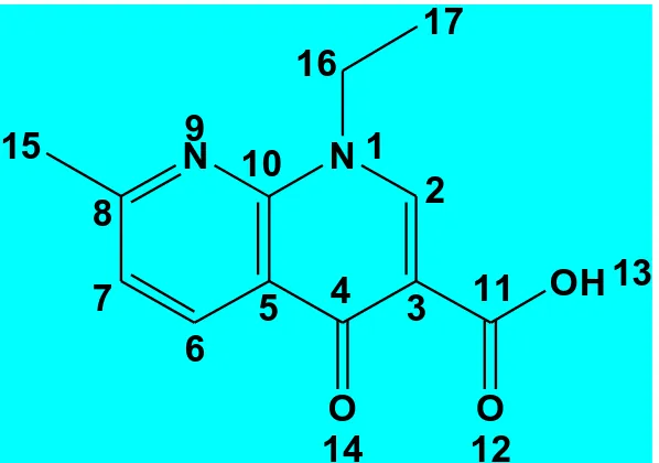
Study of the interaction between drugs and metal ions is an important and active research area in bioinorganic chemistry [1-4]. It is well known that the action of many drugs is dependent on the coordination with metal ions [1] or the inhibition [2] on the formation of metalloenzymes. Quinolones considered a class of antibacterial agents which have has been known for over 40 years [5]. Presence of metal ions considerably alters the activity of quinolones against potentially susceptible bacteria [6]. The interaction of metal ions with diverse deprotonated quinolones as ligands has been thoroughly discussed [7-16]. Quinolones have been classified in generations based on their activity [17]. Each generation presents an enhanced spectrum of activity in comparison to a previous one. Nalidixic acid (Fig. 1) is an example of first-generation quinolones that are active against Gram-negative organisms and they are used for the treatment of uncomplicated urinary tract infections [18]. The association complexes resulted between nalidixic acid and different metal ions has been described [19-25]. The copper and calcium(II) nalidixic acid complexes have been studied [19]. Nakano et al. [22] studied the corresponding complexes of nalidixic acid with Al3+, Mg2+ and Ca2+. Therefore, the importance of this paper is to study the physical and chemical structural of the interaction between the gadolinium or terbium(III) ions with NDX antibiotic ligand in nano–structural fashion. The coordination mode of nalidixic acid chelation has been investigated.
N 1
2
3
4
5
6
7
8
N
9
10
11
O
12
OH 13
O
14
15
16
[image:2.596.150.449.385.595.2]17
Figure 1.The structural formula of nalidixic acid (NDX)
2. EXPERIMENTAL
2.1. Reagents
Carbamide, GdCl3.6H2O, TbCl3.6H2O and methanol solvent were obtained from Aldrich
2.2. Synthesis of Gd(III) and Tb(III)–nalidixic acid complexes 2.2.1. Method of nano–particles preparation
The preparation can be summarized in four steps with sequence equations as follows; ia– 2[Gd((NH2)2CO)3Cl 3]+14H2O caC
o 80
. Gd
2(CO3)3.5H2O+3CO2+NH4Cl+6NH3
ib– 2[Tb((NH2)2CO)3Cl 3]+14H2O caC o 80
. Tb
2(CO3)3.5H2O+3CO2+NH4Cl+6NH3
ii– M2(CO3)3.5H2O C o 800
ca. Gd
2O3/Tb2O3
iii– Gd2O3/Tb2O3+ HCl (conc.) C o 80
ca. GdCl
3/TbCl3
iv– LnCl3 + NDX [Ln(NDX)3].6H2O
The first step of preparation was discussed previously [26, 27] as follows: Ln2(CO3)3.5H2O (Ln
= Gd and Tb(III)) were prepared by mixing aqueous solutions (100 ml) of 0.01 M of the respectively LnCl3.6H2O (Gd(III) or Tb(III)) with a volume of 100 ml of 0.1 M of carbamide. The mixtures were
heated to ca. 80 oC for 2-4 h in a water bath. The precipitated white compounds were filtered out, washed several times with hot water, dried at ca. 80 oC in an oven for 2 h and then in vacuo over anhydrous calcium chloride. Carbonate contents in Ln2(CO3)3.nH2O were determined by dissolving a
weighted sample of the products in excess standard HCl and the excess of HCl was determined by titration with standard sodium carbonate. Lanthanide metal was determined gravimetrically as sesquioxides, Ln2O3. The infrared spectra of carbamide and lanthanide carbonates were recorded in
KBr discs. At room temperature, lanthanide(III) ions react with carbamide to form the complex, Ln(carbamide)3X3 [28, 29]. The second step dealing the transfer of lanthanide(III) carbonates (Gd(III)
and Tb(III)) constructed in the first step to form of nano-oxides particles, which have been examined using X-ray. In third step lanthanum(III) oxide was converted to its chloride by treating with concentrated hydrochloric acid. An aqueous solution of lanthanum chloride in fourth step was added to a mixed methanolic solution of NDX (the mole ratio (LnCl3·xH2O): (NDX) was 1:3), the pH value of
which was adjusted to 8.0–9.0 by adding aqueous ammonia solution (5 %, V/V) with heating at 80–90 °C for about 4–5 h and continuous stirring. The precipitate was filtered off, washed with water and methanol, dried (90 °C), and then left over anhydrous calcium chloride.
2.3. Equipments
Elemental analyses of carbon, hydrogen and nitrogen elements were analyzed by a Perkin-Elmer CHN 2400 elemental analyzer. The metal contents were determined gravimetrically by converting the compounds to their corresponding oxides. The molar conductivities of freshly prepared 1.0×10⁻³
diffraction, target copper with secondary monochromate. Scanning electron microscopy (SEM) images and Energy Dispersive X-ray Detection (EDX) were taken in Joel JSM-639OLA equipment, with an accelerating voltage of 20 KV.
2.4.Biological studies
Antimicrobial activity of the tested samples was determined using a modified Kirby-Bauer disc diffusion method [30]. Briefly, 100 μl of the best bacteria/fungi were grown in 10 mL of fresh media until they reached a count of approximately108 cells/mL for bacteria or 105 cells/mL for fungi. 100 μl of microbial suspension was spread onto agar plates corresponding to the broth in which they were maintained. Isolated colonies of each organism that might be playing a pathogenic role should be selected from primary agar plates and tested for susceptibility by disc diffusion method. Of the many media available, National Committee for Clinical Laboratory Standards (NCCLS) recommends Mueller-Hinton agar due to: it results in good batch-to-batch reproducibility. Disc diffusion method for filamentous fungi tested by using approved standard method (M38-A) developed by the NCCLS for evaluating the susceptibility of filamentous fungi to antifungal agents [31-37]. Disc diffusion method for yeast developed standard method (M44-P) by the NCCLS. Plates inoculated with filamentous fungi as Aspergillus Flavus at 25 oC for 48hours; Gram (+) bacteria as Staphylococcus Aureus, Bacillus subtilis; Gram (-) bacteria as Escherichia Coli, Pseudomonas aeruginosa they were incubated at 35-37
o
C for 24-48 hours and yeast as Candida Albicans incubated at 30 oC for 24-48 hours and, then the diameters of the inhabitation zones were measured in millimeters. Standard discs of Tetracycline (Antibacterial agent), Amphotericin B (Antifungal agent) served as positive controls for antimicrobial activity but filter disc impregnated with 10 μL of solvent (distilled water, DMSO) were used as a negative control. The agar used is Meuller-Hinton agar that is rigorously tested for composition and pH. Further the depth of the agar in the plate is a factor to be considered in the disc diffusion method [31-37]. This method is well documented and standard zones of inhabitation have been determined for susceptible values. Blank paper disks (Schleicher & Schuell, Spain) with a diameter of 8.0 mm were impregnated 10 μL of tested concentration of the stock solutions. When a filter paper disc impregnated with a tested chemical is placed on agar the chemical will diffuse from the disc into the agar. This diffusion will place the chemical in the agar only around the disc. The solubility of the chemical and its molecular size will determine the size of the area of chemical infiltration around the disc. If an organism is placed on the agar it will not grow in the area around the disc if it is susceptible to the chemical. This area of no growth around the disc is known as a "Zone of inhibition" or "Clear zone". For the disc diffusion, the zone diameters were measured with slipping calipers of the National for Clinical Laboratory Standers [32-35]. Agar-based methods such as E-test disk diffusion can be good alternatives because they are simpler and faster than broth methods.
3. RESULTS AND DISCUSSION
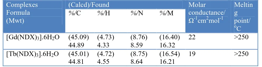
measurements like percentage of (carbon, hydrogen and nitrogen) and decomposition temperatures of the compounds are presented in Table 1. The experimental and theoretical percentages of elemental analysis are in a good agreement with each other and supported the suggested molecular formula of the synthesized nalidixic acid complexes. The complexes have high thermal decomposition points above > 250 oC. The nalidixic acid ligand behaves as bidentate ligand and coordinated to the Gd(III) and Tb(III) metal ions via one carboxylate oxygen atom and the oxygen atom of the pyridine carbonyl group. The solid NDX complexes are 1:3 molar ratio of (Gd/Tb(III): NDX).
3.1.Molar conductivities
[image:5.596.75.523.507.623.2]The molar conductance data has a supporting item, that been used in speculate the conformational structure of metal chelates. The measurement of conductance in solution form gave an idea about the degree of ionization (ions that liberated in solutions) for the resulted complexes. The higher data of molar conductivity is due to the presence of compound in electrolytic case and vice versa [38]. The molar conductivity values for the Gd(III) and Tb(III) complexes of nalidixic acid in DMSO solvent (1.00 x10⁻³ M) were found to be in the range 19–22 Ω-1.cm2.mol-1 at room temperature, suggesting them to be non-electrolytes [39]. Hence the molar conductance values indicate that no ions are present outside the coordination sphere so that chloride ions may be exhibit inside the coordination sphere or absent. The obtained results were strongly matched with the elemental analysis data where Cl- ions are absence in case of both Gd(III) and Tb(III) complexes after checked of these complexes using silver nitrate precipitated reagent. The above complexes are air-stable, insoluble in water, and soluble in dimethylsulfoxide, DMSO or dimethylformamide, DMF and concentrated mineral acids.
Table 1. Micro analytical and physical data of Gd(III) and Tb(III)/NDX complexes Complexes
Formula (Mwt)
(Calcd)/Found Molar
conductance/ Ω-1
cm2mol-1
Meltin g point/
o
C
%/C %/H %/N %/M
[Gd(NDX)3].6H2O (45.09)
44.89
(4.73) 4.33
(8.76) 8.59
(16.40) 16.32
22 >250
[Tb(NDX)3].6H2O (45.01)
44.81
(4.72) 4.55
(8.75) 8.64
(16.54) 16.21
19 >250
3.2. Electronic absorption spectra
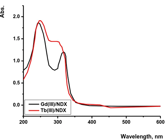
in carboxylic and ketone groups [41]. These two bands of free NDX are hypsochromically affected in the electronic spectra of Gd(III) and Tb(III) complexes. The Gd(III) and Tb(III)/NDX complexes show two absorption bands at (240 and 330) nm and (250 and 305) nm, respectively, due to π→π* and n→π* transitions. The hypsochromically change in the spectra of both NDX complexes supported that the carboxylic group and ketone groups are involved in the coordination. Also, these results are compatible with the interpretations of FT-IR and 1H-NMR spectra.
200 300 400 500 600
0.0 0.5 1.0 1.5 2.0
A
b
s
.
Wavelength, nm
[image:6.596.165.437.208.436.2]Gd(III)/NDX Tb(III)/NDX
Figure 2.Electronic spectra of Gd(III) and Tb(III)/NDX complexes
3.3. Infrared spectra
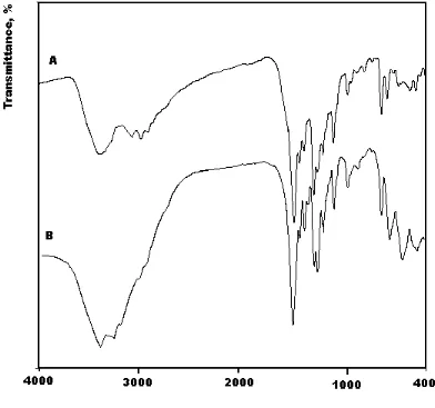
In the comparison between infrared spectra of the synthesized Gd(III) and Tb(III) complexes (Fig. 3) with that of the free NDX ligand, the place of coordination for the NDX ligand with both metal ions have been discussed and summarized in Table 2. The free NDX ligand has a strong band located at 1717 cm-1 which assigned to the stretching vibration of the carbonyl ν(CO) group for carboxylic ν(COOH) [42]. Insight into the spectrum of NDX, it was found that the absence of the νCOOH stretching
chelation take place when the difference in frequencies between asymmetric and symmetric stretching vibrations [∆ν = (νasym COO–– νsym COO–)] ≥ 200 [42-44]. From the infrared spectra of both Gd(III)
[image:7.596.97.489.318.685.2]and Tb(III) complexes the calculated ∆ν was observed at 219 and 215 cm–1, respectively, which suggests a unidentate interaction of the carboxylate group toward the central metal ions in both NDX complexes. The other reason, which confirms the place of coordination is that the carbonyl group ν(C=O) which presence at 1717 cm–1 for the free NDX was blue shifted by ∆ν = 96 and 77 cm–1 for Gd(III) and Tb(III)/NDX complexes, respectively. This discussion is strengthen the involvement of the carbonyl group in coordination of Gd(III) and Tb(III) complexes. The new band was involved in both Gd(III) and Tb(III) complexes at 451-622 cm–1 assigned to M-O stretching vibration motion [42, 44]. The uncoordinated water molecules in the Gd(III) and Tb(III) complexes were observed at the ~ 3400-3200 cm–1. From the above discussion we can say that, NDX ligand coordinated with Gd(III) and Tb(III) ions via two oxygen atoms of the carboxylate and the carbonyl groups, the most probable six coordinated fashion around each metal ion (Fig. 3) as designed below.
Table 2. Distinguish infrared bands of NDX and their complexes.
Compound (C=O) (COO) (asym)
v(COO) (sym.)
Δ
v(asy.–sym.)
v(M-O)
NDX 1717 1616 1386 230 ---
[Gd(NDX)3].6H2O 1620 1570 1351 219 546, 451
[Tb(NDX)3].6H2O 1640 1575 1360 215 522, 487
N
N
O O
O
N N
O
O O
N
N O
O
[image:8.596.172.426.235.470.2]O M
Figure 4. Suggestion structure of [M(NDX)3].6H2Ocomplexes (where, M= Gd(III) or Tb(III))
3.4. 1H-NMR spectra
The 1H-NMR data of the free nalidixic acid and its Gd(III) complex as an example were discussed. The characteristic peaks of NDX free ligand can be summarized as: 14.870 ppm (H; COOH), 9.174 ppm (H; C=H), 8.602 ppm (H; pyridine ring), 7.596 ppm (H; pyridine ring), 4.640 ppm (2H; CH2CH3), 2.712 ppm (3H; CH3-pyridine ring) and 1.425 ppm (3H; CH2CH3). Through the
comparison with the free NDX ligand, the absence of the characteristic peak for hydrogen of –COOH at δ=14.870 ppm in Gd(III) complex indicates that the coordination of NDX ligand to Gd(III) occur through the deprotonated carboxylic group [41]. The proton NMR spectrum for Gd(III) complex show peaks at the range of (3.30 - 3.50) ppm, this peaks aren’t observed in the free NDX ligand spectrum and can be assigned to the protons of H2O molecules, supporting the complex formula. The overall
3.5. Thermal analysis
The well-established thermogravimetric analysis (TGA) and differential scanning calorimetry (DSC) techniques have been reliably widely used over many decades in studying the thermal behavior and properties of various types of materials and evaluating the parameters for their degradation processes. These methods have allowed determination of the chemical steps of the investigated degradation and the evaluation, by some methods [46-52], of the kinetic parameters for each step. The evaluation of the kinetic parameters has been performed by using a thermogravimetric curve (TG curve) recorded at a certain heating rate and atmospheres (under nitrogen or air flow).
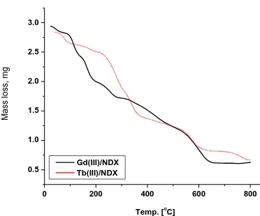
The obtained Gd(III) and Tb(III) NDX complexes were studied by thermo gravimetric and its differential analysis (TG/DTG/DTA) from ambient temperature to 800 oC under N2 atmospheres. The
TG curves were redrawn as mg mass loss versus temperature and DTG curves were redrawn as rate of loss of mass versus temperature. The thermal decomposition curves (TG/DTG/DTA) are given in Figs. (5-6).
3.5.1. [Gd(NDX)3].6H2O complex
Thermal decomposition of Gd(III)/NDX complex with general formula [Gd(NDX)3].6H2O
occurs at six steps. The first decomposition step take place at a temperature range of 35-120 oC at DTGmax=86 oC and it correspond to the loss of one of hydrated water molecule with an observed
weight loss 2.00% (calcd.=1.88%). The second step occur within a temperature range 120-290 oC at DTGmax=144, 222 oC which assigned to the loss of five molecule of hydration water + three
3-methyl-pyridine molecules with a weight loss (obs.= 39.00%, calcd.= 38.52%). The third-six steps occur within a temperature range 290-800 oC at DTGmax= (302, 571 and 605) oC and DTA= (570 and 601) oC
(exo) which assigned to the decomposition of ethyl-pyridin-4-one carboxylic acid organic moiety with a weight loss (obs.= 40.00%, calcd.= 40.70%). The ½Gd2O3 is the final product remains stable till 800 o
C as final residual with mass percentage (obs= 19.00%, calcd.= 18.90%).
3.5.2. [Tb(NDX)3].6H2O complex
The thermal decomposition of Tb(III)/NDX complex with the general formula [Gd(NDX)3].6H2O occurs at consecutive and very evenly seven steps. The first decomposition step
presence at a temperature range of 30-70 oC at DTGmax= 75 oC and it correspond to the loss of 2H2O
hydrated molecules with an observed weight loss 3.50% (calcd.= 3.75%). The second step occur within a temperature range 70-180 oC at DTGmax= 176 oC and DTA=216 oC which assigned to the loss
of 5H2O hydrated water molecules + three terminal ethyl groups of the ethyl-pyridin-4-one carboxylic
acid organic moiety with a weight loss (obs.=16.00%, calcd.=16.24%). The third-to-seven degradation steps occur within a temperature range 180-800 oC at DTGmax = (276, 336, 439, 568 and 765) oC and
product remains stable till 800 oC as final residual with mass percentage (obs= 19.04%, calcd.= 18.97%).
0 200 400 600 800
0.5 1.0 1.5 2.0 2.5 3.0
Temp. [o
[image:10.596.204.390.127.282.2]C] Ma ss lo ss, mg Gd(III)/NDX Tb(III)/NDX
Figure 5. TG curves of Gd(III) and Tb(III)/NDX complexes
0 200 400 600 800
-0.0020 -0.0015 -0.0010 -0.0005 0.0000 m g /s e c
Temp. [oC]
D T A ( u V ) DTG DTA 0 4 8 12 16 20 24 28 A
0 100 200 300 400 500 600 700 800
-0.0016 -0.0012 -0.0008 -0.0004 0.0000 m g /s e c
Temp. [oC]
DTG DTA D T A ( u V ) 0 4 8 12 16 20 B
[image:10.596.165.438.334.717.2]
3.6. Kinetic studies
The determination of the rate-dependent parameters of solid-state non-isothermal decomposition reactions were operated using analysis of TG curves [46-52]. Most commonly used methods are the differential method of Freeman and Carroll [46] integral method of Coat and Redfern [47] and the approximation method of Horowitz and Metzger [50]. In the present paper, the general thermal behaviors of the Gd(III)/Tb(III)-NDX complexes in terms of stability ranges, peak temperatures and values of kinetic parameters are discussed. The kinetic parameters have been evaluated using the Coats-Redfern equation:
2 1*
n exp( )d
0 (1- )
d T T RT E A t
(eq. 1)
This equation on integration gives;
]
ln[
]
ln[
* * 2 ) 1 ln( E AR RT E T
(eq. 2)A plot of left-hand side (LHS) against 1/T was drawn. E* is the energy of activation in J mol-1 and calculated from the slop and A in (s-1) from the intercept value. The entropy of activation ΔS* in (JK-1mol-1) was calculated by using the equation:
ΔS*
= R ln(Ah/kB Ts) (eq. 3)
Where kB is the Boltzmann constant, h is the Plank’s constant and Ts is the DTG peak
temperature [52].
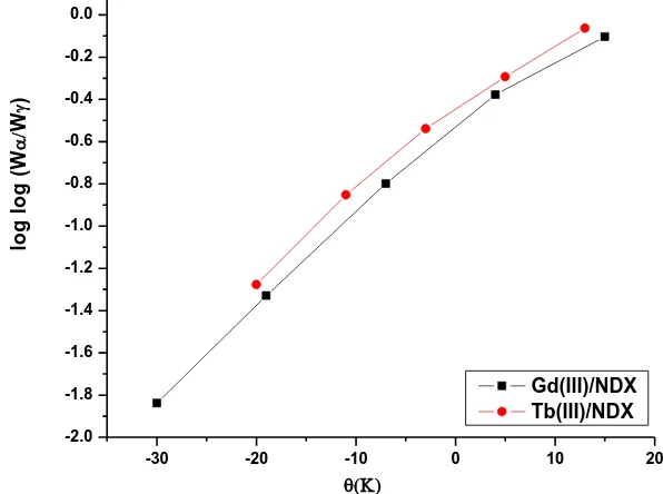
The Horowitz-Metzger equation is an illustrative of the approximation methods. log[{1-(1-α)1-n}/(1-n)] = E*θ/2.303RTs2 for n≠1 (eq. 4)
When n = 1, the LHS of equation 4 would be log[-log (1-α)]. For a first-order kinetic process the Horowitz-Metzger equation may be written in the form:
log[log(wα / wγ)] = E*θ/2.303RTs2 – log2.303 (eq. 5)
Where θ = T- Ts, wγ = wα – w, wα = mass loss at the completion of the reaction; w = mass loss
up to time t. The plot of log[log(wα / wγ)] vs θ was drawn and found to be linear from the slope of
which E* was calculated. The pre-exponential factor, A, was calculated from the equation: E* /RTs2 = A/[ φ exp(-E*/RTs)] (eq. 6)
The entropy of activation, ΔS*, was calculated from equation 3. The enthalpy activation, ΔH*, and Gibbs free energy, ΔG*, were calculated from;
ΔH*
= E* – RT and ΔG* = ΔH* – TΔS* (eq. 7)
Herein, the thermal behaviors of the nalidixic acid complexes in terms of stability ranges, peak temperatures and values of kinetic parameters, are shown in Table 3 and Figs. 7-8. The kinetic parameters have been evaluated using the Coats–Redfern and Horowitz-Metzger equations. From the kinetic and thermodynamic data resulted from the TGA curves and tabulated in Table 3, the following outcome can be discussed as follows:
- The higher values of activation energies of the NDX complexes led to thermal stability of the studied complexes.
0.0022 0.0023 0.0024 0.0025 0.0026 0.0027 0.0028 -15.5
-15.0 -14.5 -14.0 -13.5 -13.0 -12.5 -12.0 -11.5 -11.0
ln
(-ln
(1-
)
2 )
1000/T (K) Gd(III)/NDX
[image:12.596.142.454.89.337.2]Tb(III)/NDX
Figure 7. Plots of Coats–Redfern (CR) relation for Gd(III) and Tb(III)/NDX complexes
-30 -20 -10 0 10 20
-2.0 -1.8 -1.6 -1.4 -1.2 -1.0 -0.8 -0.6 -0.4 -0.2 0.0
lo
g
l
o
g
(
W
W
)
Gd(III)/NDX Tb(III)/NDX
Figure 8. Plots of Horowitz–Metzger (HM) relation for Gd(III) and Tb(III)/NDX complexes
- It is clear that the thermal decomposition process of most NDX complexes is non-spontaneous (ΔS* = negative value), i.e., the complexes are thermally stable.
[image:12.596.147.445.400.622.2]
- The values of activation energy of the second stages of decomposition are found to be higher than that of the first stages, while that of the third stage are found to be higher than that of the second stage and so. This may indicate that the rate of decomposition of the second stage is lower than that of the first stage, whereas that of the third stage is lower than that of the second stage. This may be attributed to the structural rigidity of the ligand as compared with H2O and methyl groups, which
[image:13.596.51.548.234.351.2]requires more energy for its rearrangement before undergoing any compositional change.
Table 3. Kinetic parameters using the Coats–Redfern (CR) and Horowitz–Metzger (HM) equations for the Gd(III) and Tb(III)/NDX complexes
Complex Method
Parameter r
E (J mol-1)
A (s-1)
ΔS
(J mol-1 K
-1
)
ΔH (J mol-1)
ΔG (J mol-1) Gd(III) CR
HM 7.00E+04 7.10E+04 2.81E+08 4.23E+09 -8.50E+01 -6.22E+01 6.78E+04 7.10E+04 9.50E+04 9.21E+04 0.9921 0.9861 Tb(III) CR
HM 5.12E+04 5.45E+04 5.30E+04 4.62E+05 -1.61E+02 -1.44E+02 4.77E+04 5.30E+04 1.00E+05 1.02E+05 0.9902 0.9861 r = correlation coefficient of the linear plot.
3.7. XRD and SEM studies
0 10 20 30 40 50 60 70
0 5 10 15 20 25 30 C o u n ts 2
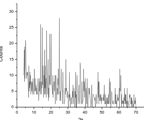
Figure 9. The XRD spectrum of Gd2O3.
The x-ray powder diffraction patterns (Fig. 9) in the range of 20o < 2θ < 70o for the residual products of thermal decomposition at 800 oC which resulted from the interaction between GdCl3 or
TbCl3 with carbamide were carried in order to obtain an idea about the lattice dynamics of the resulted
[image:13.596.181.418.431.628.2]
prominent intensity peak, relative intensity and particle size of Gd(III) oxide as a residual product have 24O, 0.2475, 100 and 6 nm, respectively. The crystallite size of the Gd(III) oxide could be estimated from XRD patterns by applying FWHM of the characteristic peaks using Deby-Scherrer equation 8 [53].
D= Kλ/βCosθ (eq. 8)
Where D is the particle size of the crystal gain, K is a constant (0.94 for Cu grid), λ is the x-ray wavelength (1.5406 Ǻ), θ is the Bragg diffraction angle and β is the integral peak width. The particle size was estimated according to the highest value of intensity compared with the other peaks. The particle size for the Gd(III) thus obtained was 6.0 nm. These data gave an impression that the particle size located within nano scale range.
[image:14.596.174.420.264.456.2]
Figure 10. SEM images of Gd(III)/NDX complexes synthetic by un-conventional method.
[image:14.596.143.450.515.740.2]
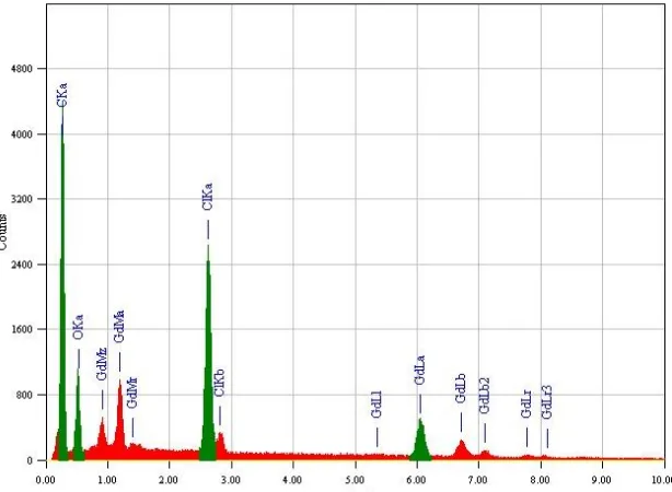
Scanning electron microscopy is a simple tool used to give an impression about the microscopic aspects of the physical behavior of the Gd(III)/NDX complex formation (Fig. 10). Energy Dispersive X-ray Detection (Fig. 11) was also operated to check the elements which consistent of gadolinium(III) oxide resulted from the second step for the method of nano–particles preparation. Although this tool is not a qualified method to confirm complex formation but it can be a reference to the presence of a single component in the synthetic products.
3.8. Microbiological investigation
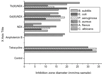
Regarding the inhibition zone diameter, Gd(III)/Tb(III)-NDX complex in nano scale has highest antimicrobial activity against all target organisms compared to other which prepared by traditional methods (Fig. 12). Also, Gd(III)/Tb(III)/NDX compounds in nano range gained approximately twice percentage rather than traditional method of preparation in comparable with antibacterial agent (Tetracycline) and antifungal agent (Amphotericin B). The most reasons for lethal action of tested compounds may due to their interactions with critical intracellular sites causing the death of cells [54]. The variety of antimicrobial activities of tested compounds may due to a different degree of tested compounds penetration through cell membrane structure of target organism. In conclusion, the interactions between the gadolinium(III) and terbium(III) ions in nano–structural form resulting developing of the effectiveness of biological characters of Gd(III) and Tb(III) ions antibiotics.
Control Tetracycline Amphotericin B NDX Gd(III)/NDX Tb(III)/NDX
0 5 10 15 20 25 30 35
Inhibition zone diameter (mm/mg sample)
X
Axi
s
T
it
le
B. subtilis E. coli
P. aeruginosa S. aureus A. flavus C. albicans
[image:15.596.124.469.447.700.2]
References
1. A. Albert, The Physico-Chemical Basis of Therapy: Selective Toxicity, 6th ed., Chapman & Hall, London, (1979).
2. M.N. Hughes (Ed.), The Inorganic Chemistry of Biological Processes, 2nd ed., Wiley, New York, (1981).
3. J. Liu, E.B. Wang, J. , Peng, and Y.S. Zhou, J. Rare Earths, 17, (1999) 139. 4. S. Chakrabarti, D. Dasgupta and D. Bhattacharyya, J. Biol. Phys., 26, (2000) 203.
5. G.A. Smita, Ranjit and V. Vikrant, International J. Pharm. Res. Development, 3, (2011) 164-171. 6. A.R. Shaikh, R. Giridhar, and M.R. Yadav, International J. Pharmaceutics, 332 (2007) 24-30. 7. I. Turel, The interaction of metal ions with quinolone antibacterial agents. Coord. Chem. Reviews,
232 (2002) 27-47.
8. G Psomas, T. Alketa K. E. Efthimiadou, S. Yiannis, P. R Catherine and K. Nikos, J. Inorg. Biochem., 100 (2006) 1764–1773.
9. E.K. Efthimiadou, T. Hellinida, S. Yiannis, C. P. Raptopoulou and A. S. Katsaros, Alexandra Karaliota, George Psomas, J. Inorg. Biochem., 101, (2007) 64–73.
10. E. K. Efthimiadou and K. Alexandra, P. George, Polyhedron, 27 (2008) 1729–1738. 11. A. Kumar and S.Y. Danveer, Int. J. Chem. Sci., 7(1) (2009)7-19.
12. K.C. Skyrianou, V. Psycharis, C.P. Raptopoulou, D.P. Kessissoglou, and G. Psomas, J. Inorg. Biochem., 105 (2011) 63–74.
13. A.M.A. Adam, J. Mat. Sci. Res., 1 (2012).
14. P. Živec, P. Franc, T.G. Iztok, P. Gerald, P. George, J. Inorg. Biochem., 117 (2012) 35–47.
15. M. A. Marianthi, G. Andreadou P. R, Catherine. Psycharis, Anastasia A. Pantazaki, George Psomas, J. Inorg. Biochem., 121 (2013) 88–99.
16. M.N.Patel, R. P. Chintan, N. J. Hardik, Inorg. Chem. Commun., 27 (2013) 51–55 17. D.E King., R. Malone and, S.H. Lilley, Am. Fam. Physician, 61(2000) 2741–2748. 18. V.T. Andriole, (Ed.), The Quinolones, 3rd Ed, Academic Press, San Diego, (2000). 19. G.Y. Lesher, and M.D. Gruett, Belg. Pat.,( 1962) 612 258.
20. E. Ruzicka, J. Lasovsky and P. Brazdil, Chem. Zvesti, 29 (1975) 517. 21. K. Timmers, R. Sternglanz, Bioinorg. Chem., 9 (1978) 145.
22. M. Nakano, M. Yamamoto and T. Arita, Chem. Pharm. Bull., 26 (19781) 1505.
23. W.R. Vicent, S.G. Schulman, J.M. Midgley, W. J. Van Ort and R.H.A. Sorel, Int. J. Pharm., 9 (1981) 191.
24. S. Panadero, A.H. Gómez and D.B. Pérez, Anal. Chim. Acta, 303 (1995) 39.
25. J. Kljun, A.K. Bytzek, W. Kandioller, C. Bartel, M.A. Jakupec, C.G. Hartinger, B.K. Keppler and I. Turel, Organometallics, 30 (2011) 2506.
26. M.S. Refat, Synth. React. Inorg. Met.-Org. Chem., 34(9) (2004) 1605. 27. S.M. Teleb and M.S. Refat, Bull. Chem. Tech. Maced., 25(1) (2006) 57.
28. Yu. Ya. Kharitonov, K.S. Sulaimankulov and N.B. Khudaibergenova, Zh. Neorg. Khim., 35 (8) (1990) 2117.
29. C.V. Deshpande and V.R. Rao, J. Indian Inst. Sci., 69(5) (1989) 329.
30. A.W. Bauer, W.M. Kirby, C. Sherris and M. Turck, Amer. J. Clinical Pathology, 45 (1966) 493. 31. M.A. Pfaller, L. Burmeister, M.A. Bartlett and M.G. Rinaldi, J. Clin. Microbiol. 26 (1988) 1437. 32. National Committee for Clinical Laboratory Standards, Performance Vol. antimicrobial
susceptibility of Flavobacteria, (1997).
34. National Committee for Clinical Laboratory Standards. (2002). Reference Method for Broth Dilution Antifungal Susceptibility Testing of Conidium-Forming Filamentous Fungi: Proposed Standard M38-A. NCCLS, Wayne, PA, USA.
35. National Committee for Clinical Laboratory Standards. (2003). Methods for Antifungal Disk Diffusion Susceptibility Testing of Yeast: Proposed Guideline M44-P. NCCLS, Wayne, PA, USA. 36. L.D. Liebowitz, H.R. Ashbee, E.G.V. Evans, Y. Chong, N. Mallatova, M. Zaidi, D. Gibbs, and
Global Antifungal Surveillance Group. Diagn. Microbiol. Infect. Dis, 4 ( 2001) 27.
37. M.J. Matar, L. Ostrosky-Zeichner, V.L. Paetznick, J.R. Rodriguez, E. Chen, J.H. Rex, Antimicrob. Agents Chemother., 47 (2003) 1647.
38. W.J. Geary, Coord. Chem. Rev., 7 (1971) 81.
39. T. Vogel, Textbook of Practical Organic Chemistry. 4th Edn., John Wiley Inc., England, (1989) 133. 40. A.V. Polishchuk, E.T. Karaseva, T.B. Emelina, M.N. Yu and V.E. Karasev, J. Stru. Chem., 50(3)
(2009) 434.
41. M.S. Refat, Spectrochimica Acta Part A, 68 (2007) 1393.
42. I. Turel, I. Leban and N. Bukovec, J. Inorg. Biochem., 66 (1997) 241. 43. G.B. Deacon and R.J. Phillips, Coord. Chem. Rev., 33 (1980) 227.
44. I. Turel, L. Golic and O.L.R. Ramirez, Acta Chem. Slov., 46(2) (1999) 203.
45. C.M. Riley, D.L. Ross, D. Van der Velde and F. Takusagawa, J. Pharm. Biomed. Analysis, 11 (1993) 49.
46. E.S. Freeman and B. Carroll, J. Phys. Chem, 62 (1958) 394. 47. A.W. Coats and J.P. Redfern, Nature, 201 (1964) 68.
48. T. Ozawa, Bull. Chem. Sot. Jpn, 38 (1965) 1881.
49. W.W. Wendlandt, Thermal Methods of Analysis, Wiley, New York, (1974). 50. H.W. Horowitz and G. Metzger, Anal. Chem., 35 (1963) 1464.
51. J.H. Flynn, and L.A. Wall, Polym. Lett., 4 (1966) 323. 52. P. Kofstad, Nature, 179 (1957) 1362.
53. C.X. Quan, L.H. Bin and G.G. Bang, Mater. Chem. Phys. 91 (2005) 317.
54. S.D. Cox, C.M. Mann, J.L.Markham, J.E.Gustafson, J.R.Warmington and S.G.Wyllie, Molecules, 6 (2001) 87.
![Figure 4. Suggestion structure of [M(NDX)3].6H2O complexes (where, M= Gd(III) or Tb(III))](https://thumb-us.123doks.com/thumbv2/123dok_us/1900225.148049/8.596.172.426.235.470/figure-suggestion-structure-ndx-complexes-gd-iii-iii.webp)
![Poly[[tetraaquatetrakis[μ3 5 (pyridine 4 carboxamido)isophthalato]nickel(II)diterbium(III)] tetrahydrate]](data:image/gif;base64,R0lGODlhAQABAIAAAP///wAAACH5BAEAAAAALAAAAAABAAEAAAICRAEAOw==)