White Rose Research Online URL for this paper:
http://eprints.whiterose.ac.uk/98717/
Version: Accepted Version
Article:
Elies, J, Johnson, E, Boyle, JP et al. (2 more authors) (2015) H2S does not regulate
proliferation via T-type Ca2+ channels. Biochemical and Biophysical Research
Communications, 461 (4). pp. 659-664. ISSN 0006-291X
https://doi.org/10.1016/j.bbrc.2015.04.087
© 2015, Elsevier. Licensed under the Creative Commons
Attribution-NonCommercial-NoDerivatives 4.0 International
http://creativecommons.org/licenses/by-nc-nd/4.0/
[email protected] https://eprints.whiterose.ac.uk/
Reuse
Unless indicated otherwise, fulltext items are protected by copyright with all rights reserved. The copyright exception in section 29 of the Copyright, Designs and Patents Act 1988 allows the making of a single copy solely for the purpose of non-commercial research or private study within the limits of fair dealing. The publisher or other rights-holder may allow further reproduction and re-use of this version - refer to the White Rose Research Online record for this item. Where records identify the publisher as the copyright holder, users can verify any specific terms of use on the publisher’s website.
Takedown
If you consider content in White Rose Research Online to be in breach of UK law, please notify us by
H
2S does not regulate proliferation via T-type Ca
2+channels
Jacobo Elies, Emily Johnson, John P Boyle, Jason L Scragg & Chris Peers*
Division of Cardiovascular and Diabetes Research, LICAMM,
Faculty of Medicine and Health, University of Leeds, Leeds LS2 9JT, U.K.
Running title: Hydrogen sulfide, proliferation and T-type Ca2+channels
*To whom correspondence should be addressed:
Prof Chris Peers, Division of Cardiovascular and Diabetes Research, LICAMM, Faculty of
Medicine and Health, University of Leeds, Clarendon Way, Leeds LS2 9JT, U.K.
ABSTRACT
T-type Ca2+ channels (Cav3.1, 3.2 and 3.3) strongly influence proliferation of various cell types,
including vascular smooth muscle cells (VSMCs) and certain cancers. We have recently shown
that the gasotransmitter carbon monoxide (CO) inhibits T-type Ca2+ channels and, in so doing,
attenuates proliferation of VSMC. We have also shown that the T-type Ca2+ channel Cav3.2 is
selectively inhibited by hydrogen sulfide (H2S) whilst the other channel isoforms (Cav3.1 and
Cav3.3) are unaffected. Here, we explored whether inhibition of Cav3.2 by H2S could account for
the anti-proliferative effects of this gasotransmitter. H2S suppressed proliferation in HEK293 cells
expressing Cav3.2, as predicted by our previous observations. However, H2S was similarly
effective in suppressing proliferation in wild type (non-transfected) HEK293 cells and those
expressing the H2S insensitive channel, Cav3.1. Further studies demonstrated that T-type Ca2+
channels in the smooth muscle cell line A7r5 and in human coronary VSMCs strongly influenced
proliferation. In both cell types, H2S caused a concentration-dependent inhibition of proliferation,
yet by far the dominant T-type Ca2+ channel isoform was the H
2S-insensitive channel, Cav3.1.
Our data indicate that inhibition of T-type Ca2+ channel-mediated proliferation by H 2S is
independent of the channels’ sensitivity to H2S.
Key words:
1. INTRODUCTION
In recent years ion channels have emerged as a major family of target proteins for modulation by
the gasotransmitters carbon monoxide (CO) and hydrogen sulfide (H2S) [1-4]. Indeed, many
beneficial and detrimental actions of these gases involve ion channel modulation [5-7]. One
particularly important cellular process that involves ion channel activity and is also modulated by
gasotransmitters is proliferation: in the vasculature, for example, vascular smooth muscle cells
(VSMCs) can undergo phenotypic change, becoming non-contractile, proliferative cells to adapt
to varying physiological and pathological situations [8-10]. This is important not only in
developmental vasculogenesis and vascular repair but also in the development of cardiovascular
diseases [8; 11; 12]. Progression of cancers is also dependent on profound cellular proliferation
[13].
Interestingly, induction of heme oxygenase-1 (HO-1), which generates CO along with biliverdin
and iron from the degradation of heme, is associated with proliferative vascular diseases [14; 15]
and much evidence suggests that CO accounts for the known anti-proliferative effects of HO-1 in
VSMCs [16-18]. HO-1 is also constitutively expressed in various types of cancer, where it may
regulate proliferation and resistance to apoptosis, in part through formation of CO [19; 20]. By
contrast, the effects of H2S on proliferation appear to be cell-type specific; In vitro studies have
shown that H2S donors such as NaHS slow proliferation of VSMCs [21] yet can increase
endothelial cell proliferation [22], and in some forms of cancer, such as colon cancer, H2S
promotes proliferation [23].
Ca2+ influx into cells is a requirement for proliferation as it regulates the activity of key
transcription factors such as NFAT (nuclear factor of activated T-cells), via Ca2+-dependent
dephosphorylation by calcineurin [24]). The relative importance of different Ca2+influx pathways
contributing to proliferation are currently under investigation but there is compelling evidence for
the involvement of voltage-gated T-type Ca2+ channels: in VSMCs, T-type Ca2+ channel
expression increases during proliferation [25; 26], and they are required for VSMC proliferation
both in vitro and in neointima formation observed following vascular injury [26-30]. In numerous
forms of cancer high expression of T-type Ca2+channels has been observed and, as in VSMCs,
their expression supports proliferation [31]. These channels therefore represent an important
therapeutic target for the treatment of both proliferative vascular diseases and cancer.
We have previously reported that CO is an effective inhibitor of all three isoforms of T-type Ca2+
proliferation via CO-mediated inhibition of T-type Ca2+ channels [5]. More recently, we have
demonstrated that H2S can also inhibit T-type Ca2+ channels, but differs from CO in that it
discriminates between subtypes; it is only effective in inhibiting Cav3.2, whilst Cav3.1 and
Cav3.3 are unaffected by this gasotransmitter [33]. Given the known effects of H2S on
proliferation and the important involvement of T-type Ca2+ channels in this process, we have
explored the possibility that H2S, like CO, may regulate proliferation via inhibition of T-type Ca2+
channels.
2. METHODS
2.1 Cell culture
HEK293 cells: Wild type (WT; untransfected) HEK293 cells were cultured in minimum essential
medium containing Earle’s salts and L-glutamine, and supplemented with 10% (v/v) foetal
bovine serum (FBS; Biosera, Ringmer UK), 1% (v/v) non-essential amino acids, 1% (v/v)
antibiotic/antimycotic, and 0.1% (v/v) gentamicin. HEK293 cells stably expressing Cav3.1 and
Cav3.2 T-type Ca2+ channels (a kind gift from Prof. E. Perez-Reyes; University of Virginia,
Virginia USA), were cultured in WT HEK293 media, additionally supplemented with 1mg/ml
G-418 to maintain selection pressure (All reagents from Gibco, Paisley UK; unless otherwise
stated). HEK293/Cav3.2 cells were used at passages between P1 and P8, and WT HEK293
cells were used at passages between P1 and P6.
A7r5 cells (a smooth muscle cell line derived from rat thoracic aorta) were obtained from the
European Collection of Cell Cultures (ECACC, Public Health England, Porton Down UK). They
were grown in A7r5 complete media, consisting of Dulbecco’s minimum essential medium
containing 10% FBS (Biosera, Ringmer UK) and 1% glutamax (Gibco, Paisley UK).
Human coronary artery smooth muscle cells (hCASMCs) were obtained from ECACC (350-05a,
Public Health England, Porton Down UK). They were grown in smooth muscle growth medium-2
supplemented with 5% FBS, growth factors (0.001% hEGF, 0.001% insulin, 0.002% hFGF-B)
and gentamicin/amphotericin-B as described by manufacturers (Clonetics™ from Lonza,
Germany). HCASMC were used at passages between P1 and P5.
All cell types were cultured in a humidified incubator at 37˚C (95% air: 5% CO2) and passaged
weekly.
2.2 Proliferation Assay
Cells were plated at 1x10⁴ / well and allowed to adhere for 6 hours in 24-well plates in complete
growth media, then exposed to serum free medium (SFM) overnight. On day 0 of the assay,
SFM was removed and replaced with 1ml of the relevant complete test media (vehicle or drug at
Dulbecco’s phosphate buffered saline (PBS) and 200µl of 0.05% trypsin-EDTA (Gibco, Paisley
UK) was added (pre-warmed to 37˚C). Post-incubation, 800µl of complete media was added and
the cell suspension centrifuged (600gfor 6 minutes). Following removal of 950µl of media, 50µl
of supernatant remained with the cell pellet, which was then re-suspended following addition of
50µl of 0.4% Trypan Blue (Thermo Scientific, Rockford USA) to exclude non-viable cells.
Media was retained from one well of each treatment, processed in the same manner as the cell
samples, and any cells present were counted as an additional quantification of non-viable cells.
Day 0 counts and media counts were performed using a hemocytometer. All other counts were
performed using a TC10 Automated Cell Counter (Bio-Rad, Hemel Hempstead UK). Repeated
counting from both test medium and trypsin suspension showed that no cells were lost in the
counting procedure.
2.3 Real-Time Polymerase Chain Reaction (RT-PCR)
To determine mRNA expression levels of Cav3.2 and Cav3.1 channels, T75 flasks containing
cells at 70-80% confluence were washed with PBS and cells dissociated using 0.5ml 0.05%
trypsin-EDTA for 3 minutes (37˚C; 95% air: 5% CO2). Enzyme activity was halted by adding
0.5ml ice-cold PBS. The cell suspension was then centrifuged (600g for 6 min) and RNA was
generated from whole cell lysates using the Aurum Total RNA Mini Kit (Bio-Rad, Hemel
Hempstead UK) following manufacturer’s instructions. A cDNA template was generated from
RNA samples using the iScript cDNA Synthesis Kit (Bio-Rad, Hemel Hempstead UK) following
manufacturer’s instructions (Reaction profile: 5 minutes at 25˚C, 30 minutes at 42˚C, 5 minutes at 85˚C, 5 minutes at 4˚C). Human Taqman probes (Applied Biosystems (ABI), UK) for Cav3.1
(CACNA1G), Cav3.2 (CACNA1H), and the endogenous housekeeper hypoxanthine
phosphoribosyltransferase (HPRT1) were used with hCASMC. In all cases, 2 l of sample cDNA and 18 l of RT-PCR reaction mix (10 l Taqman Universal PCR Master Mix, 0.5 l Taqman probes (both from ABI), and 7.5 l RNase/DNase-free water (Gibco Cambridge UK)) was added
to the required wells of a 96-well PCR plate (Applied Biosystems, Cambridge UK). RT-PCR was
carried out using an ABI 7500 Real-Time PCR system (Reaction profile: 2 minutes at 50˚C, 10 minutes at 95˚C, 15 seconds at 95˚C for 60 cycles, 1 minute at 60˚C). Data were analysed using
the 7500 software (ABI) and relative gene expression calculated using the 2- CT method with
HPRT1 as the endogenous control.
2.4 Electrophysiology
Ca2+ currents were recorded from A7r5 cells using the whole-cell configuration of the
patch-clamp technique at room temperature (21-24oC) as previously described [32] using an Axopatch
200A amplifier/Digidata 1300 interface controlled by Clampex 9.0 software (Molecular Devices,
Sunnyvale, CA, USA). Offline analysis was performed using Clampfit 9.0. Pipettes (4–6 M )
(adjusted with CsOH). To optimise recording of T-type Ca2+currents, cells were perfused with (in
mM): NaCl 95, CsCl 5, MgCl2 0.6, CaCl2 15, TEA-Cl 20, HEPES 5, D-glucose 10, sucrose 30,
pH 7.4 (adjusted with NaOH). Cells were voltage-clamped at -80mV and either repeatedly
depolarized to -20mV (200ms, 0.1Hz) or to a series of test potentials ranging from -100mV to
+60mV. All currents were low-pass filtered at 2kHz and digitised at 10kHz.
2.5 Data presentation and statistical analysis
Proliferation data are plotted example growth curves (with s.e.m., as each was performed in
triplicate) and bar graphs representing normalised mean (with s.e.m.) proliferation on the final
day of assessment, determined in at least 3 identical experiments. Statistical comparisons were
made using ANOVA with Dunnett’s post-hoc test.
RESULTS AND DISCUSSION
There is overwhelming evidence that H2S is an important modulator of both physiological and
pathological cardiovascular function. In addition to its tonic, physiological role as a regulator of
blood pressure [34], it is also a major modifier of tissue remodelling resulting from cardiovascular
diseases. Thus, for example, cardiac arteriolar hypertrophy and interstitial fibrosis observed in
spontaneously hypertensive rats was prevented by daily administration of the H2S donor NaHS
[35]. Furthermore, neointima formation and VSMC proliferation following carotid artery balloon
injury was suppressed following chronic NaHS administration [36]. Interestingly, this study also
demonstrated that expression of cystathionine -lyase (CSE), the major vascular enzyme
producing H2S, was inhibited by balloon injury, a finding in agreement with its down-regulation in
hypertension [37]. Since (a) the expression of T-type Ca2+ channels increases in VSMC
proliferation [25; 26], (b) they are a prerequisite for VSMC proliferation and neointima formation
following vascular injury [26-30] and (c) we have recently demonstrated that H2S regulates the
activity of the T-type Ca2+ channel Cav3.2 [33], the present study was conducted in order to
investigate whether the inhibitory effects of H2S on proliferation might be mediated via T-type
Ca2+channel inhibition. Our investigation was prompted not only by the importance of H 2S and
T-type Ca2+ channels in VSMC proliferation, but also by the fact that CO, another
gasotransmitter known to inhibit VSMC proliferation, appears to act in this way via inhibition of
T-type Ca2+channels [5].
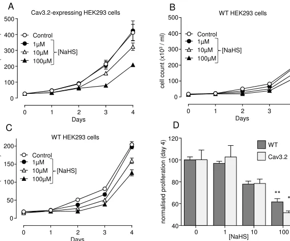
Proliferation was firstly monitored in HEK293 cells over-expressing the H2S-sensitive T-type Ca2+
channel Cav3.2 [33]. Over a 4 day period, the increase in cell number was reduced in a
concentration-dependent manner by H2S (applied as the donor NaHS; Fig. 1A), consistent with
the known ability of H2S to inhibit this class of T-type Ca2+ channel. As previously described [5],
much greater than that observed in wild-type (WT; untransfected) HEK293 cells (Fig. 1B, plotted
on the same Y axis scale as Fig. 1A for comparison). However, further reductions in this modest
rate of proliferation were observed in WT cells in the presence of NaHS (Fig. 1B); these effects
are more apparent when WT proliferation is plotted on a more restricted scale (Fig. 1C). Indeed,
the degree of inhibition of proliferation caused by NaHS was not significantly different between
WT and Cav3.2-expressing HEK 293 cells (Fig. 1D), as compared on day 4. This finding
suggests that H2S may not in fact inhibit proliferation specifically through inhibiting this class of
T-type Ca2+ channel. To explore this possibility further, we examined proliferation in HEK293
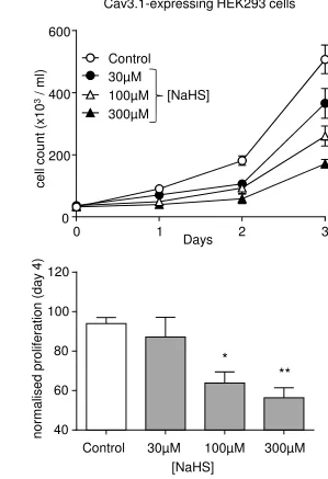
cells stably expressing the H2S insensitive T-type Ca2+ channel, Cav3.1 [33]. Our previous
studies have indicated that current densities in the Cav3.1 and Cav3.2-expressing HEK293 cells
are similar in magnitude (ca. 50-100pA/pF [32; 38]) and T-type currents are not detectable in WT
cells (data not shown). Proliferation in these Cav3.1-expressing cells was rapid and monitored
over a 3 day period. As shown in Fig. 2, H2S also reduced proliferation in these cells in a
concentration-dependent manner.
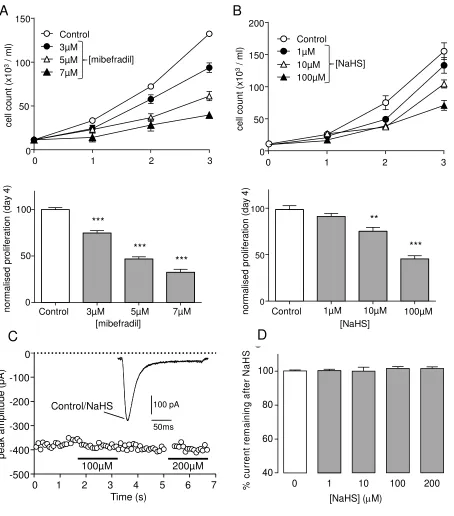
To explore any potential modulation of native T-type Ca2+ channels in VSMCs, and how this
might impact on proliferation, we first explored its action in the rat aortic smooth muscle cell line,
A7r5. We have previously shown that T-type (and not L-type) Ca2+ channels regulate
proliferation in these cells [5] and, consistent with this, we found that the T-type Ca2+ channel
inhibitor mibefradil inhibited proliferation in a concentration-dependent manner (Fig. 3A).
Exposure of cells to NaHS similarly reduced proliferation in a concentration-dependent manner
(Fig. 3B). Our recent work has suggested that A7r5 cells express both Cav3.1 and Cav3.2, but
by far the predominant channel was Cav3.1, as determined via RT-PCR [5]. To examine directly
whether T-type currents could be modulated by H2S in A7r5 cells, we recorded whole-cell Ca2+
currents under conditions designed to optimise T-type Ca2+ channel resolution (see [5] and
Methods). Under these conditions, NaHS was without effect on T-type currents, as exemplified
by Fig. 3B, and quantified in Fig. 3C. We never observed significant modulation of currents (n =
5 cells at each concentration examined). These findings strongly suggest that the ability of H2S
to inhibit A7r5 proliferation does not occur via its ability to inhibit T-type Ca2+channels expressed
in these cells.
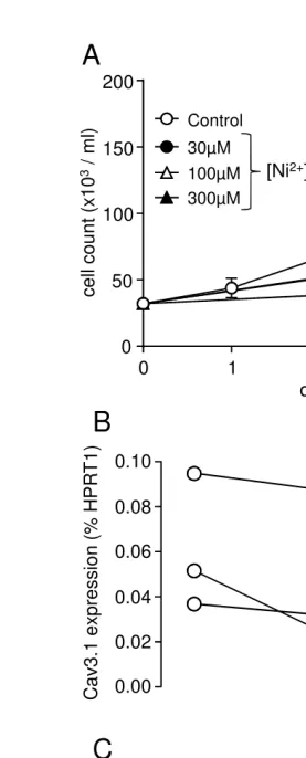
We also explored modulation of proliferation in human coronary artery smooth muscle cells
(hCASMCs). Consistent with a role for T-type Ca2+ channels in proliferation, we found that Ni2+
caused a concentration dependent reduction in hCASMC proliferation, as monitored over 4 days
(Fig. 4A). In three repeated experiments (not shown) Ni2+ only significantly (P<0.01) reduced
proliferation at > 100M, suggesting the involvement of Cav3.1 rather than Cav3.2, since Cav3.2
is much more sensitive to Ni2+ [39]. In agreement with this suggestion, we next examined the
relative expression of mRNA for the T-type Ca2+channel isoforms, Ca
RT-PCR. In three separate experiments, the Cav3.1 isoform was expressed at significantly higher
levels than the Cav3.2 isoform, but both isoforms were detected (Fig. 4B; note different scales
for each isoform). Despite the predominant expression of the H2S insensitive channel Cav3.1,
proliferation was reduced in a concentration-dependent manner by NaHS exposure (Fig. 4C). In
three repeated experiments (not shown) the effects of NaHS were significant (P<0.001) at
100M and 300M. The data in both A7r5 and hCASMCs are consistent with the idea that H2S
suppresses proliferation independently of T-type Ca2+channel modulation.
Our findings confirm and extend previous awareness that T-type Ca2+channel activity promotes
proliferation, as observed when over-expression of either Cav3.1 or Cav3.2 increases HEK293
cell proliferation (Figs 1 and 2), as we and others have shown previously [5; 40]. We also
confirm that T-type Ca2+ channels modulate proliferation in A7r5 cells, and provide new data
suggesting a similar role in hCASMCs (Fig. 4). At present, the mechanism by which T-type Ca2+
channels, specifically, can promote proliferation is not understood. In each cell type studied, we
also demonstrate that H2S inhibits proliferation, consistent with a previous report in rat aortic A10
smooth muscle [21]. Our results are also consistent with the observation that VSMCs isolated
from CSE-/- mice show increased proliferation compared to wild type (WT) VSMCs, further
indicating that H2S has a ‘breaking’ effect on proliferation [41]. The major and unexpected finding
of the present study, however, is that H2S appears to suppress proliferation independent of any
action on T-type Ca2+ channels. Thus, although the inhibition of proliferation in Cav3.2
expressing HEK293 cells by H2S (Fig. 1) is consistent with its ability to inhibit this
pro-proliferative channel, a similar degree of inhibition was also observed in WT cells which did not
express Cav3.2. Furthermore, H2S also inhibited proliferation of Cav3.1-expressing HEK293
cells (Fig. 2), despite the fact that this channel is insensitive to H2S [33], and in A7r5 and
hCASMCs, where the dominant T-type Ca2+ channel expressed is Cav3.1. It remains to be
determined how H2S suppresses proliferation, regardless of whether the proliferation is
augmented by T-type Ca2+channels.
The present data collectively suggest that, although CO can directly modulate VSMC
proliferation via regulation of T-type Ca2+ channels, H
2S clearly differs in the means by which it
exerts the same effect, despite its ability to inhibit at least one subtype of T-type Ca2+ channel.
Thus, although ion channels represent a large and growing family of target proteins through
which gasotransmitters exert their numerous, diverse biological activities, these agents clearly
target additional signalling pathways with similar, important biological outcomes.
ACKNOWLEDGEMENTS
FIGURE LEGENDS
Figure 1. NaHS inhibits proliferation in both Cav3.2-expressing HEK293 cells and wild
type HEK293 cells. A. Line graph showing proliferation of Cav3.2-expressing HEK293 cells
monitored over a 4-day period, in the absence (control, open circles) or presence of 1, 10 or
100µM NaHS as indicated.B. Line graph showing (on the same scale as (A))proliferation of wild
type HEK293 cells monitored over a 4-day period, in the absence of drug (open circles), or
during 1-100µM NaHS as indicated.C. Same data as plotted in (B) but with a magnified Y axis.
D. Bar graph showing proliferative response of both WT HEK293 cells (bold bars) and
Cav3.2-expressing HEK293 cells (open bars) on day 4 (mean ± s.e.m) in the absence and presence of
NaHS, as indicated. Note normalised proliferation (compare to control in the absence of drug,
n=3 for each cell type) is very similar for both cell types at each NaHS concentration. Statistical
significance: * p<0.05; ** P<0.01.
Figure 2. NaHS inhibits proliferation in Cav3.1-expressing HEK293 cells.Upper: Line graph
showing an example proliferation experiment using Cav3.1-expressing HEK293 cells monitored
over a 3-day period, in the absence of drug (open circles), or in the presence of 30-300µM NaHS
as indicated. Each point represents mean ± s.e.m of 3 repeats. Lower: Mean (with s.e.m.)
normalised proliferation determined on day 3 in three experiments exemplified in upper graph. *
P<0.05; ** P<0.01.
Figure 3. NaHS inhibits proliferation but does not modulate T-type Ca2+ currents in A7r5
cells. A. Upper: Line graph showing proliferation of A7r5 cells over a 3-day period in the
absence (open circles) or presence of mibefradil. Each point represents mean ± s.e.m of 3
experiments. Lower: Mean (with s.e.m.) normalised proliferation determined on day 3 in three
experiments exemplified in upper graph. *** P<0.001. B. Upper: as (A) but in the absence (open
circles) or presence of NaHS. Lower: Mean (with s.e.m.) normalised proliferation determined on
day 3 in three experiments exemplified in upper graph. ** P<0.01; *** P<0.001. C. Example,
superimposed currents (identical in amplitude and time-course) evoked in a representative A7r5
cell before (Control) and during (NaHS) exposure to 100µM NaHS. The time-series graph taken
from this cell plots successive current amplitudes (each shown by an open circle) evoked by
repeated step depolarizations (-80mV to -20mV, 200ms duration, 0.2Hz). NaHS (100µM and
200µM) was applied via the perfusate for the periods indicated by the horizontal bars. D. Bar
graph showing mean (with s.e.m., n= 5 cells in each case) effects of NaHS at 1-200M.
Figure 4. NaHS inhibits proliferation in human coronary artery smooth muscle cells
(hCASMCs). A. Line graph showing proliferation of hCASMC monitored over a 4-day period, in
and Cav3.2 mRNA determined in hCASMCs. Channel expression is plotted as percentage of
expression of the housekeeping gene, hypoxanthine phosphoribosyltransferase (HPRT1), taken
from the same samples used to detect channel mRNA individual results from 3 separate
experiments are shown. Note the difference in scales for each channel type. C. Line graph
showing proliferation of hCASMCs monitored over a 4-day period, in the absence (open circles),
or presence of NaHS as indicated.
Reference List
[1] W.J.Wilkinson, P.J.Kemp, Carbon monoxide: an emerging regulator of ion channels J Physiol 589, (2011) 3055-3062.
[2] N.R.Prabhakar, C.Peers, Gasotransmitter regulation of ion channels: a key step in o2 sensing by the carotid body Physiology.(Bethesda.) 29, (2014) 49-57.
[3] C.Peers, J.P.Boyle, J.L.Scragg, M.L.Dallas, M.M.Al-Owais, N.T.Hettiarachichi, J.Elies, E.Johnson, N.Gamper, D.S.Steele, Diverse mechanisms underlying the regulation of ion channels by carbon monoxide Br.J.Pharmacol.(2014).
[4] C.Peers, C.C.Bauer, J.P.Boyle, J.L.Scragg, M.L.Dallas, Modulation of ion channels by hydrogen sulfide Antioxid.Redox.Signal. 17, (2012) 95-105.
[5] H.Duckles, H.E.Boycott, M.M.Al-Owais, J.Elies, E.Johnson, M.L.Dallas, K.E.Porter, F.Giuntini, J.P.Boyle, J.L.Scragg, C.Peers,Heme oxygenase-1 regulates cell proliferation via carbon monoxide-mediated inhibition of T-type Ca2+channelsPflugers Archives in press, (2014).
[6] C.Peers, D.S.Steele, Carbon monoxide: A vital signalling molecule and potent toxin in the myocardium J Mol.Cell Cardiol. 52, (2012) 359-365.
[7] M.L.Dallas, Z.Yang, J.P.Boyle, H.E.Boycott, J.L.Scragg, C.J.Milligan, J.Elies, A.Duke, J.Thireau, C.Reboul, S.Richard, O.Bernus, D.S.Steele, C.Peers, Carbon Monoxide Induces Cardiac Arrhythmia via Induction of the Late Na+ Current Am.J Respir.Crit Care Med 186, (2012) 648-656.
[8] G.K.Owens, M.S.Kumar, B.R.Wamhoff, Molecular regulation of vascular smooth muscle cell differentiation in development and disease Physiol Rev. 84, (2004) 767-801.
[9] G.K.Owens, Regulation of differentiation of vascular smooth muscle cells Physiol Rev. 75, (1995) 487-517.
[10] B.R.Wamhoff, D.K.Bowles, G.K.Owens, Excitation-transcription coupling in arterial smooth muscle Circ.Res. 98, (2006) 868-878.
[12] S.J.House, M.Potier, J.Bisaillon, H.A.Singer, M.Trebak, The non-excitable smooth muscle: calcium signaling and phenotypic switching during vascular disease Pflugers Arch. 456, (2008) 769-785.
[13] D.Hanahan, R.A.Weinberg, Hallmarks of cancer: the next generation Cell 144, (2011) 646-674.
[14] S.W.Ryter, J.Alam, A.M.Choi, Heme oxygenase-1/carbon monoxide: from basic science to therapeutic applications Physiol Rev. 86, (2006) 583-650.
[15] T.Chang, L.Wu, R.Wang, Inhibition of vascular smooth muscle cell proliferation by chronic hemin treatment Am.J.Physiol Heart Circ.Physiol 295, (2008) H999-H1007.
[16] L.E.Otterbein, B.S.Zuckerbraun, M.Haga, F.Liu, R.Song, A.Usheva, C.Stachulak, N.Bodyak, R.N.Smith, E.Csizmadia, S.Tyagi, Y.Akamatsu, R.J.Flavell, T.R.Billiar, E.Tzeng, F.H.Bach, A.M.Choi, M.P.Soares, Carbon monoxide suppresses arteriosclerotic lesions associated with chronic graft rejection and with balloon injury Nat.Med. 9, (2003) 183-190.
[17] W.Durante, F.K.Johnson, R.A.Johnson, Role of carbon monoxide in cardiovascular function J.Cell Mol.Med. 10, (2006) 672-686.
[18] W.Durante, Heme oxygenase-1 in growth control and its clinical application to vascular disease J.Cell Physiol 195, (2003) 373-382.
[19] A.Jozkowicz, H.Was, J.Dulak, Heme oxygenase-1 in tumors: is it a false friend? Antioxid.Redox.Signal. 9, (2007) 2099-2117.
[20] M.M.Al-Owais, J.L.Scragg, M.L.Dallas, H.E.Boycott, P.Warburton, A.Chakrabarty, J.P.Boyle, C.Peers, Carbon monoxide mediates the anti-apoptotic effects of heme oxygenase-1 in
medulloblastoma DAOY cells via K+ channel inhibition J Biol.Chem. 287, (2012) 24754-24764.
[21] R.Baskar, A.Sparatore, S.P.Del, P.K.Moore, Effect of S-diclofenac, a novel hydrogen sulfide releasing derivative inhibit rat vascular smooth muscle cell proliferation Eur.J Pharmacol. 594, (2008) 1-8.
[22] R.Wang, Signaling pathways for the vascular effects of hydrogen sulfide Curr.Opin.Nephrol.Hypertens. 20, (2011) 107-112.
[23] C.Szabo, C.Coletta, C.Chao, K.Modis, B.Szczesny, A.Papapetropoulos, M.R.Hellmich, Tumor-derived hydrogen sulfide, produced by cystathionine-beta-synthase, stimulates bioenergetics, cell proliferation, and angiogenesis in colon cancer Proc.Natl.Acad.Sci.U.S.A 110, (2013) 12474-12479.
[24] M.Barbado, K.Fablet, M.Ronjat, W.M.De, Gene regulation by voltage-dependent calcium channels Biochim.Biophys.Acta 1793, (2009) 1096-1104.
[25] S.Richard, D.Neveu, G.Carnac, P.Bodin, P.Travo, J.Nargeot, Differential expression of voltage-gated Ca(2+)-currents in cultivated aortic myocytes Biochim.Biophys.Acta 1160, (1992) 95-104.
[27] R.Schmitt, J.P.Clozel, N.Iberg, F.R.Buhler, Mibefradil prevents neointima formation after vascular injury in rats. Possible role of the blockade of the T-type voltage-operated calcium channel Arterioscler.Thromb.Vasc.Biol. 15, (1995) 1161-1165.
[28] D.M.Rodman, K.Reese, J.Harral, B.Fouty, S.Wu, J.West, M.Hoedt-Miller, Y.Tada, K.X.Li, C.Cool, K.Fagan, L.Cribbs, Low-voltage-activated (T-type) calcium channels control proliferation of human pulmonary artery myocytes Circ.Res. 96, (2005) 864-872.
[29] L.Lipskaia, J.S.Hulot, A.M.Lompre, Role of sarco/endoplasmic reticulum calcium content and calcium ATPase activity in the control of cell growth and proliferation Pflugers Arch. 457, (2009) 673-685.
[30] B.H.Tzeng, Y.H.Chen, C.H.Huang, S.S.Lin, K.R.Lee, C.C.Chen, The Cav3.1 T-type calcium channel is required for neointimal formation in response to vascular injury in mice Cardiovasc.Res. 96, (2012) 533-542.
[31] B.Dziegielewska, L.S.Gray, J.Dziegielewski, T-type calcium channels blockers as new tools in cancer therapies Pflugers Arch. 466, (2014) 801-810.
[32] H.E.Boycott, M.L.Dallas, J.Elies, L.Pettinger, J.P.Boyle, J.L.Scragg, N.Gamper, C.Peers, Carbon monoxide inhibition of Cav3.2 T-type Ca2+ channels reveals tonic modulation by thioredoxin FASEB J. 27, (2013) 3395-3407.
[33] J.Elies, J.L.Scragg, S.Huang, M.L.Dallas, D.Huang, D.MacDougall, J.P.Boyle, N.Gamper, C.Peers, Hydrogen sulfide inhibits Cav3.2 T-type Ca2+ channels FASEB J. 28, (2014) 5376-5387.
[34] G.Yang, L.Wu, B.Jiang, W.Yang, J.Qi, K.Cao, Q.Meng, A.K.Mustafa, W.Mu, S.Zhang, S.H.Snyder, R.Wang, H2S as a physiologic vasorelaxant: hypertension in mice with deletion of cystathionine gamma-lyase Science 322, (2008) 587-590.
[35] Y.X.Shi, Y.Chen, Y.Z.Zhu, G.Y.Huang, P.K.Moore, S.H.Huang, T.Yao, Y.C.Zhu, Chronic sodium hydrosulfide treatment decreases medial thickening of intramyocardial coronary arterioles, interstitial fibrosis, and ROS production in spontaneously hypertensive rats Am.J.Physiol Heart Circ.Physiol 293, (2007) H2093-H2100.
[36] Q.H.Meng, G.Yang, W.Yang, B.Jiang, L.Wu, R.Wang, Protective effect of hydrogen sulfide on balloon injury-induced neointima hyperplasia in rat carotid arteries Am.J.Pathol. 170, (2007) 1406-1414.
[37] L.Li, P.K.Moore, Putative biological roles of hydrogen sulfide in health and disease: a breath of not so fresh air? Trends Pharmacol.Sci. 29, (2008) 84-90.
[38] I.M.Fearon, A.D.Randall, E.Perez-Reyes, C.Peers, Modulation of recombinant T-type Ca2+ channels by hypoxia and glutathione Pflugers Arch. 441, (2000) 181-188.
[39] J.H.Lee, J.C.Gomora, L.L.Cribbs, E.Perez-Reyes, Nickel block of three cloned T-type calcium channels: low concentrations selectively block alpha1H Biophys.J. 77, (1999) 3034-3042.
[40] P.Lory, I.Bidaud, J.Chemin, T-type calcium channels in differentiation and proliferation Cell Calcium 40, (2006) 135-146.
0
1
2
3
4
0
100
200
300
400
500
Days
0
1
2
3
4
0
100
200
300
400
500
Control
1µM
10µM
100µM
Days
0
1
2
3
4
0
50
100
150
200
Days
Cav3.2-expressing HEK293 cells
WT HEK293 cells
WT HEK293 cells
A
B
C
D
100µM
10µM
1µM
Control
[NaHS]
[NaHS]
Control
1µM
10µM
100µM
[NaHS]
40
60
80
100
120
0
1
10
100
[image:14.842.129.698.67.540.2]Days
0
1
2
3
0
200
400
600
Control
30µM
100µM
300µM
Cav3.1-expressing HEK293 cells
ce
ll
co
u
n
t
(x
1
0
3
/
m
l)
[NaHS]
40
60
80
100
120
*
**
Control
30µM
100µM
300µM
[NaHS]
n
o
rm
a
lise
d
p
ro
lif
e
ra
ti
o
n
(d
a
y
4
[image:15.842.240.539.99.536.2]0 1 2 3 0 50 100 150 Control 3µM 5µM 7µM [mibefradil] c e ll c o u n t (x 1 0 3/ m l)
1 2 3
50 100 150 200 0 0 10µM 1µM Control 100µM [NaHS] c e ll c o u n t (x 1 0 3/ m l)
A
B
0 50 100 ** *** 0 50 100 *** *** ***Control 3µM 5µM 7µM [mibefradil] 10µM 1µM Control 100µM [NaHS] n o rm a lis e d p ro lif e ra ti o n (d a y 4 ) n o rm a lis e d p ro lif e ra ti o n (d a y 4 )
0 1 2 3 4 5 6 7
-500 -400 -300 -200 -100 0 Time (s) 100µM 200µM p e a k a m p lit u d e (p A ) 50ms 100 pA Control/NaHS
B
% c u rr e n t re m a in in g a ft e r N a H S 40 60 80 1000 1 10 100 200
C
[NaHS] (mM)
ce
ll
co
u
n
t
(x
1
0
3/
m
l)
days
0
1
2
3
4
0
50
100
150
200
Control 300µM 30µM100µM