Declining Alphavirus Based on Complete Genome Sequences
Nicholas A. Bergren,aAlbert J. Auguste,aNaomi L. Forrester,aSurendra S. Negi,bWerner A. Braun,bScott C. Weavera
Institute for Human Infections and Immunity, Center for Tropical Diseases, and Department of Pathology, University of Texas Medical Branch, Galveston, Texas, USAa; Department of Biochemistry & Molecular Biology and Sealy Center for Structural Biology & Molecular Biophysics, University of Texas Medical Branch, Galveston, Texas, USAb
ABSTRACT
Western equine encephalitis virus (WEEV) is an arbovirus from the genus
Alphavirus
, family
Togaviridae
, which circulates in
North America between birds and mosquitoes, occasionally causing disease in humans and equids. In recent decades, human
infection has decreased dramatically; the last documented human case in North America occurred in 1994, and the virus has not
been detected in mosquito pools since 2008. Because limited information exists regarding the evolution of WEEV, we analyzed
the genomic sequences of 33 low-passage-number strains with diverse geographic and temporal distributions and performed
comprehensive phylogenetic analyses. Our results indicated that WEEV is a highly conserved alphavirus with only
approxi-mately 5% divergence in its most variable genes. We confirmed the presence of the previously determined group A and B lineages
and further resolved group B into three sublineages. We also observed an increase in relative genetic diversity during the
mid-20th century, which correlates with the emergence and cocirculation of several group B sublineages. The estimated WEEV
popu-lation size dropped in the 1990s, with only the group B3 lineage being sampled in the past 20 years. Structural mapping showed
that the majority of substitutions in the envelope glycoproteins occurred at the E2-E2 interface. We hypothesize that an event
occurred in the mid-20th century that resulted in the increased genetic diversity of WEEV in North America, followed by genetic
constriction due to either competitive displacement by the B3 sublineage or stochastic events resulting from a population
de-cline.
IMPORTANCE
Western equine encephalitis virus (WEEV) has caused several epidemics that resulted in the deaths of thousands of humans and
hundreds of thousands of equids during the past century. During recent decades, human infection decreased drastically and the
virus has not been found in mosquito pools since 2008. Because limited information exists regarding the evolution of WEEV, we
analyzed 33 complete genome sequences and conducted comprehensive phylogenetic analyses. We confirmed the presence of
two major lineages, one of which diverged into three sublineages. Currently, only one of those sublineages is found circulating in
nature. Understanding the evolution of WEEV over the past century provides a unique opportunity to observe an arbovirus that
is in decline and to better understand what factors can cause said decline.
W
estern equine encephalitis virus (WEEV) is a
mosquito-borne arbovirus and the causative agent of western equine
encephalitis (WEE). Infections of humans and horses can be fatal,
and survivors often suffer permanent neurological sequelae (1,
2).
WEEV belongs to the genus
Alphavirus
in the family
Togaviridae
and has a positive-sense, single-stranded RNA genome
approxi-mately 11.5 kb in length, including two open reading frames
(ORFs) flanked by 5
=
- and 3
=
-untranslated regions (UTRs) (3,
4).
One unusual feature of WEEV is that it is the descendant of an
ancient recombination event between Sindbis virus (SINV)-like
and eastern equine encephalitis virus (EEEV)-like ancestors (5,
6).
WEEV is found in North and South America. In North
Amer-ica, it circulates enzootically among passerine birds and is
trans-mitted by its primary mosquito vector,
Culex
(
Culex
)
tarsalis
.
Mammals can participate in a secondary cycle (7–9). Both
hu-mans and horses are thought to be dead-end hosts (10), although
some equids, such as burros and ponies, develop low to moderate
levels of viremia (slightly under 10
4PFU/ml) (11,
12), which could
allow these hosts to contribute to epizootic amplification.
In the 1930s through 1950s, WEEV produced widespread
out-breaks encompassing western North America, extending north
into Saskatchewan, Canada (10). Western states were affected by
several outbreaks during the 1930s and, by 1937, the epidemic/
epizootic reached the eastern side of the Canadian Rockies (13,
14). Sporadic outbreaks continued to occur throughout the early
20th century in the western and midwestern United States.
How-ever, the incidence of WEE has drastically decreased over the past
4 decades. The 1970s saw 209 human cases; 87 were reported
dur-ing the 1980s, only 4 cases durdur-ing the 1990s, and no cases have
been reported in the United States or Canada since 1998 (15).
Several studies investigated possible reasons for the decrease in
human WEE incidence to explain these epidemiological data (16–
21). While some suggested a reduction in mammalian virulence,
interpretations were confounded by the viral strains used
(differ-ent viral lineages, various passage histories, etc.).
Received21 May 2014Accepted29 May 2014
Published ahead of print4 June 2014
Editor:D. S. Lyles
Address correspondence to Scott C. Weaver, [email protected].
Copyright © 2014, American Society for Microbiology. All Rights Reserved.
doi:10.1128/JVI.01463-14
on November 7, 2019 by guest
http://jvi.asm.org/
Only two detailed phylogenetic studies of WEEV have been
conducted (6,
22). By sequencing partial E1 envelope glycoprotein
and nsP4 genes, Weaver et al. (6) identified two monophyletic
lineages and proposed that one had become extinct. Kramer and
Fallah (22) sequenced the E2 envelope glycoprotein gene of a large
collection of WEEV isolates from California and observed the
maintenance over time of local enzootic lineages. However, both
studies were limited by the short sequence fragments employed
and the phylogenetic methods available at that time.
To accurately assess the evolutionary history of WEEV and
identify population changes and mutations that might be related
to the historic decline in WEEV incidence, we conducted robust
phylogenetic analyses using complete WEEV genomic sequences
representing a diverse temporal and geographic distribution, with
a focus on low-passage-number virus strains. We also generated a
three-dimensional (3D) homology model of the E1 and E2
pro-teins and mapped the locations of several substitutions that define
major WEEV lineages and evolutionary events.
MATERIALS AND METHODS
Virus strain selection, propagation, and isolation of RNA.Thirty-three WEEV strains were chosen based on varied locations and years of collec-tion, with a focus on low-passage-number histories (Table 1). Viruses
were propagated on C6/36 cells (17) and precipitated with polyethylene glycol (23), and RNA was extracted using TRIzol LS (Invitrogen, Carls-bad, CA) per the manufacturer’s instructions.
RT-PCR, PCR amplification, and sequencing.cDNA was prepared using SuperScript III (Invitrogen) per the manufacturer’s instructions. Overlapping PCR amplicons covering the WEEV genome were generated using WEEV-specific primers (sequences are available on request) and Phusion high-fidelity DNA polymerase (New England BioLabs, Ipswich, MA). PCR amplicons were purified from agarose gels using a gel extrac-tion kit (Qiagen, Netherlands), and direct sequencing of amplicons was performed using WEEV-specific internal primers and a BigDye termina-tor v1.3 cycle sequencing kit (ABI, Foster City, CA) and a 3500 Genetic Analyzer (ABI). Sequences were assembled using Sequencher v5.0.1 (Gene Codes Corporation, Ann Arbor, MI).
Sequence analysis.The ORFs from 27 genomic WEEV sequences we determined were aligned with all genomic sequences from the GenBank library using Seaview v4.1 (24). MacVector v.11.0.2 (MacVector Inc., Cary, NC) was used to determine percent nucleotide and amino acid identity for the complete concatenated ORFs and for each gene. Compar-isons of all strains were made against Imperial181.
[image:2.585.42.548.78.434.2]Phylogenetic methods.A coalescent phylogenetic analysis of the WEEV sequences was performed using BEAST v.1.7.5 (25). The analysis was run once for 50 million steps, sampling every 10,000 steps and dis-carding the first 10% as burn-in; 1st, 2nd, and 3rd codon positions were TABLE 1Collection of WEEV sequences used for analysis
Strain Location Date (day-mo-yr) Host Passage historya Accession no.
AG80646 Chaco Province, Argentina 1980 Culex ocossa v (2), sm (1) GQ287646 California San Joaquin Valley, CA 1930 Horse gp (?), sm (27), C6 (1) KJ554965 McMillan Ontario Province, Canada 1941 Human mp (2), sm (2), v (2), C6 (1) GQ287640 BFS932 Bakersfield, CA 1946 Culex tarsalis sm (1), v (1) KJ554966 EP-6 Missouri 1950 Mosquito ce (1), C6 (1) KJ554967 BFS1703 Bakersfield, CA 1953 Culex tarsalis sm (1), C6 (1) KJ554968 BFS2005 Bakersfield, CA 1954 Culex tarsalis de (1) GQ287644 E1416 Kern County, CA 25-Jan-1961 Zonotrichia leucophrys bhk (4), C6 (1) KJ554969 Montana64 Montana 1967 Horse de (1), C6 (1) GQ287643 S8–122 Butte County, CA 2-Aug-1968 Sclurus griseus sm (1), C6 (1) KJ554970 BFS3060 Butte County, CA 19-Jul-1971 Culex tarsalis ce (1), sm (1), C6 (1) KJ554972 71V1658 Oregon 13-Aug-1971 Horse v (2), smb (1) GQ287645 TBT-235 Texas 1971 Gopherus berland wc (1), de (1), sm (1), bhk (1), C6 (1) KJ554971 75V9291 Wilkin City, MN 26-Jul-1975 Culex tarsalis v (2), C6 (1) KJ554973 BFS09997 Kern County, CA 30-Jun-1978 Culex tarsalis v (1), C6 (1) KJ554974 CHLV53 Riverside County, CA 19-Jul-1983 Culex tarsalis v (1), C6 (1) KJ554976 KERN5547 Kern County, CA 1983 Culex tarsalis v (1), C6 (1) KJ554975 85452NM New Mexico 1985 Culex tarsalis sm (2), C6 (1) GQ287647 PV02808A Lubbock County, TX 1990 Mosquito v (1) or sm (1), C6 (1) KJ554977 IMPR441 Imperial County, CA 21-Jul-1992 Culex tarsalis sm (1), C6 (1) KJ554978 CO921356 Larimer City, CO 30-Jul-1992 Culex tarsalis v (1), C6 (1) KJ554979 93A38 Tacna, AZ 8-Jun-1992 Mosquito v (1), C6 (1) KJ554980 93A27 Parker, AZ 9-Jun-1992 Mosquito v (1) KJ554981 93A30 Phoenix, AZ 10-Jun-1993 Mosquito v (1), C6 (1) KJ554982 93A79 Yuma, AZ 13-Jul-1993 Mosquito v (1), C6 (1) KJ554983 CNTR34 Contra Costa County, CA 1993 Culex tarsalis v (1), C6 (1) KJ554984 Lake43 Lake County, CA 1994 Culex tarsalis v (2), C6 (1) KJ554985 PV72102 El Paso County, TX 1997 Mosquito v (1) or sm (1), C6 (1) KJ554986 PV012357A El Paso County, TX 2001 Mosquito v (1) or sm (1), C6 (1) KJ554987 R02PV002957B El Paso County, TX 2002 Mosquito v (1) or sm (1), C6 (1) KJ554988 R02PV001807A El Paso County, TX 2002 Mosquito v (1) or sm (1), C6 (1) KJ554989 R05PV003422B El Paso County, TX 2005 Mosquito v (1) or sm (1), C6 (1) KJ554990 R0PV00384A El Paso County, TX 2005 Mosquito v (1) or sm (1), C6 (1) KJ554991 Imperial181 Imperial County, CA 2005 Mosquito v (2) GQ287641
aPassage numbers are in parentheses. Abbreviations: mp, mouse; sm, suckling mouse; smb, suckling mouse brain; v, Vero cells; bhk, baby hamster kidney cells; wc, wet chicks; de,
duck embryonic fibroblast; cd, chick embryonic fibroblast; C6, C6/36; p, passage in unknown medium; ?, unknown passage number.
on November 7, 2019 by guest
http://jvi.asm.org/
analyzed independently. A Bayesian skyline analysis was conducted under the strict clock, uncorrelated log-normal clock (UCLN), and uncorrelated exponential clock (UCEX) models, and convergence was assessed by exam-ining the stationary ln-likelihood and effective sample size (ESS,⬎200) pa-rameters in Tracer v1.4 (http://tree.bio.ed.ac.uk/software/tracer/). To deter-mine the best-fit substitution and clock models, path-sampling and stepping-stone analyses were used (26,27). A maximum clade credibility (MCC) tree, node heights (hypothesized dates of divergence events), evo-lutionary rates, and a Bayesian skyline plot were then generated. The BEAST output tree file was analyzed using Tree Annotator (included in the BEAST v.1.7.5 software package), discarding the first 10% as burn-in, and visualized in FigTree v.1.3.1. To verify its accuracy, a Bayesian phylogeny was inferred in MrBayes (http://mrbayes.sourceforge.net/) using the general time-reversible (GTR⫹I⫹⌫4) model, which was determined to be optimal using Modeltest
(28). The analysis was performed for 1 million steps, with sampling every 1,000 steps and discarding the first 10% as burn-in.
Nonsynonymous synapomorphic mutations of interest.To identify mutations that define major WEEV lineages, each amino acid was manu-ally traced on the inferred MCC tree using MacClade v4.08 (http: //macclade.org/macclade.html). To assess potential selective pressures ac-companying WEEV evolution, we estimated the number and locations of nonsynonymous and synonymous nucleotide substitutions per site and determined if the sites were positively or negatively selected using the Data Monkey server (29). ThedN/dSratio reflects the predominance of synon-ymous mutations, which generally reflect neutral change, versus nonsyn-onymous mutations that more often reflect phenotypic alterations. Codon-based selection analyses usedN/dSto estimate the overall impact of selection on specific codons, which, when paired with nucleotide
substi-tution models and viewed in a phylogenetic framework, can identify se-lected mutations across lineages (30). The overalldN/dSratio and selection pressure were determined by single likelihood ancestor counting (SLAC) and fast unbiased Bayesian approximation (FUBAR) methods (31,32). Positive and negative selection events at each codon also were inferred using internal fixed-effect likelihood (IFEL), FEL, and FUBAR methods, and appropriate statistical tests were run on these tests as part of the Data Monkey server package (31–33).
Molecular modeling of WEEV E1 and E2 envelope proteins. Se-quences of the E1 and E2 proteins from the BFS932 and Imperial181 WEEV strains were submitted to fold recognition servers (34) for homol-ogous sequence alignment using crystal structures of SINV (PDB entry 3MUU) and chikungunya virus (CHIKV) (PDB entry3N40) proteins as templates. Because domain 2 is missing from the SINV E2 structure, we used the SINV E1 protein to generate three-dimensional (3D) model structures of the BFS932 and Imperial181 proteins, while the CHIKV E2 protein was used to generate 3D model structures for E2. MPACK (35,36) was used to build homology model structures, which were energy mini-mized using the Fantom program (37). Finally, trimeric model structures of BFS932 and Imperial181 were obtained by fitting their E1 and E2 pro-teins into a trimeric structure of SINV (http://www.pymol.org/).
RESULTS
Percent nucleotide and amino acid identities indicated that
WEEV has maintained a highly conserved genome since 1930
(Table 2). The percent identities for the genome and individual
genes all were greater than 95%. Some genes, including E2,
con-TABLE 2Nucleotide and amino acid divergence of complete WEEV genome ORFs and individual genes compared to the Imperial181 strainStrain
% Nucleotide (amino acid) divergence from Imperial181 strain
Genome nsP1 nsP2 nsP3 nsP4 Capsid E3 E2 6K E1
California 97.3 (98.0) 98.7 (99.4) 97.1 (98.9) 97.0 (97.6) 97.5 (98.4) 97.0 (97.3) 97.2 (96.7) 96.3 (95.1) 97.3 (100) 97.0 (98.6) McMillan 97.3 (98.1) 98.7 (99.4) 97.2 (98.9) 97.0 (97.6) 97.5 (98.4) 97.0 (97.3) 97.8 (98.3) 96.3 (95.6) 97.3 (100) 97.2 (98.9) BFS932 98.0 (98.6) 99.2 (99.8) 97.8 (99.1) 97.5 (98.1) 98.1 (99.0) 97.9 (98.1) 98.3 (98.3) 97.4 (97.0) 97.3 (98.0) 97.9 (99.1) EP6 97.9 (98.6) 99.1 (99.8) 97.8 (99.1) 97.6 (98.1) 98.1 (99.2) 97.7 (97.7) 98.3 (98.3) 97.2 (97.2) 97.3 (98.0) 98 (98.9) BFS1703 97.8 (98.4) 98.9 (99.2) 97.6 (99.1) 97.2 (97.6) 97.7 (98.7) 97.8 (98.1) 98.3 (98.3) 97.4 (97.0) 97.3 (98.0) 97.7 (98.9) BFS2005 97.7 (98.4) 99 (99.4) 97.6 (99.0) 97.2 (97.6) 97.7 (98.5) 97.7 (98.1) 98.3 (98.3) 97.4 (97.0) 97.3 (98.0) 97.7 (98.9) E1416 97.8 (98.4) 98.9 (99.2) 97.6 (99.1) 97.2 (97.6) 97.7 (98.7) 97.8 (98.1) 98.3 (98.3) 97.4 (97.0) 97.3 (98.0) 97.7 (98.9) Montana64 98.4 (98.8) 99.2 (99.6) 98.3 (99.1) 98.0 (98.5) 98.7 (99.2) 98.5 (98.5) 98.3 (98.3) 97.7 (97.4) 98.7 (98.0) 98.2 (99.3) S8122 98.2 (98.8) 99.2 (99.8) 98.1 (99.0) 97.9 (98.3) 98.4 (99.3) 98.3 (98.5) 98.3 (98.3) 97.7 (97.4) 98 (100) 98 (99.3) TBT235 98.6 (99.0) 99.3 (99.6) 98.6 (99.2) 97.8 (98.5) 99.1 (99.5) 99.1 (99.2) 98.3 (98.3) 98.1 (97.7) 98.0 (98.0) 98.3 (99.1) BFN3060 98.2 (98.7) 99.2 (99.8) 98.1 (99.0) 97.9 (98.1) 98.4 (99.3) 98.1 (98.2) 98.3 (98.3) 97.7 (97.4) 98.0 (100) 97.8 (98.6) 71V1658 98.2 (98.7) 99.5 (99.8) 98.2 (99.0) 97.7 (98.1) 98.5 (99.2) 97.9 (98.1) 97.2 (96.7) 97.4 (97.0) 98.0 (98.0) 98.1 (99.3) 75V9291 98.2 (98.8) 99.2 (99.8) 98.2 (99.1) 97.6 (98.1) 98.5 (99.0) 97.8 (98.5) 97.8 (98.3) 97.7 (97.7) 98.0 (98.0) 98.2 (99.3) BFS09997 97.7 (98.4) 98.9 (99.2) 97.6 (99.1) 97.2 (97.6) 97.7 (98.7) 97.8 (98.1) 98.3 (98.3) 97.2 (96.5) 97.3 (98.0) 97.7 (98.9) KERN5547 98.9 (99.2) 99.4 (99.8) 98.7 (99.2) 98.6 (98.7) 99.2 (99.7) 99.4 (99.6) 98.3 (98.3) 98.6 (98.6) 98 (96.0) 98.6 (99.5) CHLV53 98.8 (99.1) 99.4 (99.8) 98.6 (99.1) 98.6 (98.9) 99.1 (99.5) 99.1 (99.6) 98.3 (98.3) 98.3 (98.4) 98 (96.0) 98.8 (99.5) 85452NM 99.2 (99.4) 99.7 (99.8) 99.2 (99.6) 99.0 (99.1) 99.3 (99.5) 99.5 (100) 98.3 (98.3) 98.9 (98.8) 98.7 (98.0) 99.1 (99.5) PV02808A 97.7 (98.7) 99.1 (99.8) 97.3 (99.0) 97.0 (97.9) 98.2 (99.3) 97.7 (98.1) 96.7 (98.23) 97.3 (97.2) 97.3 (96.0) 97.7 (99.3) IMPR441 99.0 (99.2) 99.2 (99.6) 99.0 (99.5) 98.8 (98.3) 99.2 (99.5) 99.0 (100) 98.3 (98.3) 98.8 (98.8) 98.7 (98.0) 98.9 (99.5) CO921356 97.7 (98.6) 99.1 (99.8) 97.3 (99.0) 96.9 (97.6) 98.2 (99.3) 97.4 (98.1) 96.7 (98.3) 97.4 (97.2) 97.3 (96.0) 97.8 (99.3) 93A38 97.7 (98.6) 99.1 (99.8) 97.4 (98.9) 97.1 (97.7) 98.0 (99.2) 97.7 (98.1) 96.7 (98.3) 97.4 (97.2) 97.3 (96.0) 97.7 (99.3) 93A27 99.0 (99.2) 99.4 (99.6) 99.0 (99.6) 98.8 (98.5) 99.0 (99.3) 99.1 (100) 98.3 (98.3) 98.5 (98.8) 98.7 (98.0) 98.9 (99.3) 93A30 99.5 (99.7) 99.8 (99.8) 99.6 (99.9) 99.4 (99.6) 99.5 (99.6) 99.6 (100) 98.3 (98.3) 99.4 (99.3) 99.3 (100) 99.2 (99.5) 93A79 99.0 (99.4) 99.6 (99.8) 98.9 (99.6) 98.8 (99.1) 99.0 (99.7) 99.2 (99.6) 98.3 (98.3) 98.7 (98.8) 98.7 (98.0) 99.0 (99.5) CNTR34 97.7 (98.6) 99.0 (99.8) 97.4 (99.0) 97.0 (97.7) 98.0 (99.3) 97.7 (98.1) 96.7 (98.3) 97.3 (97.2) 97.3 (96.0) 97.7 (99.3) Lake43 97.7 (98.6) 99.1 (99.8) 97.4 (98.9) 96.9 (97.6) 98.1 (99.3) 97.7 (98.1) 96.7 (98.3) 97.4 (97.2) 97.3 (96.0) 97.7 (99.3) PV71202 99.5 (99.7) 99.6 (99.8) 99.5 (99.9) 99.4 (99.6) 99.6 (99.8) 99.7 (100) 98.3 (98.3) 99.4 (99.3) 99.3 (100) 99.3 (99.8) PV012357A 99.6 (99.7) 99.7 (99.8) 99.6 (99.7) 99.4 (99.4) 99.8 (99.8) 99.9 (100) 99.4 (100) 99.5 (99.5) 100 (100) 99.5 (99.8) R02PV002957B 99.6 (99.8) 99.9 (100) 99.5 (99.7) 99.2 (99.4) 99.7 (99.8) 99.9 (100) 99.4 (100) 99.4 (99.5) 100 (100) 99.5 (99.8) R02PV001807A 99.6 (99.7) 99.7 (99.8) 99.6 (99.7) 99.4 (99.2) 99.8 (99.8) 99.9 (100) 99.4 (100) 99.5 (99.5) 100 (100) 99.5 (99.8) R02PV003422B 99.9 (100) 99.9 (100) 99.9 (100) 99.8 (100) 99.9 (100) 99.9 (100) 100 (100) 99.8 (100) 100 (100) 99.9 (100) R0PV003814A 99.9 (99.9) 99.7 (99.8) 99.9 (100) 99.7 (99.8) 99.9 (100) 99.9 (100) 100 (100) 99.8 (100) 100 (100) 100 (100)
on November 7, 2019 by guest
http://jvi.asm.org/
[image:3.585.45.545.79.431.2]tained higher nucleotide than amino acid identities, and E2 had
the greatest variation in both amino acids and nucleotides.
Con-versely, nsP1 was the most highly conserved gene.
Stepping-stone
and
path-sampling
analyses
indicated
GTR
⫹
I
⫹⌫
4as the best-fit nucleotide substitution model along
with an uncorrelated exponential clock model. The inferred
Bayesian MCC phylogeny showed the presence of four main
WEEV lineages (Fig. 1). The California and McMillan isolates
were not monophyletic, as in previous analyses (6), due to the
absence of a more divergent WEEV strain as an outgroup.
How-ever, group A was confirmed as monophyletic, using an Markov
chain Monte Carlo (MCMC) analysis with the South American
isolate AG80-646 as an outgroup (data not shown).
Estimated dates of lineage divergence were obtained for groups
B1 to B3. Group A sequences were difficult to resolve, and the
divergence of group A from group B1 could not be reliably
esti-mated. Based on the phylogenetic data and the history of WEE
outbreaks, this divergence probably occurred in the mid-1930s to
early 1940s. Group B2 diverged from group B1 in approximately
1944, with 95% highest posterior density (HPD) values of 1942 to
1946. Finally, group B3 diverged around 1967 (95% HPD
⫽
1965
to 1970). The overall rate of WEEV evolution was estimated at
2.8
⫻
10
⫺4(HPD
⫽
3.4
⫻
10
⫺4to 2.2
⫻
10
⫺4), with rates for
individual lineages in a narrow range of 8.0
⫻
10
⫺4to 3.0
⫻
10
⫺4substitutions/site/year.
A Bayesian skyline plot showed a slight increase in the WEEV
estimated population size between 1940 and 1965 during the era
of the last major outbreaks (Fig. 2) (10). This increase was
fol-lowed by a plateau and then by a decline beginning around 1990,
corresponding to the establishment of group B3 that contains all
recently circulating strains.
Upon manual analysis of the MCC tree and alignment file
us-ing MacClade v4.08, six nonsynonymous synapomorphic
muta-tions of interest were found that delineated the clades we resolved
on the MCC tree (Fig. 1
and
Table 3). Selection analysis showed
that the WEEV genome has evolved mainly under purifying
selec-tion (
d
N/
d
Sratio of 0.145). The IFEL analysis detected only one
positively selected site versus 39 negatively selected sites at
P
ⱕ
0.1.
The positively selected site, encoding a Val-to-Ile substitution,
involved part of group B3 (strain 93A30 and more recent
strains) as well as strains S8122, BFN3060, California, and
Mc-Millan (Fig. 1). When we looked at the mutations we manually
traced, IFEL analysis suggested positive selection; however,
P
values were
⬎
0.1 (Table 3). Positive selection on these sites
also was suggested by both FEL and FUBAR analyses (although
P
values still were
⬎
0.1).
FIG 1Maximum clade credibility tree based on 33 WEEV complete genomes. Numbers at nodes indicate posterior probabilities ofⱖ0.9. Grey bars at nodes indicate 95% confidence intervals of divergence dates, and thexaxis represents time in years. The four distinct lineages, groups A and B1 to B3, are indicated. Nonsynonymous synapomorphic mutations are indicated on the tree based on their identified nodes of occurrence. Taxon/tip labels include year of isolation, strain name, and state where the virus was isolated.
on November 7, 2019 by guest
http://jvi.asm.org/
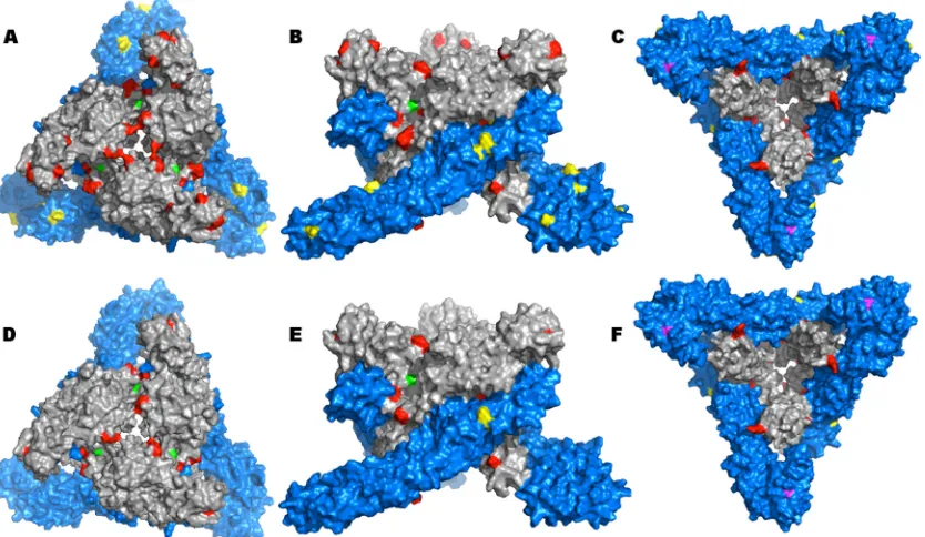
[image:4.585.41.533.66.408.2]3D models for both the BFS932 and Imperial181 E1 proteins
indicated three domains: 1 and 2 had an interlinking beta sheet
structure with a long hydrophobic fusion loop at one end of
do-main 2, while dodo-main 3 shared high structural similarity with the
immunoglobulin domain.
A complete list of amino acid positions that differed between
BFS932 and Imperial181 was obtained for the E1 and E2 proteins.
The BFS932 E1 protein had 11 differences compared to all other
WEEV strains (Fig. 3A
to
C, yellow), while there were only 4
dif-ferences compared to Imperial181 (Fig. 3D
to
F, yellow). There
were 25 differences in the BFS932 E2 protein compared to all
WEEV strains (Fig. 3A
to
C, red) and 13 differences compared to
Imperial181 (Fig. 3D
to
F, red). To visualize these amino acids, we
used a trimeric structure of the BFS932 E1-E2 heterodimer (Fig.
3). Most of the E2 substitutions were located at the E2-E2
inter-face. Nonsynonymous, synapomorphic mutations of interest
were mapped on the 3D structure and are indicated in magenta
(Thr
¡
Ser at position 374 in E1) and green (Ala
¡
Thr at position
23 in E2) (Fig. 3).
DISCUSSION
We report here the first detailed phylogenetic and evolutionary
analysis of WEEV using complete genomic sequences. We
con-firmed the prior presence of two major North American lineages
as previously described (6) and further delineated several
sub-groups (B1 to 3) within lineage B. We also identified purifying
selection as the major influence on WEEV evolution in North
America, as described previously for several other arboviruses (38,
39), and identified several mutations that define the divergence of
groups A and B1 to B3. No phylogenetically significant differences
in glycosylation sites, cysteine residues, and UTR folding patterns
were recognized.
[image:5.585.111.475.65.355.2]In North America, WEEV was first isolated in 1930 from a fatal
case of equine encephalitis (40). This California strain fell into the
FIG 2Bayesian skyline plot of WEEV strains in North America between 1930 and 2005. The centerline represents the estimate for mean genetic variation in the population through time. The upper and lower dashed lines represent the 95% HPD.TABLE 3Mutations that contribute to the definition of WEEV lineages
Gene
Amino acid change
Amino acid
position Mutation
Nucleotide position (from beginning of nonstructural protein ORF)
Codon position
IFEL analysis
Selection type Pvalue
nsP3 Thr¡Ileu 152 C¡T 4436 2 ⫹ 0.11
nsP4 Asn¡Ser 602 A¡G 7382 2 ⫹ 0.23
Capsid Lys¡Arg 89 A¡G 7714 2 ⫹ 0.16
Capsid Lys¡Trp 250 A¡T 8196 1 ⫹ 0.28
A¡G 8197 2
E2 Ala¡Thr 23 G¡A 8472 1 ⫹ 0.38
E1 Thr¡Ser 374 A¡T 10959 1 ⫹ 0.28
on November 7, 2019 by guest
http://jvi.asm.org/
[image:5.585.41.543.621.723.2]group A lineage along with the 1941 McMillan isolate. This
rela-tionship suggests WEEV’s early spread to the eastern side of the
Canadian Rockies. We hypothesize that group A became extinct in
the 1940s and was displaced by group B1, which then became
predominant. Strains from ancestral groups A and B1 generally
are more virulent than more recent strains from groups B2 and B3
(17,
19,
20).
In the late 1940s, group B2 displaced group B1, which probably
went extinct. Subsequently, in the late 1960s, group B3 lineage
emerged from group B2, eventually displacing it. The consistency
of the tightly grouped HPDs throughout the MCC tree supports
the reliability of these temporal estimates (Fig. 1).
When all 3 group B sublineages were circulating, our Bayesian
skyline analysis indicated a concurrent increase in WEEV
esti-mated population size between 1965 and the late 1980s. However,
after the late 1980s, a reduction in estimated population size
oc-curred when the group B3 viruses became predominant in North
America. These interpretations require caution, because the 95%
HPD values for 1930 to 1950 were relatively broad. There is more
confidence in the estimates between the years 1950 and 1995,
when the HPD range was tighter (Fig. 2). However, when the
estimated tMRCAs and Skyline analysis (Fig. 2) are considered in
concert, the evidence for these population size interpretations is
compelling. Interestingly, the pattern seen on our skyline analysis
is similar to that of the annual influenza A virus cycle, although
not as pronounced and over a much longer period of time (41).
This could be an effect of purifying selection on WEEV, trimming
the tree to the group B3 lineage.
The selection analyses indicated that many nucleotide sites
within the WEEV genome are under purifying selection. When
population sizes are reduced, stochastic drift could result in the
accumulation of deleterious mutations, fitness declines, and
lin-eage extinction events that could explain not only the linlin-eage
re-placements we observed but also the decline in WEEV genetic
diversity. However, manual inspection of our WEEV alignment
revealed several mutations that may represent positively selected
codons. These mutations were, for the most part, just under the
threshold of significance as determined by IFEL analysis (
P
⬎
0.1)
(Table 2). Both FEL and FUBAR flagged these same mutations at
P
values that narrowly missed the threshold of significance,
sug-gesting that they have important phenotypes. Furthermore,
cur-rent codon-based analyses sometimes lack the sensitivity to detect
positive selection. For example, analyses preformed on CHIKV
sequences failed to identify all mosquito vector-adaptive
muta-tions shown by experimental studies (38,
42–44).
Our homology models suggested that the E2-E2 and E1-E2
interfaces, locations far removed from receptor binding or
poten-tial antibody binding sites, are important sites of WEEV evolution.
Mutations at these interfaces (Fig. 3), including the substitution at
E2 position 23 (Table 3), may stabilize the E2-E2 trimer spikes and
further prevent the release of genomic RNA during fusion.
The recent epidemiology of WEE, with no reported human
cases in North America since 1998, the dearth of WEEV detected
in mosquito surveillance since 2008 (2,
15), and the pattern of
lineage displacement observed in our phylogeny (Fig. 1), with one
lineage becoming predominant along with a decline in genetic
diversity in lineage B3, raises a key question: what factor(s) caused
the apparent reduction in WEEV circulation and resulting
spill-over disease? We hypothesize that a significant disturbance in
WEEV circulation occurred roughly between 1945 and 1965. This
event affected WEEV evolution in one of two ways: (i) changes in
selective pressures altered the trajectory of WEEV evolution
dur-FIG 3Predicted E1-E2 WEEV trimers. (A to C) Mapping of all mutations on WEEV BFS932 surface compared to all WEEV strains. (A) Top view; (B) side view; (C) bottom view. (D to F) Mapping of all mutations on the WEEV BFS932 surface compared to WEEV Imperial181. (D) Top view; (E) side view; (F) bottom view. All mutations in the E1 protein are shown in yellow, and those from the E2 protein are shown in red. Amino acids 374 in E1 protein (magenta) and 23 in E2 protein (green) also are indicated.on November 7, 2019 by guest
http://jvi.asm.org/
[image:6.585.87.504.71.313.2]ing the late 20th century, or (ii) a reduction in WEEV populations
and/or diversity caused genetic drift and a decline in WEEV
fit-ness, possibly coupled with reduced mammalian virulence. The
key synapomorphic mutations we delineated, including those that
may be subject to positive selection, deserve reverse genetic
anal-yses to test these hypotheses by assessing their phenotypic
prop-erties.
In summary, using comprehensive phylogenetic analyses, we
confirmed the major group A and B lineages described previously
(6) and determined the further divergence of group B into 3
sub-lineages, two of which probably went extinct. We delineated
sev-eral mutations that define groups A and B1 to B3 and which may
have been positively selected. However, overall, WEEV’s
evolu-tion has been dominated by purifying selecevolu-tion. WEEV has
under-gone a reduction in genetic diversity coincident with the
circula-tion of only the group B3 lineage since the 1970s, suggesting that
drift reduced its fitness, levels of circulation, and possibly its
vir-ulence for mammals. These data, as well as the apparent
submer-gence of WEEV as an equine and human pathogen, provide a
unique opportunity to study a phenomenon that, compared to
studies of arboviral emergence, may be equally instructive
regard-ing their maintenance and evolution and the prediction of future
trends.
ACKNOWLEDGMENTS
We thank William Reisen of the University of California, Davis, Mary D’Anton of the Texas Department of Health and Human Services, and Robert Tesh of the World Reference Center for Emerging Viruses and Arboviruses (WRCEVA) at UTMB for providing WEEV isolates.
REFERENCES
1.Whitley RJ, Gnann JW.2002. Viral encephalitis: familiar infections and emerging pathogens. Lancet 359:507–513. http://dx.doi.org/10.1016 /S0140-6736(02)07681-X.
2.Centers for Disease Control and Prevention.1995. Arboviral disease– United States, 1994. MMWR Morb. Mortal. Wkly. Rep.44:641– 644. 3.Netolitzky DJ, Schmaltz FL, Parker MD, Rayner GA, Fisher GR, Trent
DW, Bader DE, Nagata LP.2000. Complete genomic RNA sequence of western equine encephalitis virus and expression of the structural genes. J. Gen. Virol. 81:151–159. http://vir.sgmjournals.org/content/81/1/151 .long.
4.Strauss JH, Strauss EG.1994. The alphaviruses: gene expression, repli-cation, and evolution. Microbiol. Rev.58:491–562.
5.Hahn CS, Lustig S, Strauss EG, Strauss JH. 1988. Western equine encephalitis virus is a recombinant virus. Proc. Natl. Acad. Sci. U. S. A. 85:5997– 6001.http://dx.doi.org/10.1073/pnas.85.16.5997.
6.Weaver SC, Kang W, Shirako Y, Rumenapf T, Strauss EG, Strauss JH. 1997. Recombinational history and molecular evolution of western equine encephalomyelitis complex alphaviruses. J. Virol.71:613– 623.
7.Hardy JL, Milby MM, Wright ME, Beck AJ, Presser SB, Bruen JP.1977. Natural and experimental arboviral infections in a population of blacktail jackrabbits along the Sacramento River in Butte County, California (1971–1974). J. Wildl. Dis.13:383–392.http://dx.doi.org/10.7589/0090 -3558-13.4.383.
8.Hardy JL, Reeves WC, Scrivani RP, Roberts DR.1974. Wild mammals as hosts of group A and group B arboviruses in Kern County, California. Am. J. Trop. Med. Hyg.23:1165–1177.
9.Bowers JH, Hayes RO, Hughes TB.1969. Studies on the role of mammals in the natural history of western encephalitis in Hale County, Texas. J. Med. Entomol.6:175–178.
10. Reisen WK, Monath TP. 1988. Western equine encephalomyelitis, p 89 –137.InMonath TP (ed), The arboviruses: epidemiology and ecology, vol V. CRC Press, Boca Raton, FL.
11. Byrne RJ, French GR, Yancey FS, Gochenour WS, Russell PK, Rams-burg HH, Brand OA, Scheider FG, Buescher EL.1964. Clinical and Immunological interrelationships among Venezuelan, eastern, and west-ern encephalomyelitis in burros. Am. J. Vet. Res.25:24 –31.
12. Sponseller ML, Binn LN, Wooding WL, Yager RH.1966. Field strains of western encephalitis virus in ponies: virologic, clinical, and pathologic observations. Am. J. Vet. Res.27:1591–1598.
13. Artsob H, Spence L.1979. Arboviruses in Canada, p 39.InKurstack E (ed), Arctic and tropical arboviruses. Academic Press, New York, NY. 14. Cameron GDW.1942. Western equine encephalitis. Can. Public Health J.
33:383–387.
15. CDC.2010. Western equine encephalitis virus neuroinvasive disease cases reported by state, 1964 –2010. Centers for Disease Control and Preven-tion, Atlanta, GA. http://www.cdc.gov/easternequineencephalitis/tech /epi.html.
16. Zhang M, Fang Y, Brault AC, Reisen WK.2011. Variation in western equine encephalomyelitis viral strain growth in mammalian, avian, and mosquito cells fails to explain temporal changes in enzootic and epidemic activity in California. Vector-Borne Zoonotic Dis.11:269 –275.http://dx .doi.org/10.1089/vbz.2010.0078.
17. Forrester NL, Kenney JL, Deardorff E, Wang E, Weaver SC. 2008. Western equine encephalitis submergence: lack of evidence for a decline in virus virulence. Virology380:170 –172.http://dx.doi.org/10.1016/j.virol .2008.08.012.
18. Reisen WK, Fang Y, Brault AC.2008. Limited interdecadal variation in mosquito (Diptera: Culicidae) and avian host competence for western equine encephalomyelitis virus (Togaviridae: Alphavirus). Am. J. Trop. Med. Hyg.78:681– 686.http://www.ajtmh.org/content/78/4/681.long. 19. Nagata LP, Hu W-G, Parker M, Chau D, Rayner GA, Schmaltz FL,
Wong JP.2006. Infectivity variation and genetic diversity among strains of Western equine encephalitis virus. J. Gen. Virol.87:2353–2361.http: //dx.doi.org/10.1099/vir.0.81815-0.
20. Logue CH, Bosio CF, Welte T, Keene KM, Ledermann JP, Phillips A, Sheahan BJ, Pierro DJ, Marlenee N, Brault AC, Bosio CM, Singh AJ, Powers AM, Olson KE.2009. Virulence variation among isolates of west-ern equine encephalitis virus in an outbred mouse model. J. Gen. Virol. 90:1848 –1858.http://dx.doi.org/10.1099/vir.0.008656-0.
21. Mossel EC, Ledermann JP, Phillips AT, Borland EM, Powers AM, Olson KE.2013. Molecular determinants of mouse neurovirulence and mosquito infection for western equine encephalitis virus. PLoS One 8:e60427.http://dx.doi.org/10.1371/journal.pone.0060427.
22. Kramer LD, Fallah HM.1999. Genetic variation among isolates of west-ern equine encephalomyelitis virus from California. Am. J. Trop. Med. Hyg.60:708 –713.
23. Vasilakis N, Forrester NL, Palacios G, Nasar F, Savji N, Rossi SL, Guzman H, Wood TG, Popov V, Gorchakov R, González AV, Haddow AD, Watts DM, da Rosa APAT, Weaver SC, Lipkin WI, Tesh RB.2013. Negevirus: a proposed new taxon of insect-specific viruses with wide geo-graphic distribution. J. Virol.87:2475–2488.http://dx.doi.org/10.1128 /JVI.00776-12.
24. Galtier N, Gouy M, Gautier C.1996. SEAVIEW and PHYLO_WIN: two graphic tools for sequence alignment and molecular phylogeny. Comput. Appl. Biosci.12:543–548.
25. Drummond AJ, Suchard MA, Xie D, Rambaut A.2012. Bayesian phy-logenetics with BEAUti and the BEAST 1.7. Mol. Biol. Evol.29:1969 – 1973.http://dx.doi.org/10.1093/molbev/mss075.
26. Baele G, Lemey P, Bedford T, Rambaut A, Suchard MA, Alekseyenko AV.2012. Improving the accuracy of demographic and molecular clock model comparison while accommodating phylogenetic uncertainty. Mol. Biol. Evol.29:2157–2167.http://dx.doi.org/10.1093/molbev/mss084. 27. Baele G, Li WLS, Drummond AJ, Suchard MA, Lemey P.2013. Accurate
model selection of relaxed molecular clocks in Bayesian phylogenetics. Mol. Biol. Evol.30:239 –243.http://dx.doi.org/10.1093/molbev/mss243. 28. Posada D, Crandall KA.1998. Modeltest: testing the model of DNA
substitution. Bioinformatics 14:817– 818. http://dx.doi.org/10.1093 /bioinformatics/14.9.817.
29. Pond SLK, Frost SDW.2005. Datamonkey: rapid detection of selective pressure on individual sites of codon alignments. Bioinformatics21:2531– 2533.http://dx.doi.org/10.1093/bioinformatics/bti320.
30. Pond SLK, Poon AFY, Frost SDW.2009. Estimating selection pressures on alignments of coding sequences, p 419 – 490.InLemey P, Salemi M, Vandamme A-M (ed), The phylogenetic handbook. Cambridge Univer-sity Press, New York, NY.
31. Murrell B, Moola S, Mabona A, Weighill T, Sheward D, Kosakovsky Pond SL, Scheffler K.2013. FUBAR: a fast, unconstrained Bayesian ap-proximation for inferring selection. Mol. Biol. Evol.30:1196 –1205.http: //dx.doi.org/10.1093/molbev/mst030.
on November 7, 2019 by guest
http://jvi.asm.org/
32. Kosakovsky Pond SL, Frost SDW.2005. Not so different after all: a compar-ison of methods for detecting amino acid sites under selection. Mol. Biol. Evol. 22:1208 –1222.http://dx.doi.org/10.1093/molbev/msi105.
33. Kosakovsky Pond SL, Frost SDW, Grossman Z, Gravenor MB, Rich-man DD, Brown AJL.2006. Adaptation to different human populations by HIV-1 revealed by codon-based analyses. PLoS Comput. Biol.2:e62. http://dx.doi.org/10.1371/journal.pcbi.0020062.
34. Söding J, Biegert A, Lupas AN.2005. The HHpred interactive server for protein homology detection and structure prediction. Nucleic Acids Res. 33:W244 –W248.http://dx.doi.org/10.1093/nar/gki408.
35. Mumenthaler C, Braun W.1995. Automated assignment of simulated and experimental NOESY spectra of proteins by feedback filtering and self-correcting distance geometry. J. Mol. Biol.254:465– 480.http://dx.doi .org/10.1006/jmbi.1995.0631.
36. Sanner M, Widmer A, Senn H, Braun W.1989. GEOM, a new tool for molecular modeling based on distance geometry calculations with NMR data. J. Comp. Aided Mol. Des. 3:195–210. http://dx.doi.org/10.1007 /BF01533068.
37. Schaumann T, Braun W, Wuthrich K.1990. A program, FANTOM, for energy refinement of polypeptides and proteins using a Newton-Raphson Minimizer in the torsion angle space. Biopolymers29:679 – 694.http://dx .doi.org/10.1002/bip.360290403.
38. Volk SM, Chen R, Tsetsarkin KA, Adams AP, Garcia TI, Sall AA, Nasar F, Schuh AJ, Holmes EC, Higgs S, Maharaj PD, Brault AC, Weaver SC. 2010. Genome-scale phylogenetic analyses of chikungunya virus reveal
independent emergences of recent epidemics and various evolutionary rates. J. Virol.84:6497– 6504.http://dx.doi.org/10.1128/JVI.01603-09. 39. Auguste AJ, Lemey P, Pybus OG, Suchard MA, Salas RA, Adesiyun AA,
Barrett AD, Tesh RB, Weaver SC, Carrington CVF.2010. Yellow fever virus maintenance in trinidad and its dispersal throughout the Americas. J. Virol.84:9967–9977.http://dx.doi.org/10.1128/JVI.00588-10. 40. Meyer KF, Haring CM, Howitt B.1931. The etiology of epizootic
en-cephalomyelitis of horses in the San Joaquin Valley, 1930. Science74:227– 228.http://dx.doi.org/10.1126/science.74.1913.227.
41. Rambaut A, Pybus OG, Nelson MI, Viboud C, Taubenberger JK, Holmes EC.2008. The genomic and epidemiological dynamics of human influenza A virus. Nature 453:615– 619. http://dx.doi.org/10.1038 /nature06945.
42. Tsetsarkin KA, McGee CE, Volk SM, Vanlandingham DL, Weaver SC. 2009. Epistatic roles of E2 glycoprotein mutations in adaption of Chikun-gunya virus toAdes albopictusandAe. aegyptimosquitos. PLoS One 4:e6835.http://dx.doi.org/10.1371/journal.pone.0006835.
43. Tsetsarkin K, Weaver SC.2011. Sequential adaptive mutations enhance efficient vector switching by chikungunya virus and its epidemic emer-gence. PLoS Pathog.7:e1002412.http://dx.doi.org/10.1371/journal.ppat .1002412.
44. Tsetsarkin K, Chen R, Yun R, Rossi SL, Plante K, Guerbois M, Forrester NL, Perng GC, Sreekumar E, Leal G, Huang C, Mukhopadhyay S, Weaver SC.Multi-peaked adaptive landscape for chikungunya virus evo-lution predicts continued fitness optimization in Aedes albopictus mos-quitos. Nat. Commun., in press.
on November 7, 2019 by guest
http://jvi.asm.org/