and Early in the Absence of C Protein and Can Be Destabilized by
Adenosine Deaminase Acting on RNA-1-Like Hypermutations
Christian K. Pfaller,aGeorge M. Mastorakos,a,bWilliam E. Matchett,a,bXiao Ma,a,bCharles E. Samuel,cRoberto Cattaneoa,b
Department of Molecular Medicine, Mayo Clinic, Rochester, Minnesota, USAa
; Virology and Gene Therapy Track, Mayo Graduate School, Mayo Clinic, Rochester, Minnesota, USAb
; Department of Molecular, Cellular and Developmental Biology, University of California, Santa Barbara, California, USAc
ABSTRACT
Defective interfering RNAs (DI-RNAs) of the viral genome can form during infections of negative-strand RNA viruses and
out-grow full-length viral genomes, thereby modulating the severity and duration of infection. Here we document the frequent
de
novo
generation of copy-back DI-RNAs from independent rescue events both for a vaccine measles virus (vac2) and for a
wild-type measles virus (IC323) as early as passage 1 after virus rescue. Moreover, vaccine and wild-wild-type C-protein-deficient
(C-pro-tein-knockout [C
KO]) measles viruses generated about 10 times more DI-RNAs than parental virus, suggesting that C enhances
the processivity of the viral polymerase. We obtained the nucleotide sequences of 65 individual DI-RNAs, identified breakpoints
and reinitiation sites, and predicted their structural features. Several DI-RNAs possessed clusters of A-to-G or U-to-C
transi-tions. Sequences flanking these mutation sites were characteristic of those favored by adenosine deaminase acting on RNA-1
(ADAR1), which catalyzes in double-stranded RNA the C-6 deamination of adenosine to produce inosine, which is recognized as
guanosine, a process known as A-to-I RNA editing. In individual DI-RNAs the transitions were of the same type and occurred on
both sides of the breakpoint. These patterns of mutations suggest that ADAR1 edits unencapsidated DI-RNAs that form
double-strand RNA structures. Encapsidated DI-RNAs were incorporated into virus particles, which reduced the infectivity of virus
stocks. The C
KOphenotype was dominant: DI-RNAs derived from vac2 with a C
KOsuppressed the replication of vac2, as shown
by coinfections of interferon-incompetent lymphatic cells with viruses expressing different fluorescent reporter proteins. In
con-trast, coinfection with a C-protein-expressing virus did not counteract the suppressive phenotype of DI-RNAs.
IMPORTANCE
Recombinant measles viruses (MVs) are in clinical trials as cancer therapeutics and as vectored vaccines for HIV-AIDS and other
infectious diseases. The efficacy of MV-based vectors depends on their replication proficiency and immune activation capacity.
Here we document that copy-back defective interfering RNAs (DI-RNAs) are generated by recombinant vaccine and wild-type
MVs immediately after rescue. The MV C protein interferes with DI-RNA generation and may enhance the processivity of the
viral polymerase. We frequently detected clusters of A-to-G or U-to-C transitions and noted that sequences flanking individual
mutations contain motifs favoring recognition by the adenosine deaminase acting on RNA-1 (ADAR1). The consistent type of
transitions on the DI-RNAs indicates that these are direct substrates for editing by ADAR1. The ADAR1-mediated biased
hyper-mutation events are consistent with the protein kinase R (PKR)-ADAR1 balancing model of innate immunity activation. We
show by coinfection that the C-defective phenotype is dominant.
M
easles virus (MV) is a negative-strand RNA virus of the
Paramyxoviridae
family with a genome comprising 15,894
nucleotides (nt). The MV genome encodes six genes from which
eight proteins are expressed: nucleoprotein (N), phosphoprotein
(P), matrix protein (M), fusion protein (F), hemagglutinin (H),
and large protein (L; the catalytic subunit of the viral
RNA-depen-dent RNA polymerase), as well as the host response-modulating V
and C proteins (
1–3
).
MV replication occurs in the cytoplasm of infected cells and
involves a helical ribonucleocapsid (RNP) formed by viral
genomic RNA and N protein. The polymerase complex,
consist-ing of the L and P proteins, recognizes the 3
=
end of the RNA and
generates a complementary nascent RNA strand that is
cotrans-criptionally encapsidated (
4
). Both genomic RNA and
antigeno-mic RNA can serve as a template for replication, whereas only the
genomic RNA serves as a template for the transcription of capped
and polyadenylated mRNAs (
1
).
The polymerase of negative-strand RNA viruses is error prone
and introduces nucleotide substitutions at fairly high frequencies
(about 1 substitution in 10,000 nt) (
5–8
). Defective genomes are
also generated, including abortive genomes and 5
=
copy-back
defective interfering (DI) RNAs (DI-RNAs) (
9
). DI-RNAs are
generated when the polymerase prematurely terminates genome
synthesis and then—when it is still bound to the truncated
ge-Received17 April 2015Accepted11 May 2015
Accepted manuscript posted online13 May 2015
Editor:T. S. Dermody
Address correspondence to Roberto Cattaneo, [email protected].
CitationPfaller CK, Mastorakos GM, Matchett WE, Ma X, Samuel CE, Cattaneo R. 2015. Measles virus defective interfering RNAs are generated frequently and early in the absence of C protein and can be destabilized by adenosine deaminase acting on RNA-1-like hypermutations. J Virol 89:7735–7747.doi:10.1128/JVI.01017-15. Copyright © 2015, American Society for Microbiology. All Rights Reserved. doi:10.1128/JVI.01017-15
on November 7, 2019 by guest
http://jvi.asm.org/
nome— binds back and reinitiates replication on this RNA,
thereby generating a genome-antigenome hybrid with perfectly
complementary ends. These complementary ends can form stable
panhandle duplex RNA structures, as shown for MV and Sendai
virus (SeV), a related paramyxovirus (
9
,
10
).
These double-stranded RNA (dsRNA) structures are potent
inducers of the cellular stress and innate immune response (
11
).
They trigger the activation of innate receptors, including retinoic
acid-inducible gene I (RIG-I [
12
]), melanoma
differentiation-as-sociated gene-5 (mda-5 [
13
]), and protein kinase R (PKR [
14
]).
DI-RNAs contain the 3
=
-terminal promoter sequence and
there-fore can be replicated into their complementary counterpart,
which also contains the promoter sequence at its 3
=
end. DI-RNAs,
therefore, like the genome-antigenome pair, form a replication
system, which, due to its reduced length and therefore increased
replication frequency, can interfere with full-length RNA
replica-tion and temporarily outgrow full-length RNA (
15
).
We previously reported the generation of viral DI-RNAs in
C-protein-deficient (C-protein-knockout [C
KO]) Moraten
vac-cine MV (MV-C
KO) and showed that DI-RNAs induce the
PKR-mediated stress and interferon (IFN) responses (
14
). Here we
pro-vide mechanistic insights into the generation of DI-RNAs by a
vaccine MV (vac2)-derived infectious cDNA (
16
) and a wild-type
MV (IC323) infectious cDNA (
17
) immediately after virus rescue.
We describe the molecular characteristics of DI-RNAs from
C-protein-expressing and C-defective viruses and find structural
similarities between DI-RNAs derived from all viruses. Our
anal-ysis of multiple independent virus rescues indicated that
defi-ciency in C-protein expression leads to a 10-fold increase in the
abundance of DI-RNAs. Although initially demonstrated in
seri-ally undiluted virus infections performed at a high multiplicity
(
18–20
), we detected DI-RNAs in stocks as early as passage 1 (P1)
after virus rescue. We found clusters of A-to-G or U-to-C
transi-tions in several DI-RNAs in a sequence context characteristic of
the mutations introduced by cellular adenosine deaminase acting
on RNA-1 (ADAR1) (
21–23
).
MATERIALS AND METHODS
Cells and viruses.Vero, Vero-hSLAM (a kind gift from Y. Yanagi [24]), and HeLa cells were cultivated in Dulbecco’s high-glucose modified Ea-gle’s medium (D-MEM; HyClone; GE Healthcare Life Sciences, Logan, UT) supplemented with 5% (vol/vol) fetal bovine serum (FBS; Gibco, Life Technologies, Carlsbad, CA). Vero-hSLAM cells were grown in the pres-ence of Geneticin (G418; Corning, Tewksbury, MA) at a final concentra-tion of 0.4 mg/ml. 293-3-46 cells (25), used for virus rescue, were culti-vated in D-MEM supplemented with 10% (vol/vol) FBS and 1 mg/ml G418. The mantle cell lymphoma cell line Granta-519 (26) was cultivated in RPMI 1640 (HyClone) supplemented with 10% (vol/vol) FBS.
Recombinant MVs were IC323 (isogenic to the wild-type IC-B strain [17]), IC323-CKO, vac2 (isogenic to the Moraten vaccine strain [16]), vac2-CKO, vac2(GFP) (expressing enhanced green fluorescent protein [eGFP] from an additional transcription unit downstream of H [27]), vac2-CKO(GFP), vac2(mCherryNLS), and vac2-CKO(mCherryNLS). Vi-ruses with the CKOhave specific mutations introducing early stop codons in the C open reading frame, as described previously (16).
Generation of mCherryNLS-expressing MVvac2 cDNA.A con-struct encoding mCherry with a nuclear localization signal (NLS), mCherryNLS, kindly provided by L. Enquist (28), was cloned into p(⫹) MV323(GFP)N to replace GFP, yielding p(⫹)MV323(mCherryNLS)N (Crystal Mendoza, unpublished data). mCherryNLS was amplified from this donor vector by PCR using the following primers (Integrated DNA Technologies [IDT], Coralville, IA), where the restriction sites are
under-lined: mCherryNLS EcoRI/MluI fwd (TATAGAATTCCCATCAACGCG TTACGCCACCATG) and mCherryNLS NotI/AatII rev (ATATGCGGC CGCGTTCGAGACGTCAGTTTATCTGGATCC). The PCR product was cloned into pCR3 (Life Technologies, Invitrogen, Carlsbad, CA) using the EcoRI/NotI restriction sites and then excised with MluI/AatII (New England BioLabs, Ipswich, MA). The GFP-encoding sequence in pB(⫹)MVvac2(GFP)H and pB(⫹)MVvac2-CKO(GFP)H was then replaced with the mCherryNLS fragment to yield pB(⫹)MVvac2 (mCherryNLS)H and pB(⫹)MVvac2-CKO(mCherryNLS)H, respec-tively.
Virus rescue and stock production.Recombinant viruses were gen-erated as described previously (25). Briefly, 293-3-46 cells were trans-fected with the relevant plasmid carrying full-length viral cDNA and pEMC-La, expressing the viral polymerase. After a 42°C heat shock and 2 days of incubation, cells were transferred to 100-mm dishes with Vero or Vero-hSLAM cells and incubated until syncytium formation was ob-served. Single syncytia were picked and used to inoculate Vero or Vero-hSLAM cells in 6-well plates. Cells and supernatants were harvested when the maximum cytopathic effect (CPE) was achieved (2 to 4 days), yielding passage 0 stocks. These stocks were used to generate passage 1 stocks by infecting multiple 150-mm dishes of Vero or Vero-hSLAM cells. Virus stocks were grown at 32°C (vac2) or 37°C (IC323). Cells were harvested at the time of the maximum CPE by scraping them into 1 ml Opti-MEM medium (Gibco, Life Technologies, Carlsbad, CA) per 150-mm dish, fol-lowed by three freeze-thaw cycles (liquid nitrogen and 37°C). Cleared lysates were aliquoted and stored at⫺80°C. Viral titers on Vero-hSLAM cells were determined using the 50% tissue culture infectious dose (TCID50) method. For further passages, Vero cells in 150-mm dishes were infected at a multiplicity of infection (MOI) of 0.03 and harvested, and titers were determined.
Virion purification.Vero cells (in 10 150-mm dishes) were infected with passage 1 vac2 or vac2-CKOat an MOI of 0.05. Cells were harvested at 48 h postinfection and scraped into 1 ml Opti-MEM medium per dish, followed by three freeze-thaw cycles. Cleared lysates (total volume, about 15 ml) were mixed with Coomassie blue (Brilliant Blue G; final concen-tration,⬃150g/ml; Bio-Rad, Hercules, CA), and the mixture was laid onto a 20%/60% (wt/vol) sucrose cushion (in TNE buffer [50 mM Tris-HCl {pH 7.4}, 100 mM NaCl, 0.1 mM EDTA]) and centrifuged in an SW32Ti rotor (Beckman Coulter, Indianapolis, IN) at 75,000⫻gat 4°C for 4 h. The visible blue band at the 20%-60% interface was collected with a 16.5-gauge needle and syringe in a total volume of about 6 ml, diluted with sterile phosphate-buffered saline (PBS; HyClone) supplemented with 0.5 mM MgCl2(PBS-MgCl2) to a final volume of 15 ml, and then laid onto a 20 to 60% (wt/vol) discontinuous sucrose gradient (in TNE buffer, 5% intervals). Centrifugation was to equilibrium (75,000⫻gat 4°C for 16 h) using the SW32Ti rotor. Virions that accumulated in the 30 to 40% region were collected in a total volume of 6 ml, diluted with sterile PBS-MgCl2, and concentrated using Amicon Ultra filter tubes (EMD Milli-pore, Billerica, MA) with a molecular mass exclusion of 100 kDa (3,500⫻
g, 4°C) in a tabletop centrifuge. The concentrate was washed once with 15 ml PBS-MgCl2, concentrated to a final volume of 1 ml, and stored at
⫺80°C. Titers were determined by the TCID50method. Prior to further analysis, purified stocks were subjected to treatment with Benzonase (EMD Millipore, Billerica, MA) at a final concentration of 12.5 U/l for 2 h at 37°C in 10 mM Tris, pH 8.0, buffer containing 1 mM MgCl2and10 mM NaCl.
RNA protection assay.One million TCID50s of purified vac2 or vac2-CKOvirions were treated with combinations of nonionic detergent (Tri-ton X-100), protease (trypsin), and nuclease (Benzonase). First, 1% (vol/ vol) Triton X-100 (Sigma-Aldrich, St. Louis, MO) was added to virions in reaction buffer (10 mM Tris, pH 8.0, 10 mM NaCl, 1 mM MgCl2). Next, samples were heated to 80°C for 15 min and cooled on ice for 10 min. Then, 0.1 mg/ml trypsin (Worthington, Lakewood, NJ) was added and the samples were incubated at 37°C for 15 min. To inactivate the trypsin, 3 mg/ml soybean trypsin inhibitor (Worthington, Lakewood, NJ) was
on November 7, 2019 by guest
http://jvi.asm.org/
added and the samples were incubated at 25°C for 30 min. Next, 2.5 U/l Benzonase was added and the samples were incubated at 37°C for 2.5 h. Nuclease activity was inhibited by addition of 10 mM EDTA, before sam-ple aliquots were subjected to protein analysis or RNA extraction.
RNA extraction, RT-PCR, and PCR analysis.Vero or Vero-hSLAM cells were infected with viruses at an MOI of 0.1 or left uninfected and harvested at 30 to 48 h postinfection. Total RNA was extracted using either an RNeasy minikit (Qiagen, Valencia, CA) or the TRIzol reagent (Ambion, Life Technologies, Carlsbad, CA) according to the suppliers’ instructions and stored at⫺80°C. One microgram of total RNA was sub-jected to reverse transcription-PCR (RT-PCR) using SuperScript III re-verse transcriptase (Life Technologies, Carlsbad, CA) with a mixture of MV-specific primers (IDT), where restriction sites are underlined: nega-tive-polarity primers A1SnaBI(GCGCGCTACGTAAAAGCTGGGAATA GAAACTTCG), A2vac2-NotI(TAGAGAGAGCGGCCGCTCTGGTGTAA GTCTAGTATCAGA), A2323-NotI (TAGAGAGAGCGGCCGCTCTGGT GTAAGTCTAGTACTAGA), A3NotI(TAGAGAGAGCGGCCGCGATCT AGAATTGGCGGAAACC), and A4NotI(TAGAGAGAGCGGCCGCTCC CTGATGGTCTATGATAATG) and positive-polarity primer B1 (ATGA CAGATCTCAAGGCTAAC). Briefly, RNA was mixed with primers and deoxynucleoside triphosphates and subjected to heat denaturation at 98°C for 10 min, followed by incubation on ice for 10 min. After addition of the enzyme mix, samples were incubated at 55°C for 60 min and then at 70°C for 15 min. cDNAs were stored at⫺20°C. An aliquot of the cDNA (1
l) was further amplified using GoTaq polymerase (Promega, Madison, WI) and DI-RNA-specific primer combinations (primers A1/A2, A1/A3, and A1/A4) or a control primer pair for standard genomes (primers A1/ B1). Samples were analyzed by electrophoresis using 1% (wt/vol) agarose gels and a 100-bp or 1-kb DNA ladder as a size standard (Promega, Mad-ison, WI).
Cloning and analysis of DI-RNAs.PCR products amplified from the cDNA of MV-infected cells were purified (PCR purification kit; Qiagen, Valencia, CA), digested with SnaBI and NotI-HF (New England BioLabs, Ipswich, MA), purified again, and then ligated with EcoRV/NotI-linear-ized pCR3 (Invitrogen, Life Technologies, Carlsbad, CA). Ligation prod-ucts were transformed into OneShot TOP10 cells (Life Technologies, Carlsbad, CA), and the bacteria were plated on LB-ampicillin-agar plates and incubated at 37°C for 12 to 20 h. Single colonies were picked and resuspended in 10l H2O, and 5l of the suspension was subjected to colony PCR using the primers (IDT) T7fwd(TAATACGACTCACTATA GGG) and BGHrev(TAGAAGGCACAGTCGAGG) and a 2⫻PCR mix (Promega, Madison, WI). Clones with individual inserts, as analyzed by 1% agarose gel electrophoresis, were selected and grown in 2 ml LB-ampicillin, and plasmids were extracted using a plasmid minikit (Qiagen, Valencia, CA). Sequences were determined by Sanger sequencing using the T7fwdand BGHrevprimers and analyzed by alignment to MV reference sequences with GenBank accession numbers AF266287for vac2 and AB016162for IC323.
Protein gel electrophoresis and immunoblot analysis.Protein ex-tracts of infected cells (MOI⫽0.1, 30 to 48 h postinfection) were gener-ated, and protein concentrations were determined as described previously (14). Protein (25g) was fractionated using 12% polyacrylamide gels and transferred to Immobilon-P membranes (EMD Millipore, Billerica, MA) using a semidry protocol. Membranes were blocked with 5% (wt/vol) bovine serum albumin (Sigma-Aldrich, St. Louis, MO) in Tris-buffered saline (Bio-Rad, Hercules, CA) for 1 h and incubated with primary anti-bodies at 4°C for 12 to 16 h. Membranes were washed three times with PBS with 0.5% (vol/vol) Tween 20 (PBST) for 5 to 10 min, incubated with horseradish peroxidase (HRP)-conjugated secondary antibody at room temperature for 1 h, washed again three times with PBST, and incubated with SuperSignal West Pico chemiluminescent substrate (Pierce, Thermo Fisher Scientific, Rockford, IL). Membranes were exposed to HyBlot films (Denville, South Plainfield, NJ) for various amounts of time.
Antibodies.Rabbit peptide antisera recognizing MV N (N505 antise-rum), MV C (C2antiserum), and MV H (Hcytantiserum) were described
previously (16,29,30) and were used at dilutions of 1:5,000 for primary antibody incubation. Mouse monoclonal anti-GFP antibody (eBiosci-ence, San Diego, CA) was used at a 1:1,000 dilution. HRP-conjugated antiactin (Sigma-Aldrich, St. Louis, MO) was used at a 1:25,000 dilution. Rabbit and mouse secondary antibodies conjugated with HRP (Jackson ImmunoResearch, West Grove, PA, and EMD Millipore, Billerica, MA, respectively) were used at 1:10,000 dilutions. For immunofluorescence, mouse anti-dsRNA J2 (English and Scientific Consulting, Szirák, Hungary [31]), anti-MV N505 antiserum, and secondary antibodies antimouse Alexa 488, antimouse Alexa 594, and antirabbit Alexa 405 (Molecular Probes, Life Technologies, Carlsbad, CA) were used as described previ-ously (14).
Northern blot analysis.A 250-bp fragment of the L trailer region of MV (nt 15641 to 15890) was cloned into the pCR3 vector between the HindIII and XhoI restriction sites (amplification primers [IDT], MV15641-HindIII fwd [AGACCCAAGCTTTATCTTCGTTAAGAA TCT] and MV15890 XhoI rev [TGCATGCTCGAGGACAAAGCTGG GAATAGA], where the restriction sites are underlined). Positive and negative single-stranded RNA (ssRNA) probes were generated from HindIII- or XhoI-linearized plasmids using SP6 or T7 RNA polymer-ase of a digoxigenin (DIG) RNA labeling kit (Roche Diagnostics, Indianapolis, IN). Total RNA (3g) or an amount of RNA corre-sponding to 1⫻106TCID
50s of purified virions was subjected to 1% agarose-2% formaldehyde gel electrophoresis, with DIG-labeled RNA molecular weight marker I (Roche Diagnostics, Indianapolis, IN) be-ing included as described in the DIG Northern starter kit manual (Roche Diagnostics, Indianapolis, IN). RNA was transferred to nylon membranes (Roche Diagnostics, Indianapolis, IN) by bottom-up cap-illary transfer using 20⫻SSC (3.0 M NaCl, 0.3 M sodium citrate, pH 7.0) as the buffer. RNAs were cross-linked with 125 mJ of 254-nm UV light before incubation with RNA probes (100 mg/ml) at 68°C for 16 to 20 h under constant agitation. Probe detection was performed accord-ing to the instructions in the supplier’s manual (Roche Diagnostics, Indianapolis, IN).
Confocal immunofluorescence analysis.Infection of HeLa cells and immunostaining for dsRNA and MV N protein were performed as de-scribed previously (14). Slides were analyzed using an LSM 780 inverted laser scanning confocal microscope and ZEN 2012 (Black Edition, v8.0) software (Carl Zeiss Microscopy, Jena, Germany).
Flow cytometry analysis of coinfections.Granta-519 cells (5⫻105) were infected with various combinations of GFP- and mCherryNLS-ex-pressing vac2 and vac2-CKOat an MOI of 1 for each virus and fixed with 4% paraformaldehyde (Santa Cruz Biotechnology, Santa Cruz, CA) at 48 h postinfection, and fluorescent reporter expression was analyzed using a FACSCalibur cytometer (BD Biosciences, San Jose, CA) and FlowJo soft-ware (Tree Star Inc., Ashland, OR).
RESULTS
DI-RNAs are generated at a high frequency immediately after
virus rescue.
Copy-back DI-RNAs have two characteristic
fea-tures: their breakpoint, that is, the last nucleotide incorporated by
the polymerase into the progeny genomic RNA before leaving the
template antigenome strand (
Fig. 1A
, left end of black bar), and
the reinitiation site, that is, the position of polymerase entry on the
progeny RNA. Polymerase reentry results in the synthesis of a
cRNA strand of opposite polarity (
Fig. 1A
, left end of bottom gray
bar). Even if such RNA molecules have the potential of forming a
dsRNA structure with a hairpin loop (
Fig. 1B
), they usually do not
because the replicating RNAs are cotranscriptionally
encapsi-dated. Copy-back DI-RNA can be specifically amplified by PCR
from the cDNA of infected cells by the use of primer pairs in which
one primer binds to the copy-back sequence (
Fig. 1C
, primer A1)
and the second primer (primer A2, A3, or A4) binds to the DI
sequence downstream of the breakpoint. Although only parts of
on November 7, 2019 by guest
http://jvi.asm.org/
the DI-RNA sequences are amplified with these primers (the 5
=
and 3
=
termini outside the primer binding sites are not amplified),
the amplified sequence permits calculation of the length of the
DI-RNA. Importantly, these primer combinations cannot yield a
specific PCR product with standard genomes, since there they
have the same polarity.
[image:4.585.58.263.64.224.2]To gain insights into the frequency with which DI-RNAs are
generated, we analyzed individual viral plaques obtained during
the rescue of vaccine (vac2;
Fig. 2A
, top left) or wild-type (IC323;
Fig. 2A
, top right) recombinant MV. In addition, we also isolated
plaques of mutant viruses unable to express the C protein
(vac2-C
KOand IC323-C
KO;
Fig. 2A
, bottom left and right, respectively).
We generated passage 1 (P1) stocks from these plaques and used
these to do infections at a low MOI of 0.1.
We designed primers binding to different positions in the L
gene, allowing amplification of DI-RNA sequences from less than
0.5 kb to up to 5 kb. Using the A1/A2 primer combination,
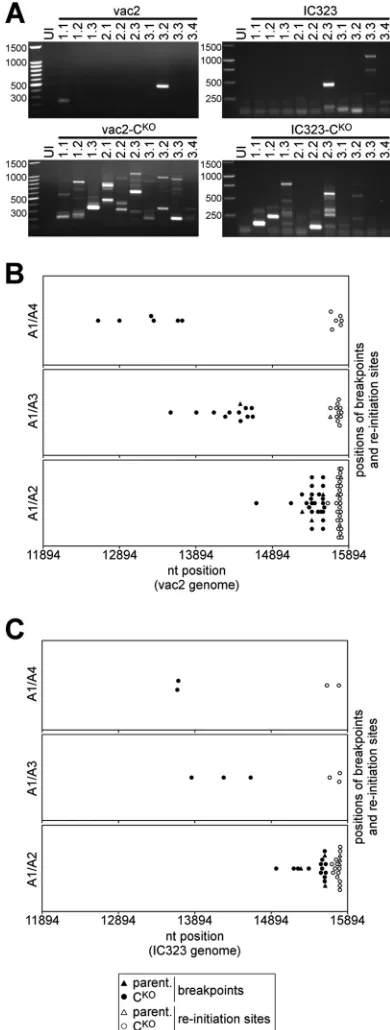
DI-RNAs were readily detected in at least 2 of 10 individual plaques of
C-expressing vac2 (
Fig. 2A
, top left) and IC323 viruses (
Fig. 2A
,
top right). DI-RNAs were prominent in nearly all plaques of both
mutant C
KOviruses: multiple DI-RNAs were identified in each
vac2-C
KOplaque (
Fig. 2A
, bottom left), and 7 of 10 IC323-C
KOplaques (
Fig. 2A
, bottom right) showed at least one DI-RNA. A
unique DI-RNA pattern was detected in each plaque.
Molecular characterization of DI-RNAs from vaccine and
wild-type MV strains.
To further characterize the DI-RNAs, we
cloned their cDNAs and obtained the sequences of 65
indepen-dent clones.
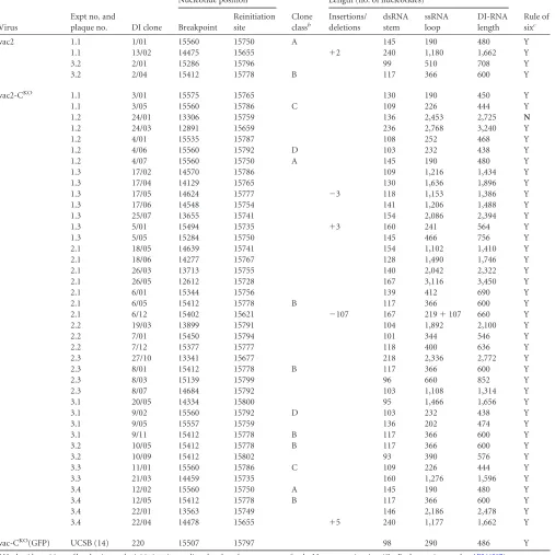
Table 1
summarizes the data for vaccine-derived
DI-FIG 1Schematic drawings of 5=copy-back DI-RNA generation, predicted secondary structure, and replication. (A) 5=copy-back DI-RNAs are hybrids of truncated genome (black) and antigenome (gray) sequences. During replica-tion, viral polymerase initiates RNA synthesis at the 3=antigenome promoter (trailer; open triangle) and prematurely terminates synthesis at the breakpoint. The polymerase then reinitiates synthesis on the newly generated strand and finishes replication by recopying the antigenomic 3=end. The two ends of the DI-RNA are therefore perfectly complementary to each other. (B) Panhandle structure that may be formed through hybridization of the complementary ends of a copy-back DI-RNA in the absence of sufficient N protein. (C) 5= copy-back DI-RNA replication. The genome DI-RNA [(⫺)DI-RNA] (top) contains the antigenome promoter in the copy-back sequence (open triangle). Therefore, it can be replicated by the viral polymerase into a complementary antigenome DI-RNA [(⫹)DI-RNA], which now contains the antigenome pro-moter at the opposite end. The polarity switch within one DI-RNA molecule allows specific amplification using combinations of primers (primer A1 with either primer A2, A3, or A4; arrows) that would bind in the same direction on a regular antigenome molecule.
FIG 2Molecular characterization of DI-RNAs generated by vaccine and wild-type MVs. (A) Analysis of DI-RNAs expressed by multiple clones of vaccine MV (vac2) and wild-type MV (IC323) able to express C protein or not (CKO). Plaques were generated in different rescue experiments, amplified, and ana-lyzed by DI-RNA-specific RT-PCR. Uninfected cells (UI) were included as a negative control. The first number above each lane is the experiment number, and the second number is the plaque number. cDNAs were amplified with the A1/A2 primer combination and analyzed by agarose gel electrophoresis. The numbers on the left of each panel indicate the sizes of the marker bands (in base pairs). (B) Nucleotide positions of the breakpoints and reinitiation sites mapped in DI-RNAs derived from vac2 and vac2-CKO(Table 1). The combi-nations of primers used for amplification are indicated on the left of the three panels. (C) Breakpoint and reinitiation site analysis of IC323 and IC323-CKO viruses (Table 2).
on November 7, 2019 by guest
http://jvi.asm.org/
[image:4.585.322.517.68.583.2]RNAs: 44 sequences were obtained, and 4 of these were derived
from parental vac2 (top four lines) and 40 were derived from
mutant vac2-C
KO.
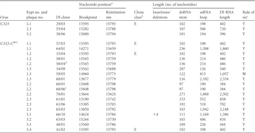
Table 2
summarizes the data for IC323
virus-derived DI-RNAs: 21 sequences were obtained, and 3 of these were
derived from parental IC323 (top three lines) and 18 were derived
from mutant IC323-C
KO. The breakpoints and reinitiation sites
for all DI sequences are summarized in
Fig. 2B
for vac2 and in
Fig.
2C
for IC323 viruses. Breakpoints were distributed broadly
(
Fig. 2B
and
C
). Their frequency of detection depended on the
[image:5.585.42.549.85.592.2]location of the primer used for amplification (primer A2, A3, or
A4) and was inversely proportional to the distance from the 5
=
end
of the genome (
Fig. 2B
and
C
; compare the top, middle, and
bot-tom panels). The average breakpoint position for vac2-derived
DI-RNAs was nucleotide 14,843, with a standard deviation of 831
nucleotides. For IC323-derived DI-RNAs, the average breakpoint
was nt 15,122
⫾
681. In contrast, deduced reinitiation sites for
TABLE 1Vaccine virus-derived DI-RNAs
Virus
Expt no. and
plaque no. DI clone
Nucleotide positiona
Clone classb
Length (no. of nucleotides)
Rule of sixc Breakpoint
Reinitiation site
Insertions/ deletions
dsRNA stem
ssRNA loop
DI-RNA length
vac2 1.1 1/01 15560 15750 A 145 190 480 Y
1.1 13/02 14475 15655 ⫹2 240 1,180 1,662 Y
3.2 2/01 15286 15796 99 510 708 Y
3.2 2/04 15412 15778 B 117 366 600 Y
vac2-CKO 1.1 3/01 15575 15765 130 190 450 Y
1.1 3/05 15560 15786 C 109 226 444 Y
1.2 24/01 13306 15759 136 2,453 2,725 N
1.2 24/03 12891 15659 236 2,768 3,240 Y
1.2 4/01 15535 15787 108 252 468 Y
1.2 4/06 15560 15792 D 103 232 438 Y
1.2 4/07 15560 15750 A 145 190 480 Y
1.3 17/02 14570 15786 109 1,216 1,434 Y
1.3 17/04 14129 15765 130 1,636 1,896 Y
1.3 17/05 14624 15777 ⫺3 118 1,153 1,386 Y
1.3 17/06 14548 15754 141 1,206 1,488 Y
1.3 25/07 13655 15741 154 2,086 2,394 Y
1.3 5/01 15494 15735 ⫹3 160 241 564 Y
1.3 5/05 15284 15750 145 466 756 Y
2.1 18/05 14639 15741 154 1,102 1,410 Y
2.1 18/06 14277 15767 128 1,490 1,746 Y
2.1 26/03 13713 15755 140 2,042 2,322 Y
2.1 26/05 12612 15728 167 3,116 3,450 Y
2.1 6/01 15344 15756 139 412 690 Y
2.1 6/05 15412 15778 B 117 366 600 Y
2.1 6/12 15402 15621 ⫺107 167 219⫹107 660 Y
2.2 19/03 13899 15791 104 1,892 2,100 Y
2.2 7/01 15450 15794 101 344 546 Y
2.2 7/12 15377 15777 118 400 636 Y
2.3 27/10 13341 15677 218 2,336 2,772 Y
2.3 8/01 15412 15778 B 117 366 600 Y
2.3 8/03 15139 15799 96 660 852 Y
2.3 8/07 14684 15792 103 1,108 1,314 Y
3.1 20/05 14334 15800 95 1,466 1,656 Y
3.1 9/02 15560 15792 D 103 232 438 Y
3.1 9/05 15557 15759 136 202 474 Y
3.1 9/11 15412 15778 B 117 366 600 Y
3.2 10/05 15412 15778 B 117 366 600 Y
3.2 10/09 15412 15802 93 390 576 Y
3.3 11/01 15560 15786 C 109 226 444 Y
3.3 21/03 14459 15735 160 1,276 1,596 Y
3.4 12/02 15560 15750 A 145 190 480 Y
3.4 12/05 15412 15778 B 117 366 600 Y
3.4 22/01 13563 15749 146 2,186 2,478 Y
3.4 22/04 14478 15655 ⫹5 240 1,177 1,662 Y
vac-CKO(GFP) UCSB (14) 220 15507 15797 98 290 486 Y
aNucleotide positions of breakpoints and reinitiation sites as aligned to the reference sequence for the Moraten vaccine virus (GenBank accession numberAF266287). b
The letters A to D indicate clone classes when identical DI-RNAs were isolated from independent plaques.
cThe rule of six indicates whether the calculated total length of the DI-RNA is a multiple of six (yes [Y]) or not (no [N]).
on November 7, 2019 by guest
http://jvi.asm.org/
most DI sequences clustered in a narrow region within the last 200
nucleotides of the MV genome (for vac2, nt 15,758
⫾
43; for
IC323, nt 15,758
⫾
50) close to the region containing the sequence
of primer A1 used for amplification. The total length of the
DI-RNAs deduced from the positions of breakpoints and reinitiation
sites (
Tables 1
and
2
, DI-RNA length columns) ranged from about
400 nt to about 3.5 kb. Interestingly, a few identical DI sequences
appeared in virus plaques generated from independent rescue
exper-iments. Identical DI-RNAs were assigned to the same class, labeled
with the letters A to E (
Tables 1
and
2
, Clone class columns).
We also assessed whether the DI-RNAs conformed to the rule
of six, which stipulates that only paramyxovirus genomes with
hexameric lengths are efficiently replicated (
10
,
32
). Indeed, the
calculated total DI-RNA length was a multiple of six for 63 out of
65 clones. However, DI-RNA clones 24/01 (vac2-C
KO) and 59/05
(IC323-C
KO) did not comply with this rule. Interestingly, 6 out of
65 DI-RNAs possessed internal deletions or insertions (
Tables 1
and
2
, Insertions/deletions columns) that made them compliant
with the rule of six (
Tables 1
and
2
, Rule of six columns).
Assum-ing that DI-RNA generation is a purely random process, about
83% (5 out of 6) of newly generated DI-RNAs are predicted to be
noncompliant with the rule of six, but because we found that only
about 3% of the DI-RNAs were noncompliant, we conclude that
DI-RNAs of hexameric length had a replicative advantage.
Clusters of A-to-G and U-to-C transitions in DI-RNA
se-quences.
DI-RNAs of negative-strand RNA viruses (
19
,
33
) and
even full-length MV genomes from diseased human brains (
34
)
can accumulate clusters of A-to-G or U-to-C transitions,
char-acteristic of the action of the cellular ADAR1 deaminase (
35
,
36
). We detected clusters of A-to-G or U-to-C transitions
(bi-ased hypermutation events) in 9 DI-RNA sequences (
Fig. 3A
and
Table 3
). The percentage of nucleotides affected by these
transitions varied from about 4 to 42% (average 21%).
Nota-bly, all identified clones exhibited only one type of transition,
and we did not observe a switch in the type of transition at the
breakpoint.
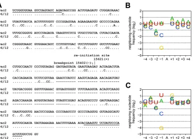
Figure 3A
illustrates one biased hypermutation
event: 27 transitions to C among 70 U residues (39%) within
the sequence prior to the breakpoint and then an additional 9
transitions to C among 41 U residues (22%) after the
reinitia-tion site. Interestingly, the DI-RNA carried an internal delereinitia-tion
in the copy-back sequence that spanned 107 nt between
posi-tions 15,653 and 15,759 (
Fig. 3A
, dashes). The internal deletion
generated a DI-RNA compliant with the rule of six.
ADAR1 is a cellular enzyme that catalyzes the deamination of
adenosines in dsRNA to produce inosine, which is recognized as
guanosine (
35
,
36
). The A-to-I editing activity of ADAR1 is
af-fected by the neighboring nucleotides of the A that is deaminated
(
Fig. 3C
) (
21
). We assessed whether the nucleotides flanking the
[image:6.585.42.545.76.339.2]modification sites in the MV DI-RNAs conformed to the
estab-lished signature for ADAR1. The neighboring nucleotide
frequen-cies (positions
⫺
4 to
⫹
4) of all observed hypermutations (
Fig. 3B
)
were in good agreement with the nearest neighbor signature
re-ported by Eggington et al. (
21
) (
Fig. 3C
is reconstructed from data
available in the supplemental material of the original publication
[
21
]). Most significantly, our data set and that of Eggington et al.
(
21
) show an underrepresentation of C and G residues at the
⫺
1
TABLE 2Wild-type virus-derived DI-RNAs
Virus
Expt no. and
plaque no. DI clone
Nucleotide positiona
Clone classb
Length (no. of nucleotides)
Rule of sixc Breakpoint
Reinitiation site
Insertions/ deletions
dsRNA stem
ssRNA loop
DI-RNA length
IC323 2.1 29/03 15595 15793 E 102 198 402 Y
2.3 55/04 15282 15788 107 506 720 Y
3.2 30/06 15600 15794 101 194 396 Y
IC323-CKO 1.1 57/03 15595 15793 E 102 198 402 Y
1.1 64/01 14271 15659 236 1,388 1,860 Y
1.2 33/04 15595 15793 E 102 198 402 Y
1.2 58/01 15545 15759 136 214 486 Y
1.2 58/04d 15545 15759 136 214 486 Y
1.3 34/09 15562 15688 207 126 540 Y
1.3 59/05 14960 15773 122 813 1,057 N
1.3 68/01 13677 15779 116 2,102 2,334 Y
2.2 60/01 15608 15798 97 190 384 Y
2.2 60/06e 15608 15798 97 190 384 Y
2.2 70/01 13664 15624 271 1,960 2,502 Y
2.3 61/01 15190 15742 153 552 858 Y
2.3 61/06 15385 15703 192 318 702 Y
2.3 65/03 13850 15792 103 1,942 2,148 Y
3.1 66/10 14624 15784 ⫹4 111 1,160 1,386 Y
3.2 63/03 15244 15730 165 486 816 Y
3.3 40/01 15560 15786 109 226 444 Y
3.4 41/02 15595 15793 E 102 198 402 Y
a
Nucleotide positions of breakpoints and reinitiation sites as aligned to the reference sequence for the IC-B wild-type virus from which IC323 is derived (GenBank accession numberAB016162).
b
The letter E indicates the clone class when identical DI-RNAs were isolated from independent plaques.
cThe rule of six indicates whether the calculated total length of the DI-RNA is a multiple of six (yes [Y]) or not (no [N]). d
This clone differed from 58/01 of the same rescue by a biased hypermutation event. eThis clone differed from 60/01 of the same rescue by a biased hypermutation event.
on November 7, 2019 by guest
http://jvi.asm.org/
position and the overrepresentation of U at the
⫺
2 position. This
suggests that ADAR1 may have acted on the DI-RNAs generated
during MV infection.
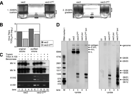
DI-RNAs are encapsidated and protected by lipid envelopes.
To assess whether DI-RNAs are incorporated into viral particles,
we generated two virus stocks: one without detectable DI-RNA
(vac2; we started with plaque 1.2;
Fig. 2A
) and the other with high
DI-RNA levels (vac2-C
KO; we started with plaque 2.1;
Fig. 2A
).
Since
⬎
90% of infectious MV remains cell associated,
intracellu-lar infectivity was purified using a two-step density gradient
pro-tocol (
Fig. 4A
). The visible bands for vac2 and vac2-C
KOparticles
had similar intensities in both steps, suggesting that the amount of
physical particles was similar for the two preparations. However,
the infectious titers of the purified virus preparations differed
be-tween vac2 and vac2-C
KOby about 10-fold (
Fig. 4B
, right
col-umns), similar to the ratio of the original stocks (
Fig. 4B
, left
columns).
To assess whether the viral RNAs were encapsidated and
pro-tected by a lipid envelope, purified virions were treated with
dif-ferent combinations of nuclease (Benzonase), protease (trypsin),
and nonionic detergent (Triton X-100). To control for enzymatic
activities, the viral envelope glycoprotein H and nucleocapsid
pro-tein N were analyzed after treatment (
Fig. 4C
, top two panels). For
both parental and C
KOvirion preparations, trypsin treatment in
the absence of detergent led to the proteolytic cleavage of most H
protein (
Fig. 4C
, lanes 4 and 9), whereas N protein was affected
only to a small extent, if at all. However, when virions were treated
with Triton X-100 prior to trypsinization, both H and N were
completely degraded by trypsin (
Fig. 4C
, lanes 5 and 10).
We also extracted RNA from the samples, reverse transcribed
it, and subjected the cDNA to PCR analysis using
DI-RNA-spe-cific primers (
Fig. 4C
, panel A1/A2) and full-length RNA-specific
primers (
Fig. 4C
, panel A1/B1). Benzonase treatment in the
[image:7.585.107.489.64.338.2]ab-FIG 3ADAR1-like hypermutations in DI-RNA clones. (A) Alignment of the sequence of DI-RNA clone 06/12 to the vac2 genome (nt 15639 to 15402) and antigenome (nt 15621 to 15887) sequences (GenBank accession numberAF266287). The binding sites of the A1 and A2 primers are underlined (top line and bottom two lines, respectively). Identical nucleotides are shown as dots. The breakpoint and reinitiation site nucleotides are indicated. Deleted nucleotides are shown as dashes. (B) Frequency analysis of nucleotides flanking hypermutated sites in DI-RNA clones (all 9 events detected here;Table 3). (C) Frequency of neighboring nucleotides flanking ADAR1 hypermutation sites definedin vitro(21).
TABLE 3ADAR1-mediated hypermutations in DI-RNA clones
Virus
Expt no. and
plaque no. DI clone Polaritya No. of hypermutations observed/no. of total nucleotidesb
% editingc
vac2-CKO 2.1 06/12 U
¡C 36/111 31.5 2.3 08/03 U¡C 6/141 4.3 3.1 20/05 U¡C 16/126 12.7
IC323-CKO 1.2 58/04 U
¡C 5/52 9.6 1.3 59/03d A
¡G 17/67 25.4
2.2 60/06 U¡C 8/21 38.1
3.1 62/04d U
¡C 19/54 35.2 3.1 62/06d A
¡G 28/67 41.8 3.1 62/11d U
¡C 11/54 20.4 a
Polarity indicates whether the hypermutations were observed as A¡G or U¡C in the genomic orientation.
b
The number of mutations observed/number of total nucleotides of the respective type (A or U) in a cluster.
c
The percentage is the calculated frequency of transitions observed over possible events.
d
These sequences were derived through semispecific hybridization of primer A2 at position⫺15670 of the MV genome.
on November 7, 2019 by guest
http://jvi.asm.org/
[image:7.585.41.286.496.651.2]sence of Triton X-100 and trypsin affected both DI-RNA- and
full-length RNA levels only to a small extent (
Fig. 4C
, lanes 2 and
7). Treatment of virions with trypsin only prior to nuclease
treat-ment did not affect the strength of the PCR signals (
Fig. 4C
, lanes
4 and 9). However, when virions were treated with Triton X-100,
the viral RNA became accessible to Benzonase, leading to the
com-plete loss of the PCR signals (
Fig. 4C
, lanes 3, 5, 8, and 10). We
conclude that both DI-RNA and full-length RNA are encapsidated
by N and that the majority of these ribonucleocapsids (RNPs) are
protected by a lipid envelope. Notably, treatment of the RNPs
with trypsin was not required when virions were treated with
de-tergent (
Fig. 4C
, lanes 3 and 8), indicating that RNPs became
nuclease sensitive after Triton X-100 treatment.
High levels of DI-RNA genomes are found in particles.
To
further characterize the viral RNP, after Benzonase treatment we
extracted RNA from 1
⫻
10
6TCID
50
s of purified virions. As a
control, we also extracted total RNA from Vero cells infected with
parental and C
KOviruses and from uninfected cells. These samples
were analyzed by Northern blotting using single-strand RNA
probes complementary to the L/trailer region of the antigenome
(
Fig. 4D
,
⫹
probe) or genome (
Fig. 4D
,
⫺
probe).
Figure 4D
shows an analysis of the antigenomic (left) and
genomic (right) RNA from uninfected cells, infected cells, and
purified virus particles. As expected, the 16-kb full-length
antige-nomic and geantige-nomic RNAs were detected in all four virus samples.
However, whereas the antigenome bands for vac2 and vac2-C
KOin infected cells had similar intensities (
Fig. 4D
, left; compare lanes
2 and 3), vac2-C
KOshowed less genomic RNA than vac2 (
Fig. 4D
,
right; compare lanes 2 and 3), correlating with the reduced
infec-tivity recovered from vac2-C
KO-infected cells compared to the
infectivity recovered from vac2-infected cells. The difference in
intensity of the genome band was not detected in purified virion
samples (
Fig. 4D
, right; compare lanes 4 and 5), since we loaded
RNA from equal numbers of infectious units of both viruses.
However, antigenome ratios were altered in purified virions (
Fig.
4D
, left; compare lanes 4 and 5), indicating that vac2-C
KOhad
incorporated higher levels of the antigenome into virions than
vac2. For total RNA samples from infected cells, the antigenome
probe also detected bands for L mRNA (about 7 kb) and two
bands that corresponded to polycistronic H-L (9 kb) and F-H-L
mRNAs (11 kb) (
37
). As expected, the L, H-L, and F-H-L mRNAs
were not detected in the purified viruses.
Using either the antigenome probe or the genome probe, we
observed multiple RNA species with a smaller molecular size that
were present only in vac2-C
KOsamples. The sizes of these bands
ranged from 0.1 kb to about 7 kb, with most being in the 0.3- to
FIG 4Analysis of nucleic acids in vac2- and vac2-CKO-infected cells and in purified vac2 and vac2-CKOparticles. (A) Two-step gradient purification of MV particles. Virus bands are indicated by brackets. (B) Titers (numbers of TCID50s per milliliter) of virus stocks prior to and after purification. (C) RNA protection assay with purified virions. Samples were treated with the indicated combinations of trypsin, Triton X-100, and Benzonase; immunoblots were against MV H and N proteins; RT-PCR/PCR analysis was performed for DI-RNAs (A1/A2 primers) and full-length RNA (A1/B1 primers). (D) Northern blot analysis of total RNA from about 3⫻105uninfected (UI; lane 1) or infected (lanes 2 and 3) Vero cells harvested at 32 h postinfection, as well as RNA extracted from 1⫻106TCID
50s of purified (pur.) and Benzonase-treated vac2 (lane 4) and vac2-CKO(lane 5) particles. The positions of the antigenome and L, H-L, and F-H-L mRNAs (⫹probe) are indicated on the right side of the left panel. The identities of the genomic RNA and the DI-RNAs (⫺probe) mapped by PCR amplification are indicated on the right side of the right panel. Lane M, molecular size marker. The numbers on the left side of the left panel indicate the sizes of the marker bands (in bases).
on November 7, 2019 by guest
http://jvi.asm.org/
[image:8.585.83.503.65.366.2]2-kb range. The sizes of some RNAs were similar to those of
spe-cific DI-RNAs of vac2-C
KOplaque 1 from experiment 2 (referred
to as 2.1), as identified by the PCR cloning approach (
Table 1
;
compare the DI-RNA clone identifiers with those marked in
Fig. 4D
). Similarly sized RNAs were also detected, but in greater
abundance, in purified vac2-C
KOvirions (
Fig. 4D
, right, lane 5).
We detected more RNA species with the genome probe than with
the antigenome probe, consistent with the notion that these RNAs
may be truncated genomes. We conclude that DI-RNAs are
incor-porated into intracellular particles since they are protected by a
lipid envelope. The 10-fold reduced infectivity of vac2-C
KOcor-relates with the enhanced incorporation of DI-RNA genomes into
intracellular particles.
C
KOviruses generate dsRNA very early.
Since DI-RNAs are
most efficiently amplified at a high MOI, we sought to limit
cell-cell fusion, a process resulting in the bulk transfer of genomes. We
therefore carried out infections on Vero cells, rather than on the
more fusogenic Vero-hSLAM cells. Because we needed viruses
expressing an alternative fluorescent reporter protein for
subse-quent competition experiments, we generated vac2-based variants
expressing mCherry (mCherryNLS [
28
]).
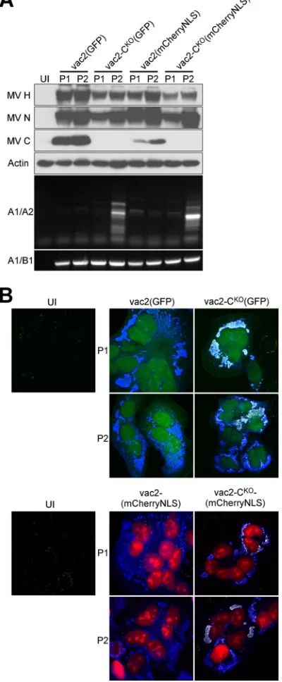
Figure 5A
shows
pro-tein expression and DI-RNA levels after two passages of the
vac2-based viruses expressing either GFP or mCherryNLS. As expected,
passage 1 DI-RNA amplification in Vero cells was minimally
de-tectable, if it was detectable at all (lanes P1, A1/A2 panel).
How-ever, significant amounts of DI-RNA-specific PCR products were
detected for both mutant C
KOviruses at passage 2 but not for
parental vac2 (lanes P2, A1/A2 panel).
Having previously documented the kinetics of dsRNA
produc-tion of standard and C
KOviruses in HeLa cells (
14
), we infected
these cells and performed immunofluorescence staining for viral
N protein and dsRNA (
Fig. 5B
). We found strong dsRNA staining
(
Fig. 5B
, yellow, seen as white when superimposed onto blue)
colocalizing with viral replication sites (revealed by N protein;
Fig.
5B
, blue signal) in passages 1 and 2 of either C
KOvirus. In contrast,
the C-expressing parental vac2 of either passage did not elicit
de-tectable dsRNA staining at viral replication sites. Thus,
immuno-fluorescence staining detects dsRNA before DI-RNA can be
de-tected by PCR amplification.
The C
KOphenotype is dominant.
To assess whether C-protein
expression in
trans
can limit DI-RNA amplification, we coinfected
cells with virus stocks possessing either high or undetectable
DI-RNA levels. These viruses expressed different fluorescent proteins
(either GFP or mCherryNLS) for easy replication tracking. We
infected interferon response-defective Granta-519 cells with each
virus at an equivalent MOI and quantified the GFP and mCherry
expression at 48 h postinoculation by fluorescence-activated cell
sorter analysis (
Fig. 6
).
Figure 6A
shows the levels of GFP expression in cells left
uninfected (panel 1) or infected only with either virus
express-ing GFP [vac2(GFP) in panel 2; vac2-C
KO(GFP) in panel 3]
or only with either virus expressing mCherryNLS [vac2
(mCherryNLS) in panel 4; vac2-C
KO(mCherryNLS) in panel 7].
Panels 5, 6, 8, and 9 document the levels of GFP expression in cells
coinfected with two viruses, as indicated on the top of each
col-umn and on the left of each row. The median fluorescence
inten-sity (MFI) of vac2(GFP) (panel 2) was about 4,500, or 110 times
over the background MFI of 41 (panel 1); the MFI of
vac2-C
KO(GFP) (panel 3) was about 70 times over the background MFI.
Infection with mCherryNLS-expressing viruses was not detected
in the GFP channel (panels 4 and 7).
We then measured GFP expression levels in cells coinfected
with 1:1 mixtures of viruses that were either DI-RNA poor
(vac2-derived stocks) or DI-RNA rich (vac2-C
KO-derived stocks). We
FIG 5DI-RNA and dsRNA generation by CKOviruses expressing either GFP or mCherryNLS. (A) Western blot and PCR analysis of P1 and P2 of fluores-cent reporter-expressing vac2 and vac2-CKO. The antisera used for detection are indicated on the left of the top four panels. Primer pairs used for amplifi-cation are indicated on the left of the bottom two panels. UI, uninfected cells. (B) Confocal microscope analysis of HeLa cells infected with fluorescent re-porter-expressing vac2 and vac2-CKO. (Top) Autofluorescence of GFP (green); (bottom) autofluorescence of mCherryNLS (red); (all panels) immu-nofluorescence staining for dsRNA (yellow) and MV N protein (blue). Mag-nifications,⫻40.
on November 7, 2019 by guest
http://jvi.asm.org/
undertook these mixed infections to assess whether DI-RNA-rich
stocks interfere with the replication of DI-RNA-poor stocks
(dominant negative effects). Alternatively, this experiment might
reveal a positive effect of DI-RNA-poor (C-protein-expressing)
stocks on the replication of DI-RNA-rich stocks, presumably
through
trans
-complementation by the C protein.
Figure 6B
illustrates the results of mixed infection analyses.
Figure 6B
, left, shows that addition of a DI-RNA-poor stock
[vac2(mCherryNLS)line] to another DI-RNA-poor stock [vac2
(GFP)] reduced the GFP MFI about 1.5-fold (compare the light
green area with the dark green dotted line). Addition of a
DI-RNA-rich stock [vac2-C
KO(mCherryNLS)] reduced the GFP MFI
of the DI-RNA-poor stock more than 8-fold (compare the light
green area with the dark green solid line). In contrast, as shown
in
Fig. 6B
, right, addition of a DI-RNA-poor stock [vac2
(mCherryNLS)line] to a DI-RNA-rich stock [vac2-C
KO(GFP)]
re-duced the GFP MFP 1.2-fold (compare the light green area with
the dark green dotted line), while addition of a DI-RNA-rich stock
[vac2-C
KO(mCherryNLS)] to another DI-RNA-rich stock had a
more pronounced negative effect (a 3.5-fold reduction of the GFP
MFI; compare the light green area with the dark green solid line).
Because DI-RNA-rich virus stocks in any combination had a
neg-ative effect on the growth of the DI-RNA-poor stocks and because
DI-RNA-poor stocks which expressed C protein had no positive
effect on DI-RNA-rich stocks, we conclude that the C
KOeffect is
dominant.
DISCUSSION
MV C-protein function.
Viruses of the
Morbillivirus
,
Henipavirus
,
and
Respirovirus
genera of the
Paramyxoviridae
family express one
or more C proteins (
2
). However, different studies have revealed
alternative functions of C proteins. In the case of MV and Nipah
virus, minigenome assays have suggested that C protein
down-regulates genome replication (
38
,
39
). Inhibition of transcription
but not of replication was described for Sendai virus (SeV) in
in
vitro
assays (
40
), although SeV C protein also is reported to be
essential for the maintenance of negative-strand RNA genomes in
virus particles (
41
) and an SeV-C
KOmutant did not undergo a
shift from early (antigenome-dominated) to late
(genome-domi-nated) replication (
42
). The generation and accumulation of
DI-RNAs in C-defective paramyxoviruses would account for many of
these observations.
In the absence of C protein, production of full-length genomes
would be reduced and defective genomes would be incorporated
into virions instead. In addition, the dsRNA structures formed by
DI-RNAs are known to activate PKR (
14
), which induces an arrest
in protein synthesis via the cellular stress response, thereby
delay-ing viral replication. While in a preceddelay-ing study we detected
DI-RNA in one MV-C
KOstock that had been passaged several times at
a low multiplicity of infection (
14
), here we provide evidence for
the generation of DI-RNAs as early as passage 1 following virus
rescue. Moreover, we observed about 10 times more DI-RNA in
C
KOvirus infection than in parental virus infection, implying a
genetic link between the C
KOphenotype and DI-RNA generation.
Notably, C
KOmutants of SeV and human parainfluenza virus type
1 have also been reported to generate double-stranded RNA and
activate PKR, but these studies did not address the origin or the
structure of these RNAs (
43
,
44
). Although C
KOmutants of several
paramyxoviruses can be rescued and are only moderately
attenu-ated in cultured cells (
16
), they are strongly attenuated
in vivo
(
45
,
46
). Thus, C proteins of diverse paramyxoviruses may share a
similar function, to enhance RNA replication processivity.
The frequency but not the quality of DI-RNA generation is
determined by the C protein. Most breakpoints of the DI-RNAs
are widely distributed along the MV genome, with the frequency
of the appearance of a breakpoint at a specific position being
in-versely proportional to the distance from the 5
=
end of the
ge-nome. In contrast, reinitiation sites are clustered in a narrow
re-gion close to the 5
=
end of the genome. We note, however, that
repositioning of the copy-back primer (
Fig. 1C
, primer A1) to a
more internal binding site resulted in the detection of DI-RNAs
with more internal reinitiation sites (data not shown).
Our Northern blot analysis showed several RNAs hybridizing
with the genome probe (
Fig. 4C
, right) that did not hybridize with
the antigenome probe (
Fig. 4C
, left) and whose sizes did not
cor-respond to the calculated sizes of any cloned DI-RNA. These
spe-cies may possibly reflect truncated genomes lacking the copy-back
FIG 6Dominance of the CKOphenotype. (A) GFP fluorescence intensities of cells coinfected with viruses indicated on the top and on the left. UI, uninfected cells. Vertical axis, cell counts (linear scale); horizontal axis, GFP fluorescence intensity (FI; log10scale). (B) Overlay of graphs from panel A. (Left) Summary of coinfections with vac2(GFP); (right) summary of coinfections with vac2-CKO(GFP). Gray area, uninfected cells (panel 1); light green area, single GFP-virus infection (panel 2 or 3); dotted green line, GFP-GFP-virus coinfected with vac2(mCherryNLS) (panel 5 or 6); solid green line, GFP-virus coinfected with vac2-CKO(mCherryNLS) (panel 8 or 9).
on November 7, 2019 by guest
http://jvi.asm.org/
[image:10.585.63.267.71.422.2]sequence. Indeed, several kinds of defective replication products
have been observed for vesicular stomatitis virus (
19
,
47
) and SeV
(
48
,
49
).
The C
KOphenotype of MV is dominant: a C-protein-defective
virus inhibited the replication efficiency of a C-protein-expressing
virus in
trans
. On the other hand, a C-protein-expressing virus did
not prevent DI-RNA replication once DI-RNA had been
gener-ated. Conceivably, the C protein might function together with the
polymerase complex in separating the nascent and template RNA
hybrid, or, alternatively, C protein might support the rapid
cotranscriptional encapsidation of the nascent RNA with N
pro-tein. Future studies will discriminate between these possibilities.
ADAR1-mediated editing and the PKR-ADAR balancing
model.
DI-RNAs of negative-strand RNA viruses are potent
trig-gers of innate immune receptors, including PKR, mda-5, and
RIG-I (
12–14
), thereby inducing the expression of type I IFNs
in
vitro
(
50
) and
in vivo
(
51
). The MV DI-RNAs characterized here, if
not cotranscriptionally encapsidated, form perfect hairpins
throughout the double-stranded structure. These hairpins are
ADAR1 substrates
in vitro
(
23
). Destabilization of these hairpins
through A-to-I editing could account for the proviral function of
ADAR1, which was documented in several viral infections and
attributed to suppression of activation of innate immune
re-sponses (
52
,
53
). For example, in cell lines deficient in ADAR1
expression, MV strongly induces IFN (
54
), PKR activation (
55
),
and the cellular stress response (
56
,
57
). Furthermore, higher
lev-els of dsRNA accumulate in ADAR1-deficient cells infected with
MV than in cells sufficient in ADAR1 expression (
14
).
Herein we provide evidence for the enzymatic activity of
ADAR1 on MV DI-RNAs. We identified several DI-RNA
se-quences bearing clusters of A-to-G or U-to-C mutations and show
through a computational approach that the sequences flanking
the edited nucleotides, as well as the average editing frequency,
were characteristic of ADAR1, but not ADAR2 (
21
). Of the three
known ADARs, only the IFN-inducible ADAR1 isoform (also
called p150) is cytoplasmic (
58
,
59
) and localizes to stress granules
(
56
). These granules are induced during MV-C
KOinfection and
form in close proximity to viral replication sites (
14
). In contrast,
the constitutively expressed isoforms of ADAR1 (p110) and
ADAR2 are exclusively found in the cell nuclei (
53
). Thus, the
subcellular cytoplasmic localization of only ADAR1-p150 is
cor-rect for editing of MV RNAs, which are exclusively replicated in
the cytoplasm (
2
).
Notably, in individual DI-RNAs the transitions were of the
same type (either A to G or U to C) and occurred on both sides of
the breakpoint and reinitiation site. This pattern indicates that the
generated DI-RNAs were direct substrates for editing by ADAR1
but not the full-length MV genomes. If the latter were the case, the
polarity switch between breakpoint and reinitiation site would
have also caused the ADAR1 transitions to change within the
DI-RNA molecule. Thus, our findings suggest that ADAR1 editing
destabilizes unencapsidated DI-RNAs that have dsRNA
struc-tures. These unencapsidated DI-RNAs may be the
double-stranded RNA that elicits innate immune responses in
negative-strand RNA virus infections. ADAR1 A-to-I editing has also been
documented in full-length RNA virus genomes (
33
,
34
,
60
,
61
)
and was recently inferred by deep sequencing analyses of several
paramyxoviruses (
62
,
63
).
DI-RNAs as a frequent by-product of wild-type and
vaccine-lineage infections.
DI-RNAs were originally detected in
labora-tory strains propagated at a high multiplicity (
9
) and later also in
commercial vaccines (
64
,
65
). Here we establish that DI-RNAs can
accumulate in the earliest passage available for analysis following
the initial virus rescue. Moreover, we documented DI-RNA
gen-eration in a wild-type MV strain, thereby providing evidence that
DI-RNAs are not restricted to vaccine-lineage viruses and have the
potential to influence virulence.
DI-RNAs may alter the characteristics of MV live-attenuated
vaccines and modulate the efficacy of recombinant MV
engi-neered for vaccination against heterologous pathogens or used as
oncolytic vectors (
27
,
66–68
). While vaccine producers seek to
avoid DI-RNAs in order to standardize efficacy, oncolytic potency
may not rely uniquely on efficient viral replication and cell lysis
but may also rely on immunostimulatory properties (
69
,
70
). The
balance between replication efficiency and immune activation of
an oncolytic vector may be an important key to a successful
ther-apy, and immunostimulatory DI-RNAs might be a desirable
fea-ture of certain oncolytic vectors. Further studies of the MV C
protein and its function may lead to the design of viruses that
strike an optimal balance between replication efficiency and
im-mune activation.
ACKNOWLEDGMENTS
This work was supported in part by research grant AI-63476 (to R.C.) and research grants AI-12520 and AI-20611 (to C.E.S.) from the National Institute of Allergy and Infectious Diseases, National Institutes of Health, U.S. Public Health Service, and by research fellowship PF791/1-1 from the Deutsche Forschungsgemeinschaft (to C.K.P.). G.M.M., W.E.M., and X.M. were supported in part by the Mayo Graduate School.
We thank Patricia Devaux, Marie Frenzke, Chanakha Navaratnarajah, Crystal Mendoza, and Ryan Donohue for critically reading and discussing the manuscript.
REFERENCES
1.Rima BK, Duprex WP.2009. The measles virus replication cycle. Curr Top Microbiol Immunol 329:77–102. http://dx.doi.org/10.1007/978-3 -540-70523-9_5.
2.Lamb RA, Parks GD.2013. Paramyxoviridae, p 957–995.InKnipe DM, Howley PM, Cohen JI, Griffin DE, Lamb RA, Martin MA, Racaniello VR, Roizman B (ed), Fields virology, 6th ed, vol 1. Lippincott Williams & Wilkins, Philadelphia, PA.
3.Griffin DE.2013. Measles virus, p 1042–1069.InKnipe DM, Howley PM, Cohen JI, Griffin DE, Lamb RA, Martin MA, Racaniello VR, Roizman B (ed), Fields virology, 6th ed, vol 1. Lippincott Williams & Wilkins, Phila-delphia, PA.
4.Longhi S.2009. Nucleocapsid structure and function. Curr Top Micro-biol Immunol329:103–128.http://dx.doi.org/10.1007/978-3-540-70523 -9_6.
5.Schrag SJ, Rota PA, Bellini WJ.1999. Spontaneous mutation rate of measles virus: direct estimation based on mutations conferring monoclo-nal antibody resistance. J Virol73:51–54.
6.Domingo E, Sheldon J, Perales C. 2012. Viral quasispecies evolution. Microbiol Mol Biol Rev76:159 –216.http://dx.doi.org/10.1128/MMBR .05023-11.
7.Domingo E, Holland JJ.1997. RNA virus mutations and fitness for survival. Annu Rev Microbiol51:151–178.http://dx.doi.org/10.1146 /annurev.micro.51.1.151.
8.Moya A, Elena SF, Bracho A, Miralles R, Barrio E.2000. The evolution of RNA viruses: a population genetics view. Proc Natl Acad Sci U S A
97:6967– 6973.http://dx.doi.org/10.1073/pnas.97.13.6967.
9.Kolakofsky D.1976. Isolation and characterization of Sendai virus DI-RNAs. Cell8:547–555.http://dx.doi.org/10.1016/0092-8674(76)90223-3. 10. Calain P, Roux L. 1993. The rule of six, a basic feature for efficient replication of Sendai virus defective interfering RNA. J Virol67:4822– 4830.
11. Holland JJ, Kennedy SIT, Semler BL, Jones CL, Roux L, Grabau EA.
on November 7, 2019 by guest
http://jvi.asm.org/
1980. Defective interfering RNA viruses and the host-cell response, p 137– 192.InFraenkel-Conrat H, Wagner RR (ed), Comprehensive virology, vol 16. Plenum Press, New York, NY.
12. Baum A, Sachidanandam R, Garcia-Sastre A.2010. Preference of RIG-I for short viral RNA molecules in infected cells revealed by next-generation sequencing. Proc Natl Acad Sci U S A107:16303–16308.http://dx.doi.org /10.1073/pnas.1005077107.
13. Runge S, Sparrer KM, Lassig C, Hembach K, Baum A, Garcia-Sastre A, Soding J, Conzelmann KK, Hopfner KP.2014. In vivo ligands of MDA5 and RIG-I in measles virus-infected cells. PLoS Pathog10:e1004081.http: //dx.doi.org/10.1371/journal.ppat.1004081.
14. Pfaller CK, Radeke MJ, Cattaneo R, Samuel CE.2014. Measles virus C protein impairs production of defective copyback double-stranded viral RNA and activation of protein kinase R. J Virol88:456 – 468.http://dx.doi .org/10.1128/JVI.02572-13.
15. Huang AS.1973. Defective interfering viruses. Annu Rev Microbiol27:
101–117.http://dx.doi.org/10.1146/annurev.mi.27.100173.000533. 16. Devaux P, von Messling V, Songsungthong W, Springfeld C, Cattaneo
R.2007. Tyrosine 110 in the measles virus phosphoprotein is required to block STAT1 phosphorylation. Virology360:72– 83.http://dx.doi.org/10 .1016/j.virol.2006.09.049.
17. Takeda M, Takeuchi K, Miyajima N, Kobune F, Ami Y, Nagata N,
Suzaki Y, Nagai Y, Tashiro M.2000. Recovery of pathogenic measles virus from cloned cDNA. J Virol74:6643– 6647.http://dx.doi.org/10.1128 /JVI.74.14.6643-6647.2000.
18. Marcus PI, Gaccione C. 1989. Interferon induction by viruses. XIX. Vesicular stomatitis virus—New Jersey: high multiplicity passages gener-ate interferon-inducing, defective-interfering particles. Virology171:
630 – 633.
19. O’Hara PJ, Nichol ST, Horodyski FM, Holland JJ. 1984. Vesicular stomatitis virus defective interfering particles can contain extensive genomic sequence rearrangements and base substitutions. Cell36:915– 924.http://dx.doi.org/10.1016/0092-8674(84)90041-2.
20. Huang AS, Baltimore D.1970. Defective viral particles and viral disease processes. Nature226:325–327.http://dx.doi.org/10.1038/226325a0. 21. Eggington JM, Greene T, Bass BL.2011. Predicting sites of ADAR editing
in double-stranded RNA. Nat Commun2:319.http://dx.doi.org/10.1038 /ncomms1324.
22. Wulff BE, Sakurai M, Nishikura K.2011. Elucidating the inosinome: global approaches to adenosine-to-inosine RNA editing. Nat Rev Genet
12:81– 85.http://dx.doi.org/10.1038/nrg2915.
23. Lehmann KA, Bass BL.2000. Double-stranded RNA adenosine deami-nases ADAR1 and ADAR2 have overlapping specificities. Biochemistry
39:12875–12884.http://dx.doi.org/10.1021/bi001383g.
24. Ono N, Tatsuo H, Hidaka Y, Aoki T, Minagawa H, Yanagi Y.2001. Measles viruses on throat swabs from measles patients use signaling lym-phocytic activation molecule (CDw150) but not CD46 as a cellular recep-tor. J Virol75:4399 – 4401.http://dx.doi.org/10.1128/JVI.75.9.4399-4401 .2001.
25. Radecke F, Spielhofer P, Schneider H, Kaelin K, Huber M, Dotsch C, Christiansen G, Billeter MA.1995. Rescue of measles viruses from cloned DNA. EMBO J14:5773–5784.
26. Jadayel DM, Lukas J, Nacheva E, Bartkova J, Stranks G, De Schouwer PJ, Lens D, Bartek J, Dyer MJ, Kruger AR, Catovsky D.1997. Potential role for concurrent abnormalities of the cyclin D1, p16CDKN2 and p15CDKN2B genes in certain B cell non-Hodgkin’s lymphomas. Func-tional studies in a cell line (Granta 519). Leukemia11:64 –72.
27. del Valle JR, Devaux P, Hodge G, Wegner NJ, McChesney MB, Catta-neo R.2007. A vectored measles virus induces hepatitis B surface antigen antibodies while protecting macaques against measles virus challenge. J Virol81:10597–10605.http://dx.doi.org/10.1128/JVI.00923-07. 28. Taylor MP, Kobiler O, Enquist LW.2012. Alphaherpesvirus axon-to-cell
spread involves limited virion transmission. Proc Natl Acad Sci U S A
109:17046 –17051.http://dx.doi.org/10.1073/pnas.1212926109. 29. Toth AM, Devaux P, Cattaneo R, Samuel CE.2009. Protein kinase PKR
mediates the apoptosis induction and growth restriction phenotypes of C protein-deficient measles virus. J Virol83:961–968.http://dx.doi.org/10 .1128/JVI.01669-08.
30. Cathomen T, Naim HY, Cattaneo R.1998. Measles viruses with altered envelope protein cytoplasmic tails gain cell fusion competence. J Virol
72:1224 –1234.
31. Schonborn J, Oberstrass J, Breyel E, Tittgen J, Schumacher J, Lukacs N.
1991. Monoclonal antibodies to double-stranded RNA as probes of RNA
structure in crude nucleic acid extracts. Nucleic Acids Res19:2993–3000. http://dx.doi.org/10.1093/nar/19.11.2993.
32. Rager M, Vongpunsawad S, Duprex WP, Cattaneo R.2002. Polyploid measles virus with hexameric genome length. EMBO J21:2364 –2372. http://dx.doi.org/10.1093/emboj/21.10.2364.
33. Cattaneo R.1994. Biased (A¡I) hypermutation of animal RNA virus genomes. Curr Opin Genet Dev 4:895–900. http://dx.doi.org/10.1016 /0959-437X(94)90076-0.
34. Cattaneo R, Schmid A, Eschle D, Baczko K, ter Meulen V, Billeter MA.
1988. Biased hypermutation and other genetic changes in defective mea-sles viruses in human brain infections. Cell55:255–265.http://dx.doi.org /10.1016/0092-8674(88)90048-7.
35. Kim U, Wang Y, Sanford T, Zeng Y, Nishikura K.1994. Molecular cloning of cDNA for double-stranded RNA adenosine deaminase, a can-didate enzyme for nuclear RNA editing. Proc Natl Acad Sci U S A91:
11457–11461.http://dx.doi.org/10.1073/pnas.91.24.11457.
36. Bass BL, Weintraub H. 1988. An unwinding activity that covalently modifies its double-stranded RNA substrate. Cell55:1089 –1098.http://dx .doi.org/10.1016/0092-8674(88)90253-X.
37. Cattaneo R, Rebmann G, Schmid A, Baczko K, ter Meulen V, Billeter MA.1987. Altered transcription of a defective measles virus genome de-rived from a diseased human brain. EMBO J6:681– 688.
38. Bankamp B, Wilson J, Bellini WJ, Rota PA. 2005. Identification of naturally occurring amino acid variations that affect the ability of the measles virus C protein to regulate genome replication and transcription. Virology336:120 –129.http://dx.doi.org/10.1016/j.virol.2005.03.009.
39. Sleeman K, Bankamp B, Hummel KB, Lo MK, Bellini WJ, Rota PA.
2008. The C, V and W proteins of Nipah virus inhibit minigenome replication. J Gen Virol89:1300 –1308.http://dx.doi.org/10.1099/vir. 0.83582-0.
40. Curran J, Marq JB, Kolakofsky D.1992. The Sendai virus nonstructural C proteins specifically inhibit viral mRNA synthesis. Virology189:647– 656.http://dx.doi.org/10.1016/0042-6822(92)90588-G.
41. Irie T, Nagata N, Yoshida T, Sakaguchi T.2008. Paramyxovirus Sendai virus C proteins are essential for maintenance of negative-sense RNA ge-nome in virus particles. Virology374:495–505.http://dx.doi.org/10.1016 /j.virol.2008.01.004.
42. Irie T, Okamoto I, Yoshida A, Nagai Y, Sakaguchi T.2014. Sendai virus C proteins regulate viral genome and antigenome synthesis to dictate the negative genome polarity. J Virol88:690 – 698.http://dx.doi.org/10.1128 /JVI.02798-13.
43. Boonyaratanakornkit J, Bartlett E, Schomacker H, Surman S, Akira S, Bae YS, Collins P, Murphy B, Schmidt A.2011. The C proteins of human parainfluenza virus type 1 limit double-stranded RNA accumulation that would otherwise trigger activation of MDA5 and protein kinase R. J Virol
85:1495–1506.http://dx.doi.org/10.1128/JVI.01297-10.
44. Takeuchi K, Komatsu T, Kitagawa Y, Sada K, Gotoh B.2008. Sendai virus C protein plays a role in