Pseudorabies Virus US3 Protein Kinase Protects Infected Cells from
NK Cell-Mediated Lysis via Increased Binding of the Inhibitory NK
Cell Receptor CD300a
K. Grauwet,aM. Vitale,bS. De Pelsmaeker,aT. Jacob,aK. Laval,aL. Moretta,cM. Parodi,dS. Parolini,eC. Cantoni,d,f,gH. W. Favoreela
Department of Virology, Parasitology, and Immunology, Faculty of Veterinary Medicine, Ghent University, Merelbeke, Belgiuma
; IRCCS AOU San Martino-IST, Genoa, Italyb ; IRCCS Ospedale Pediatrico Bambino Gesù, Rome, Italyc
; Department of Experimental Medicine (DIMES), University of Genoa, Genoa, Italyd
; Dipartimento di Medicina Molecolare e Traslazionale, University of Brescia, Brescia, Italye
; Istituto Giannina Gaslini, Genoa, Italyf
; Center of Excellence for Biomedical Research (CEBR), University of Genoa, Genoa, Italyg
ABSTRACT
Several reports have indicated that natural killer (NK) cells are of particular importance in the innate response against
herpesvi-rus infections. As a consequence, herpesviherpesvi-ruses have developed diverse mechanisms for evading NK cells, although few such
mechanisms have been identified for the largest herpesvirus subfamily, the alphaherpesviruses. The antiviral activity of NK cells
is regulated by a complex array of interactions between activating/inhibitory receptors on the NK cell surface and the
corre-sponding ligands on the surfaces of virus-infected cells. Here we report that the US3 protein kinase of the alphaherpesvirus
pseu-dorabies virus (PRV) displays previously uncharacterized immune evasion properties: it triggers the binding of the inhibitory
NK cell receptor CD300a to the surface of the infected cell, thereby providing increased CD300a-mediated protection of infected
cells against NK cell-mediated lysis. US3-mediated CD300a binding was found to depend on aminophospholipid ligands of
CD300a and on group I p21-activated kinases. These data identify a novel alphaherpesvirus strategy for evading NK cells and
demonstrate, for the first time, a role for CD300a in regulating NK cell activity upon contact with virus-infected target cells.
IMPORTANCE
Herpesviruses have developed fascinating mechanisms to evade elimination by key elements of the host immune system,
con-tributing to their ability to cause lifelong infections with recurrent reactivation events. Natural killer (NK) cells are central in the
innate antiviral response. Here we report that the US3 protein kinase of the alphaherpesvirus pseudorabies virus displays a
pre-viously uncharacterized capacity for evasion of NK cells. Expression of US3 protects infected cells from NK cell-mediated lysis
via increased binding of the inhibitory NK cell receptor CD300a. We show that this US3-mediated increase in CD300a binding
depends on aminophospholipids and on cellular p21-activated kinases (PAKs). The identification of this novel NK cell evasion
strategy may contribute to the design of improved herpesvirus vaccines and may also have significance for other PAK- and
CD300a-modulating viruses and cancer cells.
N
atural killer (NK) cells are components of innate immunity
and play a central role in the defense against viral infections
and cancer development (
1
). For herpesviruses in particular,
functional NK cells are crucial for limiting virus spread and
dis-ease symptoms. Indeed, impaired NK cell activity has been
asso-ciated with life-threatening encephalitis caused by the human
alphaherpesviruses herpes simplex virus 1 (HSV-1) and
varicella-zoster virus (VZV) (
2–4
). Given the strong antiviral potential of
NK cells against herpesviruses in particular, it comes as no
sur-prise that several herpesvirus strategies for evading NK cells have
been discovered (
5
). Interestingly, and paradoxically, such
eva-sion strategies have been reported mainly for betaherpesviruses
and gammaherpesviruses (
5–17
), while only three reports to date
have described NK cell evasion strategies for the largest
herpesvi-rus subfamily, the alphaherpesviherpesvi-ruses (
18–20
).
NK cells display on their surfaces a diversity of activating and
inhibiting germ line-encoded receptors that recognize specific
li-gands. This allows NK cells to sense a wide array of alterations in
the surface profiles of target cells (
21
,
22
). Alterations on the
sur-faces of virus-infected cells that may trigger NK cell activity
in-clude increased expression of stress-induced ligands for activating
NK cell receptors and/or suppressed levels of ligands for
inhibi-tory NK cell receptors. The latter is often a consequence of viral
evasion of cytotoxic T lymphocytes. Indeed, to interfere with
elimination by cytotoxic T lymphocytes, several viruses decrease
levels of major histocompatibility complex class I (MHC I)
mol-ecules, which represent important ligands for the KIR family of
inhibitory NK cell receptors, on the cell surface (
23
). To tilt the
activating/inhibitory NK cell receptor balance to their own
bene-fit, viruses may encode proteins that suppress the exposure of
ligands for activating NK cell receptors and/or encode viral MHC
I-like proteins that act as decoys for the inhibitory KIR receptors.
Thus far, to our knowledge, there have been no reports on viral
Received16 November 2015Accepted16 November 2015
Accepted manuscript posted online18 November 2015
CitationGrauwet K, Vitale M, De Pelsmaeker S, Jacob T, Laval K, Moretta L, Parodi
M, Parolini S, Cantoni C, Favoreel HW. 2016. Pseudorabies virus US3 protein kinase protects infected cells from NK cell-mediated lysis via increased binding of the inhibitory NK cell receptor CD300a. J Virol 90:1522–1533.doi:10.1128/JVI.02902-15.
Editor:R. M. Sandri-Goldin
Address correspondence to H. W. Favoreel, herman.favoreel@ugent.be.
C.C. and H.W.F. share senior authorship.
Copyright © 2016, American Society for Microbiology. All Rights Reserved.
on November 7, 2019 by guest
http://jvi.asm.org/
evasion of NK cells via increased binding of inhibitory NK cell
receptors that do not recognize MHC class I.
A highly conserved type of inhibitory NK cell receptor that
does not bind MHC class I is CD300a. CD300a, also known as
IRp60, is a 60-kDa glycoprotein belonging to the immunoglobulin
(Ig) superfamily and is characterized by a single V-type Ig-like
domain in the extracellular domain and several immunoreceptor
tyrosine-based inhibition motifs (ITIMs) in the cytoplasmic
do-main (
24
,
25
). CD300a recognizes cell surface-exposed
amino-phospholipids, particularly phosphatidylserine (PS) and
phos-phatidylethanolamine (PE) (
26
,
27
), and the interaction between
CD300a and its ligands suppresses the cytolytic activity of NK cells
(
28
). The inhibitory receptor CD300a and its lipid ligands are
highly conserved across animal species and have been described in
mammals, birds, and fish (
29
,
30
). To date, no viral strategies for
NK cell evasion that involve CD300a have been described.
Here we report that the US3 protein kinase of pseudorabies
virus (PRV), a porcine alphaherpesvirus, contributes to NK cell
evasion by inducing the binding of CD300a to the infected-cell
surface. This novel alphaherpesvirus mechanism for NK cell
eva-sion may shed new light on the role of CD300a and its ligands in
NK cell and virus biology.
MATERIALS AND METHODS
Viruses and cells.The wild-type (WT) virus PRV NIA3, its isogenic US3-null mutant, and the restored rescue virus have been described previously and were kindly provided by the ID-DLO, the Netherlands (31–33). The wild-type virus PRV Becker, its isogenic US3-null mutant, and a kinase-negative US3 mutant (D223A) have been described previously and were kindly provided by Greg Smith (Northwestern University, Chicago, IL) (34,35). Porcine SK cells and porcine primary epithelial cells were ob-tained and cultivated as described previously (19,36). Mouse P815 cells were maintained in RPMI medium supplemented with 10% fetal calf serum (FCS),L-glutamine, and antibiotics (penicillin and streptomycin) (37). Human HEK293 and HEK293T cells were maintained in Dulbecco’s modified Eagle’s medium (DMEM) supplemented with 10% FCS,L -glu-tamine, and antibiotics (penicillin and streptomycin) (19).
Antibodies and reagents.Antibodies directed against PRV glycopro-teins gB (mouse IgG2a [mIgG2a]; 1C11) and gD (mIgG1; 13D12) were kindly provided by H. Nauwynck (Ghent University, Ghent, Belgium) and have been described previously (38). The mouse monoclonal anti-body (MAb) raised against PRV US3 was kindly provided by L. Olsen and L. Enquist (Princeton University, Princeton, NJ). Antibodies KS153 (mIgM) and IT144 (mIgG1), directed against human CD300a, were gen-erated in the Brescia and Genoa labs, respectively, by immunizing BALB/c mice with polyclonal interleukin-2 (IL-2)-activated NK cells. Splenocytes from immunized mice were fused with P3U1 cells, and hybridomas were selected for their abilities to produce MAbs specifically recognizing CD300a on human peripheral blood mononuclear cells (hPBMC) and on cell transfectants. The anti-human CD300a antibody E59/126 (IgG1) was generated and described previously (24). Mouse monoclonal antibodies against porcine markers CD3ε(mIgG1; PPT3), CD4 (mIgG2b; 72-14-4), CD8␣(mIgG2a; 11/295/33), and CD172a (IgG1; 74-22-15) were kindly provided by E. Cox (Ghent University, Ghent, Belgium) and have all been described previously (39–41); they were used, and their titers were deter-mined, on freshly isolated porcine PBMC. Primary antibodies raised against MHC I (PT85A [mIgG2a]; VMRD), phosphatidylserine (1H6 [mIgG]; Millipore), porcine CD16 (G7 [mIgG1]; AbD Serotec), and al-pha-tubulin (DM1A [mIgG]; Abcam) were purchased. A recombinant CD300a-Fc chimera was produced as follows. The pcDNA3.1TOPO-CD300a plasmid, containing the sequence coding for the open reading frame (ORF) of CD300a, obtained by reverse transcription-PCR (RT-PCR) starting from human IL-2-activated polyclonal NK cells, was
con-structed. For this purpose, total RNA was extracted using an RNeasy mini-kit (Qiagen), and oligo(dT)-primed cDNA was prepared with a Transcriptor First Strand cDNA synthesis kit (Roche) according to the manufacturer’s instructions. PCR amplification was carried out with Plat-inumTaqDNA polymerase (Invitrogen) by using the following primers: 5=-CAAGTGCCGCCTGTGCTG (CD300a ORF up) and 5=-TGGGGCCC ATGAGAGCTC (CD300a ORF dw). Amplification was performed for 30 cycles (30 s at 95°C, 30 s at 58°C, and 1 min at 68°C). The 969-bp PCR product was subcloned into the pcDNA3.1/V5-His-TOPO expression vector (Invitrogen) to construct the pcDNA3.1TOPO-CD300a plasmid. The nucleotide sequence of the pcDNA3.1TOPO-CD300a ORF was checked using a BigDye Terminator cycle-sequencing kit, version 3.1, and an ABI Prism 3100 genetic analyzer (Applied Biosystems). Starting from this pcDNA3.1TOPO-CD300a plasmid, the sequence encoding the extracellular portion of the human CD300a receptor was amplified using the following primers: 5=-CAGGGGAACTCGAGAACGGACCATGTGG
CTGCCTTG (CD300a XhoI up) and 5=-GACTAGGATCCAAATGCTGT
GAGTTCACCACCTC (CD300a BamHI dw). Amplification was per-formed with PlatinumTaqDNA polymerase (high fidelity; Invitrogen) for 20 cycles (30 s at 95°C, 30 s at 58°C, and 1 min at 72°C), followed by a 7-min elongation step at 72°C. The PCR product was digested with the XhoI and BamHI restriction enzymes and was subcloned into the SalI-BamHI-digested pRB1-2B4Fcmut vector (kindly provided by M. Falco, Istituto Giannina Gaslini, Genoa, Italy) in frame with the sequence coding for the human IgG1 portion, which was mutagenized to pro-duce a mutated Fc that does not bind to Fc receptors (mutations Leu234Ala, Leu235Glu, and Gly237Ala) (42). The pRB1-CD300aFcmut construct was stably transfected into the HEK293 human embryonic fi-broblast cell line using FuGene 6 (Roche). Supernatants were collected from the cell transfectant cultured in Dulbecco’s modified Eagle’s me-dium supplemented with 10% ultralow IgG fetal bovine serum (Life Tech-nologies) and 0.5g/ml G418 (Calbiochem), and the CD300a-Fc mole-cule was purified by affinity chromatography using protein A–Sepharose 4 Fast Flow (Amersham Biosciences). Purified protein was checked by SDS-PAGE, followed by silver staining and enzyme-linked immunosorbent assays (ELISA) using CD300a-specific MAbs. For flow cytometric analy-sis, R-phycoerythrin (R-PE)- or Alexa Fluor 647 (AF647)-labeled goat human antibodies and R-PE- or Cy5-labeled goat mouse anti-bodies (Life Technologies) were used. R-PE-labeled goat anti-mouse IgG1, AF647-labeled goat anti-mouse IgG2a, fluorescein isothiocyanate (FITC)-labeled goat anti-mouse IgG2b (Life Technologies), and goat anti-mouse IgG MACS (magnetically activated cell sorting) beads (Milte-nyi Biotec) were used for cell sorting. Horseradish peroxidase (HRP)-labeled polyclonal goat anti-mouse antibodies (Dako) were used for Western blot detection.
Infections, transfections, and IPA-3 treatment.SK cells were inocu-lated in suspension at a multiplicity of infection (MOI) of 10, seeded in suspension flasks (Sarstedt) at 1.2⫻106cells/ml, and put on a rocking platform at 37°C basically as described previously (43). Porcine primary epithelial cells were grown in 6-well plates (Sarstedt) and were inoculated the next day at an MOI of 10, and the virus was washed away 2 h postin-oculation (hpi), as described previously (19). The pcDNA3.1TOPO-CD300a construct and the corresponding empty plasmid were transiently transfected into the HEK293T human embryonic fibroblast cell line by using jetPEI (Polyplus) according to the manufacturer’s instructions. Cells were treated with the group I PAK (p21-activated kinase) inhibitor IPA-3 (Tocris) or with dimethyl sulfoxide (DMSO) as a control, as de-scribed previously (35,44).
Flow cytometric analysis.Cells were harvested, incubated on ice for 40 min with mouse primary antibodies or recombinant CD300a-Fc (20
g/ml), and subsequently washed and incubated for 40 min on ice with PE- or Cy5-labeled goat anti-mouse secondary antibodies or with R-PE- or AF647-labeled goat anti-human secondary antibodies (Life Tech-nologies). Cells infected with PRV NIA3 strains were consistently stained using R-PE-labeled secondary antibodies. Cells infected with PRV Becker PRV Evasion of NK Cells via Increased CD300a Binding
on November 7, 2019 by guest
http://jvi.asm.org/
strains, which encode a monomeric red fluorescent protein (mRFP) ex-pression cassette, were labeled with AF647 or Cy5 to avoid spectral over-lap. Annexin V binding assays were performed according to the manufac-turer’s protocol (BD Biosciences). A total of 20,000 living cells were analyzed after washing by using a FACSAria III cell sorter and FACSDiva software (BD Biosciences). The live/dead stain Sytox Blue (Life Technol-ogies) was used to identify living cells. Primary cells were analyzed simi-larly, but 10,000 living cells were used. For statistical analysis, the mean fluorescence intensity ratio (MFIR) was calculated by dividing the mea-sured mean fluorescence intensity (MFI) by the MFI of the respective isotype control.
Western blotting.Cell lysis was performed on a shaker at 4°C for 1 h with a lysis buffer containing NP-40 (Roche) and protease inhibitors (Sig-ma-Aldrich); nuclei were removed by centrifugation (13,000⫻g, 10 min); and the protein content was measured using the bicinchoninic acid (BCA) protein assay kit (Thermo Scientific) (45). Per sample, 20g protein was loaded onto SDS-PAGE gels (10% acrylamide) and was transferred to a Hybond-P membrane (GE Healthcare), which was subsequently blocked using 5% milk powder diluted in PBS-T (phos-phate-buffered saline [PBS] supplemented with 0.1% Tween 20 [Sig-ma-Aldrich]). Incubations with primary monoclonal antibodies or HRP-labeled secondary antibodies were performed for 1 h in 5% milk powder diluted in PBS-T at room temperature. Bands were detected by chemilu-minescence using the ECL Prime kit (GE Healthcare) and were visualized by using a ChemiDoc MP imager (Bio-Rad) according to the manufac-turer’s instructions.
NK cells.Human NK cells were isolated from PBMC using the Roset-teSep NK cell enrichment kit (Stemcell Technologies), cultured in the presence of 100 U/ml human IL-2 (huIL2) (Chiron) as described previ-ously (46), and used within 3 weeks. Porcine primary NK cells were iso-lated from porcine PBMC by negative MACS depletion and a fluores-cence-activated cell sorter (FACS) purification step using antibodies against porcine CD172a, CD3, CD4, and CD8␣, as described previously (19). After isolation, porcine NK cells were incubated for 18 h in the presence of 40 U/ml huIL2 (Life Technologies). CD16 expression on sorted cells confirmedⱖ98% NK cell purity. The taking of blood for the isolation of porcine PBMC was approved by the Ethical Commission of the Faculty of Veterinary Medicine, Ghent University (EC2013/62).
Cytolytic and antibody redirected killing assays.A flow cytometric propidium iodide– carboxyfluorescein succinimidyl ester-based assay was used to quantify NK cell-mediated lytic activity against infected target cells, as described previously (19). After incubation for 4 h at 37°C with NK cells, the viability of 5,000 target cells was evaluated by flow cytometry using propidium iodide (Life Technologies). Unless stated otherwise, cy-tolytic assays with human NK cells were performed at a target-to-effector cell ratio of 1:1 and assays with porcine NK cells at a ratio of 1:25. The percentage of NK cell-mediated lysis was calculated as (% dead targetNK⫺ % dead targetspont)/(% dead targetmaximum ⫺ % dead targetspont), where % dead targetNKis the percentage of dead target cells in the presence of NK cells, % dead targetspontis the percentage of dead target cells without the addition of NK cells, and % dead targetmaximumis the maximal percentage of lysis of the target cells, which was determined by fixing and permeabilizing the cells, as described previously (19,47). To determine the level of CD300a-dependent protection against NK cell-mediated killing, NK cell-cell-mediated cytotoxicity was evaluated in the ab-sence or preab-sence of the CD300a-blocking IgM antibody KS153 (10g/ ml). The percentage of CD300a-dependent protection from NK mediated lysis was calculated by subtracting the percentage of NK cell-mediated lysis in the absence of KS153 from the percentage in the presence of KS153. Antibody redirected killing assays were performed by using the murine mastocytoma Fc␥R⫹cell line P815 in the presence of either the medium alone, the anti-CD300a antibody IT144 (mIgG1), an IgG1 iso-type control antibody, or anti-porcine CD16, and the percentage of NK cell-mediated lysis was calculated.
Statistics.Statistical analysis was performed using Prism software (GraphPad) based on the means and standard errors of the means (SEM) for at least three independent replicates using one-way analysis of variance (ANOVA).
RESULTS
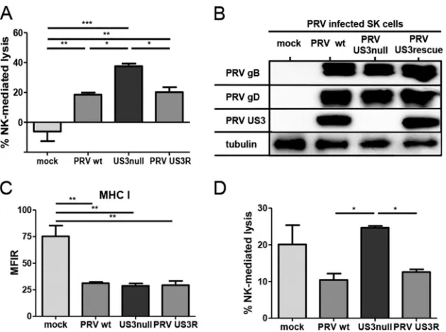
US3 reduces NK cell-mediated lysis of PRV-infected cells.
Using
a variety of gene deletion mutants of pseudorabies virus (PRV), we
recently discovered that PRV glycoprotein gD suppresses NK cell
activity via downregulation of CD112, a ligand for the activating
NK cell receptor DNAM-1 (
19
). Our initial NK cell cytotoxicity
assays with different PRV mutants indicated that US3 may also
display NK cell-evasive properties. To investigate whether PRV
US3 indeed affects the susceptibility of infected cells to NK
cell-mediated lysis, cytolytic assays were performed with SK cells
in-fected with wild-type (WT) PRV, an isogenic US3-null virus, or an
isogenic US3 rescue virus. At 10 hpi, mock-infected and
PRV-infected cells were coincubated with IL-2-primed primary porcine
NK cells for 4 h, and cells were subsequently assessed for viability
by flow cytometry (
Fig. 1A
). Mock-infected SK cells did not elicit
a significant cytolytic response from porcine NK cells; the
percent-age of NK cell-mediated lysis was not statistically different from
zero, in line with earlier data (
19
). Also, as reported previously,
PRV infection triggered porcine NK cell-mediated killing of SK
cells (
19
). Cells infected with US3-null PRV showed higher
sus-ceptibility to NK cell-mediated lysis than SK cells infected with
WT or US3 rescue PRV. The higher susceptibility of US3-null
PRV-infected cells to NK cell-mediated cell lysis than of WT or
US3 rescue PRV-infected cells was not due to differences in viral
replication (
Fig. 1B
) or to differences in the abilities of these
vi-ruses to downregulate the expression of MHC I molecules (an
important ligand for various inhibitory NK cell receptors) (
Fig.
1C
). In addition, NK cell cytotoxicity assays using a PRV strain
expressing a kinase-inactive US3 mutant PRV harboring a point
mutation (D223A) in the conserved aspartate in PRV US3 that
constitutes the catalytic base required for phosphotransfer (
34
,
35
) confirmed that kinase-intact US3 is required to increase the
protection of infected cells against NK cell-mediated lysis (data
not shown).
Because only a limited range of reagents and tools for the
in-vestigation of NK cell activation/inhibition in the porcine system
is currently available, we investigated whether PRV US3 also
gen-erated a protective effect against human NK cells. To this end, the
cytolytic activity of human IL-2-cultured NK cells against
mock-infected SK cells or SK cells mock-infected with WT, US3-null, or US3
rescue PRV was assessed (
Fig. 1D
). As observed previously,
IL-2-cultured human NK cells lysed mock-infected SK cells to a
signif-icant extent, which is in line with the known xenogeneic response
of human NK cells to porcine cells (
48–51
). US3-null
PRV-in-fected cells again showed higher susceptibility to NK
cell-medi-ated lysis than wild-type or US3 rescue PRV-infected cells. In
con-clusion, PRV US3 reduces the susceptibilities of infected cells to
both porcine and human NK cells.
PRV US3 enhances resistance to NK cell-mediated killing by
increasing the level of binding of the inhibitory NK cell receptor
CD300a to infected cells.
The protective effect of US3 against NK
cell-mediated lysis may result from a modulation of the activating/
inhibitory receptor balance on NK cells. Several inhibitory NK cell
receptors (e.g., KIR receptors) recognize MHC class I molecules
(
52
). Since we did not observe a difference in MHC class I levels on
on November 7, 2019 by guest
http://jvi.asm.org/
the cell surface between WT and US3-null PRV-infected cells (
Fig.
1C
), involvement of these NK cell receptors was unlikely. Still, the
fact that US3 provided protection to PRV-infected cells against
both porcine and human NK cells pointed to the potential
in-volvement of highly conserved NK cell receptors and ligands.
CD300a is a highly conserved inhibitory receptor that binds
to the highly conserved ligands phosphatidylserine (PS) and
phosphatidylethanolamine (PE) (
29
,
30
). Therefore, we
inves-tigated whether US3 affects the binding of CD300a to
PRV-in-fected cells. A CD300a-Fc soluble chimeric molecule was used in a
flow cytometric binding assay. As shown in
Fig. 2A
, SK cells
in-fected with WT PRV or the US3 rescue virus displayed
substan-tially higher levels of binding of recombinant CD300a than
US3-null PRV-infected or mock-infected cells. We then addressed
whether this US3-dependent increase in the binding of CD300a to
PRV-infected cells is involved in the US3-mediated protection
from NK cell-mediated lysis. To this end, cytotoxicity assays
were performed in the presence or absence of the anti-CD300a
blocking MAb KS153. The reactivity and specificity of the
KS153 antibody were confirmed by flow cytometric analysis on
CD300a-transfected 293T cells, and the ability of KS153 to
in-terfere with the binding of CD300a with its ligands was
con-firmed by blocking experiments using the recombinant
CD300a-Fc protein (data not shown). Cytotoxicity assays in
the presence or absence of KS153 allowed us to evaluate the
percentage of CD300a-dependent protection of cells from
hu-man NK cell-mediated lysis (see Materials and Methods).
Fig-ure 2B
shows that US3 increases the CD300a-mediated
protec-tion of infected cells against NK cell-mediated lysis. In
conclusion, PRV-infected cells show a US3-dependent increase
in the level of CD300a binding, and CD300a is involved in the
protective effect of PRV US3 against NK cell-mediated lysis.
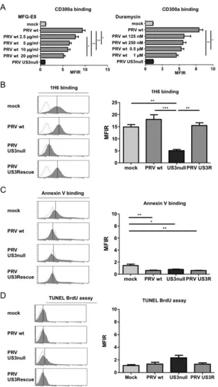
The CD300a ligands PS and PE are involved in the PRV
US3-mediated binding of CD300a.
Two highly conserved cellular
li-gands, PS and PE, have been identified for the inhibitory NK cell
receptor CD300a (
26
,
27
). To investigate whether these cellular
ligands are involved in the observed PRV US3-dependent increase
in CD300a binding to infected cells, the binding of CD300a-Fc
was assessed in the presence of increasing concentrations of milk
fat globule–EGF factor 8 (MFG-E8; also known as lactadherin) or
duramycin, agents that have been reported previously to interfere
with the ability of CD300a to bind PS or PE, respectively (
26
). As
shown in
Fig. 3A
, the addition of MFG-E8 resulted in a significant
dose-dependent reduction in the level of CD300a-Fc binding to
PRV-infected SK cells, indicating that PS is involved in the
bind-ing of CD300a to PRV-infected SK cells. Suppression of CD300a
binding was also observed by using duramycin, although the effect
appeared less dose dependent.
The involvement of PS and PE in US3-mediated CD300a
bind-ing suggests that US3 may modulate the cell surface exposure of
these CD300a ligands. No assays to specifically detect PE on the
cell surface have been described. However, PS on the cell surface
can be detected by using the PS-specific antibody 1H6 (
28
). To
determine whether US3 modulates the cell surface exposure of
PS, mock-infected SK cells or SK cells infected with WT,
US3-null, or US3 rescue PRV were analyzed by flow cytometry using
the anti-PS antibody 1H6.
Figure 3B
shows that SK cells infected
with WT or US3 rescue PRV indeed expose PS at much higher
FIG 1US3 suppresses the susceptibility of PRV-infected cells to porcine and human NK cell-mediated lysis. (A) SK cells were either mock infected or infected with WT, US3-null, or US3 rescue PRV (NIA3 strain) for 10 h and were subsequently incubated with IL-2-primed primary porcine NK cells at a target-to-effector cell ratio of 1:25 for 4 h. The viability of target cells was assessed by propidium iodide and flow cytometry, and the percentage of NK cell-mediated lysis was calculated. Data represent means⫹SEM for three independent experiments (*,P⬍0.05; **,P⬍0.01; ***,P⬍0.001). (B and C) SK cells were either mock infected or infected with WT, US3-null, or US3 rescue PRV for 12 h and were subsequently analyzed by Western blotting for the expression of PRV gB, PRV gD, PRV US3, and tubulin (B) or assessed for MHC I expression on the cell surface by flow cytometry (C). Data in panel C represent means⫹SEM for three independent repeats (**,P⬍0.01). (D) SK cells were either mock infected or infected with WT, US3-null, or US3 rescue PRV for 10 h and were subsequently incubated with IL-2-cultivated human NK cells at a target-to-effector cell ratio of 1:1 for 4 h. The viability of target cells was assessed by propidium iodide and flow cytometry, and the percentage of NK cell-mediated lysis was calculated. Data represent means⫹SEM for three independent repeats (*,P⬍0.05).PRV Evasion of NK Cells via Increased CD300a Binding
on November 7, 2019 by guest
http://jvi.asm.org/
[image:4.585.136.450.64.298.2]levels than SK cells infected with US3-null PRV. Somewhat
sur-prisingly, mock-infected cells also showed substantial PS
expo-sure. Increased PS exposure is one of the hallmarks of apoptotic
cells. In contrast to the results obtained with the PS-binding
anti-body 1H6, however, we did not observe detectable binding of
annexin V, which is widely used to detect surface-exposed PS (for
example, on apoptotic cells) (
Fig. 3C
). In support of the notion
that the observed antibody 1H6 binding did not point to increased
cell death, Sytox Blue live/dead staining indicated that none of the
conditions (mock infection, WT PRV, US3-null PRV, or US3
res-cue PRV) resulted in substantial or different quantities of dead
cells. Also in line with this finding, TUNEL (terminal
deoxynucle-otidyltransferase-mediated dUTP-biotin nick end labeling)
stain-ing, which detects apoptotic DNA fragmentation, did not indicate
substantial levels of apoptosis under any condition, or substantial
differences in apoptosis among the conditions (
Fig. 3D
).
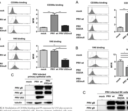
To better assess the potential biological significance of our
findings, we investigated whether PRV US3 expression also led to
increased CD300a binding to porcine primary epithelial cells and
modulated PS exposure on the surfaces of these cells.
Figure 4A
and
B
show that the effects of PRV US3 on CD300a binding to
infected porcine primary epithelial cells and PS cell surface
expo-sure are very similar to the effects observed in the SK cell line.
Again, Western blot analysis confirmed that this US3-induced
phenotype is not caused by differences in virus replication levels
(
Fig. 4C
). In conclusion, our findings show that PRV US3 triggers
CD300a binding to infected cells and indicate that US3 modulates
PS exposure on the surfaces of infected cells.
The kinase activity of PRV US3 and cellular PAKs are
re-quired for increased CD300a binding and modulation of PS
ex-posure on the cell surface.
PRV US3 is a viral serine/threonine
protein kinase and can directly phosphorylate and activate cellular
group I p21-activated kinases (PAKs), which are central regulators
in Rac1/CDC42 Rho GTPase signaling and comprise the closely
related kinases PAK1, PAK2, and PAK3 (
35
,
53
). Activation of
group I PAKs has recently been reported to trigger increased PS
exposure on the cell surface during thrombin-mediated activation
of platelets (
54
). To investigate a possible involvement of the
US3-PAK signaling axis in increased CD300a binding and the
modula-tion of PS exposure, we first assessed whether the kinase activity of
PRV US3 was required for the effects observed. For this purpose,
the kinase-inactive US3 mutant PRV, which harbors a point
mu-tation (D223A) in the conserved aspartate in PRV US3 that
con-stitutes the catalytic base required for phosphotransfer, was used.
SK cells were either mock infected or infected with WT PRV,
iso-genic US3 kinase-inactive PRV, or isoiso-genic US3-null PRV and
were assessed for the binding of recombinant CD300a and the
modulation of cell surface PS.
Figure 5A
and
B
show that cells
infected with PRV expressing kinase-inactive US3 display a
phe-notype similar to that of US3-null PRV-infected cells with regard
to CD300a binding and PS exposure on the cell surface. Western
blot analysis demonstrated similar infection efficiencies for cells
infected with WT, US3-null, and US3 kinase-inactive PRVs (
Fig.
5C
). The effect of PRV on PS exposure in the assay for which
results are shown in
Fig. 5
is somewhat less pronounced than that
in our earlier data (
Fig. 3
). This mild discrepancy can possibly be
FIG 2PRV triggers US3-dependent increased binding of the inhibitory NK cell receptor CD300a to the infected-cell surface and increased CD300a-mediated protection of infected cells against NK cell-mediated lysis. (A) SK cells were infected with WT, US3-null, or US3 rescue PRV (NIA3 strain) for 12 h and were subsequently assessed by flow cytometry for the binding of recombinant CD300a-Fc (1g/sample). (Left) Thexaxes of histogram plots indicate fluorescence intensity, and vertical lines in histograms indicate median fluorescence intensity. Dotted-line histograms represent isotype-matched antibody control signals. (Right) The graph shows means⫹SEM for three independent repeats (*,P⬍0.05; **,P⬍0.01; ***,P⬍0.001). (B) SK cells were infected with WT, US3-null, or US3 rescue PRV (NIA3 strain) for 10 h and were subsequently incubated with IL-2-cultivated human NK cells at an effector-to-target cell ratio of 1:1 in the absence or presence of the anti-CD300a antibody KS153. The viability of target cells was assessed by propidium iodide and flow cytometry. (Left) Percentage of NK cell-mediated lysis; (right) percentage of CD300a-dependent protection against NK cell-mediated lysis. Data represent means⫹SEM for three independent repeats (*,P⬍0.05).on November 7, 2019 by guest
http://jvi.asm.org/
FIG 3The CD300a ligands PS and PE are involved in the US3-dependent increased binding of CD300a to the infected-cell surface, and US3 modulates PS cell surface exposure. (A) SK cells were either mock infected or infected with WT or US3-null PRV (NIA3 strain) for 12 h, incubated with MFG-E8 or duramycin at the concentrations given, and assessed by flow cytometry for the binding of recombinant CD300a-Fc (1g/sample). Data represent means⫹SEM for three independent repeats (*,P⬍0.05; **,P⬍0.01). (B to D) SK cells were either mock infected or infected with WT, US3-null, or US3 rescue PRV (NIA3 strain) for 12 h and were subsequently assessed by flow cytometry for cell surface exposure of PS by using antibody 1H6 (B) or annexin V (C) or for apoptotic DNA fragmentation by using a TUNEL bromodeoxyuridine (BrdU) assay (D). Thexaxes of histogram plots indicate fluorescence intensity, and vertical lines in histograms indicate median fluorescence intensity. Dotted-line histograms represent isotype-matched antibody control signals. Graphs show means⫹SEM for three independent repeats (*,P⬍0.05; **,P⬍0.01; ***,P⬍0.001).
PRV Evasion of NK Cells via Increased CD300a Binding
on November 7, 2019 by guest
http://jvi.asm.org/
[image:6.585.136.452.63.625.2]attributed to the fact that in the assays to determine the kinase
involvement of US3, PRV strain Becker and isogenic mutants were
used, whereas in the former experiments, the highly virulent field
strain NIA3 (and isogenic mutants) was utilized.
To investigate whether group I PAKs are involved in PRV
US3-mediated effects on CD300a binding and PS exposure on the cell
surface, group I PAKs were inhibited using the selective allosteric
group I PAK inhibitor IPA-3 (
44
,
55
). As shown in
Fig. 6A
,
treat-ment with IPA-3 abrogated the increase in CD300a binding and
reduced PS cell surface exposure in WT PRV-infected cells and
mock-infected cells to the levels observed in US3-null
PRV-in-fected cells. The inhibitory effects of IPA-3 are not caused by
sup-pressive effects on PRV infection or the expression level of US3, as
indicated by the Western blots shown in
Fig. 6B
. In conclusion,
these experiments indicate that the PRV US3-mediated increase in
CD300a binding and the modulation of PS exposure on the cell
surface depend on the kinase activity of US3 and on the group I
PAK cell-signaling pathway.
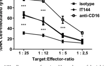
CD300a in primary porcine NK cells.
The CD300a receptor is
highly conserved across mammals and recently was also
charac-terized as an inhibitory receptor in birds (
29
,
30
). The natural host
of PRV is the pig, and currently, porcine NK cell receptors are
poorly characterized. The function of CD300a in porcine NK cells
has not yet been addressed.
To assess whether CD300a also serves as an inhibitory receptor
in porcine NK cells, a P815-based antibody redirected killing assay
using porcine NK cells as effector cells was performed. This assay
FIG 4Modulation of CD300a binding and PS exposure by US3 also occurs inPRV-infected primary epithelial cells. Porcine primary epithelial cells were either mock infected or infected with WT or US3-null PRV (NIA3 strain) for 12 h. (A and B) Cells were assessed by flow cytometry for recombinant CD300a-Fc (1g/sample) binding (A) and PS exposure on the cell surface (by using antibody 1H6) (B). (Left) Thexaxes of histogram plots indicate fluores-cence intensity, and vertical lines in histograms indicate median fluoresfluores-cence intensity. Dotted-line histograms represent isotype-matched antibody control signals. (Right) Graphs show means⫹SEM for three independent repeats (**,
P⬍0.01; ***,P⬍0.001). (C) Cells were assessed by Western blotting for the expression of PRV gB, PRV gD, PRV US3, and tubulin.
FIG 5The kinase activity of PRV US3 is required for the modulation of CD300a binding and PS exposure. SK cells were either mock infected or in-fected with WT PRV, kinase-inactive D223A US3 PRV, or US3-null PRV (Becker strain) for 12 h. (A and B) Cells were assessed by flow cytometry for the binding of recombinant CD300a-Fc (1g/sample) (A) and for PS exposure on the cell surface (by using antibody 1H6) (B). (Left) Thexaxes of histogram plots indicate fluorescence intensity, and vertical lines in histograms indicate median fluorescence intensity. Dotted-line histograms represent isotype-matched antibody control signals. (Right) Graphs show means⫹SEM for three independent repeats (*,P⬍0.05; **,P⬍0.01; ***,P⬍0.001). (C) Cells were assessed by Western blotting for the expression of PRV gB, PRV gD, PRV US3, and tubulin.
on November 7, 2019 by guest
http://jvi.asm.org/
[image:7.585.46.473.66.436.2] [image:7.585.291.544.69.464.2]is typically used to determine the activating or inhibitory nature of
a given NK cell receptor and has been used to demonstrate that
CD300a serves as an inhibitory receptor in human NK cells (
24
,
28
,
56
). Murine P815 cells express receptors for the constant (Fc)
domain of IgG antibodies (Fc
␥
receptors) on their surfaces.
Hence, when monoclonal IgG antibodies against particular NK
cell receptors are added to the assay mixture, the antibodies will
bind to their respective receptors on the NK cells via their antigen
binding (Fab) domains and, at the same time, to the Fc
␥
receptors
on the P815 cells via their Fc domains, thereby bridging the NK
cells and P815 cells. Depending on whether the antibody
recog-nizes an activating or an inhibitory NK cell receptor, this antibody
bridging will trigger increased or decreased NK cell-mediated lysis
of P815 cells, respectively. In the case of IgG antibodies directed
against CD300a, this assay results in reduced killing of P815 cells
by human NK cells (
24
,
28
,
56
).
Here (
Fig. 7
) a similar redirected killing assay was performed
using primary porcine NK cells instead of human NK cells to
evaluate the cross-reactivity of the anti-human CD300a antibody
IT144 with the porcine CD300a homologue and to determine
whether, as in human NK cells, CD300a serves as an inhibitory
receptor in porcine NK cells. The redirected killing assay was
per-formed in the presence of either mouse monoclonal IgG1
anti-body IT144, directed against huCD300a, an isotype-matched
mouse IgG1 control antibody, a mouse monoclonal IgG1
anti-porcine CD16 antibody (which generates an activating effect on
NK cells), or medium alone, and the percentage of NK
cell-medi-ated lysis of P815 cells was calculcell-medi-ated.
Figure 7
shows that, as
reported previously (
57
), the porcine CD16 receptor serves as an
activating NK cell receptor, since triggering of CD16 resulted in
substantially increased porcine NK cell-mediated killing of P815
cells. Importantly, the CD300a-directed antibody triggered
signif-icant inhibition of porcine NK cell-mediated lysis of P815 cells
compared to lysis with the isotype-matched control or medium
alone, indicating that in the porcine system also, CD300a serves as
an inhibitory NK cell receptor.
DISCUSSION
In the current report, we describe a previously uncharacterized
viral strategy for evading NK cells and reveal the involvement of
FIG 6Group I PAKs are involved in US3-mediated modulation of CD300a binding and PS exposure. SK cells were either mock infected or infected with WT or US3-null PRV (NIA3 strain). At 2 hpi, the group I PAK inhibitor IPA-3 (10M) or DMSO (as a diluent control) was added. (A) At 12 hpi, cells were assessed by flow cytometry for the binding of recombinant CD300a-Fc (1g/sample) (left) and for PS exposure on the cell surface (by using antibody 1H6) (right). Graphs show means⫹SEM for three independent repeats (***,P⬍0.001). (B) At 12 hpi, cells were assessed by Western blotting for the expression of PRV gB, PRV gD, PRV US3, and tubulin.FIG 7Porcine NK cells express a functional homologue of the inhibitory NK cell receptor CD300a. An antibody redirected killing assay using Fc receptor-bearing P815 cells was performed with IL-2-activated porcine primary NK cells, at the indicated target-to-effector cell ratios, in the presence of either medium, anti-CD300a antibody IT144, an anti-porcine CD16 antibody, or an isotype-matched control antibody. The viability of target cells was assessed by propidium iodide and flow cytometry, and the percentage of NK cell-mediated lysis was calculated. Data represent means⫹SEM for three independent re-peats (*,P⬍0.05; ***,P⬍0.001).
PRV Evasion of NK Cells via Increased CD300a Binding
on November 7, 2019 by guest
http://jvi.asm.org/
[image:8.585.135.453.68.316.2] [image:8.585.335.505.528.636.2]CD300a in the recognition of virus-infected cells. By using PRV
mutants and specific inhibitors, we demonstrate that this trait
depends on the CD300a ligands phosphatidylserine (PS) and
phosphatidylethanolamine (PE), on the expression of catalytically
intact US3, and on the activation of group I PAKs. Interestingly,
different tumor cell lines have recently been reported to show
increased PS exposure and CD300a binding (
28
). In addition,
blocking of PS using MFG-E8 enhanced NK cell-mediated killing
of tumor cells, leading to the hypothesis that tumor cells may
subvert NK cell-mediated lysis via increased CD300a binding
(
28
). Our data, obtained by using CD300a-blocking antibodies,
demonstrate directly, for the first time, that manipulation of
CD300a may indeed represent a bona fide NK cell evasion
strat-egy, indicating that viruses as well as tumor cells may manipulate
this NK cell-inhibitory pathway for their own benefit.
CD300a is expressed not only on NK cells but also on several
other immune cell populations, where it typically functions as an
inhibitory receptor. Thus, CD300a has been implicated in the
in-hibition not only of NK cell activity (
24
,
28
) but also of the
activ-ities of mast cells (
58
), neutrophils (
59
), eosinophils (
60
), and B
and T cells (
61
,
62
). As such, the consequences of viral triggering of
the CD300a inhibitory receptor may stretch beyond the effects on
NK cells described here. On the other hand, CD300a displays
sig-nificant overlap regarding ligand specificity with the closely
re-lated activating receptor CD300c, which is specifically expressed
on monocytes and mast cells (
63
,
64
). Hence, in future research, it
will be interesting to study whether the US3-mediated modulation
of CD300a binding has consequences for other immune cells and
whether or not US3 also affects CD300c binding.
The polar lipid PS and the neutral lipid PE have been described
as the main CD300a ligands (
26
). Both PS and PE are typically
distributed asymmetrically in the plasma membrane lipid bilayer
and are enriched at the inner, cytoplasmic leaflet (
65
). Although
externalization of PS is a hallmark of early stages of apoptosis,
there is increasing evidence that cells may also show PS exposure
independently of programmed cell death, as exemplified by
re-ports on apoptosis-independent PS exposure on tumor cells and
during activation of mast cells, B cells, T cells, and platelets (
28
,
66–70
). Also along these lines, apoptosis is considered to trigger an
eat-me signal for phagocytosis, and PS exposure was found not to
be sufficient to induce this process
in vivo
(
71
). Our results are in
line with the possibility that under certain circumstances, PS
ex-posure may be independent of apoptotic cell death. Although we
observed PS exposure by using the PS-specific antibody 1H6, this
was not accompanied by an obvious apoptotic cellular phenotype,
since we did not detect differences in viability (using the Sytox
Blue dead/live marker) and apoptotic DNA fragmentation (using
the TUNEL assay). Remarkably, antibody 1H6 reactivity was not
accompanied by increased binding of annexin V, a reagent
com-monly used to detect PS exposure. The reasons underlying the
differential reactivity between annexin V and 1H6 are currently
unclear. One could argue that 1H6 may not specifically bind PS.
However, several reports indicate that antibody 1H6 specifically
binds to PS and not to other lipids (
72–74
). Nevertheless, we
can-not formally rule out the possibility that 1H6 binds acan-nother
lip-id(s) in the cell membrane that is modulated by PRV US3 during
infection. Irrespective of whether this is the case, we found that
exposure of 1H6-reactive lipids is not sufficient to trigger
substan-tial recombinant CD300a binding. Indeed, mock-infected cells
showed a level of 1H6 reactivity comparable to (and sometimes
even higher than) that observed for PRV-infected cells but, unlike
PRV-infected cells, did not show significant CD300a binding.
Hence, additional CD300a ligands appear to be involved in
US3-triggered CD300a binding. This is in line with the findings of our
blocking assays, which showed that not only the PS-blocking
re-agent MFG-E8, but also the PE-blocking re-agent duramycin,
sup-pressed the US3-mediated increase in CD300a binding. Also in
line with the view that additional ligands may be involved in the
binding of CD300a to cells, we found that US3-null PRV-infected
cells, which do not show obvious reactivity with the PS
anti-body 1H6, display a level of CD300a binding comparable to that of
mock-infected cells. Functional recognition reporter assays
indi-cated that CD300a may bind more strongly to PE than to PS (
26
,
64
,
75
). This could account for the discrepancies in the observed
correlation of CD300a binding and PS exposure. Unfortunately,
due to the lack of reagents for the detection of PE, potential
dif-ferences in PE exposure between mock-, WT PRV-, and US3-null
PRV-infected cells could not be assessed. In any case, our data
suggest that US3 expression affects the cell surface exposure of
CD300a ligands, which are associated with substantially increased
CD300a binding.
We showed previously that PRV US3 directly phosphorylates
and thereby activates group I p21-activated kinases, critical
down-stream effectors of the Cdc42/Rac1 signaling pathways (
35
). Here
we report that inhibition of group I PAK activity inhibits the
abil-ity of US3 to trigger increased CD300a binding or modulate PS
exposure. Group I PAK activity has been reported previously to be
critically involved in PS exposure during platelet activation (
54
).
Our current data therefore suggest that group I PAKs may be
linked to PS exposure/CD300a binding in different cell types. In
this context, it is interesting that several viruses, including HIV,
have been reported to trigger group I PAK activity (
76
) and may
therefore modulate effects similar to those we describe here. In
line with this, several viruses have been reported to trigger the
exposure of aminophospholipids, such as PS (
77
). This has been
speculated to enable viruses to evade immune recognition and
dampen inflammatory responses to infection (
77
). Our current
report demonstrates that viral manipulation of the exposure of
phospholipids, such as PS, may indeed allow viruses to subvert
important components of the antiviral immune response.
Target-ing of aminophospholipid exposure and signalTarget-ing pathways, such
as those mediated by group I PAKs, and identification of the viral
factors that trigger these events may therefore hold promise as
therapeutic strategies for viral diseases (
76
,
77
). The viral US3
protein kinase may be of particular interest in this respect, since it
is an important alphaherpesvirus virulence factor, and since US3
does not appear to be closely related genetically to any known
cellular kinase, which may make it an attractive candidate to target
for the development of antiviral drugs (
78
).
Interestingly, PRV strains lacking the US3 protein kinase show
substantially reduced virulence in pigs (
79–81
). Despite this
atten-uation, pigs infected with US3-null PRV were protected against
clinical signs upon challenge infection with a virulent wild-type
virus (
79
,
80
). Although further studies will be needed to elucidate
how attenuated US3-null PRV may generate a protective immune
response, our current findings on the immune evasion properties
of the US3 protein may be significant in this context.
Our findings may also have relevance for cancer therapy, since
several types of cancer have been associated with upregulated
group I PAK activity and nonapoptotic PS exposure (
82–84
). Our
on November 7, 2019 by guest
http://jvi.asm.org/
observation that noninfected primary epithelial cells are
substan-tially recognized by the PS-binding antibody 1H6 may indicate
that perhaps caution should be exercised in targeting PS for
anti-cancer or antiviral therapy (
82
). We found that, despite showing
similar binding of 1H6, PRV-infected cells show substantially
higher levels of CD300a binding than mock-infected cells.
Com-bined with the observation that several types of cancer cells also
display substantially increased CD300a binding (
28
,
77
), this may
indicate that targeting CD300a binding may be a more stringent
strategy for identifying virus-infected or tumor cells than
target-ing PS exposure.
Finally, our data indicate that, as in humans and other
mam-mals, CD300a serves as an inhibitory receptor in swine. Several
studies have reported that the CD300 receptor family is highly
conserved across multiple species (
29
,
30
). This is particularly true
for CD300a, as illustrated by its recent identification and
charac-terization in chickens, where it shows inhibitory activity and
af-finity for PS and PE, as described for mammals (
30
). Our data also
indicate that the human inhibitory NK cell receptor CD300a
rec-ognizes porcine cells, suggesting that, under certain
circum-stances, human CD300a can be involved in the recognition of
porcine cells, which may be relevant to the study of the human NK
cell response to pig xenografts.
In conclusion, we report a novel alphaherpesvirus strategy for
evading NK cells, consisting of US3-dependent increased binding
of the inhibitory NK cell receptor CD300a, which is orchestrated
by group I PAK activity and phospholipids such as PS and PE. Our
data provide novel insights in alphaherpesvirus and CD300a
biol-ogy and may have implications for antiviral and antitumor
thera-pies.
ACKNOWLEDGMENTS
We thank C. Van Waesberghe, S. Brabant, H. Vereecke, C. Helsmoortel, and L. Sys for excellent technical assistance, R. Cooman for animal man-agement, and G. Smith (Northwestern University, Chicago, IL), L. En-quist (Princeton University, Princeton, NJ), ID-DLO (Lelystad, the Neth-erlands), and H. Nauwynck and E. Cox (Ghent University, Ghent, Belgium) for reagents.
This research was supported by grants from the Special Research Fund of Ghent University (grants 01J29110, 01J11611, and 01G01311) and F.W.O.-Vlaanderen (grants 1.5.077.11N and G.0176.15N), Hercules Foundation grant AUGE-035, and AIRC:IG project 15428 (to M.V.).
FUNDING INFORMATION
Hercules Foundation provided funding to Herman W. Favoreel under grant number AUGE-035. Ghent University, Special Research Fund pro-vided funding to Herman W. Favoreel under grant numbers 01J29110, 01J11611, and 01G01311. F.W.O.-Vlaanderen provided funding to Her-man W. Favoreel under grant numbers 1.5.077.11N and G.0176.15N. Associazione Italiana per la Ricerca sul Cancro (AIRC) provided funding to Massimo Vitale under grant number IG 15428.
REFERENCES
1.Vivier E, Raulet DH, Moretta A, Caligiuri MA, Zitvogel L, Lanier LL, Yokoyama WM, Ugolini S. 2011. Innate or adaptive immunity? The example of natural killer cells. Science331:44 – 49.http://dx.doi.org/10 .1126/science.1198687.
2.Etzioni A, Eidenschenk C, Katz R, Beck R, Casanova JL, Pollack S.2005. Fatal varicella associated with selective natural killer cell deficiency. J Pe-diatr146:423– 425.http://dx.doi.org/10.1016/j.jpeds.2004.11.022. 3.Almerigogna F, Fassio F, Giudizi MG, Biagiotti R, Manuelli C,
Chiap-pini E, Galli L, Romagnani S, De Martino M.2011. Natural killer cell
deficiencies in a consecutive series of children with herpetic encephalitis. Int J Immunopathol Pharmacol24:231–238.
4.Biron CA, Byron KS, Sullivan JL.1989. Severe herpesvirus infections in an adolescent without natural killer cells. N Engl J Med320:1731–1735. http://dx.doi.org/10.1056/NEJM198906293202605.
5.Babic´ M, Krmpotic´ A, Jonjic´ S.2011. All is fair in virus-host interactions: NK cells and cytomegalovirus. Trends Mol Med17:677– 685.http://dx.doi .org/10.1016/j.molmed.2011.07.003.
6.Fielding CA, Aicheler R, Stanton RJ, Wang EC, Han S, Seirafian S, Davies J, McSharry BP, Weekes MP, Antrobus PR, Prod’homme V, Blanchet FP, Sugrue D, Cuff S, Roberts D, Davison AJ, Lehner PJ, Wilkinson GW, Tomasec P.2014. Two novel human cytomegalovirus NK cell evasion functions target MICA for lysosomal degradation. PLoS Pathog10:e1004058.http://dx.doi.org/10.1371/journal.ppat.1004058. 7.Wilkinson GW, Tomasec P, Stanton RJ, Armstrong M, Prod’homme V,
Aicheler R, McSharry BP, Rickards CR, Cochrane D, Llewellyn-Lacey S, Wang EC, Griffin CA, Davison AJ.2008. Modulation of natural killer cells by human cytomegalovirus. J Clin Virol41:206 –212.http://dx.doi .org/10.1016/j.jcv.2007.10.027.
8.Arase H, Mocarski ES, Campbell AE, Hill AB, Lanier LL.2002. Direct recognition of cytomegalovirus by activating and inhibitory NK cell receptors. Science296:1323–1326.http://dx.doi.org/10.1126/science.1070884. 9.Dunn C, Chalupny NJ, Sutherland CL, Dosch S, Sivakumar PV,
John-son DC, Cosman D.2003. Human cytomegalovirus glycoprotein UL16 causes intracellular sequestration of NKG2D ligands, protecting against natural killer cell cytotoxicity. J Exp Med197:1427–1439.http://dx.doi .org/10.1084/jem.20022059.
10. Tomasec P, Wang EC, Davison AJ, Vojtesek B, Armstrong M, Griffin C, McSharry BP, Morris RJ, Llewellyn-Lacey S, Rickards C, Nomoto A, Sinzger C, Wilkinson GW.2005. Downregulation of natural killer cell-activating ligand CD155 by human cytomegalovirus UL141. Nat Immu-nol6:181–188.http://dx.doi.org/10.1038/ni1156.
11. Nachmani D, Stern-Ginossar N, Sarid R, Mandelboim O.2009. Diverse herpesvirus microRNAs target the stress-induced immune ligand MICB to escape recognition by natural killer cells. Cell Host Microbe5:376 –385. http://dx.doi.org/10.1016/j.chom.2009.03.003.
12. Smith W, Tomasec P, Aicheler R, Loewendorf A, Nemcovicova I, Wang EC, Stanton RJ, Macauley M, Norris P, Willen L, Ruckova E, Nomoto A, Schneider P, Hahn G, Zajonc DM, Ware CF, Wilkinson GW, Benedict CA.2013. Human cytomegalovirus glycoprotein UL141 targets the TRAIL death receptors to thwart host innate antiviral defenses. Cell Host Microbe13:324 –335.http://dx.doi.org/10.1016/j.chom.2013.02.003. 13. Madrid AS, Ganem D.2012. Kaposi’s sarcoma-associated herpesvirus
ORF54/dUTPase downregulates a ligand for the NK activating receptor NKp44. J Virol86:8693– 8704.http://dx.doi.org/10.1128/JVI.00252-12. 14. Thomas M, Boname JM, Field S, Nejentsev S, Salio M, Cerundolo V,
Wills M, Lehner PJ. 2008. Down-regulation of NKG2D and NKp80 ligands by Kaposi’s sarcoma-associated herpesvirus K5 protects against NK cell cytotoxicity. Proc Natl Acad Sci U S A105:1656 –1661.http://dx .doi.org/10.1073/pnas.0707883105.
15. Krmpotic´ A, Busch DH, Bubic´ I, Gebhardt F, Hengel H, Hasan M, Scalzo AA, Koszinowski UH, Jonjic´ S.2002. MCMV glycoprotein gp40 confers virus resistance to CD8⫹T cells and NK cells in vivo. Nat Immu-nol3:529 –535.http://dx.doi.org/10.1038/ni799.
16. Zarama A, Pérez-Carmona N, Farré D, Tomic A, Borst EM, Messerle M, Jonjic´ S, Engel P, Angulo A.2014. Cytomegalovirus m154 hinders CD48 cell-surface expression and promotes viral escape from host natural killer cell control. PLoS Pathog10:e1004000.http://dx.doi.org/10.1371 /journal.ppat.1004000.
17. Stanton RJ, Prod’homme V, Purbhoo MA, Moore M, Aicheler RJ, Heinzmann M, Bailer SM, Haas J, Antrobus R, Weekes MP, Lehner PJ, Vojtesek B, Miners KL, Man S, Wilkie GS, Davison AJ, Wang EC, Tomasec P, Wilkinson GW.2014. HCMV pUL135 remodels the actin cytoskeleton to impair immune recognition of infected cells. Cell Host Microbe16:201–214.http://dx.doi.org/10.1016/j.chom.2014.07.005. 18. Schepis D, D’Amato M, Studahl M, Bergstrom T, Karre K, Berg L.2009.
Herpes simplex virus infection downmodulates NKG2D ligand expres-sion. Scand J Immunol 69:429 – 436. http://dx.doi.org/10.1111/j.1365 -3083.2009.02241.x.
19. Grauwet K, Cantoni C, Parodi M, De Maria A, Devriendt B, Pende D, Moretta L, Vitale M, Favoreel HW.2014. Modulation of CD112 by the alphaherpesvirus gD protein suppresses DNAM-1-dependent NK cell-PRV Evasion of NK Cells via Increased CD300a Binding
on November 7, 2019 by guest
http://jvi.asm.org/
mediated lysis of infected cells. Proc Natl Acad Sci U S A111:16118 – 16123.http://dx.doi.org/10.1073/pnas.1409485111.
20. Campbell TM, McSharry BP, Steain M, Slobedman B, Abendroth A.20 May 2015. Varicella-zoster virus and herpes simplex virus 1 differentially modulate NKG2D ligand expression during productive infection. J Virol http://dx.doi.org/10.1128/JVI.00292-15.
21. Vivier E, Tomasello E, Baratin M, Walzer T, Ugolini S.2008. Functions of natural killer cells. Nat Immunol9:503–510.http://dx.doi.org/10.1038 /ni1582.
22. Sivori S, Carlomagno S, Pesce S, Moretta A, Vitale M, Marcenaro E. 2014. TLR/NCR/KIR: which one to use and when? Front Immunol5:105. http://dx.doi.org/10.3389/fimmu.2014.00105.
23. van de Weijer ML, Luteijn RD, Wiertz EJ.2015. Viral immune evasion: lessons in MHC class I antigen presentation. Semin Immunol27:125–137. http://dx.doi.org/10.1016/j.smim.2015.03.010.
24. Cantoni C, Bottino C, Augugliaro R, Morelli L, Marcenaro E, Castriconi R, Vitale M, Pende D, Sivori S, Millo R, Biassoni R, Moretta L, Moretta A.1999. Molecular and functional characteriza-tion of IRp60, a member of the immunoglobulin superfamily that functions as an inhibitory receptor in human NK cells. Eur J Immunol 29:3148 –3159. http://dx.doi.org/10.1002/(SICI)1521-4141(199910) 29:10⬍3148::AID-IMMU3148⬎3.0.CO;2-L.
25. Green BJ, Clark GJ, Hart DN.1998. The CMRF-35 mAb recognizes a second leukocyte membrane molecule with a domain similar to the poly Ig receptor. Int Immunol10:891– 899.http://dx.doi.org/10.1093/intimm/10 .7.891.
26. Simhadri VR, Andersen JF, Calvo E, Choi SC, Coligan JE, Borrego F. 2012. Human CD300a binds to phosphatidylethanolamine and phospha-tidylserine, and modulates the phagocytosis of dead cells. Blood119: 2799 –2809.http://dx.doi.org/10.1182/blood-2011-08-372425.
27. Nakahashi-Oda C, Tahara-Hanaoka S, Honda S, Shibuya K, Shibuya A. 2012. Identification of phosphatidylserine as a ligand for the CD300a im-munoreceptor. Biochem Biophys Res Commun417:646 – 650.http://dx .doi.org/10.1016/j.bbrc.2011.12.025.
28. Lankry D, Rovis TL, Jonjic´ S, Mandelboim O.2013. The interaction between CD300a and phosphatidylserine inhibits tumor cell killing by NK cells. Eur J Immunol43:2151–2161.http://dx.doi.org/10.1002/eji .201343433.
29. Cannon JP, O’Driscoll M, Litman GW.2012. Specific lipid recognition is a general feature of CD300 and TREM molecules. Immunogenetics64: 39 – 47.http://dx.doi.org/10.1007/s00251-011-0562-4.
30. Sperling B, Viertlboeck BC, Gobel TW.2015. Chicken CD300a homolog is found on B lymphocytes, various leukocytes populations and binds to phospholipids. Dev Comp Immunol 50:121–128. http://dx.doi.org/10 .1016/j.dci.2015.02.004.
31. Baskerville A.1973. Ultrastructural changes in the lungs of pigs infected with Aujeszky’s disease virus. Res Vet Sci14:229 –233.
32. Kimman TG, Pol JM, de Wind N, Oei-Lie N, Berns AJ, Gielkens AL. 1992. Role of different genes in the virulence and pathogenesis of Aujesz-ky’s disease virus. Vet Microbiol 33:45–52. http://dx.doi.org/10.1016 /0378-1135(92)90034-Q.
33. van Zijl M, van der Gulden H, de Wind N, Gielkens A, Berns A.1990. Identification of two genes in the unique short region of pseudorabies virus; comparison with herpes simplex virus and varicella-zoster virus. J Gen Virol71(Part 8):1747–1755.http://dx.doi.org/10.1099/0022-1317-71 -8-1747.
34. Coller KE, Smith GA.2008. Two viral kinases are required for sustained long distance axon transport of a neuroinvasive herpesvirus. Traffic 9:1458 –1470.http://dx.doi.org/10.1111/j.1600-0854.2008.00782.x. 35. Van den Broeke C, Radu M, Deruelle M, Nauwynck H, Hofmann C,
Jaffer ZM, Chernoff J, Favoreel HW. 2009. Alphaherpesvirus US3-mediated reorganization of the actin cytoskeleton is US3-mediated by group A p21-activated kinases. Proc Natl Acad Sci U S A106:8707– 8712.http://dx .doi.org/10.1073/pnas.0900436106.
36. Geenen K, Favoreel HW, Nauwynck HJ. 2005. Higher resistance of porcine trigeminal ganglion neurons towards pseudorabies virus-induced cell death compared with other porcine cell types in vitro. J Gen Virol 86:1251–1260.http://dx.doi.org/10.1099/vir.0.80760-0.
37. Takeda K, Oshima H, Hayakawa Y, Akiba H, Atsuta M, Kobata T, Kobayashi K, Ito M, Yagita H, Okumura K. 2000. CD27-mediated activation of murine NK cells. J Immunol164:1741–1745.http://dx.doi .org/10.4049/jimmunol.164.4.1741.
38. Nauwynck HJ, Pensaert MB.1995. Effect of specific antibodies on the
cell-associated spread of pseudorabies virus in monolayers of different cell types. Arch Virol140:1137–1146.http://dx.doi.org/10.1007/BF01315422. 39. Yang H, Oura CA, Kirkham PA, Parkhouse RM.1996. Preparation of monoclonal anti-porcine CD3 antibodies and preliminary characteriza-tion of porcine T lymphocytes. Immunology88:577–585.http://dx.doi .org/10.1046/j.1365-2567.1996.d01-682.x.
40. Pescovitz MD, Lunney JK, Sachs DH.1984. Preparation and character-ization of monoclonal antibodies reactive with porcine PBL. J Immunol 133:368 –375.
41. Jonjic´ S, Koszinowski UH.1984. Monoclonal antibodies reactive with swine lymphocytes. I. Antibodies to membrane structures that define the cytolytic T lymphocyte subset in the swine. J Immunol133:647– 652. 42. Canfield SM, Morrison SL.1991. The binding affinity of human IgG for
its high affinity Fc receptor is determined by multiple amino acids in the CH2 domain and is modulated by the hinge region. J Exp Med173:1483–
1491.http://dx.doi.org/10.1084/jem.173.6.1483.
43. Favoreel HW, Nauwynck HJ, Van Oostveldt P, Mettenleiter TC, Pen-saert MB.1997. Antibody-induced and cytoskeleton-mediated redistri-bution and shedding of viral glycoproteins, expressed on pseudorabies virus-infected cells. J Virol71:8254 – 8261.
44. Deacon SW, Beeser A, Fukui JA, Rennefahrt UE, Myers C, Chernoff J, Peterson JR.2008. An isoform-selective, small-molecule inhibitor targets the autoregulatory mechanism of p21-activated kinase. Chem Biol15: 322–331.http://dx.doi.org/10.1016/j.chembiol.2008.03.005.
45. Deruelle M, Geenen K, Nauwynck HJ, Favoreel HW. 2007. A point mutation in the putative ATP binding site of the pseudorabies virus US3 protein kinase prevents Bad phosphorylation and cell survival following apoptosis induction. Virus Res 128:65–70.http://dx.doi.org/10.1016/j .virusres.2007.04.006.
46. Balsamo M, Vermi W, Parodi M, Pietra G, Manzini C, Queirolo P, Lonardi S, Augugliaro R, Moretta A, Facchetti F, Moretta L, Mingari MC, Vitale M. 2012. Melanoma cells become resistant to cell-mediated killing when exposed to cell numbers compatible with NK-cell infiltration in the tumor. Eur J Immunol42:1833–1842.http://dx.doi .org/10.1002/eji.201142179.
47. Pintaric M, Gerner W, Saalmuller A.2008. Synergistic effects of IL-2, IL-12 and IL-18 on cytolytic activity, perforin expression and IFN-␥ pro-duction of porcine natural killer cells. Vet Immunol Immunopathol121: 68 – 82.http://dx.doi.org/10.1016/j.vetimm.2007.08.009.
48. Forte P, Lilienfeld BG, Baumann BC, Seebach JD.2005. Human NK cytotoxicity against porcine cells is triggered by NKp44 and NKG2D. J Immunol175:5463–5470. http://dx.doi.org/10.4049/jimmunol.175.8 .5463.
49. Sommaggio R, Cohnen A, Watzl C, Costa C.2012. Multiple receptors trigger human NK cell-mediated cytotoxicity against porcine chondro-cytes. J Immunol 188:2075–2083. http://dx.doi.org/10.4049/jimmunol .1100433.
50. Tran PD, Christiansen D, Winterhalter A, Brooks A, Gorrell M, Lil-ienfeld BG, Seebach JD, Sandrin M, Sharland A.2008. Porcine cells express more than one functional ligand for the human lymphocyte acti-vating receptor NKG2D. Xenotransplantation15:321–332.http://dx.doi .org/10.1111/j.1399-3089.2008.00489.x.
51. Kim TJ, Kim N, Kim EO, Choi JR, Bluestone JA, Lee KM. 2010. Suppression of human anti-porcine natural killer cell xenogeneic re-sponses by combinations of monoclonal antibodies specific to CD2 and NKG2D and extracellular signal-regulated kinase kinase inhibitor. Immu-nology130:545–555.http://dx.doi.org/10.1111/j.1365-2567.2010.03253.x. 52. Parham P, Moffett A.2013. Variable NK cell receptors and their MHC
class I ligands in immunity, reproduction and human evolution. Nat Rev Immunol13:133–144.http://dx.doi.org/10.1038/nri3370.
53. Pacheco A, Chernoff J.2010. Group I p21-activated kinases: emerging roles in immune function and viral pathogenesis. Int J Biochem Cell Biol 42:13–16.http://dx.doi.org/10.1016/j.biocel.2009.09.006.
54. Aslan JE, Baker SM, Loren CP, Haley KM, Itakura A, Pang J, Greenberg DL, David LL, Manser E, Chernoff J, McCarty OJ. 2013. The PAK system links Rho GTPase signaling to thrombin-mediated platelet activa-tion. Am J Physiol Cell Physiol 305:C519 –C528.http://dx.doi.org/10 .1152/ajpcell.00418.2012.
55. Van den Broeke C, Deruelle M, Nauwynck HJ, Coller KE, Smith GA, Van Doorsselaere J, Favoreel HW.2009. The kinase activity of pseudo-rabies virus US3 is required for modulation of the actin cytoskeleton. Virology385:155–160.http://dx.doi.org/10.1016/j.virol.2008.11.050. 56. Lankry D, Simic H, Klieger Y, Levi-Schaffer F, Jonjic´ S, Mandelboim O.
on November 7, 2019 by guest
http://jvi.asm.org/
2010. Expression and function of CD300 in NK cells. J Immunol185: 2877–2886.http://dx.doi.org/10.4049/jimmunol.0903347.
57. Mair KH, Essler SE, Patzl M, Storset AK, Saalmuller A, Gerner W.2012. NKp46 expression discriminates porcine NK cells with different func-tional properties. Eur J Immunol42:1261–1271.http://dx.doi.org/10 .1002/eji.201141989.
58. Bachelet I, Munitz A, Moretta A, Moretta L, Levi-Schaffer F.2005. The inhibitory receptor IRp60 (CD300a) is expressed and functional on hu-man mast cells. J Immunol175:7989 –7995.http://dx.doi.org/10.4049 /jimmunol.175.12.7989.
59. Alvarez Y, Tang X, Coligan JE, Borrego F.2008. The CD300a (IRp60) inhibitory receptor is rapidly up-regulated on human neutrophils in re-sponse to inflammatory stimuli and modulates CD32a (Fc␥RIIa) medi-ated signaling. Mol Immunol 45:253–258. http://dx.doi.org/10.1016/j .molimm.2007.05.006.
60. Munitz A, Bachelet I, Eliashar R, Moretta A, Moretta L, Levi-Schaffer F. 2006. The inhibitory receptor IRp60 (CD300a) suppresses the effects of IL-5, GM-CSF, and eotaxin on human peripheral blood eosinophils. Blood107:1996 –2003.http://dx.doi.org/10.1182/blood-2005-07-2926. 61. Silva R, Moir S, Kardava L, Debell K, Simhadri VR, Ferrando-Martinez
S, Leal M, Pena J, Coligan JE, Borrego F.2011. CD300a is expressed on human B cells, modulates BCR-mediated signaling, and its expression is down-regulated in HIV infection. Blood117:5870 –5880. http://dx.doi .org/10.1182/blood-2010-09-310318.
62. Simhadri VR, Mariano JL, Zhou Q, DeBell KE, Borrego F. 2011. Differential expression of CD300a/c on human TH1 and TH17 cells. BMC Immunol12:62.http://dx.doi.org/10.1186/1471-2172-12-62.
63. Simhadri VR, Mariano JL, Gil-Krzewska A, Zhou Q, Borrego F.2013. CD300c is an activating receptor expressed on human monocytes. J Innate Immun5:389 – 400.http://dx.doi.org/10.1159/000350523.
64. Takahashi M, Izawa K, Kashiwakura J, Yamanishi Y, Enomoto Y, Kaitani A, Maehara A, Isobe M, Ito S, Matsukawa T, Nakahara F, Oki T, Kajikawa M, Ra C, Okayama Y, Kitamura T, Kitaura J.2013. Human CD300C delivers an Fc receptor-␥-dependent activating signal in mast cells and monocytes and differs from CD300A in ligand recognition. J Biol Chem288:7662–7675.http://dx.doi.org/10.1074/jbc.M112.434746. 65. Hankins HM, Baldridge RD, Xu P, Graham TR.2015. Role of flippases,
scramblases and transfer proteins in phosphatidylserine subcellular distri-bution. Traffic16:35– 47.http://dx.doi.org/10.1111/tra.12233.
66. Lentz BR.2003. Exposure of platelet membrane phosphatidylserine reg-ulates blood coagulation. Prog Lipid Res42:423– 438.http://dx.doi.org/10 .1016/S0163-7827(03)00025-0.
67. Dillon SR, Mancini M, Rosen A, Schlissel MS.2000. Annexin V binds to viable B cells and colocalizes with a marker of lipid rafts upon B cell receptor activation. J Immunol164:1322–1332.http://dx.doi.org/10.4049 /jimmunol.164.3.1322.
68. Elliott JI, Surprenant A, Marelli-Berg FM, Cooper JC, Cassady-Cain RL, Wooding C, Linton K, Alexander DR, Higgins CF.2005. Membrane phosphatidylserine distribution as a non-apoptotic signalling mechanism in lymphocytes. Nat Cell Biol7:808 – 816.http://dx.doi.org/10.1038 /ncb1279.
69. Elliott JI, Sardini A, Cooper JC, Alexander DR, Davanture S, Chimini G, Higgins CF.2006. Phosphatidylserine exposure in B lymphocytes: a role for lipid packing. Blood108:1611–1617.http://dx.doi.org/10.1182 /blood-2005-11-012328.
70. Rysavy NM, Shimoda LM, Dixon AM, Speck M, Stokes AJ, Turner
H, Umemoto EY.2014. Beyond apoptosis: the mechanism and func-tion of phosphatidylserine asymmetry in the membrane of activating mast cells. Bioarchitecture 4:127–137. http://dx.doi.org/10.1080/19490992 .2014.995516.
71. Segawa K, Suzuki J, Nagata S.2011. Constitutive exposure of phospha-tidylserine on viable cells. Proc Natl Acad Sci U S A108:19246 –19251. http://dx.doi.org/10.1073/pnas.1114799108.
72. Mourdjeva M, Kyurkchiev D, Mandinova A, Altankova I, Kehayov I, Kyurkchiev S.2005. Dynamics of membrane translocation of phosphati-dylserine during apoptosis detected by a monoclonal antibody. Apoptosis 10:209 –217.http://dx.doi.org/10.1007/s10495-005-6076-5.
73. Prudovsky I, Vary CP, Markaki Y, Olins AL, Olins DE.2012. Phospha-tidylserine colocalizes with epichromatin in interphase nuclei and mitotic chromosomes. Nucleus3:200 –210.http://dx.doi.org/10.4161/nucl.19662. 74. Mandinov L, Mandinova A, Kyurkchiev S, Kyurkchiev D, Kehayov I,
Kolev V, Soldi R, Bagala