Emerging Roles of Protein Deamidation in Innate Immune Signaling
Jun Zhao, Junhua Li, Simin Xu, Pinghui Feng
Department of Molecular Microbiology and Immunology, Norris Comprehensive Cancer Center, Keck School of Medicine, Los Angeles, California, USA
Protein deamidation has been considered a nonenzymatic process associated with protein functional decay or “aging.” Recent studies implicate protein deamidation in regulating signal transduction in fundamental biological processes, such as innate im-mune responses. Work investigating gammaherpesviruses and bacterial pathogens indicates that microbial pathogens deploy deamidases or enzyme-deficient homologues (pseudoenzymes) to induce deamidation of key signaling components and evade host immune responses. Here, we review studies on protein deamidation in innate immune signaling and present several immi-nent questions concerning the roles of protein deamidation in infection and immunity.
I
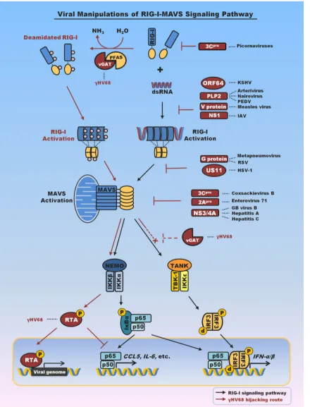
nnate immunity is the first line of defense against invading pathogens. Central to host immune responses is the detection of pathogen-associated molecular patterns (PAMPs) by cellular pat-tern recognition receptors (PRRs) (1). Retinoic acid-induced gene I (RIG-I) is a cytosolic receptor that senses double-stranded RNA (dsRNA) originating from pathogens such as viruses (2–5). Bind-ing to dsRNA disrupts an intramolecular interaction that keeps RIG-I in an autoinhibitory state (6,7), triggering an overall con-formational change that releases the N-terminal CARD domain (8,9). The N-terminal CARD of RIG-I undergoes homotypic oli-gomerization and heteroolioli-gomerization with that of the mito-chondrion antiviral signaling (MAVS) adaptor molecule (10). Oligomerized MAVS forms prion-like filaments that are capable of activating two kinase complexes, IB kinase alpha beta gamma (IKK␣␥) and IKKε–TBK-1, which, in turn, activate NF-B and interferon (IFN) regulatory factors (IRFs) (11–13). Along with other transcription factors, NF-B and IRFs upregulate the ex-pression of intrinsic antiviral molecules (e.g., Mx and viperin) and the secretion of various cytokines (e.g., interferon) that further induce the expression of a network of a few hundred antiviral genes (14). Given the potent activity of RIG-I in inducing inflam-matory responses, it is not surprising that RIG-I activation is reg-ulated by multiple mechanisms in response to viral infection. For example, noncovalent binding and covalent conjugation of the Lys63-linked polyubiquitin chain to the CARD domain are re-ported to activate RIG-I (15–17), whereas phosphorylation by protein kinase C and casein kinase represses and dephosphoryla-tion promotes RIG-I-mediated signaling (18–20). These are key cellular events that have been evolved to tightly regulate RIG-I-mediated immune activation in response to viral infection.Viruses often evolve intricate mechanisms to deflect host im-mune responses. While RNA viruses deploy various proteins to blunt RIG-I-mediated innate defenses by hampering key signaling components such as RIG-I and MAVS, DNA viruses can manip-ulate the signaling cascade to benefit their infection (21–23) (Fig. 1). Studies of RNA viruses have identified distinct viral factors that target RIG-I and MAVS. Influenza virus NS1 derails RIG-I ubiq-uitination by nullifying the essential TRIM25 E3 ligase (24). No-tably, hepatitis C virus uses its NS3/4A protease to cleave MAVS and release it from the mitochondrial membrane (25–27), thereby halting RIG-I-dependent antiviral immune responses. A similar strategy is employed by hepatitis G virus, hepatitis A virus, entero-virus 71, and coxsackieentero-virus to derail IFN production (28–31). DNA viruses utilize strategies that are more intricate than those
utilized by RNA viruses to evade these innate immune signaling cascades. The manipulation of innate and adaptive immune re-sponses by herpesviruses has been previously well reviewed (21). One interesting example is murine gammaherpesvirus 68 (␥HV68), which requires MAVS for efficient lytic replication.
␥HV68 is a model herpesvirus for human Kaposi’s sarcoma-asso-ciated herpesvirus (KSHV) and Epstein-Barr virus (EBV). With a combination of genetic and biochemical analyses, Dong et al. showed that the downstream IKKkinase is usurped to phos-phorylate viral replicationtrans-activator (RTA) and thereby pro-motes viral lytic gene expression (32). Additionally, IKKis also coopted to phosphorylate p65 (also known as RelA), which primes p65 for proteasome-mediated degradation in conjunction with the RTA E3 ligase, thereby terminating NF-B activation (33,34). Thus, MAVS-dependent signaling is critical for efficient produc-tive replication of␥HV68.
HIJACKING RIG-I TO EVADE CYTOKINE PRODUCTION VIA DEAMIDATION
In order to characterize the virus-host interactions that instigate MAVS-dependent signaling, a screen for open reading frames from␥HV68 that activate MAVS-dependent signaling revealed that ORF75C is a potent activator of RIG-I and MAVS signaling (35). Genetic and biochemical studies demonstrated functional and direct physical interactions between ORF75c and RIG-I. ORF75 genes are highly conserved in all gammaherpesviruses. While the KSHV genome encodes ORF75 and EBV encodes BNRF1, ORF75 genes are duplicated in herpesvirus saimiri (ORF3 and ORF75) and triplicated in murine␥HV68 (i.e., ORF75a, -b, and -c). Accumulating studies describe antagonism of members of the promyelocytic leukemia (PML) family as a common immune evasion function of gammaherpesvirus ORF75 proteins (36–38). These studies have been extensively reviewed elsewhere (70). Here, we discuss the deamidating activity of ORF75 proteins of gammaherpesviruses.
Accepted manuscript posted online17 February 2016
CitationZhao J, Li J, Xu S, Feng P. 2016. Emerging roles of protein deamidation in innate immune signaling. J Virol 90:4262– 4268.doi:10.1128/JVI.01980-15. Editor:M. Gack
Address correspondence to Pinghui Feng, pinghui.feng@med.usc.edu.
Copyright © 2016, American Society for Microbiology. All Rights Reserved. GEM
on November 7, 2019 by guest
http://jvi.asm.org/
FIG 1Summary of viral factors that interfere with or hijack RIG-I-mediated innate immune signaling. Emphasis is placed on viral proteins that interfere with RIG-I or MAVS to evade antiviral cytokine production. Notably, viral proteins that target TBK-1 and IRF3 to block interferon production are not included here. vGAT proteins of KSHV and␥HV68 recruit PFAS to deamidate and activate RIG-I. Activation of RIG-I and its downstream signaling events, specifically, those associated with IKK, result in p65 degradation and suppress inflammatory cytokine production (33–35). vGAT appears to blunt IRF activation by an unknown mechanism (indicated by dashed inhibition sign). PEDV, porcine epidemic diarrhea virus; IAV, influenza A virus; RSV, respiratory syncytial virus; HSV-1, herpes simplex virus 1; Hepatitis A, hepatitis A virus; Hepatitis C, hepatitis C virus; GB virus, hepatitis G virus; pro, protease; NEMO, NF-B essential modulator; TANK, TRAF family member-associated NF-B activator. RTA, replication and transcription activator.
on November 7, 2019 by guest
http://jvi.asm.org/
[image:2.585.77.517.67.644.2]The gammaherpesvirus ORF75 shares homology with phos-phoribosyl-formylglycinamidine synthetase (PFAS; also known as FGARAT of phosphoribosyl-formylglycinamidine [FGAM]), a cellular glutamine amidotransferase (GAT), and is thus also re-ferred to as vGAT. PFAS catalyzes reaction 4 of the 10 steps of purinede novosynthesis. However, the vGAT proteins of␥HV68 cannot complement cells deficient in PFAS (40). The carboxyl-terminal GAT domain of vGAT is sufficient to interact with RIG-I but fails to activate RIG-I. Coupled with the fact that vGAT pro-teins share homology with cellular GATs, this observation sug-gests that vGAT-induced RIG-I activation may require the enzy-matic activity of GAT. Indeed, treatment of cells expressing vGAT with a GAT inhibitor specifically diminished signaling down-stream of RIG-I, but not that downdown-stream of IKK, indicating that GAT activity is specifically required for events upstream of IKK (e.g., RIG-I). Two-dimensional gel electrophoresis showed that vGAT reduced the RIG-I charge, and mass spectrometry analysis identified three site-specific deamidations within RIG-I: Q10 in the CARD domain and N245 and N445 within the helicase 1 do-main. Simultaneous deamidation of all three residues led to con-stitutive activation of RIG-I in the absence of RNA (35), revealing a new means to activate the RIG-I receptor. Consistent with pre-vious findings on MAVS and IKK(32,33), activated RIG-I is usurped to evade antiviral cytokine production via inducing the degradation of p65, the key subunit of the transcriptionally active NF-B dimer. Whether deamidated and activated RIG-I is re-quired for efficient replication of␥HV68 remains an open ques-tion.
vGAT proteins lack the catalytic triad essential for amidotrans-fer/deamidation catalysis (35), suggesting that vGAT may recruit a cellular enzyme(s) to induce the deamidation and concomitant activation of RIG-I. Enzymes involved in nucleotide biosynthesis have a propensity to form homo-oligomers in order to regulate enzymatic activity (39,41), so it is possible that vGAT proteins recruit cellular PFAS to deamidate RIG-I. Indeed, vGATs of KSHV, EBV, and␥HV68 interact with PFAS, but only vGATs of KSHV and␥HV68 induce RIG-I deamidation and activation. De-pletion or pharmacological inhibition of PFAS recapitulates the phenotype of fibroblasts deficient in RIG-I and MAVS, i.e., in-creased cytokine production in response to␥HV68 infection (33– 35). Furthermore, purified vGAT and PFAS deamidated RIG-Iin vitro. Neither vGAT nor PFAS alone was sufficient to induce RIG-I deamidation, suggesting that vGAT activates PFAS by an intrinsic mechanism. For the first time, these studies demonstrated that a PRR is activated by a deamidase consisting of a metabolic enzyme and a viral pseudoenzyme rather than by its conventional ligand. This work also describes a new function of the cellular metabolic PFAS enzyme in deamidating asparagines and glutamines of RIG-I to regulate innate immune signaling (35), suggesting that protein deamidation could play pivotal roles in regulating innate immune signaling.
PROTEIN DEAMIDATION
Protein deamidation was initially reported more than half a cen-tury ago (42). Early work focused on the nonenzymatic deamida-tion of asparaginyl and, to a lesser extent, glutaminyl residues of proteinsin vivo andin vitro. Analyzing a large set of proteins, Robinson and Robinson showed that the rate of asparaginyl deamidation was determined by the primary sequence, secondary and tertiary structures of the protein, and cellular environment
(such as pH) (43). The ubiquitous distribution of asparaginyl/ glutaminyl residues and frequent deamidation thereof in proteins, coupled with the finding that surrounding sequences determine the rate of protein deamidation, prompted the postulation that nonenzymatic protein deamidation serves as an internal clock to time the biological events of a particular protein (44). Thus, non-enzymatic deamidation may be a built-in clock to time protein functional decay or “aging.”
(i) Enzymes (deamidases) that catalyze protein deamidation. Emerging studies indicate that microbes deploy protein deamida-tion to manipulate key signaling components to promote their infection. Microbial enzymes constitute the founding members of the protein deamidase family and offer an opportunity to answer fundamental questions concerning protein deamidation. Work on the PFAS-vGAT protein-deamidating complex has described a new regulatory function of the metabolic PFAS enzyme, and likely other glutamine amidotransferases, in innate immune signal transduction. This work raises a number of imminent questions that are fundamental to enzymology and innate immune signal-ing. First, how does the amidotransferase activity of PFAS in pu-rine synthesis correlate with its protein-deamidating activity in virus-infected cells? PFAS is known to catalyze amidotransfer from free glutamine to synthesize phosphoribosyl-formylglyci-namidine (FGAM) in purine synthesis (45), whereas the PFAS-vGAT complex is capable of deamidating asparaginyl and glu-taminyl residues of RIG-I (35). Cellular GAT enzymes catalyze the biosynthesis of nucleotides, amino acids, glycoprotein, and NAD (46). The fact that PFAS can participate in both purine synthesis and RIG-I activation suggests that nucleotide metabolism is linked to innate immune signaling. Indeed, inhibitors targeting dihydroorotate dehydrogenase, a key enzyme of thede novo py-rimidine synthesis pathway, reduced viral replication (47–49). Later, it was shown that this antiviral activity relies on an elevated cellular immune response (50). The molecular link between re-duced pyramidine biosynthesis and increased antiviral gene ex-pression remains unknown. Presumably, the nucleotide pool within cells infected by a virusin vivois small, so activating nucle-otide biosynthesis is imperative for efficient viral transcription and genome replication. An elegant example is how cellular SAMHD1 nucleotide hydrolase restricts human immunodefi-ciency virus (HIV) via depleting the nucleotide pool available for HIV replication (51). If vGAT activates PFAS for purine biosyn-thesis and RIG-I-dependent evasion of antiviral cytokine produc-tion, a proper division between the two enzymatic activities (i.e., protein deamidation and purine synthesis) is critical for viral lytic replication. In support of this notion, murine␥HV68 robustly upregulates the protein expression of PFAS, partly by increas-ing its mRNA level (35). Future work concerning PFAS regu-lation in virus-infected cells will likely further elucidate the possible cross talk between innate immune signaling and nu-cleotide biosynthesis.
Second, how does RIG-I access the active site of the PFAS deamidase domain? Cellular glutamine amidotransferases consist of a GAT domain and a metabolite synthetase domain connected by an ammonia channel. Structural analysis of theSalmonella en-tericaserovar Typhimurium homolog of PFAS revealed a rela-tively buried catalytic triad for deamidation facing the internal ammonia channel (39). If human PFAS is structurally similar to SalmonellaPFAS, binding to vGAT perhaps triggers a conforma-tional change that exposes the catalytic triad to accommodate
on November 7, 2019 by guest
http://jvi.asm.org/
paraginyl and glutaminyl residues of RIG-I. The GAT domain is loosely connected to the FGAM synthetase domain (39), which likely provides flexibility and enables substrate accessibility. Such a conformational change may uncouple deamidation from am-monia channeling to the catalytic center of the FGAM synthetase, thereby facilitating the release of free ammonia. Interestingly, mu-tations blocking the ammonia channel between the GAT domain and the neighboring synthetase domain of imidazole-glycerol-phosphate synthase increased the deamidating activity of the glu-taminase domain by more than 3 orders of magnitude (52), sug-gesting that ammonia release is a mechanism to significantly elevate glutamine hydrolysis. vGAT may deploy a similar
mecha-nism of uncoupling the ammonia channel from the enzymatic domain to deamidate RIG-I, although the detailed mechanism of vGAT activation of PFAS requires further investigation.
Third, it is unclear whether PFAS, and other glutamine amido-transferases, can deamidate proteins in the absence of gammaher-pesvirus vGAT proteins. RIG-I deamidation was not observed in cells infected with Sendai virus or␥HV68 deficient in vGAT. If PFAS and other GATs can deamidate proteins in mammalian cells, it would be interesting to quantify how viral infection im-pacts the spectrum of deamidated proteins. This would be best assessed by a proteome-wide deamidation analysis. The herpesvi-rus proteome offers an excellent platform to systematically ana-TABLE 1Emerging discoveries of mammalian protein deamidations
Functional consequence Deamidated cellular target(s) Deamidase/other mechanism(s) Deamidase-encoding species Reference
G protein signaling pathways
Activation of G protein signaling Rho GTPases Cytotoxic necrotizing factors (CNFs) Escherichia coli(EPEC) 60
●RhoA
●Rac
●Cdc42 Yersinia pseudotuberculosis 53
Heterotrimeric G proteins (G␣i, G␣q/11, and G␣12/13)
Pasteurella multocidatoxin (PMT) Pasteurella multocida 61
Rho GTPases VopC Vibrio parahaemolyticus 62
●Rac
●Cdc42
Translation/cell cycle progression/apoptosis
Inhibition of translation Eukaryotic initiation factor 4A (eIF4A)
Burkholderialethal factor 1 (BLF1) Burkholderia pseudomallei 63
Critical switch (checkpoint) of apoptosis induced by DNA damage
B-cell lymphoma—extra large (Bcl-XL)
pH change NAa 64
Inhibition of cell cycle progression
Ubiquitin Cycle-inhibiting factors (Cifs) Escherichia coli 58
Yersinia pseudotuberculosis
NEDD8 Burkholderia pseudomallei
Photorhabdus asymbiotica Photorhabdus luminescens
Innate immunity and inflammatory responses
Inactivation of ubiquitin system and its related pathways, e.g., NF-B signaling pathway
Ubiquitin Cycle-inhibiting factors (Cifs) Escherichia coli 58
Yersinia pseudotuberculosis Burkholderia pseudomallei Photorhabdus asymbiotica Photorhabdus luminescens
Inhibition of UBC13/TRAF6-dependent inflammatory responses
Ubiquitin-conjugating enzyme 13 (UBC13)
OspI Shigella flexneri 59
Activation of RIG-I signaling to evade inflammatory signaling
Retinoic acid-inducible gene 1 (RIG-I)
vGAT (ORF75/ORF75c)⫹
phosphoribosylformylglycinamidine synthase (PFAS)
Murine herpesvirus 68 (ORF75c) 35
Human herpesvirus 8 (ORF75)
Homo sapiens(PFAS) Recognition by gut-derived T
cells to promote intestinal inflammation
Gliadin (wheat), etc. Tissue transglutaminase (TG2) Homo sapiens 65
Others
N-end rule pathway of protein degradation
Model substrate of N-end rule pathway
N-terminal glutamine amidase (NTQA)
Mus musculus 66
Alteration of the kinetics of excitatory synaptic transmission
Eukaryotic initiation factor 4E-binding protein 2 (4E-BP2)
pH change NA 67
aNA, not applicable.
on November 7, 2019 by guest
http://jvi.asm.org/
[image:4.585.41.540.77.555.2]lyze protein deamidation. vGATs of KSHV, EBV, and␥HV68 dis-play similar interactions with PFAS, but only EBV vGAT failed to deamidate RIG-I, implying that vGATs and PFAS may have other functions shared by all three gammaherpesviruses such as nucle-otide metabolism and evasion of intrinsic antiviral immunity. Al-though the genomes of herpes simplex viruses (HSV) contain no homolog of vGAT, HSV-1 infection induced a robust reduction in RIG-I’s charge, indicative of deamidation. This suggests that her-pes simplex viruses may have evolved a different mechanism for inducing RIG-I deamidation. Future studies may reveal a new example of protein deamidation in innate immune signaling.
In contrast to viral pseudoenzymes, several bacterial proteins appear to possess intrinsic deamidase activity toward multiple sig-naling molecules. Cytotoxic necrotizing factors (CNFs) produced by uropathogenic (CNF1) or enteropathogenic (CNF2) Esche-richia colideamidate and constitutively activate small G proteins (53–55). Similarly to chemotaxis D (CheD) deamidase, CNFs form a common␣//sandwich that contains the catalytic dyad in a shallow cavity at the top of the protein (56,57). The members of the Cif family of effectors secreted byBurkholderia pseudomallei and enteropathogenicE. coliand the OspI effector secreted by Shigella flexnerican deamidate ubiquitin/Nedd8 and the UBC13 E2 enzyme, respectively (58,59). Deamidation of these signaling components by bacterial effectors is essential for evading cellular immune responses and the pathogenesis of these microbes. These studies define the structure and function of protein deamidases in pathogen infection.
(ii) Protein targets of enzyme-catalyzed deamidation. Pro-tein deamidation catalyzed by enzymes is generally rapid and tightly regulated. To date, we have understood the functional con-sequences of deamidation of a small subset of proteins in pro-karyotes and mammalian cells (Table 1). Initial studies showed that key cellular signaling molecules are deamidated by patho-genic microbes to facilitate their invasion, which underpins their pathogenesis (68). Small G proteins such as RhoA and Cdc42 re-quire a key glutamine residue for GTP hydrolysis. Deamidation of the conserved glutamine by pathogenicE. colilocks these G pro-teins in a GTP-bound state, resulting in the constitutive activation of G proteins and stress fiber formation (53,60,62). Interestingly, deamidation is also crucial for regulating signal transduction in bacterial chemotaxis. In nonenteric bacteria, the chemotaxis C (CheC) phosphatase and methyl-accepting chemotaxis proteins (known as MCPs) are deamidated by the CheD polypeptide and are required for directional chemotaxis (57). Mammalian ubiq-uitins Nedd8 and UBC13 have recently been shown to be deami-dated by effectors secreted by enteric pathogenicE. coliand Shi-gella, respectively (58,59). Deamidation of these cellular signaling molecules inactivates the ubiquitin proteasome system that is crit-ical for signal transduction downstream of tumor necrosis factor alpha (TNF-␣), an important component of the host immune defense system. In response to DNA damage, anti-apoptotic Bcl-xLis targeted for degradation via deamidation (64). Recently,
the deamidation of 4E-BP2 was shown to promote its association with mammalian target of rapamycin (mTOR) and modulate neuronal excitatory synaptic transmission (67). It was postulated that the deamidation of Bcl-xLand 4E-BP2 is a nonenzymatic
process and results from an increase in cellular pH. Deamidation of other key proteins is implicated in regulating some fundamen-tal processes (e.g., cell-matrix interaction) and underpins medi-cally important diseases (e.g., Alzheimer’s disease) (69).
CONCLUSION AND PERSPECTIVES
Recent studies of enterobacterial effectors and gammaherpesviral pseudoenzymes implicate a new function of protein deamidation in regulating innate immune signal transduction. This is likely just the tip of iceberg concerning the general regulatory roles of pro-tein deamidation in fundamental biological processes. That met-abolic glutamine amidotransferases, such as the vGAT-PFAS complex, are capable of deamidating proteins is interesting in that these enzymes may provide a physical and functional link to other biological processes. However, these studies have generated more questions than answers in regard to fundamental principles con-cerning protein deamidation. For example, how are these cellular GATs regulated and delegated between metabolism and signal transduction in response to infection? What sequence and struc-tural elements of GATs (e.g., PFAS) enable their dual functionality or substrate promiscuity in deamidating free glutamine and glu-taminyl/asparaginyl residues? Is there sequence specificity of tar-get deamidated proteins? If yes, is the specificity defined by the primary, secondary, or tertiary structure of the deamidated pro-tein? Last, and importantly, is protein deamidation involved in other fundamental biological processes that are intrinsically linked to nucleotide biosynthesis, such as DNA damage and re-pair? These microbial studies have unveiled a new function of the simplest posttranslational modification of proteins, deamidation, in immune regulation and will certainly instruct us more in the years to come.
ACKNOWLEDGMENTS
Work in the Feng laboratory is supported by grants from NIDCR (DE021445) and NCI (CA134241).
We apologize to those whose work is not cited here due to limited space.
FUNDING INFORMATION
This work, including the efforts of Pinghui Feng, was funded by HHS | NIH | National Cancer Institute (NCI) (CA134241). This work, including the efforts of Pinghui Feng, was funded by HHS | NIH | National Institute of Dental and Craniofacial Research (NIDCR) (DE021445).
REFERENCES
1.Takeuchi O, Akira S.2010. Pattern recognition receptors and inflamma-tion. Cell140:805– 820.http://dx.doi.org/10.1016/j.cell.2010.01.022. 2.Hornung V, Ellegast J, Kim S, Brzozka K, Jung A, Kato H, Poeck H,
Akira S, Conzelmann KK, Schlee M, Endres S, Hartmann G. 2006. 5=-Triphosphate RNA is the ligand for RIG-I. Science314:994 –997.http: //dx.doi.org/10.1126/science.1132505.
3.Kato H, Takeuchi O, Sato S, Yoneyama M, Yamamoto M, Matsui K, Uematsu S, Jung A, Kawai T, Ishii KJ, Yamaguchi O, Otsu K, Tsujimura T, Koh CS, Reis e Sousa C, Matsuura Y, Fujita T, Akira S. 2006. Differential roles of MDA5 and RIG-I helicases in the recognition of RNA viruses. Nature441:101–105.http://dx.doi.org/10.1038/nature04734. 4.Pichlmair A, Schulz O, Tan CP, Naslund TI, Liljestrom P, Weber F,
Reis e Sousa C. 2006. RIG-I-mediated antiviral responses to single-stranded RNA bearing 5=-phosphates. Science314:997–1001.http://dx .doi.org/10.1126/science.1132998.
5.Schmidt A, Schwerd T, Hamm W, Hellmuth JC, Cui S, Wenzel M, Hoffmann FS, Michallet MC, Besch R, Hopfner KP, Endres S, Rothen-fusser S.2009. 5=-triphosphate RNA requires base-paired structures to activate antiviral signaling via RIG-I. Proc Natl Acad Sci U S A106:12067– 12072.http://dx.doi.org/10.1073/pnas.0900971106.
6.Saito T, Hirai R, Loo YM, Owen D, Johnson CL, Sinha SC, Akira S, Fujita T, Gale M, Jr.2007. Regulation of innate antiviral defenses through a shared repressor domain in RIG-I and LGP2. Proc Natl Acad Sci U S A
104:582–587.http://dx.doi.org/10.1073/pnas.0606699104.
7.Takahasi K, Yoneyama M, Nishihori T, Hirai R, Kumeta H, Narita R,
on November 7, 2019 by guest
http://jvi.asm.org/
Gale M, Jr, Inagaki F, Fujita T.2008. Nonself RNA-sensing mechanism of RIG-I helicase and activation of antiviral immune responses. Mol Cell
29:428 – 440.http://dx.doi.org/10.1016/j.molcel.2007.11.028.
8.Kowalinski E, Lunardi T, McCarthy AA, Louber J, Brunel J, Grigorov B, Gerlier D, Cusack S.2011. Structural basis for the activation of innate immune pattern-recognition receptor RIG-I by viral RNA. Cell147:423– 435.http://dx.doi.org/10.1016/j.cell.2011.09.039.
9.Luo D, Ding SC, Vela A, Kohlway A, Lindenbach BD, Pyle AM.2011. Structural insights into RNA recognition by RIG-I. Cell147:409 – 422.
http://dx.doi.org/10.1016/j.cell.2011.09.023.
10. Wu B, Peisley A, Tetrault D, Li Z, Egelman EH, Magor KE, Walz T, Penczek PA, Hur S.2014. Molecular imprinting as a signal-activation mechanism of the viral RNA sensor RIG-I. Mol Cell55:511–523.http://dx .doi.org/10.1016/j.molcel.2014.06.010.
11. Fitzgerald KA, McWhirter SM, Faia KL, Rowe DC, Latz E, Golenbock DT, Coyle AJ, Liao SM, Maniatis T.2003. IKKepsilon and TBK1 are essential components of the IRF3 signaling pathway. Nat Immunol4:491– 496.http://dx.doi.org/10.1038/ni921.
12. Sharma S, tenOever BR, Grandvaux N, Zhou GP, Lin R, Hiscott J.2003. Triggering the interferon antiviral response through an IKK-related path-way. Science300:1148 –1151.http://dx.doi.org/10.1126/science.1081315. 13. Hou F, Sun L, Zheng H, Skaug B, Jiang QX, Chen ZJ.2011. MAVS forms functional prion-like aggregates to activate and propagate antiviral innate immune response. Cell146:448 – 461.http://dx.doi.org/10.1016/j .cell.2011.06.041.
14. Panne D, Maniatis T, Harrison SC. 2007. An atomic model of the interferon-beta enhanceosome. Cell129:1111–1123.http://dx.doi.org/10 .1016/j.cell.2007.05.019.
15. Gack MU, Shin YC, Joo CH, Urano T, Liang C, Sun L, Takeuchi O, Akira S, Chen Z, Inoue S, Jung JU. 2007. TRIM25 RING-finger E3 ubiquitin ligase is essential for RIG-I-mediated antiviral activity. Nature
446:916 –920.http://dx.doi.org/10.1038/nature05732.
16. Zeng W, Sun L, Jiang X, Chen X, Hou F, Adhikari A, Xu M, Chen ZJ.
2010. Reconstitution of the RIG-I pathway reveals a signaling role of un-anchored polyubiquitin chains in innate immunity. Cell141:315–330.
http://dx.doi.org/10.1016/j.cell.2010.03.029.
17. Oshiumi H, Miyashita M, Inoue N, Okabe M, Matsumoto M, Seya T.
2010. The ubiquitin ligase Riplet is essential for RIG-I-dependent innate immune responses to RNA virus infection. Cell Host Microbe8:496 –509.
http://dx.doi.org/10.1016/j.chom.2010.11.008.
18. Maharaj NP, Wies E, Stoll A, Gack MU.2012. Conventional protein kinase C-alpha (PKC-alpha) and PKC-beta negatively regulate RIG-I an-tiviral signal transduction. J Virol 86:1358 –1371. http://dx.doi.org/10 .1128/JVI.06543-11.
19. Sun Z, Ren H, Liu Y, Teeling JL, Gu J.2011. Phosphorylation of RIG-I by casein kinase II inhibits its antiviral response. J Virol85:1036 –1047.
http://dx.doi.org/10.1128/JVI.01734-10.
20. Wies E, Wang MK, Maharaj NP, Chen K, Zhou S, Finberg RW, Gack MU.2013. Dephosphorylation of the RNA sensors RIG-I and MDA5 by the phosphatase PP1 is essential for innate immune signaling. Immunity
38:437– 449.http://dx.doi.org/10.1016/j.immuni.2012.11.018.
21. Feng P, Moses A, Fruh K.2013. Evasion of adaptive and innate immune response mechanisms by gamma-herpesviruses. Curr Opin Virol3:285– 295.http://dx.doi.org/10.1016/j.coviro.2013.05.011.
22. Sparrer KM, Gack MU.2015. Intracellular detection of viral nucleic acids. Curr Opin Microbiol 26:1–9. http://dx.doi.org/10.1016/j.mib.2015.03 .001.
23. Loo YM, Gale M, Jr.2011. Immune signaling by RIG-I-like receptors. Immunity34:680 – 692.http://dx.doi.org/10.1016/j.immuni.2011.05.003. 24. Gack MU, Albrecht RA, Urano T, Inn KS, Huang IC, Carnero E, Farzan M, Inoue S, Jung JU, Garcia-Sastre A.2009. Influenza A virus NS1 targets the ubiquitin ligase TRIM25 to evade recognition by the host viral RNA sensor RIG-I. Cell Host Microbe5:439 – 449.http://dx.doi.org/10.1016/j .chom.2009.04.006.
25. Li XD, Sun L, Seth RB, Pineda G, Chen ZJ.2005. Hepatitis C virus protease NS3/4A cleaves mitochondrial antiviral signaling protein off the mitochondria to evade innate immunity. Proc Natl Acad Sci U S A102:
17717–17722.http://dx.doi.org/10.1073/pnas.0508531102.
26. Meylan E, Curran J, Hofmann K, Moradpour D, Binder M, Barten-schlager R, Tschopp J.2005. Cardif is an adaptor protein in the RIG-I antiviral pathway and is targeted by hepatitis C virus. Nature437:1167– 1172.http://dx.doi.org/10.1038/nature04193.
27. Li K, Foy E, Ferreon JC, Nakamura M, Ferreon AC, Ikeda M, Ray SC,
Gale M, Jr, Lemon SM.2005. Immune evasion by hepatitis C virus NS3/4A protease-mediated cleavage of the Toll-like receptor 3 adaptor protein TRIF. Proc Natl Acad Sci U S A102:2992–2997.http://dx.doi.org /10.1073/pnas.0408824102.
28. Chen Z, Benureau Y, Rijnbrand R, Yi J, Wang T, Warter L, Lanford RE, Weinman SA, Lemon SM, Martin A, Li K.2007. GB virus B disrupts RIG-I signaling by NS3/4A-mediated cleavage of the adaptor protein MAVS. J Virol81:964 –976.http://dx.doi.org/10.1128/JVI.02076-06. 29. Yang Y, Liang Y, Qu L, Chen Z, Yi M, Li K, Lemon SM. 2007.
Disruption of innate immunity due to mitochondrial targeting of a picor-naviral protease precursor. Proc Natl Acad Sci U S A104:7253–7258.http: //dx.doi.org/10.1073/pnas.0611506104.
30. Wang B, Xi X, Lei X, Zhang X, Cui S, Wang J, Jin Q, Zhao Z.2013. Enterovirus 71 protease 2Apro targets MAVS to inhibit anti-viral type I interferon responses. PLoS Pathog9:e1003231.http://dx.doi.org/10.1371 /journal.ppat.1003231.
31. Mukherjee A, Morosky SA, Delorme-Axford E, Dybdahl-Sissoko N, Oberste MS, Wang T, Coyne CB.2011. The coxsackievirus B 3C protease cleaves MAVS and TRIF to attenuate host type I interferon and apoptotic signaling. PLoS Pathog 7:e1001311. http://dx.doi.org/10.1371/journal .ppat.1001311.
32. Dong X, Feng H, Sun Q, Li H, Wu TT, Sun R, Tibbetts SA, Chen ZJ, Feng P.2010. Murine gamma-herpesvirus 68 hijacks MAVS and IKKbeta to initiate lytic replication. PLoS Pathog6:e1001001.http://dx.doi.org/10 .1371/journal.ppat.1001001.
33. Dong X, Feng P.2011. Murine gamma herpesvirus 68 hijacks MAVS and IKKbeta to abrogate NFkappaB activation and antiviral cytokine produc-tion. PLoS Pathog 7:e1002336. http://dx.doi.org/10.1371/journal.ppat .1002336.
34. Dong X, He Z, Durakoglugil D, Arneson L, Shen Y, Feng P. 2012. Murine gammaherpesvirus 68 evades host cytokine production via repli-cation transactivator-induced RelA degradation. J Virol86:1930 –1941.
http://dx.doi.org/10.1128/JVI.06127-11.
35. He S, Zhao J, Song S, He X, Minassian A, Zhou Y, Zhang J, Brulois K, Wang Y, Cabo J, Zandi E, Liang C, Jung JU, Zhang X, Feng P.2015. Viral pseudo-enzymes activate RIG-I via deamidation to evade cytokine production. Mol Cell 58:134 –146. http://dx.doi.org/10.1016/j.molcel .2015.01.036.
36. Ling PD, Tan J, Sewatanon J, Peng R.2008. Murine gammaherpesvirus 68 open reading frame 75c tegument protein induces the degradation of PML and is essential for production of infectious virus. J Virol82:8000 – 8012.http://dx.doi.org/10.1128/JVI.02752-07.
37. Tsai K, Thikmyanova N, Wojcechowskyj JA, Delecluse HJ, Lieberman PM.2011. EBV tegument protein BNRF1 disrupts DAXX-ATRX to acti-vate viral early gene transcription. PLoS Pathog7:e1002376.http://dx.doi .org/10.1371/journal.ppat.1002376.
38. Full F, Jungnickl D, Reuter N, Bogner E, Brulois K, Scholz B, Sturzl M, Myoung J, Jung JU, Stamminger T, Ensser A.2014. Kaposi’s sarcoma associated herpesvirus tegument protein ORF75 is essential for viral lytic replication and plays a critical role in the antagonization of ND10-instituted intrinsic immunity. PLoS Pathog10:e1003863.http://dx.doi .org/10.1371/journal.ppat.1003863.
39. Anand R, Hoskins AA, Stubbe J, Ealick SE.2004. Domain organization of Salmonella typhimurium formylglycinamide ribonucleotide amido-transferase revealed by X-ray crystallography. Biochemistry43:10328 – 10342.http://dx.doi.org/10.1021/bi0491301.
40. Gaspar M, Gill MB, Losing JB, May JS, Stevenson PG.2008. Multiple functions for ORF75c in murid herpesvirus-4 infection. PLoS One
3:e2781.http://dx.doi.org/10.1371/journal.pone.0002781.
41. Guy HI, Schmidt B, Herve G, Evans DR.1998. Pressure-induced disso-ciation of carbamoyl-phosphate synthetase domains. The catalytically ac-tive form is dimeric. J Biol Chem273:14172–14178.
42. Mycek MJ, Waelsch H.1960. The enzymatic deamidation of proteins. J Biol Chem235:3513–3517.
43. Robinson NE, Robinson AB.2001. Prediction of protein deamidation rates from primary and three-dimensional structure. Proc Natl Acad Sci U S A98:4367– 4372.http://dx.doi.org/10.1073/pnas.071066498. 44. Robinson NE, Robinson AB.2001. Molecular clocks. Proc Natl Acad Sci
U S A98:944 –949.http://dx.doi.org/10.1073/pnas.98.3.944.
45. Patterson D, Bleskan J, Gardiner K, Bowersox J.1999. Human phos-phoribosylformylglycineamide amidotransferase (FGARAT): regional mapping, complete coding sequence, isolation of a functional genomic
on November 7, 2019 by guest
http://jvi.asm.org/
clone, and DNA sequence analysis. Gene239:381–391.http://dx.doi.org /10.1016/S0378-1119(99)00378-9.
46. Massière F, Badet-Denisot MA.1998. The mechanism of glutamine-dependent amidotransferases. Cell Mol Life Sci54:205–222.http://dx.doi .org/10.1007/s000180050145.
47. Bonavia A, Franti M, Pusateri Keaney E, Kuhen K, Seepersaud M, Radetich B, Shao J, Honda A, Dewhurst J, Balabanis K, Monroe J, Wolff K, Osborne C, Lanieri L, Hoffmaster K, Amin J, Markovits J, Broome M, Skuba E, Cornella-Taracido I, Joberty G, Bouwmeester T, Hamann L, Tallarico JA, Tommasi R, Compton T, Bushell SM.2011. Identifica-tion of broad-spectrum antiviral compounds and assessment of the drug-gability of their target for efficacy against respiratory syncytial virus (RSV). Proc Natl Acad Sci U S A108:6739 – 6744.http://dx.doi.org/10.1073/pnas .1017142108.
48. Wang QY, Bushell S, Qing M, Xu HY, Bonavia A, Nunes S, Zhou J, Poh MK, Florez de Sessions P, Niyomrattanakit P, Dong H, Hoffmaster K, Goh A, Nilar S, Schul W, Jones S, Kramer L, Compton T, Shi PY.2011. Inhibition of dengue virus through suppression of host pyrimidine bio-synthesis. J Virol85:6548 – 6556.http://dx.doi.org/10.1128/JVI.02510-10. 49. Hoffmann HH, Kunz A, Simon VA, Palese P, Shaw ML.2011. Broad-spectrum antiviral that interferes with de novo pyrimidine biosynthesis. Proc Natl Acad Sci U S A108:5777–5782.http://dx.doi.org/10.1073/pnas .1101143108.
50. Lucas-Hourani M, Dauzonne D, Jorda P, Cousin G, Lupan A, Helynck O, Caignard G, Janvier G, Andre-Leroux G, Khiar S, Escriou N, Despres P, Jacob Y, Munier-Lehmann H, Tangy F, Vidalain PO.2013. Inhibition of pyrimidine biosynthesis pathway suppresses viral growth through in-nate immunity. PLoS Pathog 9:e1003678. http://dx.doi.org/10.1371 /journal.ppat.1003678.
51. Goldstone DC, Ennis-Adeniran V, Hedden JJ, Groom HC, Rice GI, Christodoulou E, Walker PA, Kelly G, Haire LF, Yap MW, de Carvalho LP, Stoye JP, Crow YJ, Taylor IA, Webb M.2011. HIV-1 restriction factor SAMHD1 is a deoxynucleoside triphosphate triphosphohydrolase. Nature480:379 –382.http://dx.doi.org/10.1038/nature10623.
52. List F, Vega MC, Razeto A, Hager MC, Sterner R, Wilmanns M.2012. Catalysis uncoupling in a glutamine amidotransferase bienzyme by un-blocking the glutaminase active site. Chem Biol19:1589 –1599.http://dx .doi.org/10.1016/j.chembiol.2012.10.012.
53. Flatau G, Lemichez E, Gauthier M, Chardin P, Paris S, Fiorentini C, Boquet P.1997. Toxin-induced activation of the G protein p21 Rho by deamidation of glutamine. Nature 387:729 –733. http://dx.doi.org/10 .1038/42743.
54. Lerm M, Selzer J, Hoffmeyer A, Rapp UR, Aktories K, Schmidt G.1999. Deamidation of Cdc42 and Rac by Escherichia coli cytotoxic necrotizing factor 1: activation of c-Jun N-terminal kinase in HeLa cells. Infect Immun
67:496 –503.
55. Sugai M, Hatazaki K, Mogami A, Ohta H, Peres SY, Herault F, Hori-guchi Y, Masuda M, Ueno Y, Komatsuzawa H, Suginaka H, Oswald E.
1999. Cytotoxic necrotizing factor type 2 produced by pathogenic Esche-richia coli deamidates a gln residue in the conserved G-3 domain of the rho family and preferentially inhibits the GTPase activity of RhoA and rac1. Infect Immun67:6550 – 6557.
56. Buetow L, Flatau G, Chiu K, Boquet P, Ghosh P.2001. Structure of the Rho-activating domain of Escherichia coli cytotoxic necrotizing factor 1. Nat Struct Biol8:584 –588.http://dx.doi.org/10.1038/89610.
57. Chao X, Muff TJ, Park SY, Zhang S, Pollard AM, Ordal GW, Bilwes AM, Crane BR.2006. A receptor-modifying deamidase in complex with a
signaling phosphatase reveals reciprocal regulation. Cell124:561–571.
http://dx.doi.org/10.1016/j.cell.2005.11.046.
58. Cui J, Yao Q, Li S, Ding X, Lu Q, Mao H, Liu L, Zheng N, Chen S, Shao F.2010. Glutamine deamidation and dysfunction of ubiquitin/NEDD8 induced by a bacterial effector family. Science329:1215–1218.http://dx .doi.org/10.1126/science.1193844.
59. Sanada T, Kim M, Mimuro H, Suzuki M, Ogawa M, Oyama A, Ashida H, Kobayashi T, Koyama T, Nagai S, Shibata Y, Gohda J, Inoue J, Mizushima T, Sasakawa C. 2012. The Shigella flexneri effector OspI deamidates UBC13 to dampen the inflammatory response. Nature483:
623– 626.http://dx.doi.org/10.1038/nature10894.
60. Schmidt G, Sehr P, Wilm M, Selzer J, Mann M, Aktories K.1997. Gln 63 of Rho is deamidated by Escherichia coli cytotoxic necrotizing factor-1. Nature387:725–729.http://dx.doi.org/10.1038/42735.
61. Orth JH, Preuss I, Fester I, Schlosser A, Wilson BA, Aktories K.2009. Pasteurella multocida toxin activation of heterotrimeric G proteins by deamidation. Proc Natl Acad Sci U S A106:7179 –7184.http://dx.doi.org /10.1073/pnas.0900160106.
62. Zhang L, Krachler AM, Broberg CA, Li Y, Mirzaei H, Gilpin CJ, Orth K.2012. Type III effector VopC mediates invasion for Vibrio species. Cell Rep1:453– 460.http://dx.doi.org/10.1016/j.celrep.2012.04.004. 63. Cruz-Migoni A, Hautbergue GM, Artymiuk PJ, Baker PJ,
Bokori-Brown M, Chang CT, Dickman MJ, Essex-Lopresti A, Harding SV, Mahadi NM, Marshall LE, Mobbs GW, Mohamed R, Nathan S, Ngugi SA, Ong C, Ooi WF, Partridge LJ, Phillips HL, Raih MF, Ruzheinikov S, Sarkar-Tyson M, Sedelnikova SE, Smither SJ, Tan P, Titball RW, Wilson SA, Rice DW.2011. A Burkholderia pseudomallei toxin inhibits helicase activity of translation factor eIF4A. Science334:821– 824.http: //dx.doi.org/10.1126/science.1211915.
64. Deverman BE, Cook BL, Manson SR, Niederhoff RA, Langer EM, Rosova I, Kulans LA, Fu X, Weinberg JS, Heinecke JW, Roth KA, Weintraub SJ.2002. Bcl-xL deamidation is a critical switch in the regula-tion of the response to DNA damage. Cell111:51– 62.http://dx.doi.org/10 .1016/S0092-8674(02)00972-8.
65. Molberg O, McAdam SN, Korner R, Quarsten H, Kristiansen C, Mad-sen L, Fugger L, Scott H, Noren O, Roepstorff P, Lundin KE, Sjostrom H, Sollid LM.1998. Tissue transglutaminase selectively modifies gliadin peptides that are recognized by gut-derived T cells in celiac disease. Nat Med4:713–717.http://dx.doi.org/10.1038/nm0698-713.
66. Wang H, Piatkov KI, Brower CS, Varshavsky A. 2009. Glutamine-specific N-terminal amidase, a component of the N-end rule pathway. Mol Cell34:686 – 695.http://dx.doi.org/10.1016/j.molcel.2009.04.032. 67. Bidinosti M, Ran I, Sanchez-Carbente MR, Martineau Y, Gingras AC,
Gkogkas C, Raught B, Bramham CR, Sossin WS, Costa-Mattioli M, DesGroseillers L, Lacaille JC, Sonenberg N.2010. Postnatal deamidation of 4E-BP2 in brain enhances its association with raptor and alters kinetics of excitatory synaptic transmission. Mol Cell37:797– 808.http://dx.doi .org/10.1016/j.molcel.2010.02.022.
68. Washington EJ, Banfield MJ, Dangl JL.2013. What a difference a Dalton makes: bacterial virulence factors modulate eukaryotic host cell signaling systems via deamidation. Microbiol Mol Biol Rev77:527–539.http://dx .doi.org/10.1128/MMBR.00013-13.
69. Weintraub SJ, Deverman BE. 2007. Chronoregulation by asparagine deamidation. Sci STKE2007:re7.
70. Tsai K, Messick TE, Lieberman PM.2015. Disruption of host antiviral resistances by gammaherpesvirus tegument proteins with homology to the FGARAT purine biosynthesis enzyme. Curr Opin Virol14:30 – 40.
http://dx.doi.org/10.1016/j.coviro.2015.07.008.