Cryo-electron Microscopy Reconstruction and Stability Studies of the
Wild Type and the R432A Variant of Adeno-associated Virus Type 2
Reveal that Capsid Structural Stability Is a Major Factor in Genome
Packaging
Lauren M. Drouin,a*Bridget Lins,a*Maria Janssen,c*Antonette Bennett,aPaul Chipman,aRobert McKenna,aWeijun Chen,b Nicholas Muzyczka,bGiovanni Cardone,c*Timothy S. Baker,cMavis Agbandje-McKennaa
Department of Biochemistry and Molecular Biology, Center for Structural Biology, The McKnight Brain Institute, College of Medicine, University of Florida, Gainesville, Florida, USAa
; Department of Molecular Genetics and Microbiology, University of Florida Genetics Institute, College of Medicine, University of Florida, Gainesville, Florida, USAb
; Department of Chemistry and Biochemistry and Division of Biological Sciences, University of California—San Diego, San Diego, California, USAc
ABSTRACT
The adeno-associated viruses (AAV) are promising therapeutic gene delivery vectors and better understanding of their capsid
assembly and genome packaging mechanism is needed for improved vector production. Empty AAV capsids assemble in the
nu-cleus prior to genome packaging by virally encoded Rep proteins. To elucidate the capsid determinants of this process, structural
differences between wild-type (wt) AAV2 and a packaging deficient variant, AAV2-R432A, were examined using cryo-electron
microscopy and three-dimensional image reconstruction both at an
⬃
5.0-Å resolution (medium) and also at 3.8- and 3.7-Å
reso-lutions (high), respectively. The high resolution structures showed that removal of the arginine side chain in AAV2-R432A
elimi-nated hydrogen bonding interactions, resulting in altered intramolecular and intermolecular interactions propagated from
un-der the 3-fold axis toward the 5-fold channel. Consistent with these observations, differential scanning calorimetry showed an
⬃
10°C decrease in thermal stability for AAV2-R432A compared to wt-AAV2. In addition, the medium resolution structures
re-vealed differences in the juxtaposition of the less ordered, N-terminal region of their capsid proteins, VP1/2/3. A structural
rear-rangement in AAV2-R432A repositioned the

A strand region under the icosahedral 2-fold axis rather than antiparallel to the

B strand, eliminating many intramolecular interactions. Thus, a single amino acid substitution can significantly alter the AAV
capsid integrity to the extent of reducing its stability and possibly rendering it unable to tolerate the stress of genome packaging.
Furthermore, the data show that the 2-, 3-, and 5-fold regions of the capsid contributed to producing the packaging defect and
highlight a tight connection between the entire capsid in maintaining packaging efficiency.
IMPORTANCE
The mechanism of AAV genome packaging is still poorly understood, particularly with respect to the capsid determinants of the
required capsid-Rep interaction. Understanding this mechanism may aid in the improvement of AAV packaging efficiency,
which is currently
⬃
1:10 (10%) genome packaged to empty capsid in vector preparations. This report identifies regions of the
AAV capsid that play roles in genome packaging and that may be important for Rep recognition. It also demonstrates the need to
maintain capsid stability for the success of this process. This information is important for efforts to improve AAV genome
pack-aging and will also inform the engineering of AAV capsid variants for improved tropism, specific tissue targeting, and host
anti-body escape by defining amino acids that cannot be altered without detriment to infectious vector production.
T
he adeno-associated viruses (AAVs) have emerged as
attrac-tive gene therapy vectors because they can package foreign
genes and achieve stable, long-term gene expression in a broad
range of tissues, with no known pathogenicity (
1–3
). AAVs are
small, nonenveloped, icosahedral viruses (
⬃
260 Å in diameter)
that package
⬃
4.7 kb of single-stranded DNA (ssDNA) (
4
). This
genome contains the
rep
,
cap
, and
aap
open reading frames
(ORFs) flanked by inverted terminal repeats of
⬃
145 bp. The
rep
gene expresses four nonstructural proteins (Rep78, Rep68, Rep52,
and Rep40) required for viral replication and genome packaging.
Three overlapping structural proteins (VP1, VP2, and VP3) share
a VP3 region and are encoded by the
cap
gene. The unique
N-ter-minal region of VP1 (VP1u) contains a phospholipase A2 domain,
and the VP1u and VP1/2 common regions contain nuclear
local-ization signals (NLS). The PLA2 and NLS are required for viral
infection (
5
,
6
). Sixty VPs form the T
⫽
1 viral capsid in a VP1/
VP2/VP3 ratio of 1:1:10 (
7–10
). The more recently identified
aap
ORF encodes the assembly-activating protein, which is necessary
for capsid assembly (
11–13
).
The AAV VP3 common structure is comprised of a

A strand,
an eight-stranded

-barrel core (

B-

I), and a small
␣
-helix
(
␣
A), which are conserved in all parvovirus structures, and
con-tain large loop insertions that link these secondary structure
ele-ments and are located on the capsid surface (
14–23
). The tops of
these insertion loops are classified into nine major variable regions
(VRs) that vary in sequence and structure among the different
serotypes. These VRs are associated with specific functional roles,
including receptor attachment, transduction phenotype, and
an-tigenicity, for each of the AAV serotypes (
15
,
24–34
). The VPs
assemble a capsid whose morphology is characterized by surface
depressions at the icosahedral 2-fold axes of symmetry, three
pro-trusions surrounding a depression at each 3-fold axis, and a pore
at each 5-fold axis surrounded by a shallow (canyon-like)
depres-sion (
14–23
).
on November 7, 2019 by guest
http://jvi.asm.org/
AAVs package their ssDNA genomes into preformed capsids
through the use of Rep (
35–39
). The current model for packaging
is that (i) empty capsids assemble in the nucleoli and then
associ-ate with Rep52 and Rep78 complexes; (ii) Rep78/68 proteins
as-sociate with the 5
=
end of the AAV genome to be packaged, which
is then docked onto the preexisting Rep-capsid complex; and (iii)
the helicase activities of both Rep proteins load the ssDNA into the
capsid in a 3
=
to 5
=
direction. However, exactly how these processes
occur is poorly understood and the capsid determinants of
suc-cessful genome packaging are not well characterized.
In a charge-to-alanine mutagenesis screen of AAV2, a single
residue change, R432A, was identified as having a severe
genome-packaging defect (a 5-log decrease) compared to wild-type AAV2
(wt-AAV2) (
34
). This residue is buried within the capsid at a
3-fold symmetric VP-VP interface. This site is not accessible from
the exterior surface of the capsid, where it could potentially
inter-act with Rep, nor is it accessible from the interior surface of the
capsid, where it could interact with the packaged DNA. Thus, the
role of residue 432 in genome packaging is difficult to reconcile.
Further studies of AAV2-R432A reported an indirect effect on
packaging due to tighter association with Rep52 and Rep78
com-pared to wt-AAV2 rather than a lack of interaction with the
pack-aging machinery (
40
). AAV2-R432A was also reported to have its
VP1 and VP2 N termini abnormally exposed on the capsid surface
at room temperature, in contrast to wt-AAV2 (
37
). Based on these
observations, it was postulated that the VP N termini of
AAV2-R432A are extruded through the 5-fold pores, one of which is
proposed to be the portal for genome packaging in parvoviruses
(
6
,
35
,
40
,
41
), thus blocking the route for DNA encapsulation.
However, chimeric AAV2 variants with externalized VP1 and VP2
still maintain wt packaging efficiencies (
5
,
42
,
43
). Therefore, if the
5-fold channel indeed functions as the packaging portal, perhaps
not all 12 channels are blocked by extruded N termini, and this
might suggest that a different mechanism of action occurs in
AAV2-R432A. It is noteworthy that a second residue in AAV2,
R513, which when mutated to alanine also abrogates DNA
pack-aging, is located close to R432 from a 3-fold related VP monomer
(
44
). These observations suggest a role for the 3-fold interface
interactions in genome packaging and possibly Rep association.
In this report, the structures of wt-AAV2 and AAV2-R432A
virus-like particles (VLPs) produced in a baculovirus/sf9 system
were determined by cryo-electron microcopy (cryo-EM) and
three-dimensional (3D) image reconstruction (cryo-EM
recon-struction) to
⬃
3.8- and
⬃
3.7-Å resolutions, respectively. The
R432A mutation resulted in a loss of intra- and inter-VP
mono-mer interactions, as well as conformational side- and main-chain
rearrangements propagated from the 3-fold region to the base of
the 5-fold channel. We also compared the wt and AAV2-R432A
structures studied at
⬃
5.0-Å resolution. This enabled the
visual-ization and interpretation of less well-ordered VP features that are
not visible in the higher resolution cryo-EM reconstruction owing
to signal loss at high spatial frequency. A striking difference was
observed in the repositioning of the

A strand in AAV2-R432A
compared to wt-AAV2. In AAV2-R432A, residues within this
strand are repositioned near the 2-fold axis, which is distinct from
its antiparallel pairing with strand

B in the capsid interior of
wt-AAV2. Consistent with the loss of side chain and interface
interactions in AAV2-R432A, analysis of capsid stability using
dif-ferential scanning calorimetry (DSC) showed an
⬃
10°C decrease
in thermostability. Overall, the observations indicate that residues
at the 2-, 3-, and 5-fold interface regions of the capsid must
main-tain a cermain-tain topology in addition to interactions that control Rep
binding and confer a level of capsid stability sufficient for ssDNA
packaging to occur.
MATERIALS AND METHODS
Generation of wt-AAV2 and AAV2-R432A baculovirus constructs.The pFBDVPm11 baculovirus donor plasmid (45), containing a mutation in the AAV2capstart site to ensure that correct ratios of VP1, VP2, and VP3 are expressed (a generous gift from Sergei Zolotukhin, Department of Pediatrics, University of Florida), was used for the generation of the bac-ulovirus construct for AAV2-R432A. To insert the R432A mutation into thecapgene, the pFBDVPm11 and available pIM45-AAV2-R432A (34) plasmids were double digested with the XcmI and BsiWI restriction enzymes (New England BioLabs), and the appropriate DNA fragments were excised from an agarose gel and purified. The purified vector (pFBDVPm11) and insert (AAV2-R432A) DNA were ligated between the BsiWI and XcmI restriction endonuclease sites in a 1:2 ratio, and the resultant plasmid was used to transform JM109 electrocompetent cells (Stratagene). The resulting colonies were grown overnight, followed by plasmid DNA purification using a MiniPrep kit (Qiagen). The isolates were digested with BsiWI and electrophoresed on an agarose gel to con-firm the presence of the AAV2-R432A insert, and one positive isolate was selected for DNA sequencing using AAV2cap-specific primers.
The original pFBDVPm11 and pFBDVPm11-AAV2-R432A donor plasmids were used to generate recombinant baculovirus according to the manufacturer’s specifications (Invitrogen Bac-to-Bac baculovirus expres-sion system). Individual baculovirus clones were amplified inSpodoptera frugiperda(Sf9) cells maintained in Sf900 II SFM media (Gibco) and sup-plemented with 1% antibiotic-antimycotic (Gibco). The resulting clones were plaque purified and amplified to the P1 stage (passage 1) and then assayed for VP production by Western blotting. The titers of the highest-producing clones were determined by plaque assay, and these clones were then amplified to produce P2 stocks. In turn, the titers of the P2 stocks were determined, and the clone with the highest concentration was am-plified to produce a P3 stock. The titer of this P3 stock was also deter-mined, and this stock was then used for VLP production.
Large-scale expression and purification of wt-AAV2 and AAV2-R432A VLPs.The P3 stocks of the wt-AAV2 and AAV2-R432A baculovi-rus constructs were used to infect 1 liter of Sf9 cells (2⫻106cells/ml) in suspension at 27°C with a multiplicity of infection of 5. The infected cells were harvested 72 h postinfection and centrifuged at 3,000 rpm in a JS-5.3 rotor for 15 min to produce a pellet. The cell pellet was resuspended in lysis buffer (137 mM NaCl, 0.7 mM Na2HPO4, 5 mM KCl, and 25 mM
Received27 March 2016Accepted12 July 2016
Accepted manuscript posted online20 July 2016
CitationDrouin LM, Lins B, Janssen M, Bennett A, Chipman P, Mckenna R, Chen W, Muzyczka N, Cardone G, Baker TS, Agbandje-Mckenna M. 2016. Cryo-electron microscopy reconstruction and stability studies of the wild type and the R432A variant of adeno-associated virus type 2 reveal that capsid structural stability is a major factor in genome packaging. J Virol 90:8542– 8551.doi:10.1128/JVI.00575-16.
Editor:L. Banks, International Centre for Genetic Engineering and Biotechnology
Address correspondence to Timothy S. Baker, [email protected], or Mavis Agbandje-McKenna, [email protected].
L.M.D., B.L., and M.J. contributed equally to this article.
*Present address: Lauren M. Drouin, Voyager Therapeutics, Inc., Cambridge, Massachusetts, USA; Bridget Lins, Division of Viral Products, CBER/FDA, Silver Spring, Maryland, USA; Maria Janssen, The Scripps Research Institute, La Jolla, California, USA; Giovanni Cardone, The Facility of Optical Imaging, Max Planck Institute of Biochemistry, Martinsried, Germany.
Copyright © 2016, American Society for Microbiology. All Rights Reserved.
on November 7, 2019 by guest
http://jvi.asm.org/
Tris-HCl [pH 7.4] plus 0.2% Triton X-100) in the presence of protease inhibitor (Roche) and subjected to three cycles of rapid freezing and thaw-ing. Benzonase (Sigma-Aldrich) was added to the sample after the final thaw, followed by incubation at 37°C for 30 min, with periodic vortexing. The crude cell lysate was clarified by centrifugation in a JA-20 rotor at 9,000 rpm for 20 min, and the supernatant was collected.
The supernatant was purified using an iodixanol step gradient (Op-tiPrep medium; Sigma-Aldrich) in 32.4-ml OptiSeal tubes (Beckman Coulter). Samples were layered on top of the gradient tubes and centri-fuged at 59,000 rpm for 2 h in a 60 Ti rotor at 18°C. Fractions were collected from the bottom of the tube by needle puncture and dripping into collection tubes. Each fraction was further purified by anion ex-change using a 5-ml prepacked HiTrap Q HP column (GE Healthcare) on an AKTA FPLC/Unicorn system (GE Healthcare). After washing with 20 mM Tris-HCl (pH 8.5)–15 mM NaCl, 1-ml fractions were eluted with a linear gradient of 20 mM Tris-HCl (pH 8.5)–500 mM NaCl.
The fractions were monitored for the presence of the wt-AAV2 and AAV2-R432A VPs (VP1 [87 kDa], VP2 [72 kDa], and VP3 [62 kDa]) by SDS-PAGE using 4 to 15% Criterion TGX precast gels (Bio-Rad) that were stained with Bio-Safe Coomassie stain (Bio-Rad). The specificity of the visualized VP bands and the presence of assembled capsids were ver-ified by Western and native dot blot analyses, respectively, using the B1 and A20 or C37-B antibodies (33), respectively. The integrity of the puri-fied VLPs was confirmed by negative-stain (2% uranyl acetate) electron microscopy using an FEI Spirit transmission electron microscope (TEM) operating at 120 kV. The micrographs were collected using a Gatan 2Kx2K charged-coupled device camera.
Cryo-EM.Cryo-electron micrographs were recorded from wt-AAV2 and AAV2-R432A samples vitrified on Quantifoil grids using an FEI Po-lara microscope. Small aliquots (3.5l) of each sample, at⬃1 mg/ml, were applied to glow-discharged, holey Quantifoil grids and vitrified by manual plunging into liquid ethane. The grids were transferred into a precooled FEI Polara multispecimen holder, which maintained the spec-imen at liquid nitrogen temperature. Cryo-EM images were recorded on Kodak SO163 films under low-dose conditions (⬃20 e⫺/Å2) at 1.1 to 3.7
m and 1.2 to 3.2m under focus for wt-AAV2 and AAV2-R432A, re-spectively, on an FEI Tecnai G2 (Polara) TEM operated at 300 kV and a nominal magnification of⫻59,000 with parallel beam illumination. Mi-crographs were digitized on a Nikon Super Coolscan 8000 microdensi-tometer scanner at 6.35-m intervals (representing 1.12 Å pixels at the specimen level) and saved in the PIF format for further processing and structure determination.
Data processing and 3D image reconstruction.For both data sets, the application CTFFIND (46) within the AUTO3DEM software package (47) (http://cryoem.ucsd.edu/programs.shtm) was used to determine the defocus value and astigmatism parameters for each micrograph. Micro-graphs that exhibited minimal astigmatism and specimen drift were se-lected for further processing. The RobEM subprogram in AUTO3DEM was used to extract individual particle images and to preprocess them as described previously (48). The random-model computation procedure (49) was used to generate an initial reconstructed model at⬃25-Å reso-lution from 150 particle images. This map was used to initiate full deter-mination of the orientations and origins of all particles and refinement of the entire set of images utilizing AUTO3DEM (47). To reduce noise, the maps were masked during alignment procedures. The microscope con-trast transfer function (CTF) was refined using AUTO3DEM for each image. The phases but not the amplitudes of the structure factor data were corrected for effects caused by the CTF prior to the final map calculation. The wt-AAV2 data included in the final cryo-EM reconstruction con-sisted of 23,039 particle images selected from 64 micrographs. The 3D map was estimated to be reliable to at least a 3.8-Å resolution (Fourier shell correlation [FSC] threshold⫽0.5) (50). The AAV2-R432A data set consisted of 19,457 particle images selected form 49 micrographs, which yielded a final 3D map that was estimated to be reliable to at least 3.7-Å resolution (FSC⫽0.5). Maps were computed with inverse temperature
factors (51) of 1/150 Å2and 1/100 Å2for the wt-AAV2 and AAV2-R432A structures, respectively, to enhance the higher spatial frequency details. These values were empirically determined by screening a range of values from 1/50 to 1/500 Å2. For both structures, the Fourier amplitudes were multiplied by a noise suppression factor to filter out spatial frequencies in the map according to FSC resolution estimates to prevent excessive am-plification of noise. In addition to the final high-resolution maps, density maps at the medium resolution of 5.0 Å, generated in earlier steps of the structure determination process, were utilized for comparative analyses of less well-ordered VP features. A 60mer coordinate file was generated from the AAV2 VP crystal structure (PDB1LP3) with the Oligomer Generator subroutine in VIPERdb (52) and docked into the high- and medium-resolution wt-AAV2 and AAV2-R432A maps using the Chimera program (53) to determine the absolute hand of each map prior to comparative analysis.
Comparison of cryo-EM reconstructed density maps, model build-ing, and model refinement.The 3D density maps were normalized and visualized in Chimera at a density threshold of 1.0for analysis and interpretation. Model building into the wt-AAV2 and AAV2-R432A maps was performed using the program COOT (54). This procedure made use of the available AAV2 crystal structure coordinates (PDB1LP3) as a tem-plate and was guided by the cryo-reconstructed maps. Each of the result-ing VP monomer models was utilized to generate a 60mer capsid model, as described above, and refined against the high resolution cryo-recon-structed maps using the newly developed “Real_space Refinement” sub-routine within the PHENIX program (55). The refined models were com-pared to each other and the AAV2 crystal structure in COOT (54). The figures were generated using the PyMOL (56) and Chimera (53) pro-grams.
Differential scanning calorimetry. wt-AAV2 and AAV2-R432A VLPs, purified from the 25/40% and 40% iodixanol fractions, respec-tively, were dialyzed into 1⫻phosphate-buffered saline (PBS; pH 7.4), using 150-kDa nominal cutoff Apollo concentrators (Orbital Biosciences) to characterize capsid stability. Approximately 0.5 ml of wt-AAV2 at 0.3 mg/ml, AAV2-R432A at 0.4 mg/ml, and 1⫻PBS (pH 7.4; as the reference buffer) were subjected to heating from 10 to 100°C at a scan rate of 60°C/h in a MicroCal VP-DSC instrument (GE Healthcare). The reference buffer and samples were loaded into two separate chambers for comparison, and each experiment was conducted in triplicate. The data obtained from the three thermal scans were plotted, normalized, and analyzed using the Origin software suite (OriginLab). The melting temperatures from each scan were averaged to improve the signal-to-noise ratio, and the standard deviations were calculated to provide error values.
Accession number(s).The wt-AAV2 and AAV2-R432A density maps and models built into the maps have been deposited at the EMDB data-base as data set EMD-8099 and PDB5IPIand data set EMD-8100 and PDB5IPK, respectively.
RESULTS
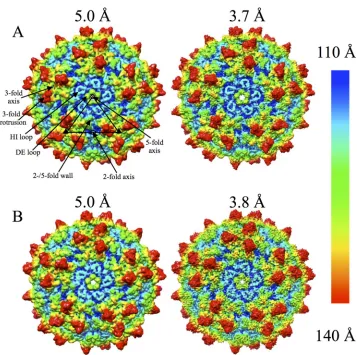
Structures of wt-AAV2 and AAV2-R432A.
Analysis of purified
VLPs by SDS-PAGE, Western and native dot blots, and
negative-stain EM showed that the wt-AAV2 and AAV2-R432A
baculovi-rus constructs were both capable of producing pure intact
parti-cles suitable for structural analysis (data not shown). These
samples were vitrified and used to generate cryo-reconstructed
structures at two different resolutions: 5.0/3.8 Å for wt-AAV2 and
5.0/3.7 Å for AAV2-R432A (
Fig. 1
). Comparison of the maps for
each virus showed an increase in structural detail of the capsid
surface features with increasing resolution, as expected. All maps
clearly showed the characteristic features of an AAV capsid: a
de-pression at each 2-fold axis and surrounding each 5-fold axis, and
three protrusions surrounding each 3-fold axis (
Fig. 1
). The
den-sity for the HI (between

-strands H and I) and DE (between

-strands D and E) loops were also clearly defined (
Fig. 1
).
on November 7, 2019 by guest
http://jvi.asm.org/
Amino acid side chains were clearly represented in the
wt-AAV2 and wt-AAV2-R432A high-resolution maps (
Fig. 2A
).
Resi-dues 225 to 735 (VP1 numbering) and 236 to 735 (here referred to
as VP3) were interpretable in the wt-AAV2 and AAV2-R432A
maps, respectively. The correlation coefficients for the real-space
refinement of the models were 0.71 (wt-AAV2) and 0.73
(AAV2-R432A). The lack of ordering of the VP1u, VP1/2 common region,
and first 22/33 residues of VP3 is consistent with previous AAV
structure reports (
57
). The observation is predicted to be due to
low copies of VP1 and VP2 within the capsid and conformational
variation of the overlapping VP1/2/3 N termini. The models built
into the wt-AAV2 and AAV2-R432A cryo-EM reconstructed
den-sity maps superpose with a root mean square deviation (RMSD) of
0.93 Å (
Fig. 2B
). The wt-AAV2 VP model built into the 3.8-Å
structure superposed onto the available AAV2 crystal structure
with an RMSD of 1.25 Å. Differences were observed at the top of
the 3-fold protrusions, notably within VRs IV and V (not shown).
The AAV2-R432A VP model superposed onto the AAV2 crystal
structure with an RMSD of 1.13 Å.
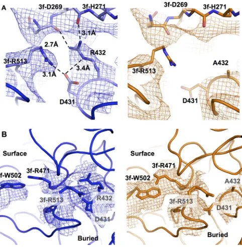
Comparison of AAV2-R432A to wt-AAV2 reveals differences
at high and medium resolutions.
A residue level comparison of
the VP3 models built onto the high-resolution maps showed the
ordered R432 side chain in the wt-AAV2 map, which was absent in
AAV2-R432A, a finding consistent with the genome mutation
(
Fig. 2B
and
3A
). In addition, other changes occurred in the
im-mediate vicinity of residue A432, along with some at distant
posi-tions. Immediately adjacent to A432, the side chains for D431 and
R513 are also disordered (
Fig. 3
). In the wt-AAV2 structure, the
capsid is stabilized by polar interactions between the guanidine
group of R432 on the reference VP monomer and the main-chain
carbonyl oxygens of D269 and H271 of a 3-fold-related monomer.
Residues 269 and 271 are situated within a surface loop located on
the 2/5-fold wall of the capsid (
Fig. 3A
). In addition, the epsilon
nitrogen of R432 participates in a polar interaction with one of the
oxygen atoms of the carboxyl group of D431 on the same VP
monomer. The D431 carboxyl group oxygen also interacts with
the guanidine group of R513 on the 3-fold related VP monomer
(
Fig. 3A
). These interactions do not occur in AAV2-R432A owing
to side chain conformational changes/disorder in D431 and R513,
likely arising from the lack of stabilization from the side chain of
R432. Although residue 432 and the disrupted contacts with
resi-dues 431 and 513 are buried within the capsid, surface differences
immediately above residue 432 occur at R471 and W502 (
Fig. 3B
and
4
). Arginine 471 of the reference monomer is disordered in
FIG 1Cryo-EM reconstructions of wt-AAV2 and AAV2-R432A. Radially colored AAV2-R432A (A) and wt-AAV2 (B) density maps at medium (⬃5.0 Å) and high (3.7/3.8 Å) resolutions, respectively. A viral asymmetric unit is delineated by the triangle on the capsid surface of the medium-resolution AAV2-R432A map in panel A. The positions of select 2-, 3-, and 5-fold axes are indicated by oval, triangle, and pentagon symbols, respectively. The positions of an HI loop, a DE loop, a 3-fold protrusion, and the 2-/5-fold wall are indicated. The density in panels A and B are shown at a sigma threshold of 1.0.
on November 7, 2019 by guest
http://jvi.asm.org/
[image:4.585.116.473.61.416.2]wt-AAV2 but becomes ordered in AAV2-R432A, which is
con-comitant with a shift in the side chain of W502 of the
3-fold-related monomer that appears to stabilize R471 (
Fig. 3B
). These
two side chain shifts are the only surface-exposed conformational
changes in the AAV2-R432A structure and are located
immedi-ately adjacent to the side chain of D269 at the base of the
protru-sions surrounding the 3-fold axes on the wall facing the 5-fold axis
(
Fig. 4
).
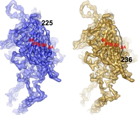
The substitution of an alanine for an arginine at position 432
propagates changes in two VP regions that are far from the
mu-tated residue in the AAV2-R432A structure. One of these occurs in
the conserved

A strand of VP3. In the AAV2-R432A map, the
first ordered residue is 236, which is 11 and 19 amino acids
up-stream from that observed in the wt-AAV2 structure determined
in this study and that in the previously determined AAV2 crystal
structure, respectively (
Fig. 5
). Thus, maps at a lower resolution
(5.0 Å) generated from fewer particle images were analyzed in an
effort to visualize the less-ordered VP features of the capsid
be-cause the signal for less structurally ordered regions of
macromol-ecules (or components of complexes with a reduced copy
num-ber) can be lost at high resolution. This is due to the finer spatial
frequency of data sampling which can be overcome at lower
res-olution. Models built into these low resolution maps showed the
main chain of VP3 to be traceable, and the BIDG and CHEF

sheets, as well as the
␣
A helix, were visible, but side chain densities
were not resolved (not shown). The AAV2-R432A map showed a
buildup of density projecting into the capsid interior from residue
236 (located on the interior surface) and at the 2-fold symmetry
axis (
Fig. 6A
). The main chain for four additional residues could
be modeled into this 5.0-Å density map, extending the
AAV2-R432A VP3 chain to N-terminal residue 232 (
Fig. 6B
). However,
this main chain is rotated
⬃
60° toward the 2-fold rather than
extending toward the 5-fold axis as in the wt-AAV2
cryo-recon-structed and crystal structures (
Fig. 2B
and
6B
). In the wt-AAV2
map, additional density is observed in the interior surface at the
3-fold axis, which is absent in the AAV2-R432A map (
Fig. 6A
).
This additional density is proximal to but does not overlap with
the position of the nucleotide density observed in the capsid
inte-rior of the majority of other wt-AAV serotype capsid structures
determined to date (
58
). While a nucleotide model was not built
into this 3-fold density, its absence in the AAV2-R432A is
consis-tent with its DNA packaging defect.
The second VP site that differs far away from R432A in the
AAV2-R432A structure is residue R404 located at the interior
cap-sid opening of the 5-fold channel (
Fig. 7A
). This residue
under-goes a rearrangement which positions the terminal amino groups
of its side chain away from the opening of the channel (
Fig. 7A
). In
the wt-AAV2 cryo-reconstructed structure, the first ordered
resi-due, 225, is positioned immediately adjacent to R404 from a
5-fold-related VP (
Fig. 7B
). The reorganization of the

A strand
and N terminus toward the 2-fold axis in AAV2-R432A creates a
space that the R404 side chain occupies. Of note, an AAV2 R404A
variant has been shown to produce a 14-fold reduction in DNA
packaging (unpublished data).
FIG 2Density map and model of AAV2-R432A. (A) Small region of the AAV2-R432A cryo-EM density map (gray mesh) with modeled residues 407 to 414. These residues were readily interpretable in the map. (B) Superposition of the AAV2-R432A (orange) and wt-AAV2 (blue) ribbon models built into the cryo-reconstructed density maps. The enlarged inset image shows the location and orientation of residue 432 in the wt-AAV2 (blue mesh) and AAV2-R432A (orange mesh) density maps. The densities in panels A and B are shown at a sigma threshold of 2.0.
FIG 3Residue level structural rearrangements in AAV2-R432A. (A) Intra-and inter-VP monomer interactions in wt-AAV2 structure (blue density mesh and model) between R432 (on the reference VP monomer) with D431 (on the same VP monomer), with 3f-D269 and 3f-H271 (of a 3-fold-related mono-mer), which likely serve to stabilize the native capsid, are lost in AAV2-R432A (orange density mesh and model). In this vicinity the interaction between D431 with 3f-R513 in wt-AAV2 is absent in AAV2-R432A. The loss of inter-actions in AAV2-R432A is due to the lack of the arginine side chain in A432 and side chain conformational changes/disorder in D431 and R513. (B) Sur-face differences between wt-AAV2 and AAV2-R432A at positions R471 and W502 located above D431, R432, and R513. R471 is disordered in wt-AAV2 but ordered in AAV2-R432A, concomitant with a shift in the side chain of W502 of the 3-fold-related monomer. The densities in panels A and B are shown at a sigma threshold of 1.5.
on November 7, 2019 by guest
http://jvi.asm.org/
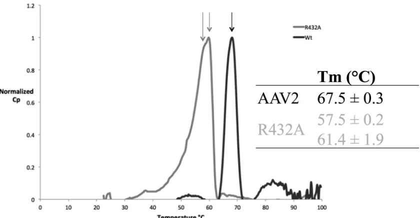
[image:5.585.302.543.66.312.2] [image:5.585.42.294.69.294.2]Wild-type and AAV2-R432A capsid stability.
The loss of
in-terface interactions suggested that AAV2-R432A would be less
stable than wt-AAV2. Differential scanning calorimetry was thus
used to measure the thermostability of AAV2-R432A for
compar-ison with wt-AAV2. The melting temperature (
T
m) determined
for wt-AAV2 at pH 7.4 was 67.5
⫾
0.3°C and the native capsid
exhibited a melting profile with a single, sharp denaturation peak
(
Fig. 8
). This observation is congruent with a previously reported
T
mof 67.8
⫾
0.2°C (
59
). In contrast, the AAV2-R432A melting
profile exhibited a “shoulder” prior to the major peak, resulting in
two melting temperatures: peak 1
⫽
57.5
⫾
0.2°C and peak 2
⫽
61.1
⫾
1.9°C (
Fig. 8
). This dual melting profile suggests a two-step
denaturation process for the mutant, which was not observed for
wt-AAV2.
DISCUSSION
Cryo-EM and image reconstruction yielding medium and
high-resolution structures of wt-AAV2 and the packaging deficient
variant AAV2-R432A revealed capsid properties required for
ge-nome encapsulation to occur. At 3.8 and 3.7 Å, respectively, these
structures are among the highest-resolution structures
deter-mined to date of viruses with T
⫽
1 capsids using this experimental
approach. With the high-resolution density maps, models of the
wt-AAV2 and AAV2-R432A structures could be built using the
known AAV2 crystal structure coordinates as a template. A
com-parison of these models showed an overall conservation of the
VP3 monomer backbone structure. Side chain and main-chain
differences visualized the altered capsid interactions inhibiting
ge-nome packaging. Significantly, the change at R432A, situated at a
3-fold VP:VP interface, showed structural changes that propagate
to regions of the capsid near the 2- and 5-fold axes. Density maps
at 5.0-Å resolution showed a large, main-chain rearrangement in
AAV2-R432A that alters the juxtaposition of residues under the
5-fold and 2-fold axes. The data demonstrate that alterations at
the 2-, 3-, and 5-fold regions of the capsid contribute to producing
a packaging defect and highlight a tight connection between the
entire capsid in maintaining packaging efficiency.
Sequence analysis of the VP3 of several AAV serotypes
(AAV1-AAV9) indicated that R432 and the surrounding region
(includ-ing D431) are highly conserved, AAV5 be(includ-ing the only exception
(not shown). Amino acids W502 and R513 are also highly
con-served among these AAVs, except in AAV4, which contains no
structurally equivalent residue to position 502, and in AAV5,
which contains F489 and A500, respectively. Residues
511-NGR-513, conserved in AAV1 to AAV13, except in AAV4, AAV5, and
AAV11, form a partially exposed, capsid surface loop at the base of
the 3-fold protrusions on the side facing the 2-fold depression.
FIG 4Location of capsid surface differences between wt-AAV2 and AAV2-R432A. R471 and W502 are highlighted in cyan at the base of the 3-fold protrusion on the 2-/5-fold wall. Residues 269 to 271, which participate in main-chain interactions with R432A, and R513 (side chain interactions in wt-AAV2) are also highlighted. The densities in panels A and B are shown at a sigma threshold of 1.0.
FIG 5Wild-type and AAV2-R432A N-terminal differences at high resolution. Density maps of wt-AAV2 (blue) and AAV2-R432A (orange) monomers viewed from the capsid interior are shown with the docked main chain (black line) of the AAV2 crystal structure coordinates (1LP3). Densities for theB,
I,D, andG strands are well-ordered (labeled) and structurally equivalent in wt-AAV2 and AAV2-R432A, but theA strand is absent in the AAV2-R432A density map. The interpretable density for the wt-AAV2 structure is ordered starting at residue 225, whereas the AAV2-R432A map only reveals the density starting at residue 236. The densities in panels A and B are shown at a sigma threshold of 2.5.
on November 7, 2019 by guest
http://jvi.asm.org/
[image:6.585.117.475.66.252.2] [image:6.585.42.284.427.628.2]This NGR motif is reportedly essential for integrin
␣
5

1
corecep-tor binding by AAV2 and AAV9 (
44
,
60
). A previous study showed
that modification of this motif produced AAV2 capsids that were
unable to package genome, and this phenotype was proposed to
arise owing to the loss of interaction with residues D431 and R432
(
44
). The R432A and R513A mutation information suggests that
the interactions of residues 511 to 513 and residues 431 to 432 play
a critical role in ssDNA genome packaging (
44
). This implies that
the region of the capsid surface lying above these residues serves as
a contact point for Rep during the packaging process. The
altera-tions to the AAV2-R432A variant capsid surface above R432 may
be responsible for the increased Rep loading reported by Bleker et
al. (
40
). Thus, although a role for residues equivalent to R432 in
genome packaging has not been investigated for other AAVs, this
residue is critical in AAV2 packaging and thermodynamics, and is
predicted to perform a similar role in other AAVs.
DSC experiments provided physical evidence of the decreased
stability of the AAV2-R432A with a significantly lower
T
mthan
wt-AAV2. Prior DSC studies have demonstrated that the specific
heat capacity of the AAV capsid directly correlates to its structural
stability, and so it follows that a lower
T
mindicates a capsid with
decreased stability (
59
). In addition, the mutant AAV displayed
two melting peaks, suggesting dual denaturation events. Of note,
the melting profile of AAV8 also demonstrates two transitions,
separated by
⬃
4.3
°
C (
59
). A plausible explanation for the
AAV2-R432A melting behavior is that two populations of capsids exist,
for example, one with altered

A conformation (as observed) and
another in transition from the wt-AAV2 conformation. This
would explain the lack of or reduced ordering of the altered
AAV2-R432A VP N terminus to the same position as for the
wt-AAV2 cryo-reconstructed structure despite these residues being
within the common VP3 region. Density consistent with the
con-formational change was seen only at a low sigma threshold (0.5
)
in the high-resolution map and when resolution was reduced to 5
Å, suggesting a reduced occupancy for the N-terminal amino acids
observed beyond position 236. It is also possible that the reduced
stability of AAV2-R432A results in dissociation into an
interme-diate VP oligomeric state such as trimers or pentamers prior to
complete capsid disassembly, which appears to be a one step
pro-cess for AAV2 and other serotypes (
59
).
In summary, the data reported here suggest that the AAV2
FIG 6Wild-type AAV2 and AAV2-R432A structural changes at the 2-fold symmetry axis seen at medium resolution. (A) Cross-section of the wt-AAV2 (left, blue) and AAV2-R432A (right, orange) capsid density maps showing positive difference density (cryo-reconstructed density minus AAV2 crystal structure capsid model density) primarily confined to the interior capsid sur-face. In wt-AAV2, extra density occurs at the 3-fold axes, whereas in AAV2-R432A, extra density occurs at the 2-fold axes. (B) Ribbon representations of a VP3 monomer fitted within the AAV2-R432A density map (orange) (left) and superposed wt-AAV2 (blue) and AAV2-R432A (orange) surface densities (right) close to the 2-fold axis. TheBIDG strands, as well as strandA, are labeled in the left side of panel B. The reposition of theA strand in AAV2-R432A and the build-up of density at the 2-fold axis are evident. wt-AAV2 is ordered from residue 225, whereas in AAV2-R432A the residues can only be assigned from residue 236 at a resolution of 3.7 Å and from residue 232 at a resolution of 5.0 Å. This density for the ordered N terminus of AAV2-R432A projects into the capsid interior. In wt-AAV2 the VP1/2/3 N termini from 5-fold-related neighboring monomers interact, whereas in AAV2-R432A this interaction is lost (seeFig. 7). The densities in panels A and B are shown at sigma thresholds of 2.5 and 2.0, respectively.
FIG 7Wild-type and AAV2-R432A differences at the base of the 5-fold chan-nel. (A) A closeup view of a pentamer of wt-AAV2 (blue ribbon and mesh density) and AAV2-R432A (orange ribbon and mesh density) exhibits the alternative positions of the R404 side chain. The R404 side chain moves away from the opening of the channel in AAV2-R432A. The view is from the interior of the capsid. (B) Pentamer of superposed wt-AAV2 and AAV2-R432A from the capsid interior showing the reposition of the R404 side chain adjacent to the first ordered N-terminal residue from a 5-fold-related VP monomer in wt-AAV2. The AAV2-R432A structure is disordered up to this position. The density in panel A is shown at a sigma threshold of 2.0.
on November 7, 2019 by guest
http://jvi.asm.org/
[image:7.585.317.523.65.362.2] [image:7.585.42.286.66.313.2]R432 residue plays an important role in maintaining AAV2 capsid
interactions and stability that is important for packaging
compe-tency. The loss of contacts between the

A and

B strands below
the 5-fold axis would contribute to decreased capsid stability. The
5-fold pore has been suggested to be a site for genome packaging
and uncoating from assembled capsids for the AAVs, as well as
other parvoviruses. Thus, the structural changes at the base of the
5-fold pore of the AAV2-R432A may contribute to the observed
packaging defect. The structural rearrangements observed at the
3-fold region of the AAV2-R432A capsid caused loss of
inter-monomer interactions which could also lead to global capsid
de-stabilization. The AAV2-R432A structure revealed a buildup of
density underneath the 2-fold axis, a process that has been
sug-gested to occur with the VP1/2 common region and VP1u N
ter-minus prior to extrusion through the 5-fold pore (
61
). This would
also suggest that the VP1/2 common region and VP1u are
perma-nently displayed on the capsid surface. However, no density was
observed within the 5-fold pore of AAV2-R432A at medium or
high resolution. Moreover, density observed at the 2-fold axis was
ascribed to residues within the VP1/2/3 common region. The lack
of density blocking the pore could merely be a consequence of
icosahedral symmetry averaging effects, which diminish the
visu-alization of elements of structure such as VP1u that are present in
low copy number. Interestingly, density proximal to the region
previously reported to be a nucleotide binding site was observed in
wt-AAV2 but not AAV2-R432A, consistent with the inability of
the variant to package ssDNA. Finally, the global effect of the
single R432A amino change, at a buried capsid location, highlights
the plasticity of the assembled capsid and the need to conserve
structural fidelity in all functions, including capsid stability and
genome packaging.
ACKNOWLEDGMENTS
We thank the University of Florida (UF) Interdisciplinary Center for Bio-technology Research (ICBR) electron microscopy lab for providing neg-ative-stain EM services.
Financial support was provided by the UF Division of Sponsored Re-search and the College of Medicine to establish cryo-EM facilities at the UF ICBR and by the University of California—San Diego (UCSD) and the Agouron Foundation (to T.S.B.) to establish cryo-EM facilities at UCSD. This project was funded in part by NIH R01 GM109524 (M.A.-M., R.M., and N.M.), NIH P01 HL59412 (M.A.-M. and N.M.), NIH R37 GM33050 (T.S.B.), NIH 1S10 RR020016 (T.S.B.), and NIH T32 GM008799 (L.M.D.).
FUNDING INFORMATION
This work, including the efforts of Lauren M. Drouin, Bridget Lins, An-tonette Bennett, Paul Chipman, Robert McKenna, Weijun Chen, Nicho-las Muzyczka, and Mavis Agbandje-McKenna, was funded by HHS | Na-tional Institutes of Health (NIH) (GM109524 and HL59412). This work, including the efforts of Maria Janssen, Giovanni Cardone, and Timothy S. Baker, was funded by HHS | National Institutes of Health (NIH) (GM33050). This work, including the efforts of Lauren M. Drouin, was funded by HHS | National Institutes of Health (NIH) (GM008799).
REFERENCES
1.Asokan A, Schaffer DV, Samulski RJ.2012. The AAV vector toolkit: poised at the clinical crossroads. Mol Ther20:699 –708.http://dx.doi.org /10.1038/mt.2011.287.
2.Daya S, Berns KI.2008. Gene therapy using adeno-associated virus vec-tors. Clin Microbiol Rev 21:583–593. http://dx.doi.org/10.1128/CMR .00008-08.
3.Flotte TR, Carter BJ.1995. Adeno-associated virus vectors for gene ther-apy. Gene Ther2:357–362.
4.Chapman M, Agbandje-McKenna M.2006. Atomic structure of viral particles, p 107–123.InKerr CS, Jr, Bloom ME, Linden RM, Parrish CR (ed), Parvoviruses. Edward Arnold, Ltd, New York, NY.
5.Grieger JC, Johnson JS, Gurda-Whitaker B, Agbandje-McKenna M, Samulski RJ. 2007. Surface-exposed adeno-associated virus Vp1-NLS capsid fusion protein rescues infectivity of noninfectious wild-type Vp2/ Vp3 and Vp3-only capsids but not that of fivefold pore mutant virions. J Virol81:7833–7843.http://dx.doi.org/10.1128/JVI.00580-07.
6.Bleker S, Sonntag F, Kleinschmidt JA. 2005. Mutational analysis of narrow pores at the fivefold symmetry axes of adeno-associated virus type 2 capsids reveals a dual role in genome packaging and activation of phos-pholipase A2 activity. J Virol79:2528 –2540.http://dx.doi.org/10.1128 /JVI.79.4.2528-2540.2005.
FIG 8Stability of AAV2 and AAV2-R432A. Melting profile for wt-AAV2 (in black) and AAV2-R432A (in gray). Normalized representations for three experiments are shown.
on November 7, 2019 by guest
http://jvi.asm.org/
[image:8.585.83.503.65.283.2]7.Buller RM, Rose JA. 1978. Characterization of adenovirus-associated virus-induced polypeptides in KB cells. J Virol25:331–338.
8.Rose JA, Maizel JV, Jr, Inman JK, Shatkin AJ.1971. Structural proteins of adenovirus-associated viruses. J Virol8:766 –770.
9.Johnson FB, Ozer HL, Hoggan MD.1971. Structural proteins of adeno-virus-associated virus type 3. J Virol8:860 – 863.
10. Snijder J, van de Waterbeemd M, Damoc E, Denisov E, Grinfeld D, Bennett A, Agbandje-McKenna M, Makarov A, Heck AJ.2014. Defining the stoichiometry and cargo load of viral and bacterial nanoparticles by Orbitrap mass spectrometry. J Am Chem Soc136:7295–7299.http://dx .doi.org/10.1021/ja502616y.
11. Sonntag F, Schmidt K, Kleinschmidt JA.2010. A viral assembly factor promotes AAV2 capsid formation in the nucleolus. Proc Natl Acad Sci U S A107:10220 –10225.http://dx.doi.org/10.1073/pnas.1001673107. 12. Sonntag F, Kother K, Schmidt K, Weghofer M, Raupp C, Nieto K, Kuck
A, Gerlach B, Bottcher B, Muller OJ, Lux K, Horer M, Kleinschmidt JA.
2011. The assembly-activating protein promotes capsid assembly of dif-ferent adeno-associated virus serotypes. J Virol85:12686 –12697.http://dx .doi.org/10.1128/JVI.05359-11.
13. Naumer M, Sonntag F, Schmidt K, Nieto K, Panke C, Davey NE, Popa-Wagner R, Kleinschmidt JA. 2012. Properties of the adeno-associated virus assembly-activating protein. J Virol86:13038 –13048.
http://dx.doi.org/10.1128/JVI.01675-12.
14. DiMattia MA, Nam HJ, Van Vliet K, Mitchell M, Bennett A, Gurda BL, McKenna R, Olson NH, Sinkovits RS, Potter M, Byrne BJ, Aslanidi G, Zolotukhin S, Muzyczka N, Baker TS, Agbandje-McKenna M.2012. Structural insight into the unique properties of adeno-associated virus serotype 9. J Virol86:6947– 6958.http://dx.doi .org/10.1128/JVI.07232-11.
15. Govindasamy L, Dimattia MA, Gurda BL, Halder S, McKenna R, Chiorini JA, Muzyczka N, Zolotukhin S, Agbandje-McKenna M.2013. Structural insights into adeno-associated virus serotype 5. J Virol87:
11187–11199.http://dx.doi.org/10.1128/JVI.00867-13.
16. Govindasamy L, Padron E, McKenna R, Muzyczka N, Kaludov N, Chiorini JA, Agbandje-McKenna M. 2006. Structurally mapping the diverse phenotype of adeno-associated virus serotype 4. J Virol80:11556 – 11570.http://dx.doi.org/10.1128/JVI.01536-06.
17. Kronenberg S, Kleinschmidt JA, Bottcher B. 2001. Electron cryo-microscopy and image reconstruction of adeno-associated virus type 2 empty capsids. EMBO Rep2:997–1002.http://dx.doi.org/10.1093/embo -reports/kve234.
18. Lerch TF, Xie Q, Chapman MS.2010. The structure of adeno-associated virus serotype 3B (AAV-3B): insights into receptor binding and immune evasion. Virology 403:26 –36. http://dx.doi.org/10.1016/j.virol.2010.03 .027.
19. Nam HJ, Gurda BL, McKenna R, Potter M, Byrne B, Salganik M, Muzyczka N, Agbandje-McKenna M.2011. Structural studies of ad-eno-associated virus serotype 8 capsid transitions associated with en-dosomal trafficking. J Virol 85:11791–11799. http://dx.doi.org/10 .1128/JVI.05305-11.
20. Nam HJ, Lane MD, Padron E, Gurda B, McKenna R, Kohlbrenner E, Aslanidi G, Byrne B, Muzyczka N, Zolotukhin S, Agbandje-McKenna M.2007. Structure of adeno-associated virus serotype 8, a gene therapy vector. J Virol81:12260 –12271.http://dx.doi.org/10.1128/JVI.01304-07. 21. Ng R, Govindasamy L, Gurda BL, McKenna R, Kozyreva OG, Samulski RJ, Parent KN, Baker TS, Agbandje-McKenna M. 2010. Structural characterization of the dual glycan binding adeno-associated virus sero-type 6. J Virol84:12945–12957.http://dx.doi.org/10.1128/JVI.01235-10. 22. Xie Q, Bu W, Bhatia S, Hare J, Somasundaram T, Azzi A, Chapman
MS.2002. The atomic structure of adeno-associated virus (AAV-2), a vector for human gene therapy. Proc Natl Acad Sci U S A99:10405–10410.
http://dx.doi.org/10.1073/pnas.162250899.
23. Xie Q, Lerch TF, Meyer NL, Chapman MS.2011. Structure-function analysis of receptor-binding in adeno-associated virus serotype 6 (AAV-6). Virology420:10 –19.http://dx.doi.org/10.1016/j.virol.2011.08.011. 24. McCraw DM, O’Donnell JK, Taylor KA, Stagg SM, Chapman MS.2012.
Structure of adeno-associated virus-2 in complex with neutralizing monoclonal antibody A20. Virology 431:40 – 49. http://dx.doi.org/10 .1016/j.virol.2012.05.004.
25. Gurda BL, DiMattia MA, Miller EB, Bennett A, McKenna R, Weichert WS, Nelson CD, Chen WJ, Muzyczka N, Olson NH, Sinkovits RS, Chiorini JA, Zolotutkhin S, Kozyreva OG, Samulski RJ, Baker TS, Parrish CR, Agbandje-McKenna M.2013. Capsid antibodies to different
adeno-associated virus serotypes bind common regions. J Virol87:9111– 9124.http://dx.doi.org/10.1128/JVI.00622-13.
26. Gurda BL, Raupp C, Popa-Wagner R, Naumer M, Olson NH, Ng R, McKenna R, Baker TS, Kleinschmidt JA, Agbandje-McKenna M.
2012. Mapping a neutralizing epitope onto the capsid of adeno-associated virus serotype 8. J Virol86:7739 –7751.http://dx.doi.org/10 .1128/JVI.00218-12.
27. Kern A, Schmidt K, Leder C, Muller OJ, Wobus CE, Bettinger K, Von der Lieth CW, King JA, Kleinschmidt JA. 2003. Identification of a heparin-binding motif on adeno-associated virus type 2 capsids. J Virol
77:11072–11081.http://dx.doi.org/10.1128/JVI.77.20.11072-11081.2003. 28. Levy HC, Bowman VD, Govindasamy L, McKenna R, Nash K, War-rington K, Chen W, Muzyczka N, Yan X, Baker TS, Agbandje-McKenna M.2009. Heparin binding induces conformational changes in adeno-associated virus serotype 2. J Struct Biol165:146 –156.http://dx.doi.org /10.1016/j.jsb.2008.12.002.
29. Lochrie MA, Tatsuno GP, Christie B, McDonnell JW, Zhou S, Surosky R, Pierce GF, Colosi P.2006. Mutations on the external surfaces of adeno-associated virus type 2 capsids that affect transduction and neutral-ization. J Virol80:821– 834.http://dx.doi.org/10.1128/JVI.80.2.821-834 .2006.
30. O’Donnell J, Taylor KA, Chapman MS.2009. Adeno-associated virus-2 and its primary cellular receptor-cryo-EM structure of a heparin complex. Virology385:434 – 443.http://dx.doi.org/10.1016/j.virol.2008.11.037. 31. Opie SR, Warrington KH, Jr, Agbandje-McKenna M, Zolotukhin S,
Muzyczka N.2003. Identification of amino acid residues in the capsid proteins of adeno-associated virus type 2 that contribute to heparan sul-fate proteoglycan binding. J Virol 77:6995–7006.http://dx.doi.org/10 .1128/JVI.77.12.6995-7006.2003.
32. Tseng YS, Gurda BL, Chipman P, McKenna R, Afione S, Chiorini JA, Muzyczka N, Olson NH, Baker TS, Kleinschmidt J, Agbandje-McKenna M.2015. Adeno-associated virus serotype 1 (AAV1)- and AAV5-antibody complex structures reveal evolutionary commonalities in parvovirus an-tigenic reactivity. J Virol 89:1794 –1808. http://dx.doi.org/10.1128/JVI .02710-14.
33. Wobus CE, Hugle-Dorr B, Girod A, Petersen G, Hallek M, Klein-schmidt JA.2000. Monoclonal antibodies against the adeno-associated virus type 2 (AAV-2) capsid: epitope mapping and identification of capsid domains involved in AAV-2-cell interaction and neutralization of AAV-2 infection. J Virol74:9281–9293.http://dx.doi.org/10.1128/JVI.74.19.9281 -9293.2000.
34. Wu P, Xiao W, Conlon T, Hughes J, Agbandje-McKenna M, Ferkol T, Flotte T, Muzyczka N.2000. Mutational analysis of the adeno-associated virus type 2 (AAV2) capsid gene and construction of AAV2 vectors with altered tropism. J Virol74:8635– 8647.http://dx.doi.org/10.1128/JVI.74 .18.8635-8647.2000.
35. King JA, Dubielzig R, Grimm D, Kleinschmidt JA.2001. DNA helicase-mediated packaging of adeno-associated virus type 2 genomes into pre-formed capsids. EMBO J20:3282–3291.http://dx.doi.org/10.1093/emboj /20.12.3282.
36. Chiorini JA, Wiener SM, Owens RA, Kyostio SR, Kotin RM, Safer B.
1994. Sequence requirements for stable binding and function of Rep68 on the adeno-associated virus type 2 inverted terminal repeats. J Virol68:
7448 –7457.
37. Chiorini JA, Yang L, Safer B, Kotin RM.1995. Determination of adeno-associated virus Rep68 and Rep78 binding sites by random sequence oligonucleotide selection. J Virol69:7334 –7338.
38. Im DS, Muzyczka N.1990. The AAV origin binding protein Rep68 is an ATP-dependent site-specific endonuclease with DNA helicase activity. Cell61:447– 457.http://dx.doi.org/10.1016/0092-8674(90)90526-K. 39. Kyostio SR, Wonderling RS, Owens RA.1995. Negative regulation of the
adeno-associated virus (AAV) P5 promoter involves both the P5 rep bind-ing site and the consensus ATP-bindbind-ing motif of the AAV Rep68 protein. J Virol69:6787– 6796.
40. Bleker S, Pawlita M, Kleinschmidt JA.2006. Impact of capsid confor-mation and Rep-capsid interactions on adeno-associated virus type 2 ge-nome packaging. J Virol80:810 – 820.http://dx.doi.org/10.1128/JVI.80.2 .810-820.2006.
41. Plevka P, Hafenstein S, Li L, D’Abrgamo A, Jr, Cotmore SF, Ross-mann MG, Tattersall P. 2011. Structure of a packaging-defective mutant of minute virus of mice indicates that the genome is packaged via a pore at a 5-fold axis. J Virol85:4822– 4827.http://dx.doi.org/10 .1128/JVI.02598-10.
on November 7, 2019 by guest
http://jvi.asm.org/
42. Warrington KH, Jr, Gorbatyuk OS, Harrison JK, Opie SR, Zolo-tukhin S, Muzyczka N. 2004. Adeno-associated virus type 2 VP2 capsid protein is nonessential and can tolerate large peptide insertions at its N terminus. J Virol78:6595– 6609.http://dx.doi.org/10.1128/JVI .78.12.6595-6609.2004.
43. Lux K, Goerlitz N, Schlemminger S, Perabo L, Goldnau D, Endell J, Leike K, Kofler DM, Finke S, Hallek M, Buning H.2005. Green fluo-rescent protein-tagged adeno-associated virus particles allow the study of cytosolic and nuclear trafficking. J Virol79:11776 –11787.http://dx.doi .org/10.1128/JVI.79.18.11776-11787.2005.
44. Asokan A, Hamra JB, Govindasamy L, Agbandje-McKenna M, Samul-ski RJ. 2006. Adeno-associated virus type 2 contains an integrin alpha5beta1 binding domain essential for viral cell entry. J Virol80:8961– 8969.http://dx.doi.org/10.1128/JVI.00843-06.
45. Kohlbrenner E, Aslanidi G, Nash K, Shklyaev S, Campbell-Thompson M, Byrne BJ, Snyder RO, Muzyczka N, Warrington KH, Zolotukhin S.
2005. Successful production of pseudotyped rAAV vectors using a modi-fied baculovirus expression system. Mol Ther12:1217–1225.
46. Mindell JA, Grigorieff N.2003. Accurate determination of local defocus and specimen tilt in electron microscopy. J Struct Biol142:334 –347.http: //dx.doi.org/10.1016/S1047-8477(03)00069-8.
47. Yan X, Sinkovits RS, Baker TS.2007. AUTO3DEM: an automated and high-throughput program for image reconstruction of icosahedral parti-cles. J Struct Biol157:73– 82.http://dx.doi.org/10.1016/j.jsb.2006.08.007. 48. Baker TS, Olson NH, Fuller SD.1999. Adding the third dimension to virus life cycles: three-dimensional reconstruction of icosahedral viruses from cryo-electron micrographs. Microbiol Mol Biol Rev63:862–922. 49. Yan X, Dryden KA, Tang J, Baker TS.2007. Ab initio random model
method facilitates 3D reconstruction of icosahedral particles. J Struct Biol
157:211–225.http://dx.doi.org/10.1016/j.jsb.2006.07.013.
50. Rosenthal PB, Henderson R.2003. Optimal determination of particle orientation, absolute hand, and contrast loss in single-particle electron cryomicroscopy. J Mol Biol333:721–745.http://dx.doi.org/10.1016/j.jmb .2003.07.013.
51. Havelka WA, Henderson R, Oesterhelt D. 1995. Three-dimensional structure of halorhodopsin at 7-Å resolution. J Mol Biol247:726 –738.
http://dx.doi.org/10.1016/S0022-2836(05)80151-2.
52. Carrillo-Tripp M, Shepherd CM, Borelli IA, Venkataraman S, Lander
G, Natarajan P, Johnson JE, Brooks CL, III, Reddy VS.2009. VIPERdb2: an enhanced and web API enabled relational database for structural virol-ogy. Nucleic Acids Res 37:D436 –D442. http://dx.doi.org/10.1093/nar /gkn840.
53. Pettersen EF, Goddard TD, Huang CC, Couch GS, Greenblatt DM, Meng EC, Ferrin TE.2004. UCSF Chimera: a visualization system for exploratory research and analysis. J Comput Chem25:1605–1612.http: //dx.doi.org/10.1002/jcc.20084.
54. Emsley P, Cowtan K.2004. Coot: model-building tools for molecular graphics. Acta Crystallogr D Biol Crystallogr60:2126 –2132.http://dx.doi .org/10.1107/S0907444904019158.
55. Adams PD, Afonine PV, Bunkoczi G, Chen VB, Davis IW, Echols N, Headd JJ, Hung LW, Kapral GJ, Grosse-Kunstleve RW, McCoy AJ, Mo-riarty NW, Oeffner R, Read RJ, Richardson DC, Richardson JS, Terwilliger TC, Zwart PH.2010. PHENIX: a comprehensive Python-based system for macromolecular structure solution. Acta Crystallogr D Biol Crystallogr66:
213–221.http://dx.doi.org/10.1107/S0907444909052925.
56. DeLano WL.2008. The PyMOL molecular graphics system. DeLano Sci-entific LLC, New York, NY.http://www.pymol.org.
57. Halder S, Ng R, Agbandje-McKenna M.2012. Parvoviruses: structure and infection. Future Virol7:253–278.http://dx.doi.org/10.2217/fvl .12.12.
58. Halder S, Van Vliet K, Smith JK, Duong TT, McKenna R, Wilson JM, Agbandje-McKenna M.2015. Structure of neurotropic adeno-associated virus AAVrh.8. J Struct Biol192:21–36.http://dx.doi.org/10.1016/j.jsb .2015.08.017.
59. Rayaprolu V, Kruse S, Kant R, Venkatakrishnan B, Movahed N, Brooke D, Lins B, Bennett A, Potter T, McKenna R, Agbandje-McKenna M, Bothner B.2013. Comparative analysis of adeno-associated virus capsid stability and dynamics. J Virolhttp://dx.doi.org/10.1128/JVI.01415-13. 60. Shen S, Berry GE, Castellanos Rivera RM, Cheung RY, Troupes AN,
Brown SM, Kafri T, Asokan A.2015. Functional analysis of the putative integrin recognition motif on adeno-associated virus 9. J Biol Chem290:
1496 –1504.http://dx.doi.org/10.1074/jbc.M114.608281.
61. Kronenberg S, Bottcher B, von der Lieth CW, Bleker S, Kleinschmidt JA.2005. A conformational change in the adeno-associated virus type 2 capsid leads to the exposure of hidden VP1 N termini. J Virol79:5296 – 5303.http://dx.doi.org/10.1128/JVI.79.9.5296-5303.2005.
on November 7, 2019 by guest
http://jvi.asm.org/