Membrane Requirements for Uridylylation of the Poliovirus VPg
Protein and Viral RNA Synthesis In Vitro
Mark H. Fogg,† Natalya L. Teterina, and Ellie Ehrenfeld*
National Institutes of Health, Bethesda, Maryland 20892
Received 7 April 2003/Accepted 10 July 2003
Efficient translation of poliovirus (PV) RNA in uninfected HeLa cell extracts generates all of the viral proteins required to carry out viral RNA replication and encapsidation and to produce infectious virus in vitro. In infected cells, viral RNA replication occurs in ribonucleoprotein complexes associated with clusters of vesicles that are formed from preexisting intracellular organelles, which serve as a scaffold for the viral RNA replication complex. In this study, we have examined the role of membranes in viral RNA replication in vitro. Electron microscopic and biochemical examination of extracts actively engaged in viral RNA replication failed to reveal a significant increase in vesicular membrane structures or the protective aggregation of vesicles observed in PV-infected cells. Viral, nonstructural replication proteins, however, bind to heterogeneous mem-brane fragments in the extract. Treatment of the extracts with nonionic detergents, a memmem-brane-altering inhibitor of fatty acid synthesis (cerulenin), or an inhibitor of intracellular membrane trafficking (brefeldin A) prevents the formation of active replication complexes in vitro, under conditions in which polyprotein synthesis and processing occur normally. Under all three of these conditions, synthesis of uridylylated VPg to form the primer for initiation of viral RNA synthesis, as well as subsequent viral RNA replication, was inhibited. Thus, although organized membranous structures morphologically similar to the vesicles observed in infected cells do not appear to form in vitro, intact membranes are required for viral RNA synthesis, including the first step of forming the uridylylated VPg primer for RNA chain elongation.
All known positive-strand RNA viruses modify and utilize rearranged intracellular membranes to create structures to support replication of their genomes (see reference 21 and references within). Apparently, this membrane association is critical for the generation of active replication complexes, al-though the morphology of the structures formed differs for different positive-strand viruses, and the mechanism or mech-anisms by which the structures are formed have not yet been established.
The importance of membranes for viral RNA replication has been most extensively demonstrated for the prototype member of thePicornaviridae, Poliovirus(PV). The rearrangement of intracellular membranes to generate vesicles is dependent upon expression of viral nonstructural proteins (1, 15, 20, 43). Upon infection of cells with PV, the viral RNA (vRNA) is translated into a single polypeptide that is processed by virally encoded proteases into both structural (capsid) and nonstruc-tural proteins. All viral proteins containing 2B, 2C, or 3A sequences (1, 18, 48, 49, 51) can bind to and induce alterations in membrane morphology, and 2BC, when expressed alone (15) or together with 3A (43), can induce the formation of membranous structures morphologically similar to those ob-served during PV infection. All nonstructural proteins and viral RNA become associated with the membranes to form an active replication complex, although how vRNA or proteins
associate with and are maintained within these areas is un-known. Early studies demonstrated an increase in phosphati-dylcholine synthesis during PV infection of HeLa cells (50). Since phosphatidylcholine is required for new membrane syn-thesis, this observation suggested that membrane proliferation occurs during virus replication. Subsequent studies using ceru-lenin, an inhibitor of fatty acid synthesis, showed that it inhib-ited [3H]uridine incorporation into vRNA in infected cells
(26), as well as in a cell-free system (34). Members of other families of positive-strand RNA viruses, such as Semliki Forest virus and cowpea mosaic virus, also show sensitivity to cerule-nin during replication in cultured mammalian cells (39) or plant protoplasts (14), respectively. In PV-infected cells and in cell extracts, addition of the unsaturated fatty acid oleic acid, which alters membrane fluidity, inhibited replication of vRNA (25, 34); at higher concentrations, viral protein synthesis was inhibited as well. Interestingly, brome mosaic virus requires unsaturated fatty acids for replication of its genome (30), and replication of Japanese encephalitis virus was enhanced in the presence of oleic acid (31). Finally, brefeldin A (BFA), which inhibits intracellular membrane trafficking, is a potent inhibitor not only of PV replication in both cell culture (28, 32) and cell-free systems (16) but also of rhinovirus (28), hepatitis A virus (10), and echovirus 11 (24) in cultured cells. However, replication of another picornavirus, encephalomyocarditis vi-rus, is unaffected by the presence of BFA (28), and parecho-virus 1 is partially resistant (24).
Crude replication complexes (CRCs) have been isolated from PV-infected HeLa cells, generally 4 h following infection (22), and their structural and functional properties have been characterized (9, 12, 13, 22). The majority of vRNA synthesis catalyzed by the CRCs in vitro results from elongation of RNA
* Corresponding author. Mailing address: Laboratory of Infectious Diseases, NIAID, National Institutes of Health, Building 50, Room 6120, 9000 Rockville Pk., Bethesda, MD 20892. Phone: (301) 594-1654. Fax: (301) 435-6021. E-mail: eehrenfeld@niaid.nih.gov.
† Present address: Department of Medicine, Brigham and Women’s Hospital, Harvard Medical School, Boston, MA 02115.
11408
on November 8, 2019 by guest
http://jvi.asm.org/
chains initiated prior to isolation from infected cells, rather than initiation of new chains de novo in vitro.
An alternative system for study of PV RNA replication in vitro utilizes uninfected HeLa cell extracts translating purified vRNA (33). This generates all viral proteins necessary for replication of the input vRNA, as well as subsequent encapsi-dation to produce infectious virus. The formation and role of membrane structures in RNA synthesis in this system have not been studied extensively, although several lipophilic agents were shown to inhibit virus production (34), and components of the cell’s membrane trafficking system have been implicated in viral RNA synthesis in vitro (16).
To gain insight into the formation of PV replication com-plexes in vitro, we have analyzed the membrane structures utilized to support viral RNA synthesis in this assay and also sought to determine what steps in PV RNA replication require membranes. Morphological structures similar to those ob-served in PV-infected cells and upon isolation from infected cells are not observed in vitro; however, the membranes with which the replication reaction is associated are essential for even the first step of RNA synthesis, the uridylylation of VPg to generate a primer for chain elongation. This initial reaction is also dependent upon vesicular membrane trafficking, as ev-idenced by its inhibition by BFA.
MATERIALS AND METHODS
Purification of vRNA.vRNA was isolated from purified, CsCl-banded PV by phenol-chloroform-isoamyl alcohol extraction and ethanol precipitation and quantified by determination ofA260.
In vitro translation/replication reactions.HeLa cell S10 extract and HeLa cell ribosomal salt wash as a source of translation initiation factors (IFs) were pre-pared as described previously (4). Translation/replication reactions contained 51% (by volume) HeLa S10 extracts, 18% (by volume) ribosomal salt wash, 10% (by volume) 10⫻reaction mix (10 mM ATP, 1.5 mM GTP, 1.5 mM CTP, 1.5 mM UTP, 600 mM KCH3CO2, 300 mM creatine phosphate, 4-mg/ml creatine kinase, 155 mM HEPES-KOH [pH 7.4]), and purified vRNA at 25g/ml. Translation was monitored in 10-l reaction mixtures by the addition of 15Ci of [35 S]me-thionine (1,000 Ci/mmol; Amersham) after incubation at 34°C for 3.5 h. Labeled proteins were separated by sodium dodecyl sulfate-polyacrylamide gel electro-phoresis (SDS-PAGE) (12.5% polyacrylamide) and were detected by autora-diography. vRNA synthesis was analyzed in 40-l reaction mixtures. Preinitia-tion-replication complexes were formed in the presence of 2 mM guanidine HCl (GuHCl) for3.5 h at 34°C and were isolated by centrifugation at 16,000⫻gfor 20 min at 4°C. Pellets were resuspended in 25l of replication buffer (see method 4 in reference 5) containing 25Ci of [32P]CTP (400 Ci/mmol; Amer-sham) and incubated for 1 h at 37°C. Total RNA in the reaction mixtures was isolated with the RNeasy kit (Qiagen) and denatured with glyoxal denaturing buffer (Ambion) for 40 min at 60°C. RNA was resolved by electrophoresis in 0.8% agarose. Gels were dried, and labeled RNA was detected and quantified by PhosphorImager (Molecular Dynamics). VPg uridylylation was monitored by using 40-l preinitiation-replication complexes generated in the same way as for vRNA synthesis and which were resuspended in reaction buffer as described for vRNA replication, except they contained 25Ci of [32P]UTP (3,000 Ci/mmol; Perkin-Elmer). Reaction mixtures were incubated for 60 min at 37°C and cen-trifuged at 16,000⫻gfor 20 min to pellet the membranous complexes. Pellets were resuspended in SDS buffer and analyzed by electrophoresis on a 12.5% PAGE Tris-Tricine gel (17, 42). Radiolabeled products were detected by auto-radiography and quantitated by phosphorimaging.
CRCs.CRCs were isolated from PV-infected HeLa cells as described previ-ously (13, 22, 44). For analysis of vRNA synthesis, replication reactions were assembled essentially as in vitro translation/replication reactions, except S10 extract and IFs were replaced by 20% (by volume) CRC and 49% (by volume) S10 buffer (see method 4 in reference 5) containing 25Ci of [32P]CTP (400 Ci/mmol; Amersham). Uridylylated VPg was detected in an identical way to that described above for preinitiation-replication complexes.
RNase, BFA, NP-40, and cerulenin treatment.Assembled reaction mixtures were treated with 100g of RNase A (Boehringer) per ml at 30°C for 30 min
either prior to isolation of preinitiation-replication complexes or following RNA synthesis in these reactions. CRCs were treated with RNase A prior to or following RNA synthesis. NP-40, BFA, and cerulenin (the latter two dissolved in dimethyl sulfoxide [DMSO]) were purchased from Sigma and were added to reaction mixtures during preinitiation-replication complex formation or during RNA synthesis or VPg uridylylation reactions. BFA and cerulenin control reac-tion mixtures contained DMSO alone.
EM and IEM.For electron microscopy (EM) and immunoelectron microscopy (IEM), membranes were pelleted at 16,000⫻gat 4°C for 20 min and fixed in 2% glutaraldehyde; samples were processed and stained with antibodies, if required, as described in reference 9. Staining and microscopy were performed by Kunio Nagashima at the National Cancer Institute—Frederick Electron Microscopy Facility.
[3H]glycerol incorporation.[3H]glycerol (3 Ci/mmol; Amersham) incorpora-tion into fatty acids was estimated by the method described by Folch et al. (23). Briefly, membranes from in vitro translation/replication reactions containing 0.5 Ci of [3H]glycerol were pelleted (13,000⫻g, 20 min, 4°C) and resuspended in 1 volume of methanol by rapid vortexing. Two volumes of chloroform were added, and tubes were vortexed once again. A total of 0.9 volume of 0.88% KCl (wt/vol) in water was added, and this mixture was vortexed for 1 min, followed by centrifugation at 200⫻gfor 10 min. The lipid-containing chloroform fraction was removed, filter paper was spotted with it, and radioactivity was counted by scintillation spectroscopy.
RESULTS
Characterization of replication complexes formed in vitro.
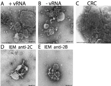
PV infection leads to a block in protein secretory transport and results in an accumulation of membranous vesicles in the cy-toplasm of the infected cell. Previous work has demonstrated that replicating PV RNA is associated with these vesicular membranes, which contain all viral and cellular components required for replication of the viral genome (11–13), and EM autoradiography demonstrated direct association of replicating RNA with the membranes (6). The isolation of these CRCs from PV-infected HeLa cells and their subsequent analysis by EM demonstrated that these vesicles aggregate into clusters described as “rosettes” when isolated from infected HeLa cells (7, 9, 19). We utilized a cell-free system derived from unin-fected HeLa cell S10 extracts that translate and replicate ex-ogenous PV RNA in vitro (33). We collected the membranous structures and examined their morphology by EM after nega-tive staining (Fig. 1A). Although some membranous structures with irregular shapes and heterogeneous sizes were present, no detectable differences were observed between these structures and those isolated from control HeLa cell extracts incubated without vRNA (Fig. 1B). The membrane structures observed after synthesis of viral proteins in the presence of GuHCl to prevent RNA replication also appeared the same (data not shown); similarly, no effect of BFA on the morphology of isolated membrane aggregates was discernible. Membrane structures representing CRCs from PV-infected cells are illus-trated in Fig. 1C for comparison, showing a cluster of vesicles. To determine whether viral replication proteins were asso-ciated with the observed membrane aggregates, we used IEM to examine the localization of viral proteins 2B and 2C (7, 9), which are usually used as markers for the replication complex. IEM of pelleted membranes from reactions undergoing trans-lation and replication of vRNA shows the localization of both 2B- and 2C-containing proteins on the surface of the mem-branes (Fig. 1D and E). Membrane pellets from control reac-tions (containing no vRNA) showed no reactivity with antibod-ies against 2B- or 2C-containing proteins (data not shown). Thus, although characteristic vesicles were not seen in the
on November 8, 2019 by guest
http://jvi.asm.org/
HeLa cell extracts that were synthesizing vRNA, viral proteins 2B and 2C did associate with the membrane aggregates that were present, as expected from previous results showing inher-ent membrane binding properties of proteins containing 2B and 2C sequences (1, 18, 48, 51).
Our microscopic analysis of the membrane structures de-scribed above suggested that the characteristic vesicle clusters observed in PV-infected cells did not form in vitro, even during viral RNA replication, or were present in concentrations too low to be readily observed. To investigate the properties of the structures supporting RNA synthesis in vitro, we examined the RNase sensitivity of both template and product RNA present in and synthesized by the CRCs isolated from PV-infected cells and by the membrane-associated replication complexes found in vitro during translation/replication of vRNA in HeLa ex-tracts.
Template RNA present in the CRCs isolated from PV-infected cells is substantially protected from digestion with RNase (19), as evidenced by the ability of RNase-treated CRCs to support viral RNA synthesis after reisolation by sed-imentation (Fig. 2A, compare lanes 1 and 3). Similarly, the product RNA synthesized by untreated CRCs is also protected from digestion with RNase A (Fig. 2A, lane 2). In contrast,
RNase treatment of membrane-associated preinitiation com-plexes formed during translation of viral RNA in vitro in the presence of GuHCl completely eliminated their capacity to support subsequent RNA replication after reisolation (Fig. 2B, lane 1). These data suggest that the membrane association of preinitiation-replication complexes does not protect template RNA from RNase digestion. Similarly, product RNA present at the end of the replication assay in the cell-free translation/ replication reaction was completely sensitive to RNase A di-gestion (Fig. 2B, lane 2). Product RNA synthesized by the membrane-associated complexes in the translation/replication reaction, in the absence of RNase A digestion either before or after synthesis, is shown in Fig. 2B, lane 3. The lack of protec-tive vesicle organization in HeLa cell S10 extracts that are replicating viral RNA—suggested by our failure to observe such structures by electron microscopy and by the sensitivity of both template and product RNA to RNase A—may contribute to reduced efficiency of viral RNA synthesis in vitro but does not preclude viral RNA synthesis.
Replication of PV RNA in vitro is detergent sensitive.
[image:3.603.81.510.70.401.2]Al-though viral proteins containing 2B and 2C sequences were observed to associate with membrane fragments present in the HeLa S10 extracts, the results described above prompted us to
FIG. 1. EM and IEM analysis of membranes pelleted from in vitro translation/replication reactions. Negatively stained preparations from PV RNA translation/replication reactions (A) or control reactions without vRNA (B). For comparison, a negatively stained sample of CRCs isolated from infected cells is shown (C). (D and E) IEM using antibodies against PV 2C and 2B proteins, respectively, to stain membranes from in vitro translation/replication reactions.
on November 8, 2019 by guest
http://jvi.asm.org/
question the requirement for membranes in viral RNA synthe-sis in vitro. We therefore examined the sensitivity of vRNA synthesis in cell extracts to detergent. Previous data have shown that elongation of initiated strands of RNA in CRCs is not detergent sensitive, whereas initiation of new RNA mole-cules fails to occur in the presence of detergent (22). We used the cell-free translation/replication system, separated into two steps. Inclusion of 2 mM GuHCl during an initial incubation period allows translation and formation of preinitiation com-plexes to occur but prevents VPg uridylylation (5) and vRNA synthesis (4). After collection of the preinitiation complexes by sedimentation and resuspension to remove the GuHCl, the reactions can proceed with uridylylation of VPg and synthesis of vRNA. Recovery of the active preinitiation-replication
com-plexes in the membranous pellet is consistent with their having become associated with membrane structures during the initial incubation (5). To examine the effect of detergents, we in-cluded the nonionic detergent NP-40 at 0.1% either during the translation step in the presence of GuHCl or during VPg uridylylation and replication, after reversal of GuHCl inhibi-tion. While total protein synthesis and polyprotein processing were unaffected when NP-40 was added during translation (Fig. 3A), subsequent RNA synthesis, upon removal of GuHCl, was completely inhibited (Fig. 3B). (Higher concentrations of NP-40 [1%], however, resulted in alterations in translation and polyprotein processing.) If detergent was added only after translation and formation of preinitiation complexes, RNA replication was somewhat reduced, but not completely inhib-ited (Fig. 3C). These data suggest that membranes are re-quired for the formation of a preinitiation-replication complex and perhaps as well for the subsequent replication of vRNA within the replication complex. Similar effects were observed after addition of other nonionic detergents (Triton X-100 and Tween 20). It is not known whether there was a differential effect on positive- versus negative-strand synthesis during the subsequent replication step, after preinitiation complexes were formed.
[image:4.603.303.537.68.307.2]Dependence of RNA replication on the presence of mem-branes was further tested by removal of membranous material from the cell extracts by centrifugation prior to the start of the
FIG. 2. RNase A sensitivity of template and product vRNAs. To examine template sensitivity, freshly isolated CRCs (A) or in vitro-generated preinitiation-replication complexes (B) were treated with RNase A (see Materials and Methods) and then incubated under RNA synthesis conditions in the presence of [32P]CTP (lane 1). Product
sensitivity was measured by treatment with RNase A after RNA syn-thesis (lane 2). Control samples were untreated with RNase (lane 3). Labeled RNA was isolated and analyzed under denaturing conditions on 0.8% agarose gels and was detected by PhosphorImager analysis.
FIG. 3. Detergent sensitivity of vRNA synthesis. (A) Translation reaction mixtures containing GuHCl and [35S]methionine were
incu-bated for 3.5 h in the presence (⫹) or absence (⫺) of 0.1% NP-40. Samples were analyzed by SDS-PAGE, and labeled proteins were detected by PhosphorImager. (B) vRNA synthesis was measured by preinitiation-replication complexes generated in the presence (⫹) or absence (⫺) of 0.1% NP-40. (C) vRNA synthesis was measured by preinitiation-replication complexes formed in the absence of deter-gent, but subsequently incubated in the presence (⫹) or absence (⫺) of NP-40. vRNA synthesis was measured as described in the legend to
Fig. 2.
on November 8, 2019 by guest
http://jvi.asm.org/
reaction. No functional replication complexes were found, as evidenced by the absence of any RNA replication, despite normal levels of viral protein synthesis (data not shown).
Detergent sensitivity of VPg uridylylation.An essential step
between the formation of a replication complex and the initi-ation of vRNA repliciniti-ation is the synthesis of the primer VPgpUpU. This reaction occurs in vitro with purified viral protein 3B (VPg), viral polymerase (3D), and UTP, using ei-ther a cis-acting replication element (cre) in the 2C coding region of vRNA or poly(A) as a template. The cre-dependent reaction is markedly enhanced by viral protein 3CD (37). The poly(A)-dependent reaction was shown to be unaffected by 0.1% NP-40 (38). When NP-40 was included in the cell-free replication system during translation-preinitiation complex formation, subsequent uridylylation of VPg was completely inhibited (Fig. 4A). Detergent treatment during the uridylyla-tion reacuridylyla-tion, after GuHCl removal, resulted in reduced uri-dylylation (Fig. 4B). Thus, the effects of NP-40 on VPg uridy-lylation and on viral RNA synthesis were parallel (compare Fig. 4B and 3C). VPg uridylylation was previously detected in CRCs isolated from infected cells (44) and was reported to be inhibited by concentrations of 0.1% NP-40 and higher (45). This finding demonstrates that the formation of membranous
structures during preinitiation-replication complex formation in vitro in the presence of GuHCl is required for the synthesis of the uridylylated VPg primer.
The membrane-altering compound cerulenin inhibits VPg
uridylylation.Previous studies with infected cells and cell-free
reactions have demonstrated the sensitivity of viral RNA rep-lication or virus production to many compounds thought to affect membrane formation or structure (4, 16, 25, 26, 29, 32, 34), although the exact basis of their modes of action has not been identified in the majority of cases. We examined the effect of a membrane-altering compound, cerulenin, during transla-tion and replicatransla-tion reactransla-tions in vitro.
Cerulenin irreversibly inhibits fatty acid synthesis by co-valently binding to the active site cysteine of-ketoacyl-ACP synthase, a key enzyme in the fatty acid synthesis pathway (27). HeLa cell S10 extracts convert [3H]glycerol into
chloroform-soluble material, and this reaction is inhibited by 200 to 500
M cerulenin (Table 1). Background radioactivity was deter-mined in samples containing no membranes or by preincuba-tion of S10 extracts with 500M cerulenin for 1 h prior to the addition of [3H]glycerol. These data provided a measure of
cerulenin-sensitive fatty acid synthesis in HeLa S10 extracts. Cerulenin up to 200 M had no effect on translation or polyprotein processing in vitro, as observed previously (34), and did not alter the localization of proteins or vRNA to the membrane fraction (data not shown). However, preinitiation-replication complexes that were generated in the presence of cerulenin were unable to support vRNA synthesis (Fig. 5A, lanes 1 to 4) or VPg uridylylation (Fig. 5B).
Figure 6 shows a cerulenin dose response comparison be-tween [3H]glycerol incorporation, uridylylation of VPg, and
vRNA synthesis in HeLa S10 extracts. Our data show that the membrane-altering compound cerulenin has no effect on events prior to vRNA replication (translation and polyprotein processing) but that cerulenin-sensitive fatty acid synthesis cor-relates with the synthesis of the viral primer VPgpUpU and thus vRNA synthesis.
BFA inhibits VPg uridylylation.BFA is an inhibitor of
in-tracellular membrane trafficking that has been demonstrated to inhibit replication of PV both in cultured cells and in cell-free systems (16, 28, 32). A previous study with the translation/ replication system suggested that the activity of the ADP-ribosylation factor (ARF) family of GTP-binding proteins involved in the generation of secretory transport vesicles is required for PV RNA replication (16). ARFs are required for the formation of several classes of secretory vesicles, and some family members are indirectly inactivated by BFA.
FIG. 4. Detergent sensitivity of VPg uridylylation. (A) VPg uri-dylylation in preinitiation-replication complexes generated in the pres-ence (⫹) or absence (⫺) of 0.1% NP-40. (B) VPg uridylylation in preinitiation-replication complexes formed in the absence of deter-gent, but subsequently resuspended in the presence (⫹) or absence (⫺) of 0.1% NP-40. VPg uridylylation was detected by the inclusion of [32P]UTP for 1 h following resuspension of the
[image:5.603.64.259.69.366.2]preinitiation-replica-tion complexes. Labeled VPg was detected by analysis on a 12.5% Tris–Tricine gel and PhosphorImager analysis.
TABLE 1. Effect of cerulenin on [3H]glycerol incorporation into
chloroform-soluble material in HeLa cell S10 extracts
Cerulenin concn (M) incorporation (cpm)[3H]glycerol
0 ... 1,953 25 ... 1,614 50 ... 1,305
100 ... 959
200 ... 411
500 ... 387
Control (no S10 extract)... 315
on November 8, 2019 by guest
http://jvi.asm.org/
We examined the effect of BFA addition on both VPg uri-dylylation and vRNA synthesis in HeLa cell extracts (Fig. 7). After translation and formation of preinitiation-replication complexes in the presence of GuHCl, the complexes were sedimented and suspended in the absence of guanidine to allow uridylylation of VPg or vRNA synthesis to proceed (Fig. 7A and B, respectively, lane 1). The continued presence of GuHCl in the second incubation prevents both uridylylation of VPg and vRNA synthesis (Fig. 7A and B, lane 2). Addition of BFA to the translation reaction in the presence of GuHCl, during formation of the preinitiation complexes, reduced sub-sequent VPg uridylylation and vRNA synthesis by 80 to 90% (Fig. 7A and B, lanes 3 and 4). This concentration of BFA (100
g/ml) had no effect on translation or protein processing (data not shown). DMSO, the solvent for BFA, caused a slight re-duction in VPg uridylylation when added alone (Fig. 7A,
com-pare lane 4 with lane 1). This effect of BFA would be expected if both VPg uridylylation and vRNA synthesis were dependent upon the formation of a membranous preinitiation complex that required a BFA-sensitive step of membrane vesiculariza-tion or rearrangement. When BFA was added after the
[image:6.603.306.543.74.221.2]for-FIG. 5. Effect of cerulenin on vRNA synthesis and VPg uridylyla-tion. Cerulenin (100, 50, 12.5, or 0M [lanes 1 through 4, respective-ly]) was included during formation of preinitiation-replication com-plexes. Complexes were assayed for vRNA synthesis (A) or VPg uridylylation (B). vRNA synthesis and VPg uridylylation were mea-sured as described in the legend to Fig. 4.
FIG. 6. Dependence of vRNA synthesis, VPg uridylylation, and [3H]glycerol incorporation on cerulenin concentration. Values are
ex-pressed as percent of control samples (no cerulenin present).Œ, vRNA
[image:6.603.303.539.369.629.2]synthesis;䊐, VPg uridylylation;⽧, [3H]glycerol.
FIG. 7. Effect of guanidine and BFA on VPg uridylylation (A) and vRNA synthesis (B) in vitro. Preinitiation-replication complexes were sedimented and resuspended in either the absence (lane 1) or presence (lane 2) of 2 mM GuHCl, or BFA was added during generation of preinitiation-replication complexes (lane 3) or after their sedimenta-tion and resuspension of preinitiasedimenta-tion-replicasedimenta-tion complexes (lane 5). Control reaction mixtures contained DMSO during the generation (lane 4) or after resuspension (lane 6) of preinitiation-replication com-plexes. vRNA synthesis and VPg uridylylation were detected as de-scribed in the legends to Fig. 2 and 4.
on November 8, 2019 by guest
http://jvi.asm.org/
mation of the preinitiation complexes (after removal of GuHCl inhibition), a similar inhibition of both reactions was observed (Fig. 7A and B, lanes 5 and 6). The extent of the inhibition observed when BFA was added after isolation of preinitiation complexes was quite variable in different experiments. This suggests that the process or processes inhibited by BFA during formation of preinitiation complexes may continue to a vari-able extent after their isolation and resuspension under con-ditions used for assay of VPg uridylylation and vRNA synthe-sis.
DISCUSSION
The apparent requirement for membranes in the reactions involved in PV RNA synthesis has hindered previous attempts to reconstitute an RNA replication reaction from purified components in vitro. Absent this means of dissecting the indi-vidual biochemical steps that occur during viral RNA-depen-dent RNA synthesis, the coupled translation/replication system first described by Molla et al. (33) has provided a useful tool for analysis of many aspects of viral RNA synthesis (for exam-ples, see references 4, 47, and 52). Since the products of this in vitro system include infectious virus, it is generally assumed that all steps in translation, polyprotein processing, RNA syn-thesis, encapsidation, and assembly mimic the reactions that occur inside an infected cell.
Previous studies have included only preliminary observa-tions regarding the role of membranes in the various reacobserva-tions contributing to virus production in the HeLa cell S10 extracts (34). Our EM examination of extracts actively engaged in viral RNA replication failed to reveal either a demonstrable in-crease in vesicular membrane structures or the rosette-like aggregation of vesicles previously described for replication complexes isolated from PV-infected cells (8).
IEM, however, revealed PV nonstructural proteins associ-ated with morphologically heterogeneous membrane frag-ments similar to those seen in extracts not engaged in viral RNA replication, which appear to serve as some sort of scaf-fold or anchor for viral RNA synthesis. Additional evidence for a difference in membrane organization between the replication complexes utilized in vitro and in vivo is the difference in RNase sensitivity of both template and product vRNAs. In the in vitro translation/replication system, both RNAs are sensitive to RNase, whereas the template and product vRNA in repli-cation complexes isolated from PV-infected cells are signifi-cantly protected from RNase digestion, consistent with the rosette-like aggregation of vesicles surrounding the sites of vRNA synthesis described previously (19). The lack of mem-brane rearrangement to form vesicle-protected compartments may contribute to the relative inefficiency of virion production in the in vitro system (33).
The overall process of viral RNA replication includes nu-merous biochemical steps and reactions occurring in a coordi-nated fashion. Several of these individual reactions have been reproduced in isolation in vitro; for example, template-depen-dent uridylylation of VPg to generate a primer for initiation of RNA chain synthesis is catalyzed by purified 3Dpol in vitro
(38); similarly, purified 3D-catalyzed RNA chain elongation from a primed template also has been studied in some detail (2, 3, 40, 41). In both of these examples, the reactions occur in
the absence of membranes and are not inhibited by nonionic detergents or by BFA (unpublished observations). Admittedly, VPg uridylylation assays utilizing purified components in vitro are performed at quite high concentrations of substrates and enzyme. On the other hand, replication complexes isolated from infected cells, as well as those generated in the transla-tion/replication system utilized in this study, manifest marked inhibition of VPg uridylylation and initiation of RNA synthesis by detergent (22, 44) (Fig. 3 and 4), and VPg uridylylation in the translation/replication system is also sensitive to treatment with BFA (Fig. 7); thus, it is likely that the requirement for intact membrane structures for vRNA synthesis represents provision of a scaffold that positions all of the essential com-ponents, rather than contribution of catalytic functions. In addition, the reduction in viral RNA synthesis observed in response to membrane-altering agents may result from the inhibitory effect on the first step of the reaction, synthesis of the primer to initiate RNA chain synthesis, as was suggested previously (45).
Although addition of detergent in this study to the transla-tion reactransla-tion during formatransla-tion of preinitiatransla-tion complexes com-pletely abolished subsequent synthesis of uridylylated VPg and initiation of vRNA synthesis, addition of detergent after for-mation of the complexes inhibited synthesis to a lesser degree. The reduced effect of detergent in the second step may be due to a clumping of the membranes when centrifuged to isolate the preinitiation complexes, rendering them less sensitive to detergent action.
Morphological reorganization of the membrane fragments present in the homogenized cell extracts was not evident by EM. Nevertheless, inhibition of primer synthesis and vRNA synthesis by BFA and cerulenin suggests that some sort of membrane rearrangement or reorganization may be required for these reactions to occur. Molla et al. (34) had previously reported that cerulenin inhibited vRNA synthesis, but not translation of vRNA, in the translation/replication system. Cerulenin has been shown to inhibit fatty acid biosynthesis via its interaction with the enzyme fatty acid synthase, although it likely has other effects as well, such as its suggested inhibition of membrane vesicular transport in BFA-resistant Vero cells (36). In this study, we showed that cerulenin-induced inhibition of VPg uridylylation and vRNA synthesis correlated with the inhibition of incorporation of [3H]glycerol into
chloroform-soluble material. Previous studies showed stimulation of phos-phatidylcholine synthesis in PV-infected cells (50), suggesting a requirement for de novo membrane synthesis. A mechanistic basis for these early studies was not pursued. More recently, however, another VPg-containing positive-strand RNA virus, cowpea mosaic virus, has been reported to be sensitive to cerulenin (14).
It is generally thought that the inhibitory action of BFA on cellular membrane transport was likely responsible for its in-hibition of PV replication. A specific role for ARF, a key protein involved in vesicular transport, was suggested by stud-ies performed with HeLa cell extracts translating and replicat-ing viral RNA (16). Competition of ARF with an inhibitory peptide led to inhibition of vRNA replication, adding more support for a role for membrane transport in virus replication. Our observation that BFA treatment prevents the specific step of VPg uridylylation was initially surprising; ARF proteins
on November 8, 2019 by guest
http://jvi.asm.org/
interact with multiple molecules involved in several different vesicular trafficking pathways and also activate specific phos-pholipase Ds (for a review, see reference 35). PV may utilize some aspect of the reaction for vesicular transport as a mech-anism involved in the synthesis of uridylylated VPg, rather than as a requirement for membrane transport per se.
Relatively high concentrations of BFA (⬃0.3 mM) were utilized to inhibit VPg uridylylation and vRNA synthesis in vitro. BFA concentrations utilized to inhibit viral RNA repli-cation or cellular membrane trafficking in intact cultured cells are generally 10- to 40-fold lower. There may be active trans-port systems that concentrate BFA in cells; alternatively, the difference in inhibitory concentration may reflect a BFA-sen-sitive process occurring in vitro different from that in infected cells. Indeed, one previous report (46) failed to show any inhibition of virus production in cell extracts treated with up to 1-mg/ml (3.6 mM) BFA. The reason for this variability is not clear; it is possible that the cell disruption process during extract preparation in that laboratory generated a loss of sen-sitivity to BFA.
Not all picornaviruses manifest the same sensitivity to BFA (24, 28). Apparently, some picornaviruses either require less membrane activity, or they may be able to bypass the BFA-sensitive step(s). Further studies with a wider selection of pi-cornaviruses may reveal more information on the sensitivity and mechanism of this process.
ACKNOWLEDGMENTS
We thank M. Rinaudo for preparation of the HeLa cell extracts.
REFERENCES
1. Aldabe, R., and L. Carrasco.1995. Induction of membrane proliferation by poliovirus proteins 2C and 2BC. Biochem. Biophys. Res. Commun.206:64– 76.
2. Arnold, J. J., and C. E. Cameron.2000. Poliovirus RNA-dependent RNA polymerase (3D(pol)). Assembly of stable, elongation-competent complexes by using a symmetrical primer-template substrate (sym/sub). J. Biol. Chem. 275:5329–5336.
3. Arnold, J. J., and C. E. Cameron.1999. Poliovirus RNA-dependent RNA polymerase (3Dpol) is sufficient for template switching in vitro. J. Biol. Chem.274:2706–2716.
4. Barton, D. J., E. P. Black, and J. B. Flanegan.1995. Complete replication of poliovirus in vitro: preinitiation RNA replication complexes require soluble cellular factors for the synthesis of VPg-linked RNA. J. Virol.69:5516–5527. 5. Barton, D. J., and J. B. Flanegan.1997. Synchronous replication of poliovi-rus RNA: initiation of negative-strand RNA synthesis requires the guani-dine-inhibited activity of protein 2C. J. Virol.71:8482–8489.
6. Bienz, K., D. Egger, and L. Pasamontes.1987. Association of polioviral proteins of the P2 genomic region with the viral replication complex and virus-induced membrane synthesis as visualized by electron microscopic im-munocytochemistry and autoradiography. Virology160:220–226.
7. Bienz, K., D. Egger, T. Pfister, and M. Troxler.1992. Structural and func-tional characterization of the poliovirus replication complex. J. Virol.66: 2740–2747.
8. Bienz, K., D. Egger, Y. Rasser, and W. Bossart.1980. Kinetics and location of poliovirus macromolecular synthesis in correlation to virus-induced cyto-pathology. Virology100:390–399.
9. Bienz, K., D. Egger, M. Troxler, and L. Pasamontes.1990. Structural orga-nization of poliovirus RNA replication is mediated by viral proteins of the P2 genomic region. J. Virol.64:1156–1163.
10. Blank, C. A., D. A. Anderson, M. Beard, and S. M. Lemon.2000. Infection of polarized cultures of human intestinal epithelial cells with hepatitis A virus: vectorial release of progeny virions through apical cellular membranes. J. Virol.74:6476–6484.
11. Butterworth, B. E., E. J. Shimshick, and F. H. Yin.1976. Association of the polioviral RNA polymerase complex with phospholipid membranes. J. Virol. 19:457–466.
12. Caliguiri, L. A., and I. Tamm.1969. Membranous structures associated with translation and transcription of poliovirus RNA. Science166:885–886. 13. Caliguiri, L. A., and I. Tamm.1970. The role of cytoplasmic membranes in
poliovirus biosynthesis. Virology42:100–111.
14. Carette, J. E., M. Stuiver, J. Van Lent, J. Wellink, and A. B. Van Kammen. 2000. Cowpea mosaic virus infection induces a massive proliferation of endoplasmic reticulum but not Golgi membranes and is dependent on de novo membrane synthesis. J. Virol.74:6556–6563.
15. Cho, M. W., N. Teterina, D. Egger, K. Bienz, and E. Ehrenfeld.1994. Membrane rearrangement and vesicle induction by recombinant poliovirus 2C and 2BC in human cells. Virology202:129–145.
16. Cuconati, A., A. Molla, and E. Wimmer.1998. Brefeldin A inhibits cell-free, de novo synthesis of poliovirus. J. Virol.72:6456–6464.
17. Dayhuff, T. J., R. F. Gesteland, and J. F. Atkins.1992. Electrophoresis, autoradiography and electroblotting of peptides: T4 gene 60 hopping. Bio-Techniques13:499–502, 505.
18. Echeverri, A. C., and A. Dasgupta.1995. Amino terminal regions of polio-virus 2C protein mediate membrane binding. Virology208:540–553. 19. Egger, D., L. Pasamontes, R. Bolten, V. Boyko, and K. Bienz.1996.
Revers-ible dissociation of the poliovirus replication complex: functions and inter-actions of its components in viral RNA synthesis. J. Virol.70:8675–8683. 20. Egger, D., N. Teterina, E. Ehrenfeld, and K. Bienz.2000. Formation of the
poliovirus replication complex requires coupled viral translation, vesicle pro-duction, and viral RNA synthesis. J. Virol.74:6570–6580.
21. Egger, D., B. Wolk, R. Gosert, L. Bianchi, H. E. Blum, D. Moradpour, and K. Bienz.2002. Expression of hepatitis C virus proteins induces distinct membrane alterations including a candidate viral replication complex. J. Vi-rol.76:5974–5984.
22. Etchison, D., and E. Ehrenfeld.1981. Comparison of replication complexes synthesizing poliovirus RNA. Virology111:33–46.
23. Folch, J., M. Lees, and G. H. Stanley.1957. A simple method for the isolation and purification of total lipids from animal tissues. J. Biol. Chem. 226:497–509.
24. Gazina, E. V., J. M. Mackenzie, R. J. Gorrell, and D. A. Anderson.2002. Differential requirements for COPI coats in formation of replication com-plexes among three genera ofPicornaviridae. J. Virol.76:11113–11122. 25. Guinea, R., and L. Carrasco.1991. Effects of fatty acids on lipid synthesis
and viral RNA replication in poliovirus-infected cells. Virology185:473–476. 26. Guinea, R., and L. Carrasco.1990. Phospholipid biosynthesis and poliovirus
genome replication, two coupled phenomena. EMBO J.9:2011–2016. 27. Heath, R. J., S. W. White, and C. O. Rock.2001. Lipid biosynthesis as a target
for antibacterial agents. Prog. Lipid Res.40:467–497.
28. Irurzun, A., L. Perez, and L. Carrasco.1992. Involvement of membrane traffic in the replication of poliovirus genomes: effects of brefeldin A. Virol-ogy191:166–175.
29. Irurzun, A., S. Sa´nchez-Palomino, I. Novoa, and L. Carrasco.1995. Monen-sin and nigericin prevent the inhibition of host translation by poliovirus, without affecting p220 cleavage. J. Virol.69:7453–7460.
30. Lee, W. M., M. Ishikawa, and P. Ahlquist.2001. Mutation of host⌬9 fatty acid desaturase inhibits brome mosaic virus RNA replication between tem-plate recognition and RNA synthesis. J. Virol.75:2097–2106.
31. Makino, S., and H. M. Jenkin.1975. Effect of fatty acids on growth of Japanese encephalitis virus cultivated in BHK-21 cells and phospholipid metabolism of the infected cells. J. Virol.15:515–525.
32. Maynell, L. A., K. Kirkegaard, and M. W. Klymkowsky.1992. Inhibition of poliovirus RNA synthesis by brefeldin A. J. Virol.66:1985–1994. 33. Molla, A., A. V. Paul, and E. Wimmer.1991. Cell-free, de novo synthesis of
poliovirus. Science254:1647–1651.
34. Molla, A., A. V. Paul, and E. Wimmer.1993. Effects of temperature and lipophilic agents on poliovirus formation and RNA synthesis in a cell-free system. J. Virol.67:5932–5938.
35. Moss, J., and M. Vaughan.1998. Molecules in the ARF orbit. J. Biol. Chem. 273:21431–21434.
36. Oda, T., and H. C. Wu.1993. Cerulenin inhibits the cytotoxicity of ricin, modeccin,Pseudomonastoxin, and diphtheria toxin in brefeldin A-resistant cell lines. J. Biol. Chem.268:12596–12602.
37. Paul, A. V., E. Rieder, D. W. Kim, J. H. van Boom, and E. Wimmer.2000. Identification of an RNA hairpin in poliovirus RNA that serves as the primary template in the in vitro uridylylation of VPg. J. Virol.74:10359– 10370.
38. Paul, A. V., J. H. van Boom, D. Filippov, and E. Wimmer.1998. Protein-primed RNA synthesis by purified poliovirus RNA polymerase. Nature393: 280–284.
39. Perez, L., R. Guinea, and L. Carrasco.1991. Synthesis of Semliki Forest virus RNA requires continuous lipid synthesis. Virology183:74–82.
40. Richards, O. C., S. Baker, and E. Ehrenfeld.1996. Mutation of lysine resi-dues in the nucleotide binding segments of the poliovirus RNA-dependent RNA polymerase. J. Virol.70:8564–8570.
41. Richards, O. C., and E. Ehrenfeld.1998. Effects of poliovirus 3AB protein on 3D polymerase-catalyzed reaction. J. Biol. Chem.273:12832–12840. 42. Schagger, H., and G. von Jagow. 1987. Tricine-sodium dodecyl
sulfate-polyacrylamide gel electrophoresis for the separation of proteins in the range from 1 to 100 kDa. Anal. Biochem.166:368–379.
43. Suhy, D. A., T. H. Giddings, Jr., and K. Kirkegaard.2000. Remodeling the endoplasmic reticulum by poliovirus infection and by individual viral
on November 8, 2019 by guest
http://jvi.asm.org/
teins: an autophagy-like origin for virus-induced vesicles. J. Virol.74:8953– 8965.
44. Takeda, N., R. J. Kuhn, C.-F. Yang, T. Takegami, and E. Wimmer.1986. Initiation of poliovirus plus-strand RNA synthesis in a membrane complex of infected HeLa cells. J. Virol.60:43–53.
45. Takegami, T., R. J. Kuhn, C. W. Anderson, and E. Wimmer.1983. Mem-brane-dependent uridylylation of the genome-linked protein VPg of polio-virus. Proc. Natl. Acad. Sci. USA80:7447–7451.
46. Tang, R. S., D. J. Barton, J. B. Flanegan, and K. Kirkegaard.1997. Polio-virus RNA recombination in cell-free extracts. RNA3:624–633.
47. Teterina, N. L., D. Egger, K. Bienz, D. M. Brown, B. L. Semler, and E. Ehrenfeld.2001. Requirements for assembly of poliovirus replication com-plexes and negative-strand RNA synthesis. J. Virol.75:3841–3850. 48. Teterina, N. L., A. E. Gorbalenya, D. Egger, K. Bienz, and E. Ehrenfeld.
1997. Poliovirus 2C protein determinants of membrane binding and rear-rangements in mammalian cells. J. Virol.71:8962–8972.
49. Towner, J. S., T. V. Ho, and B. L. Semler.1996. Determinants of membrane association for poliovirus protein 3AB. J. Biol. Chem.271:26810–26818. 50. Vance, D. E., E. M. Trip, and H. B. Paddon.1980. Poliovirus increases
phosphatidylcholine biosynthesis in HeLa cells by stimulation of the rate-limiting reaction catalyzed by CTP: phosphocholine cytidylyltransferase. J. Biol. Chem.255:1064–1069.
51. van Kuppeveld, F. J., J. G. Hoenderop, R. L. Smeets, P. H. Willems, H. B. Dijkman, J. M. Galama, and W. J. Melchers.1997. Coxsackievirus protein 2B modifies endoplasmic reticulum membrane and plasma membrane per-meability and facilitates virus release. EMBO J.16:3519–3532.
52. Walter, B. L., T. B. Parsley, E. Ehrenfeld, and B. L. Semler.2002. Distinct poly(rC) binding protein KH domain determinants for poliovirus translation initiation and viral RNA replication. J. Virol.76:12008–12022.

![TABLE 1. Effect of cerulenin on [3H]glycerol incorporation intochloroform-soluble material in HeLa cell S10 extracts](https://thumb-us.123doks.com/thumbv2/123dok_us/258490.59402/5.603.64.259.69.366/effect-cerulenin-glycerol-incorporation-intochloroform-soluble-material-extracts.webp)

