COMPARISON OF CT BASED AND INTEGRATED F 18 FDG PET CT SCAN
BASED GROSS TUMOUR VOLUME IN HEAD AND NECK CANCERS AND
EVALUATION OF THE DIFFERENT SEGMENTATION METHOD FOR
DELINEATION OF THE TARGET ON PET SCAN
DEPARTMENT OF RADIOTHERAPY CHRISTIAN MEDICAL COLLEGE
VELLORE 632004
DISSERTATION SUBMITTED IN PARTIAL FULFILLMENT OF MD BRANCH IX RADIOTHERAPY
EXAMINATION APRIL 2016
TAMIL NADU DR. M.G.R MEDICAL UNIVERSITY
CHENNAI - 600032
CHRISTIAN MEDICAL COLLEGE, VELLORE
CERTIFICATE
CERTIFICATE
CERTIFICATE
CERTIFICATE
This is to certify that the dissertation entitled “COMPARISON OF CT BASED AND INTEGRATED F 18 FDG PET CT SCAN BASED GROSS TUMOUR VOLUME IN HEAD AND NECK CANCERS AND EVALUATION OF THE DIFFERENT SEGMENTATION METHOD FOR DELINEATION OF THE TARGET ON PET SCAN” is a bonafide work done by Dr. PAUL GOPU G, Post Graduate Student in the Department of Radiotherapy, Christian Medical College, Vellore during the period from April 2014 to April 2016 and is being submitted to The Tamil Nadu Dr. M. G. R Medical University in partial fulfilment of the MD Branch IX Radiotherapy examination conducted in April 2016.
Guide
Dr. Subhashini John
Professor
Department of Radiotherapy
Christian Medical College
Vellore, India – 632004
CERTIFICATE
CERTIFICATE
CERTIFICATE
CERTIFICATE
This is to certify that the dissertation entitled “COMPARISON OF CT BASED AND INTEGRATED F 18 FDG PET CT SCAN BASED GROSS TUMOUR VOLUME IN HEAD AND NECK CANCERS AND EVALUATION OF THE DIFFERENT SEGMENTATION METHOD FOR DELINEATION OF THE TARGET ON PET SCAN” is a bonafide work done by Dr. PAUL GOPU G, Post Graduate Student in the Department of Radiotherapy, Christian Medical College, Vellore during the period from April 2014 to April 2016 and is being submitted to The Tamil Nadu Dr. M. G. R Medical University in partial fulfilment of the MD Branch IX Radiotherapy examination conducted in April 2016.
Dr. Selvamani B
Dr. Alfred Job Daniel Prof and Head of the department
Principal Department of Radiotherapy
Christian Medical College Christian Medical College
CERTIFICATE
CERTIFICATE
CERTIFICATE
CERTIFICATE
I, Paul Gopu G, PG Registrar, Department of Radiation therapy, Christian Medical College Vellore hereby declare that the dissertation titled ‘COMPARISON OF CT BASED AND INTEGRATED F 18 FDG PET CT SCAN BASED GROSS TUMOUR VOLUME IN HEAD AND NECK CANCERS AND EVALUATION OF THE DIFFERENT SEGMENTATION METHOD FOR DELINEATION OF THE TARGET ON PET SCAN’ is a bonafide work done by me for partial fulfilment towards MD Radiotherapy (Branch IX) Degree examination of the Tamil Nadu Dr M G R Medical University to be held in April 2016.
DR. Paul Gopu G PG REGISTRAR,
Acknowledgements
I would like to sincerely thank my guide, Dr. Subhashini John for her invaluable guidance, constant encouragement and valuable ideas without whose help, this dissertation would not be possible.
I would like to thank my co guide Dr Devakumar Devadas for his encouragement, creative and comprehensive advises.
I would like thank my co guides, Dr. Rajesh I, Dr Saikat Das, Dr Julie Hephzibah, Dr Jeba Karunya, Dr Pavithra Mannan and Mr Ebenezer Suman Babu. Without their help the completion of this work would have been immeasurably more difficult.
I would like to thank my patients who consented to this study and enabled research to happen.
I would like to thank all the support staff in the Nuclear Medicine Department who helped me with my data collection and all technical assisstance.
I would also like to thank the Department of Biostatistics for their help in the statistical evaluation.
Contents
Introduction ... 9
Aim ... 12
Review of Literature ... 13
Anatomy ... 15
Lymphatics of head and Neck ... 22
Etiology ... 28
Histology of head and neck cancers ... 31
Natural History(13) ... 34
Evaluation and Diagnosis ... 37
Prognostic factors ... 41
Management of Locally advanced head and neck cancers ... 45
PET CT in Head and Neck cancers ... 72
Methods and Materials ... 78
Results ... 86
Discussion... 108
Conclusion ... 116
Reference ... 118
ABSTRACT TITLE: COMPARISON OF CT BASED AND INTEGRATED F 18 FDG PET CT SCAN BASED GROSS TUMOUR VOLUME IN HEAD AND NECK CANCERS AND EVALUATION OF THE DIFFERENT SEGMENTATION METHOD FOR DELINEATION OF THE TARGET ON PET SCAN
DEPARTMENT: Department of Radiotherapy, Dr Ida B Scudder Cancer Centre
NAME OF THE CANDIDATE: Dr. Paul Gopu G
DEGREE& SUBJECT: MD Radiotherapy
NAME OF THE GUIDE: Dr Subhashini John
Objective:
To evaluate the differences in radiotherapy gross tumour volumes (Primary and lymph nodes) delineated on CT and PET for radiotherapy planning of Head and neck cancers.
Methods and materials:
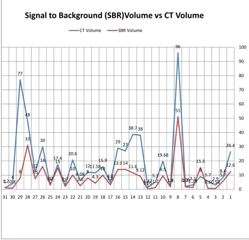
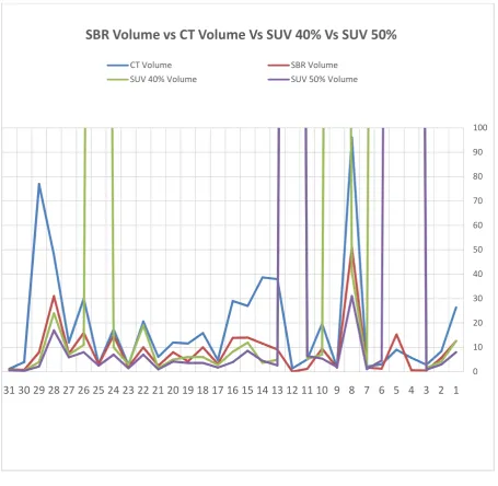
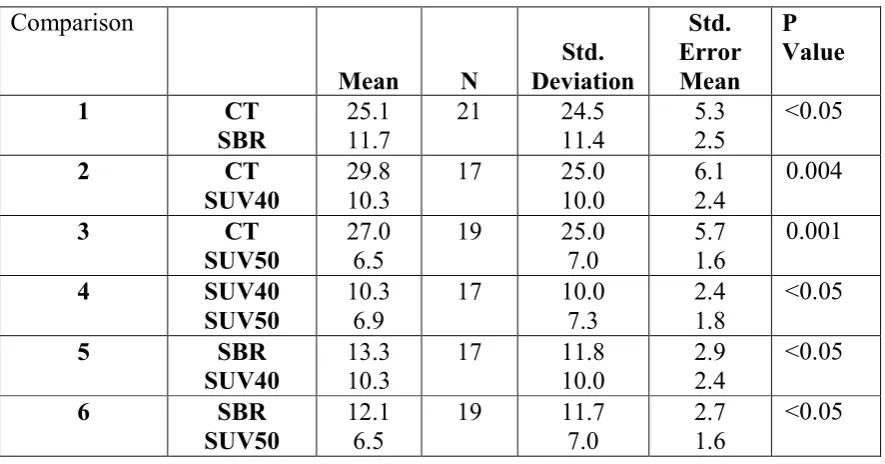
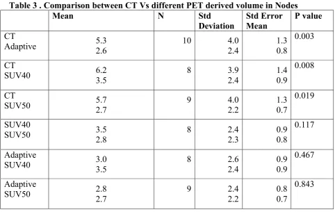
was calculated and the contours were done with the help of 3D slicer. The SUV-based delineation was obtained by applying an isocontour around the tumor with two thresholds which were based on fixed percentages of the maximum signal intensity in the primary tumor; 40% (GTV40%) and 50% (GTV50%). The absolute volumes of tumour (primary and lymph nodes) obtained using the CT scan and the PET data were documented. The CT volume was compared with PET volume that was got through the SBR technique. Volume was also segmented using fixed SUV technique with SUV 40% and SUV 50%, and these volumes were also compared with each other.
Results:
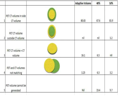
50%. i.e adaptive method was better than the fixed threshold methods. In some instances the nodal volumes derived using the SBR technique was grossly less than the CT volumes. The necrosis part of the node failed to pick up FDG and thus the contoured metabolic tumour volume was very different from the anatomical volume.
Conclusion:
Introduction
Head and neck malignancy are a major cause of morbidity and mortality throughout the world. The incidence of head and neck malignancies are very high in India and it ranks third commonest, after breast and cervical cancer.(1)(2)
A patient diagnosed with stage I or II head and neck cancer is offered single modality treatment in the form of surgery or radiotherapy. Multidisciplinary approach with surgery, radiation therapy and chemotherapy forms the mainstay of treatment of locally advanced HNSCC. Radiotherapy forms a major treatment modality in the form of radical radiotherapy, concurrent chemoirradiation as part of organ preservation protocol, post operative radiotherapy with or without chemotherapy and palliative radiotherapy.
With the use of IMRT the dose to the primary tumor can be escalated while keeping the dose to the adjacent normal structures at the minimum. There is a sharp fall of in dose at the edges of the tumour volume which results in reducing the doses to normal structures. So appropriate imaging for precise delineation of target volume and organs at risk is required for planning in conformal radiotherapy, otherwise there is a chance of geographical miss.(5)
Aim
-
Primary objective:
To evaluate the differences in radiotherapy gross tumour volumes (Primary and lymph nodes) delineated on CT and PET for radiotherapy planning of Head and neck cancers
Secondary objective:
• To assess the impact of the addition of PET scan on staging and thus the change in management
1. Change in nodal tumour volume
Review of Literature
Head and Neck squamous cell cancers are a major cause of morbidity and mortality throughout the world. Although the trend of incidence and prevalence of head and neck malignancies had decreased throughout other parts of the world, the incidence and prevalence is still very high in India.(9)(10)
WHO GLOBOCAN report 2012 reported the most common malignancies seen in india are breast, cervix, lip and oral cavity cancers. When we consider cancers of the head and neck region, lip and oral cavity cancers had an incidence of 11.6%, laryngeal cancers 4.8%, nasopharynx 0.6% and other pharynx 6.6%. In India this constitute about 23.6% of new diagnosed cancers .(10)
The WHO GLOBOCAN Report 2012 also showed a very high 5 year prevalence rates for head and neck cancers in India. The 5 year prevalence rate of lip and oral cavity was 12.6%, while for Laryngeal cancers it was 6.8%, nasopharynx 1.1% and other pharynx 7%. This clearly points out the high burden of head and neck cancers in India with 5 year prevalence rates of nearly 27%.(10)
Anatomy
Diagram 1 showing anatomy of Head and Neck subsites.
Nasopharynx
Oropharynx
Hypopharynx
Oral Cavity:
The oral cavity is divided into a number of areas namely: The Lip
Buccal mucosa - Membrane lining inner surface of the lip and cheeks to the attachment of mucosa to alveolar ridges and pterygomandibular raphe
Lower alveolar ridge - Extends from lower buccal gutter to free mucosa of the floor of the mouth and goes to ascending ramus of the mandible posteriorly.
Upper alveolar ridge - Extends from upper buccal gutter to junction of hard palate and it posteriorly goes to upper end of pterygopalatine arch
Retro molar trigone – extends from the mucosa overlying ascending ramus of mandible from the posterior surface of last molar tooth to the apex, adjacent to tuberosity of maxilla
Hard palate – It is a semilunar area between the upper alveolar ridge and mucous membrane which is covering the palatine process of maxillary palatine bones. It is the region from inner surface of superior alveolar ridge to posterior edge of the palatine bone
Oral tongue – Anatomically it extends anteriorly from circumvallate papillae to under surface of the tongue at the junction of the floor of mouth.
Oropharynx :
The oropharynx is bounded anteriorly by the anterior pillars of the pharyngeal fauces (the palatoglossus muscle), the circumvallate papillae (sulcus terminales) or the junction of the hard and soft palates. Posterior and lateral boundaries are formed by the muscular pharyngeal wall (superior and middle constrictors). The superior extent is the level of the soft palate. The inferior extent is the level of the base of tongue (level of the hyoid). The oropharynx is subdivided into five areas. These include lateral pharyngeal walls, tonsillar regions, posterior wall, base of tongue, and soft palate.
Hypopharynx:
cartilage. Hypopharynx can be divided into four subsites: the two pyriform sinus, the post cricoid area, and the posterior pharyngeal wall. The pyriform sinus is a funnel shaped structure that is bounded superiorly at the glossoepiglottic fold and extends inferiorly with its apex at the level of the cricopharyngeus. It is bounded posteriorly by the lateral wall of the hypopharynx and laterally by the thyroid lamina. Its medial boundary is the lateral surface of the arytenoid. The third area is the post cricoid area. This includes the posterior surface of the aryepiglottic fold and posterior surface of the arytenoid to the inferior border of the cricoid cartilage. The fourth region is the posterior pharyngeal wall, which extends from a plane drawn at the level of the tip of the epiglottis to a plane at the inferior border of the cricoid. The superior and inferior margins of the hypopharynx blend with the posterior wall of the oropharynx and esophagus, respectively.
Larynx:
The Larynx consists of 3 parts – The Supraglottic larynx, the Glottic larynx and the Sub Glottic larynx. The Supraglottic larynx consists of epiglottis, aryepiglottic folds, arytenoid cartilages and false cord.
The Glottic larynx is made up of true vocal cords and anterior & posterior commissures down to 5 mm below free margin of vocal cords.
Nasopharynx:
The Nasopharynx begins superiorly border is the cribriform plate and sphenoid sinus. Anteriorly it extends from the end of the nasal cavity, at the posterior choana. It extends along plane of the airway to the level of free border of the soft palate posteriorly. Lateral walls consist of the torus tubarius (opening of eustachian tube), pharyngeal recess (Fossa of Rosenmuller) posterior to torus tubarius, and behind these are the superior pharyngeal constrictor muscles, and behind this is the medial pterygoid plate. The posterior boundary of the nasopharynx consist of clivus which is part of the sphenoid bone (behind the sinus, the tail end of the sella turcica) and part of the occipital bon. Hard and soft palates (the inferior border) sits about at the level of C2.
Clinical examination of nasopharyngeal cancers includes cranial nerve examination because of the anatomical relationship of the nasopharynx to cranial nerves and commonly extend along them intracranially:
Cavernous sinus – Cranial nerves III, IV, V1 + V2, VI - (all pass through superior orbital fissure except V2 is through foramen rotundum)
Foramen rotundum – Cranial nerve V2
Foramen ovale – Cranial nerve V3 - anterolateral to clivus
Hypoglossal canal – Cranial nerve XII - lateral to foramen magnum
Lymphatics of head and Neck
Level Ia
Level Ia nodes are the submental group of nodes. Anotomically these are placed between the anterior belly of the digastric muscles. There is no actual medial boundary for level Ia and it is continuous with level Ia nodal region on the opposite. The region from which level Ia nodes drain are floor of the mouth (mainly the anterior part), tip of the anterior tongue, the middle third of the lower lip and skin of the chin. Submental nodes has the potential risk of developing metastases from cancer arising from or involving the anterior portion of floor of the mouth, the anterior oral tongue, the lower lip and the anterior mandibular alveolar ridge.(12)
Level Ib
node also receive efferent from anterior nasal cavity and soft tissue structures of the mid-face.(12)
Level II
oropharynx, and less frequent of cancer sites of efferents are the oral cavity, larynx, hypopharynx or oral cavity.(12)
Level III
Level III forms the middle jugular nodes located around the mid third of the the IJV. Cranio – caudally it extends from the caudal edge of the body of the hyoid bone to the caudal edge of the cricoid cartilage. The anterior boundary is the anterior edge of the sternocleidomastoid muscle or the posterior third of the thyro-hyoid muscle, and the posterior margin is the posterior edge of the sternocleidomastoid muscle. Medially, level III is limited by medial edge of the common carotid artery and the scalenius muscles and laterally by the deep surface of the sternocleidomastoid muscle. Level III receives efferent lymphatics from levels II and V, and some efferent lymphatics from the pretracheal, recurrent laryngeal and retropharyngeal nodes. It collects the lymphatics from the larynx, hypopharynx, base of the tongue, tonsils and thyroid gland.(12)
Level IV
caudally; the posterior margin is the posterior edge of the sternocleidomastoid muscle cranially and the scalenius muscles caudally. Laterally the level IVa is limited by the medial surface of the sternocleidomastoid muscle cranially and the lateral edge of that muscle caudally; the medial margin of level IVa is the medial border of the common carotid artery, the medial edge of the thyroid gland and the scalenius muscle in the upper part, and the medial edge of the sternocleidomastoid muscle in the lower part. Level IVa receives efferent lymphatics primarily from levels III and V, some efferent lymphatics from the recurrent laryngeal, pretracheal and retropharyngeal, and collecting lymphatics from the larynx , hypopharynx, and thyroid gland.(12)
Level V (Va and Vb)
Level Va and Vb include the nodes of the posterior triangle group which are located posterior to the sternocleidomastoid muscle. It is found around the lower part of the spinal accessory nerve and the transverse cervical vessels. It extends from a plane at the level of the cranial edge of the body of the hyoid bone to a plane crossing the cervical transverse vessels caudally. Laterally the level V is limited by the platysma muscle and the skin, and medially by the levator scapulae (cranially) and the posterior scalenius (caudally) muscles. Posterior margin is the limit set at the anterior border of the trapezius muscles. Level V is subdivided into levels Va and Vb using the caudal edge of the cricoid cartilage as an anatomic landmark. Level V receives efferent lymphatics from the retroauricular and occipital nodes as well as those from the parietal and occipital scalp, the skin of the lateral and posterior neck and shoulder, the oropharynx, the nasopharynx, and the thyroid gland. Level V lymph nodes are at high risk for developing metastases from cancers of the nasopharynx, oropharynx and thyroid gland.(12)
previsceral space, pre-tracheal, the pre-laryngeal, and para-tracheal (recurrent laryngeal nerve) nodes (level VIb)(12)
Level VIIa
Level VIIa are the retropharyngeal nodes, extending superiorly from the upper edge of the first cervical vertebrae and inferiorly upto the cranial edge of the body of the hyoid bone. Anteriorly the margin is the pharyngeal constrictor muscles, and posterior margin is the longus capitis and longus colli muscles. Lateral margin is the medial edge of the internal carotid artery. Retropharyngeal node receives efferent lymphatics from the Eustachian tube, the mucosa of the nasopharynx and the soft palate. These nodes are at risk of harboring metastases from cancers of the the oropharynx (mainly the tonsillar fossa and the soft palate)., nasopharynx and the posterior pharyngeal wall.
Etiology
Some of the known causes of head and neck cancers are consumption of tobacco and tobacco products, alcohol consumption, exposure to chemicals , human papilloma virus infection, precancerous conditions, dietary factors, and some other factors like sharp tooth and consumption of spicy food(13) .Out of all these, the most important factor is consumption of tobacco and tobacco products. The various forms in which tobacco is smoked are in cigarettes, cigars, bides, hukkas and pipes. The other forms of smokeless tobacco are also having high consumption rates and form a major cause of head and neck malignancies. Some of the popular modes are chewing tobacco, gutkha, betel quid or pan and moist snuff. All these varieties of tobacco products have been found to have carcinogens and have been implicated to be the main causative agent in large number of malignancies including head and neck malignancies.(13)
One other important agent in causation of head and neck malignancies is consumption of alcohol. It has been linked to various vitamin deficiencies and that also may leads to the causation of head and neck malignancies. Consumption of alcohol along with tobacco smoking has got a synergistic effect and will increases the risk of head and neck malignancies by many folds.(13)
synchronous malignancies since the entire mucosal tract of had and neck region was under the exposure of various carcinogens.
Recently there are lot of studies which shows the association of Human papilloma virus 16 (HPV 16) for causation of oral cavity and oropharyngeal cancers in particular. (15,16) HPV are small DNA viruses and are widely distributed in vertebrates. However, in the past few years there is an increased incidence of head and neck cancers even though there is decreased usage of tobacco products. HPV has been seen to be associated with a variety of head and neck cancers, especially the oropharyngeal cancers (base of the tongue, soft palate, tonsils and tonsillar fossa). The reason for the increase in the incidence of HPV associated oral cancers is attributed to the sexual practice of genito-oral sex. Cigarette smoking adds an additive role in the causation of these cancers associated with HPV.
There are many precancerous lesions which have been implicated in the causation of head and neck cancers. Oral cancers in particular can be linked to precancerous conditions.(24) Erythroplakia and leucoplakia are the two most common premalignant lesions seen which can transform into invasive carcinoma of the oral cavity.(25) The term Leucoplakia is defined by WHO as white plaques of questionable risk having excluded other known diseases or disorders that carry no increased risk for cancer.(26) The term Erythroplakia is defined as a red patch on the oral mucosa that cannot be accounted for by any specific disease entity; it exists on a continum both in appearance and behaviour with leukoplakia and mixed erythroleukoplakia (a lesion that is both white and red). The other types of lesions which have also been implicated are actinic keratosis, submucosal fibrosis and lichen planus. Erythroplakia has a 20 times more chance for transformation into invasive cancer when compared to leucoplakia.
Histology of head and neck cancers
The World Health Organisation (WHO) has given a classification for the different histological classification of
head and neck malignancies. (27)
According to this classifications, head and neck cancers are divided into:
1. Malignant epithelial tumours: Squamous cell carcinoma, Basaloid squamous cell carcinoma, Adenosquamous carcinoma, Verrucous carcinoma, Papillary squamous cell carcinoma, Acantholytic squamous cell carcinoma, Spindle cell carcinoma, Lymphoepithelial carcinoma, Giant cell carcinoma
2. Malignant salivary gland-type tumors: Mucoepidermoid carcinoma, Adenoid cystic carcinoma,
3. Neuroendocrine tumours: neuroendocrine type, Combined small cell carcinoma, neuroendocrine type, typical carcinoid, Atypical carcinoid, Small cell carcinoma,
4. Benign epithelial tumours: Papillomatosis , Papilloma
5. Salivary gland-type adenomas: Pleomorphic adenoma, Oncocytic papillary cystadenoma
6. Soft tissue tumours
a. Malignant tumours: Liposarcoma, Leiomyosarcoma, Fibrosarcoma, Malignant fibrous histiocytoma, Rhabdomyosarcoma, Angiosarcoma, Kaposi sarcoma, Malignant peripheral nerve sheath tumour, Synovial sarcoma
7. Benign tumours: Lipoma, Leiomyoma, Rhabdomyoma, Schwannoma, Neurofibroma, Hemangioma, Lymphangioma, Granular cell tumour, Haematolymphoid tumours
8. Tumours of bone and cartilage: Chondroma, Giant cell tumour, Chondrosarcoma, Osteosarcoma
9. Haematolymphoid tumours: Diffuse large B-cell lymphoma, Extranodal NK/T cell lymphoma, Follicular dendritic cell sarcoma/tumour, Hodgkin lymphoma, Extramedullary plasmacytoma
10. Mucosal malignant melanoma 11. Secondary tumours
Amongst all these, the most common malignancies arise from the epithelium. Squamous cell carcinomas and its variants (lymphoepithelioma, verrucous carcinoma, spindle cell carcinoma, and undifferentiated carcinoma) are the most common epithelial malignancies seen in head and neck region.(13) (Other tumors which are also commonly seen are salivary gland tumors, sarcomas and lymphoma). However, the incidence of these tumors is very less as compared to squamous cell carcinomas.
Nasopharyngeal carcinomas have been classified by the WHO as the following three subtypes:(27)
1. Nonkeratinizing Carcinoma
Of all these subtypes histology, non-keratinizing carcinomas of nasopharynx have been found to have the best prognosis.
Natural History(13)
Most common cancer of the head and neck region are malignant epithelial tumors. Squamous cell carcinoma is the most common histology which is seen in this region. These tumors usually start as surface lesions, but sometimes originate below the surface of the mucosa. Very early surface lesions may show only a superficial erythema and a slightly elevated mucosa.
The local spread is governed by the anatomical location of tumour, and thus varies by each site. Muscular invasion is common, and tumor spread along muscle or fascial planes. Tumor may abut the periosteum or perichondrium, but bone or cartilage invasion is usually a late event as bone and cartilage usually act as a barrier to spread. Tumors that encounter these structures are often diverted and spreads along a path of least resistance.
Tumor extension into the parapharyngeal space fascilitate superior or inferior spread from the skull base to the low neck.
Vascular space invasion is also attributed with an increased risk for regional and distant metastases.
Lymphatic spread
The risk of lymph nodal metastasis can be predicted by the size of the primary lesion, differentiation of the tumor, presence of vascular space invasion, perineural invasion and density of capillary lymphatics.(28) Recurrent cancers have an increased risk of lymph nodal involvement.
Lymphatic spread is also influenced by the the histology of the tumor. Sarcomas and low-grade minor salivary gland tumors have a lower risk of lymph node metastases than squamous cell carcinomas.
The probability of spread to a nodal region is is determined by the primary site and T stage of the tumor. Well-lateralized cancers spread more to ipsilateral neck nodes. Lesions near the midline like tongue base and nasopharyngeal lesions, may spread to both sides of the neck, although the risk is higher to the side occupied by the bulk of the lesion. If there is a positive ipsilateral neck nodes then there is high risk for contralateral disease, especially if the nodes are large or multiple.
The probability of retropharyngeal nodal spread is related to the presence of clinically involved lymph nodes and primary site, and is particularly high for nasopharyngeal carcinomas. (13)
Distant Spread:
Evaluation and Diagnosis
All patient presenting with diagnosis of head and neck malignancy should undergo a thorough general clinical evaluation, including a thorough head and neck examination endoscopy. The site and extent of the primary tumor with its dimensions (T staging) and all clinically positive lymph nodes should be documented. Physical examination is not complete without examination of cranial nerve, percussion and auscultation of the chest, palpation of the abdomen for possible liver involvement, and percussion of the spine and bones for possible bone metastasis.(29)
Head and neck cancer patient is evaluated by multidisciplinary team and hence patient is also needs dental evaluation, nutritional assessment, swallowing and speech therapy, counseling, audiology, addiction services, etc. (30) These work up will help patient in maintaining a good quality of life.
anaesthesia should be performed to determine the extent of the tumor and to obtain a tissue diagnosis. (29,31)
Those patients who present with a metastatic node from a primary of unknown site can undergo fine-needle aspiration (FNA) of the enlarged node to obtain a tissue diagnosis. FNA Biopsy of node is only done in cases were the primary lesion can only be biopsied under general anaesthesia and patient is unfit for anaesthesia. Incisional biopsies are avoided in head and neck cancer to prevent tumor spread along the biopsy tracks. Excisional biopsy is performed usually in cases suspecting lymphoma or if the FNA results are inconclusive. (31)
compored with CT scan and it was seen that sensitivity of MRI was higher in conditions like skull base involvement (60% vs. 40%), intracranial involvement (57% vs. 36%), retropharyngeal node (58% vs. 21%), and tumor infiltration of prevertebral muscles (51% vs. 22%) compared to CT. MR studies also changed the T-staging in 27% of patients, with 22% being upstaged and 4% being downstaged.(32)
MRI scan is the imaging of choice in Parotid gland malignancies where the incidence of peri neural invasion/spread is very high. MRI scan with contrast help in visualising the spread of the tumor along the nerves. As a general rule MRI is imaging of choice in suprahyoid neck tumours (nasopharynx, oropharynx , base of tongue, anterior tongue and hard palate) and CT with contrast for infrahyoid lesion. MRI scans(both non contrast and post gadolinium) from base of skull to root of neck is advised.(33)
USG of neck for assessment of node is done in cases where the primary cannot be biopsied and we use ultrasound guided nodal FNA as a method of histological diagnosis. USG neck is not used as a staging imaging of a neck in HNSCC.
in patients with high alkaline phosphatase or bony tenderness. This is offered to patients who are not willing for whole body PET CT.
Role of PET CT
18
F-fluorodeoxy-D-glucose positron emission tomography- computed tomography (18 F-FDG PET-CT) has gained lot of importance as a diagnostic tool for evaluation of head and neck squamous cell carcinomas (HNSCCs). Its application ranges from pre-treatment staging to radiotherapy planning, treatment response assessment and post-therapy on follow-up.(34) The 18F-FDG PET-CT plays an important role in patients with cervical lymph node metastasis from a carcinoma of unknown origin and it is a useful diagnostic tool to detect the primary tumour, with a detection rate of 25-38.5%. (35–37) It is well documented in many studies that there is a superiority of PET-CT over anatomical imaging in detecting lymph node involvement.(34,38) Synchronous primaries are can be diagnosed with the help of PET CT scan. It is important to screen for distant metastases in patients with advanced disease, especially in nasopharyngeal carcinomas and with nodal involvement. (34) Major limitation of PET CT is that it shows false positive results in inflammatory or infective condition.(34)
pathologists, dentists, speech and swallowing therapists, and social workers. The treatment options with pros and cons are discussed and recommendations are presented to the patient who makes the final decision.
Prognostic factors
Progostic factors varies in head and neck cancers according to the subsites. In general the T stage of the disease and the presence or absence of nodal metastasis which are the 2 most important prognostic factors related to survival. Histology of the tumor and occasionally the sex predilection of tumor have also been seen as important prognosic indicators.
Nasopharyngeal Carcinoma
The important prognostic factors are:
Extent of local invasion(T stage)
Regional lymphatic spread(N stage), and
Distant metastasis (M)
Presence of distant metastasis (M1) at the time of presentation is an indicator of poor prognosis. The presence of lower nodal level, bone erosion or a cranial nerve palsy are all poor prognostic factors.
Histology wise - Undifferentiated carcinomas and Nonkeratinizing are more radiosensitive and shows better prognosis after treatment than keratinizing squamous cell carcinoma
Oral cavity and Oropharynx(28)
In oral cavity carcinoma the most important prognostic factor is the presence of cervical nodal metastases. When there is positive cervical metastases the 5-year survival is reduced by nearly 50% as compared to those without cervical metastases. When there is multiple levels of nodal involvement or extra capsular extension of the tumor the prognosis is much worse.
Histopathologic factors in the primary lesion that have shown prognostic significance are-
Perineural invasion – is associated with higher chance of cervical lymph node metastases and extracapsular extension all of which then leads to decreased survival.
Microvascular invasion has also been correlated significantly with higher cervical lymph node metastases
Hypopharynx
In hypopharyngeal tumors, age and sex have been associated as a progostic factors for survival. Age, particularly more than 70 years, has been seen as an unfavorable predictor of outcome. Women have been found to have relatively better outcomes compared to men. Tumor location has also shown an important impact on outcome. Cancers of the pyriform sinus generally respond better than cancers arising in the postcricoid or posterior pharyngeal wall regions.
Larynx
Management of Locally advanced head and neck cancers
The treatment options for a patient with squamous cell carcinoma of the head and neck depends on the site and stage of the disease and on the overall performance status of the patient. In most cases of stage I or II cancers, the single modality treatment in the form of surgery or radiotherapy is the initial treatment offered. (39)
Before 1980, the initial treatment of patients with locally advanced stage III or IV (M0) carcinoma of the head and neck was surgery and/or radiation therapy. The choice depended on the site of the disease, the resectability of the cancers and the performance status patient. The results obtained with “traditional” therapy in this group, especially those with stage IV disease or unresectable cancers were poor. Hence systemic chemotherapy was tried in the mid 1970s as part of combined modality treatment to improve the treatment efficacy. Later, chemotherapy was introduced in patients with earlier disease stages and with resectable disease for organ preservation and better cure rates. The use of systemic chemotherapy alone is usually with palliative intent to patients with advanced stage IV disease, metastatic cancers, or recurrent disease beyond local salvage treatment.(40)
combination of Cisplatin and Bleomycin was introduced, which was administered as a single course before local therapy. Later two or three cycles of cisplatin plus bleomycin were given as part of combined modality treatment. Methotrexate alone and/or vinca alkaloids (vincristine or vinblastine) were then added in combination with cisplatin plus bleomycin. In 1980, the combination of cisplatin and continuous infusion of 5-fluorouracil (5FU) over 96-120 hours was introduced, which has become a widely used combination chemotherapy in patients with HNSCC. In 1980s the concept of concurrent chemotherapy with radiation therapy was revisited and cisplatin was given concurrently with radiotherapy as the primary treatment for inoperable and/or unresectable head and neck cancers.(39,40)
In the last quarter of a century, clinical trials for squamous cell carcinoma of the head and neck have shown improvement in treatment outcomes, including local control, lower incidence of systemic recurrences, better disease-free survival and, improved overall survival. The quality of life has improved for head and neck patients, especially when the larynx and voice function is preserved in cancers of the larynx or hypopharynx. (39)
PRINCIPLES OF SURGERY: (30)
involvement of the mandible and the proximity to the primary tumor, to get adequate tumor free margins.
Surgery in laryngeal tumors may either be total laryngectomy or conservative surgeries like trans oral resection, hemi laryngectomy or supraglottic laryngectomy. These subsites of tumours can also be taken up for the organ preservation protocol in which these can be treated with radiation therapy and concurrent chemotherapy with surgery reserved as a salvage. The organ preservation protocols in this sub group of head and neck tumors has shown similar overall survival benefits with improved quality of life and larynx preservation. The role of surgery is limited only for a biopsy proof of malignancy in nasopharyngeal canceersn and occasionally as a salvage option in cases of recurrence. Radical radiation therapy with or without the use of chemotherapy forms the mainstay of management of nasopharyngeal carcinomas.
All tumours are not resectable and there is criteria which aids surgeon to help to define the resectibility of the tumors.
Criteria of Unresectability : (30)
2. Involvement of the pterygoid muscles especially when associated with severe trismus and pterygopalatine fossa involvement with cranial neuropathy .
3. Direct extension of the tumor into superior nasopharynx or lateral nasopharyngeal walls and deep extension into Eustachian tubes.
4. Invasion or encasement of common or internal carotid artery. Encasement of artery is assessed radiologically and is defined as tumor encasing the carotid artery by 270 degrees or greater.
5. Direct extension to mediastinal structures, prevertebral fascia or cervical vertebrae
6. Presence of subdermal metastasis
Based on the anatomic extent of the tumor which is assessed both clinically and radiologically, surgery is planned which best suit the patient and give an adequate resection margin.
Chemotherapy in head and neck cancers
concurrently to radiotherapy. The addition of chemotherapy concurrently to radiotherapy results in a reduction of the risk of death of 13%, which was consistent in all 4 sites. The 5-year absolute benefits associated with concomitant chemotherapy added to radiotherapy was around 8% for oral cavity and oropharynx cancers, and around 5% for larynx and hypopharyngeal cancers.(42)
In meta-analysis of chemotherapy in Nasopharynx Carcinoma analyzed the benefit of chemotherapy in concurrent and adjuvant setting. The meta-analysis of data of individual nasopharyngeal carcinoma patients showed significant and clinically relevant improvements in overall survival and progression-free survival, reductions in locoregional failure, distant failure, and nasopharyngeal carcinoma-related mortality. Even though this study was very large, it did not completely answer the question whether there is a benefit of the adjuvant chemotherapy in the concomitant setting.(43)
cancer.The results of this study showed no difference in survival between the 2 groups.(45)
Radiotherapy
During the second half of the twentieth century, key technological innovations in
diagnostic imaging, computer science and radiotherapy technology greatly changed the
routine practice of radiotherapy, leading to substantial improvements in treatment
delivery and outcome.(46) During the 1970s and 1980s, the treatment planning was based on the use of planar diagnostic x rays. The “simulator,” a specialized imaging unit for
radiotherapy employing an x-ray imaging system and having the same geometry and
degrees of freedom as a rotational 60Co unit or LINAC, was used as a tool for planning
the treatment delivery. The planar x rays showed only bony anatomy, but the location of
soft tissues including tumors was difficult to ascertain and was deduced from correlating
with bony landmarks, air cavities and sometimes contrast enhanced images. With the
advent of use of x-ray computed tomography (CT) in the 1980s and magnetic resonance
imaging (MRI) in the 1990s enabled much more accurate three-dimensional (3D)
characterization of the location and extent of the disease. With these imaging
improvements when applied with advances in treatment-planning techniques it became
practical to design treatment fields that conformed more closely to the regions of disease.
the intensity will be relatively uniform or smoothly varying across the field. They also
employed low-melting-point heavy cast-metal alloys which allowed the treatment fields
to be more easily custom shaped than with lead blocks. With the advent of Multileaf
collimators (MLCs), the heavy metal blocks were replaced and this made it easier to use
multiple complex-shaped fields even in the same treatment session.(47)
Linacs were equipped with electronic portal-imaging systems which helped in verifing
patient position and thus improving conformity between the planned and delivered doses.
Digitally reconstructed radiographs (DRRs) were build from the CT scan data set by
digitally simulating the passage of x rays through the patient’s CT representation in the
same geometry as the treatment. (48) The Digitally reconstructed radiographs were comparable with x-ray images acquired at the time of treatment to verify the treatment
position. All these technical innovations made sure more accurate treatment were
delivered to tumors, potentially allowed higher absorbed doses to tumor and thus lead to
increased local tumor control and reduced absorbed doses to the normal tissues around.
The techniques of 3D planning and special delivery systems to shape the field are made
use to reduce normal tissue damage close to the target volume, the technique is usually
referred to as conformal radiotherapy (CRT) or three-dimensional conformal therapy
(3D-CRT). (47)
The concept of intensity-modulated radiation therapy (IMRT) arose because radiotherapy
treatment-planning optimization algorithms predicted that the optimal radiation pattern
homogeneity inside the tumor similar to that from conventional radiotherapy but with
better conformality. It holds good more in cases of concave or other complex-shaped
target volumes and there will be better sparing nearby normal tissues (50). IMRT also makes it easier to produce non-uniform absorbed-dose distributions if required for
treatment of a volume within another defined volume (also known as simultaneous
integrated boost techniques) . IMRT tries to achieve the best possible optimal
absorbed-dose distributions by varying the beam intensity (fluence) within each incident beam, by
subdividing the beam into a number of smaller segments and modulating each beam to
achieve its selected fluence contribution. Modulation of the beam is mostly achieved by
the use of MLCs or of binary collimators.(49–51)
Calculation of the fluence required from each beam segment is made possible with the
use of high-performance computers which uses algorithms taking an iterative approach to
dose calculation and referred to as “inverse treatment planning”(47). In Inverse treatment planning system the planner starts by describing a goal, which is a series of descriptors
characterizing the desired absorbed-dose distribution within the tumor, with additional
descriptors designed to spare normal tissues. The process of inverse-planning works
iteratively to determine beam shapes and fluence patterns to attain an acceptable or
optimal absorbed-dose distribution. The descriptors include the dose–volume
specifications for both tumor and organs at risk (OAR), minimum absorbed dose to the
There are several ways for delivering IMRT, which are Segmental MLC (step and
shoot), Dynamic MLC (sliding window), Intensity-modulated arc therapy (IMAT) ,
Serial tomotherapy , Helical tomotherapy and Robotic radiotherapy.(52)(53)(54)(55) In head and neck malignancies, IMRT is the technique that is routinely opted. Tumors close to the base of the skull, such as nasopharyngeal and sinonasal cancers, showed higher rate of local control and a lower incidence of complications with IMRT in comparison with standard two-dimensional (2D) techniques in retrospective comparisons.(56) There was a substantially lower rate of late radiation-induced toxicity, such as xerostomia, which has been documented following the use of IMRT for pharyngolaryngeal squamous cell carcinomas (SCCs)(57). Some retrospective studies has reported that, despite the high conformality in dose distribution, geographical miss is rather an uncommon event in IMRT for pharyngolaryngeal tumors, provided that an adequate selection of target volumes is made(58,59).
Therapeutic Index or Therapeutic Ratio(60) is a concept which compares the tumor control probability (TCP) with the normal tissue complication probability (NTCP), this is an important and integral part of any radiation therapy plan.
complications are measured by another parameter called as Normal Tissue Complication Probability (NTCP).
It is never possible to avoid the whole normal tissue during a course of radiation therapy.(61) This is because the normal tissue always lies in surrounding areas to the tumor cells and radiation dose given to the tumor cells for eradication of the tumor will always cause some damage to the normal tissues in close proximity.
The newer techniques allows radiation therapy plan to take into consideration the normal tissue complications with respect to the tumor dose. It tends to give us a higher the therapeutic index, i.e. high tumor control probability with less normal tissue complication probability.
adequate dose to the tumor. The documented advantages of IMRT in head and neck cancers is parotid sparing and prevention of late dysphagia with dose escalation to the tumor.(3,62) Intensity Modulated Radiation Therapy (IMRT), thus, is presently the radiation therapy technique which is considered as the standard of care in managing head and neck malignancies.(63)
Radical Radiotherapy
Radical radiotherapy is considered as treatment of choice in early stage cancers in sites like nasopharynx, oropharynx, hypopharynx and larynx.(33,64) There is good local control and better quality of life for patients who are treated with RT alone.
Radiotherapy with concurrent chemotherapy
Radiotherapy with concurrent Biological therapy
Cetuximab is a humanized monoclonal mouse antibody that gets attached to the extracellular ligand binding domain of the EGFR. It prevent the activation and dimerization of the receptor and this blockade disrupts EGFR signal transduction. This inhibits the tumor growth and metastasis Majority of HNSCC expresses high level of EGFR. This activation of EGFR results in phosphorylation of its intracytoplasmic tyrosine kinase domain, leads the cell to a cascade of signal transduction and result in synthesis of DNA, proliferation of cells, anti-apoptosis and transcription of various growth factors. Cetuximab causes blockade of the EGFR pathway and forms an effective anti-neoplastic strategy.(66)
In locally advanced cancers of oropharynx, hypopharynx and larynx can be offered concurrent biological agent with Cetuximab if patient is having poor creatinine clearance. Studies have shown concurrent cetuximab with radiotherapy was better than radiotherapy alone in locally advanced oropharynx, hypopharynx and larynx.(67)
Radiotherapy in post operative HNSCC
chemotherapy is added to adjuvant RT when the post op HPE shows extracapsular extension or positive margins.(68–70)
Target Volume Delineation
The target volumes of the radiation therapy field were defined by International Commission on Radiation Unit (ICRU) reports. ICRU in reports 50 and 62 gave the concept of Gross Tumor Volume (GTV), Clinical Target Volume (CTV) and Planning Target Volume (PTV). ICRU 50 (71) defined 5 target volumes which were the GTV, CTV, PTV, Treated Volume (TV) and the Irradiated Volume (IrV). Normal tissue was considered as Organs at Risk (OAR). ICRU report 62 (72)was a supplementary article to the earlier published report 50 and introduced the concept of Internal target motion(ITV) for the CTV with respect to anatomic variations with time (eg: movement with respiration, bladder filling, rectal emptying, etc.). It defined a new concept which was known as the Internal Margin (IM) for Internal Target Motion Volume (ITV).
The GTV can be categorized into a primary tumor (primary tumor G TV or GTV-T),
metastatic regional node(s) (nodal GTV or GTV-N), or distant metastasis (metastatic
GTV, or GTV-M). Ideally different GTVs are defined for the primary tumor and the
regional node(s). In clinical practice we come across situations were the metastatic node
cannot be distinguished from the primary tumor, e.g., a nasopharyngeal undifferentiated
carcinoma infiltrating into the retropharyngeal space, including possible infiltrated nodes.
In these kind of situations, a single GTV encompassing both the primary tumor and the
node(s) may be delineated. (73)
The GTV should be delineated and reported in a complete and accurate way. It is
required for staging, e.g., according to the TNM system (prognostic significance). We
must ensure that adequate absorbed dose must be delivered to the whole GTV to obtain a
good local tumor control. The evaluation of the regression of the GTV might be needed
for redefining the CTV and the PTV during the course of treatment. And changes of the
GTV during treatment might be predictive of treatment outcome.(73)
ICRU 83 recommends to report which modality of imaging is used for delineating GTV.
Until recently, anatomic imaging with CT or magnetic resonance (MR) scans was the
most commonly used technique to define the extent of the GTV. The use of functional
imaging with positron emission tomography (PET) using various tracers such as
18F-fluoro-2-deoxy-D-glucosea (18F-FDG) , 18F-fluoro-ethyl-tyrosinea (18F-FET) ,
11C-methioninea (11C-MET) 11C-acetate, 18F-fluoro-misonidazole (18F-FMISO) give
information regarding glucose metabolism, protein synthesis, cell proliferation and
hypoxia. Functional MRI can also reveal some biological factors like metabolic status,
hypoxia, cellular proliferation that are likely to impact on the treatment outcome.(73) Some studies have recommended the use of functional information to define sub-GTVs
that are to receive some additional absorbed dose.(74)
Clinical target Volume (CTV): It is the tissue target volume that contains the GTV and/or subclinical microscopic malignant disease, which has a certain probability of occurrence considered relevant for therapy. In the absence of any general consensus on what
probability is considered relevant for therapy, a likelhood of occult disease higher than
from 5 % to 10 % is assumed to require treatment. The ambiguities in GTV delineation generally propagate to CTV delineation and therefore this volume has to be treated adequately in order to achieve the aim of therapy: cure or palliation. The CTV is an anatomical-clinical concept and therefore it depends upon the clinical judgment were we consider the type of malignancy, the consequence of failure, and the expected feasibility
of salvage treatment.(73)
In head and neck squamous cell carcinoma, the probability of pathologic lymph-node
involvement has been well studied, and the distribution follows a predictable pattern
follows anatomical compartments ( para-laryngeal, para-pharyngeal, pre-epiglottic spaces
in the head-and-neck area) bounded by anatomical barriers (bone cortex, muscular fascia,
ligaments). (73,76)
Planning Target Volume (PTV): It is a geometrical concept which was brought for planning and evaluation. PTV helps to select appropriate beam sizes and beam arrangements, taking into consideration the net effect of all the possible geometrical variations such as organ motion and setup errors in order to ensure that the prescribed dose is actually absorbed in the CTV. Its size and shape is given by CTV but it also consider the treatment technique used, to compensate for the effects of organ and patient movement, and inaccuracies in beam and patient setup.(73)
Internal Margin (IM): This is a margin provided to the CTV to account for the anatomical changes with respect to physiological functions (eg. movements with respiration, bladder filling and rectal emptying)
Setup margin (SM): This was another margin which was given to the CTV to account for the various setup errors which may have occurred from the time of planning the patient till the delivery of radiation therapy.
Planning Target Volume (PTV): PTV which was defined as per ICRU report 62 comprises of
Aa absorbed dose variation of +7% and -5% within the PTV was considered to be acceptable. (72)
Treated Volume: This is the volume enclosed by an isodose surface (e.g. 95% isodose), selected and specified by radiation oncology team as being appropriate to achieve the purpose of treatment. In an ideal condition Treated Volume would be identical to PTV.(73)
Organs at Risk (OAR):(73) These comprise the normal tissues which can cause significant morbidity and are within the radiation therapy field and thus may significantly influence treatment planning and/or prescribed dose.
Planning Organ at Risk Volume (PRV):(73) This was defined as the planning organ at risk volume which was analogous to the PTV for tumor and accounted for the organ motion and setup errors for OAR during treatment.
to dysfunction of organ below that level(eg. Spinal cord, nerves, gastrointestinal organs etc). However, in parallel organs, the functional units are stacked in parallel and the disruption of a single or a small group of FSU does not lead to organ dysfunction. But when a considerable number of FSU’s are damaged, that leads to organ dysfunction. (73)
The response to radiation therapy in different types of organs is different. In serial organs, as discussed the FSU’s are arranged in a series, there is a binary all or none response to radiation therapy. Doses below the threshold level of that organ do not cause any significant effect, but once that threshold dose is reached, the link is broken and the entire organ becomes dysfunctional below that level. On the other hand, the response of radiation therapy is entirely different in parallel organs. Small areas within the organs can receive very high doses of radiation and still the organ may be functioning well. Dysfunction of few FSU’s does not lead to complete organ dysfunction as other FSU’s can compensate. Organ dysfunction occurs only when a specific number of FSU’s are inactivated by the action of radiation therapy following which organ dysfunction becomes clinically quantified. This is a graded response in which as the radiation therapy mean dose is increased or the volume that receives an absorbed dose is in excess of some defined value and this predicts the loss of function.(80,81)
treatment volumes warrants the use of modern techniques in head and neck cancers and thus it is important to know about the spatial organization of these organs.
The serial organs found in the head and neck region are:
1. Brainstem
2. Optic Nerves and Optic Chiasm
3. Spinal cord etc.
The important parallel organs of the head and neck region is:
1. Parotid glands
tissue tolerance is always evolving and the current guideline which defined the dose volume relationship of toxicity to various organs at risk was published in 2010. The QUANTEC(82)study (Quantitative Analysis of Normal Tissue Effects in the Clinic). This paper has tried to give a quantitative measure of radiation toxicity to various organs at risk on the dose volume relationship. In this 3 dimensional era it mentioned the probability of developing a radiation induced toxicity depending on the dose-volume relationships of normal tissue irradiated or on the maximum dose received by the organ at risk.
IMRT Planning and execution:
1. Pre radiotherapy work up
2. Radiotherapy preparation –
• Patient history
• Diagnosis
• 3D Imaging and Staging
• Multi disciplinary tumour board decision
• Positioning and Immobilisation of the patient
• Imaging in the position planned with the
3. Planning
4.Treatment
Patient is decided for radiotherapy after clinical workup and decision in multidisciplinary tumour board. The patient positioning and immobilization forms and important step in IMRT planning. Proper immobilization devices are used which help in reproducing the same position over the entire course of the treatment and care should be taken to see that patient is comfortable in the immobilized position. The immobilization devices which is used in head and neck region is ray casts. The main aim of an immobilization device is to help reproduce the same position throughout the course of the treatment so that the chances of missing the target is minimal. After the immobilization device has been custom made for the patient, the patient undergoes a planning CT scan or a planning PET CT scan with the immobilization device. The CT scan is then used to contour the
• Defining dose constraints to the target volumes and the organs at risk
• Inverse Planning of IMRT
• Data transfer to treatment machine
• Dosimetry prior to starting treatment
structures which are the target and also the organs at risk, based on the anatomical details on the CT scan. Proper discussion with the Radiologist, head and neck surgeon(especially in post op patients) and ENT surgeons helps to ensure no areas are being missed and all information regarding the disease condition has been obtained. Initially the Gross tumor volume (GTV) is contoured. Then depending on the pattern of spread of the disease and clinical judgement of the clinician, the clinical target volume (CTV) is contoured which comprises of the GTV, microscopical disease region and all potential areas of risk. The Planning target volume (PTV), is then created based on institutional protocols. It consider the site of cancer and the amount of variation which is expected with the immobilization device. The organs at risk (OAR) are also contoured and a PRV is given to it.
Dose is prescribed to target volumes and we prescribe the tumoricidal dose to PTV . Dose to OAR’s needs to be minimised and proper dose constraints are given based on the guidelines which have been provided.
The QUANTEC (83) in 2010 published dose constraint guidelines for the organs at risk in case of conformal radiotherapy planning and provides us with a benchmark guide to give the dose constraints to the organs at risk.
Adequate weightage are given to the target organs and organs at risk, the planner also takes into consideration of hot spots and target volume coverage. The plan is computed in an iterative process by generating fluence maps for the various selected beam angles and Dose Volume Histogram (DVH) graphs are produced. Once the desired DVH is attained, the iterative process is stopped and leaf motion calculation is done which defines the movement and positioning of the multileaf collimators (MLC). Now the plan is evaluated by the Medical Physicist and the Radiation Oncologist.
The patient is positioned on the couch of the Liner accelerator with the same immobilization and in the same position in which he was during the process of planning. A final check is made by taking an on board image which can either be by 2 dimensional verification method (X Rays) or by 3 dimensional volumetric verification method (Cone beam CT scans - CBCT) of the patient in treatment position and this verify that there are no setup errors. As IMRT gives very sharp dose gradients along the target areas, there are high chances of a geographical miss to the tumor and excessive radiation dose being delivered to the in proximity organ at risk if the patient positioning is not correct.
Unacceptable ranges of error leads to the abandoning of current treatment plan and recheck/replanning are carried out so that the patient can be treated more accurately. Usually on the first 3 days of treatment on board imaging will be done for verifying proper target volume coverage and if the target coverage is found to be adequate during the first 3 days of verification, then weekly verification checks are made. However, if there is gross random error requiring shift before treatment seen during the first 3 days of verification, then the verification process is carried out daily to ensure proper delivery of radiation therapy.
PET CT in Head and Neck cancers
Positron emission tomography (PET) using the radiotracer 18-fluorodeoxyglucose (FDG) is a widely applied mode of imaging for the better evaluation of head and neck squamous cell carcinomas (HNSCC). Cancer cells demonstrate an increased glucose metabolism as compared to normal tissues. [18F]- fluoro-2-deoxy-D-glucose is a glucose analogue that is delivered intravenously and preferentially transported into cancer cells by glucose transporters. Neoplastic cells make use of anaerobic glycolysis more than surrounding normal tissues, due to intracellular signaling abnormalities, high metabolic rate, and poor vascular supply. [18F]- fluoro-2-deoxy-D-glucose is converted within these cells to 2- deoxyglucose-6-phosphate after undergoing phosphorylation by hexokinase, which cannot be utilized by the glycolytic pathway and becomes trapped within the cells. This trapping of FDG inside the cell allows tissue with preferential uptake to be imaged by PET, which detects positrons emitted by the 18F incorporated into the glucose analogue. However, FDG is also incorporated in some of the normal tissue like the brain, active muscle, lymphoid tissue, and salivary glands. Some of the benign tumors that show accumulation of FDG are the salivary oncocytomas, salivary Warthin’s tumors, and thyroid Hurtle cell adenomas. The major hurdle in PET CT scan is that areas of inflammation, infection, and trauma may also lead to increased FDG uptake. (84,85)
PET-CT plays an important role in patients with cervical lymph node metastasis from a carcinoma of unknown origin and it is a useful diagnostic tool to detect the primary tumour, with a detection rate of 25-38.5%. It is well documented in many studies that there is a superiority of PET-CT over anatomical imaging in detecting lymph node involvement(74.7% vs 52.6%)(86). It is important to screen for distant metastases in patients with advanced disease, especially in nasopharyngeal carcinomas and with nodal involvement. (34–37)
Role of PET CT in radiotherapy contouring
who utilise PET information for contouring the lesion is the typical blurred and noisy functional images.(88)
The increased diagnostic accuracy of 18F-FDG PET-CT is made use in RT planning, reducing interobserver variability in target delineation, and modifying the extension of gross tumor volume (GTV), clinical tumor volume (CTV) and planning target volume (PTV) for both primary tumor and regional lymph nodes. With the dual -modality integrated PET/CT scanning systems we have an opportunity of improving target localization and facilitating treatment planning for radiation therapy. (6) One advantage of FDG-PET for HNSCC appears to be in identifying metastatic nodal disease that is equivocal on CT scans for inclusion in the IMRT planning.(89) The decision to designate a node as involved or not with disease translates into the difference between delivering tumoricidal doses applicable for gross versus delivering prophylactic dose for elective at-risk nodes. Several studies that have examined the role of PET/CT in the context of radiotherapy planning have concluded that that there are significant quantitative and qualitative differences between PET-derived and CT-derived tumor volumes in a large proportion of these patients.(89,90)
not drawn on the CT slices, and the algorithm will try to spare regions that are not contoured. Significant inter-physician variability exists in producing target volumes and radiation treatment plans for conformal radiotherapy. One study comparing target volumes delineated by three diagnostic radiologists and eight radiation oncologists showed up to a three-fold variation in volumes outlined by the different clinicians.(91) To minimize such variability, it is strongly encouraged that GTV delineation be done in a multidisciplinary fashion, including a team consisting of a radiation oncologist, a radiologist, and, whenever necessary, a head and neck surgeon, particularly in the postoperative setting. When such a team is not available, fusion of the diagnostic MRI, CT, and/or positron emisssion tomography (PET)/CT scans with the treatment planning CT should be implemented to further assist the radiation oncologist in GTV delineation. (4) Inter observer variability when PET CT is used for radiotherapy planning is much less compared to CT based plan.(7)
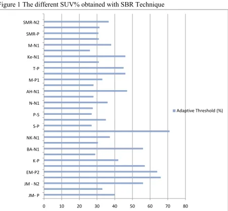
methods used are visual method, SUV 2.5 isocontour, 40% and 50% threshold of maximum tumor SUV (SUVmax), and an adaptive threshold based on the signal-to-background (S/B) ratio that was specific for each case. Adaptive threshold algorithms appears promising with regard to segmentation, and may reduce variability among radiation oncologists.(93) Comparison of PET-GTVs with CT-GTVs has limited value unless it is in the context of pathology and true disease. The investigators reported that PET was superior to CT for detecting primary tumors with a sensitivity of 94% and 82%, respectively, and superior for staging of the neck with a sensitivity of 90% and 67%, respectively.(94) To better understand the optimal segmentation for GTV delineation using PET data, investigators have correlated GTVs with pathologic specimens. No single SUV threshold gives a metabolic tumor volume that adequately captures pathologic tumor volume but the gradient-based volume p