EFFICACY, SAFETY AND COST EFFECTIVENESS OF ORAL
DOXOFYLLINE AND THEOPHYLLINE FOR MILD TO
MODERATE PERSISTENT BRONCHIAL ASTHMA: A
RANDOMIZED PROSPECTIVE OPEN LABELED
COMPARATIVE STUDY
Dissertation Submitted to
THE TAMILNADU DR. M.G.R. MEDICAL UNIVERSITY
In partial fulfilment of the
regulations for the award of the degree of
M.D. (PHARMACOLOGY)
BRANCH – VI
GOVT. CHENGALPATTU MEDICAL COLLEGE & HOSPITAL
THE TAMILNADU DR. M.G.R. MEDICAL UNIVERSITY
CHENNAI, INDIA.
CERTIFICATE
This to certify that this dissertation entitled, “Efficacy, safety and cost
effectiveness of oral Doxofylline and Theophylline for mild to moderate
persistent bronchial asthma: A randomized prospective open labeled comparative study’’ by the candidate Dr.M.Nandhini Priya for M.D
(Pharmacology) is a bonafide record of the research work done by her, under
the guidance of Dr.S.Purushothaman,MD., Professor, Department of Pharmacology, Chengalpattu Medical College, during the period of study
(2013-2016), in the Department of Pharmacology, Chengalpattu Medical
College, Chengalpattu - 603001. I also certify that this dissertation is the result
of the independent work on the part of the candidate.
Dr .K.Muthuraj, M.S.,
Dean
Chengalpattu Medical College Chengalpattu
Dr. K.Baskaran, M.D.,
CERTIFICATE
This is to certify that the dissertation entitled, “Efficacy, safety and
cost effectiveness of oral Doxofylline and Theophylline for mild to
moderate persistent bronchial asthma: A randomized prospective open labeled comparative study’’ submitted by the candidate Dr.M.Nandhini Priya
in partial fulfilment for the award of the degree of Doctor of Medicine in
Pharmacology by The Tamilnadu Dr.M.G.R. Medical University, Chennai is a
bonafide record of original work done by her under my guidance and
supervision in the Department of Pharmacology, Chengalpattu Medical
College, Chengalpattu during the academic year 2013-16.
Place: Chengalpattu
Date:
Dr.S.Purushothaman,MD.,
Professor,
Department of Pharmacology,
Chengalpattu Medical College,
DECLARATION
I Dr.M.Nandhini Priya, solemnly declare that the dissertation titled
“Efficacy, safety and cost effectiveness of oral Doxofylline and
Theophylline for mild to moderate persistent bronchial asthma: A randomized prospective open labeled comparative study’’ has been done
by me, in the Department of Pharmacology, Chengalpattu Medical College,
Chengalpattu under the guidance of Dr.S.Purushothaman,MD., Professor,
Department of Pharmacology, Chengalpattu Medical College, Chengalpattu.
This dissertation is submitted to The Tamilnadu Dr. M.G.R. Medical
University, Chennai, in partial fulfilment of the rules and regulations for the
award of M.D degree branch VI (pharmacology) to be held in April 2016.
I also declare that this bonafide work was not submitted by me on any previous
occasion for the award of any degree or diploma to any other university.
Place: Chengalpattu
Date:
Signature of the Candidate
ACKNOWLEDGEMENT
I express my sincere gratitude to Dr. K.Muthuraj.,M.S.,Dean, Chengalpattu Medical College, for permitting me to undertake this research
work as a part of my MD curriculum.
I would like to convey my gratitude to my guide Dr.S.Purushothaman
M.D., Professor, Department of Pharmacology, Chengalpattu Medical College
for his unfailing guidance, persuasion and constant support throughout the
study.
I sincerely thank Dr.K.Baskaran.,M.D., Professor and Head,
Department of Pharmacology, Chengalpattu Medical College who gave
encouragement and support to the study.
I am extremely thankful to Dr.R.Sivagami., M.D. Professor,
Department of Pharmacology, Chengalpattu Medical College for her valuable
support, guidance and genuine concern in my work.
I immensely thank Dr.B.Sharmila M.D., Professor, Department of
Pharmacology, Chengalpattu Medical College for her intense support and
I convey my gratitude to Dr.N.NaliniJayanthi,M.D., Professor
and Head, Department of Thoracic Medicine, Chengalpattu Medical College
for permitting me to carry out the study in the Thoracic Medicine OPD.
I express my sincere thanks to my Assistant Professors
Dr.T.Ragupathy, M.D., Dr.T.Siyamala Devi, M.D., K.Arumugasamy.M.Sc.,
Dr.B.Bhuvaneswari M.D., Dr.A.VinothKumar, M.D., Dr.R.Ranjini, M.D., and
Dr.K.Rani, D.G.O., Tutor, Department of Pharmacology, Chengalpattu
Medical College for their advice and encouragement.
I have great pleasure in thanking Mrs.Jenifer, Statistician, for helping
me in the statistical analysis. I thank my fellow post graduates
Dr.M.NithyaPriya, Dr.Sweetlin, Dr.SanuSain, Dr.G.Amutha, Dr.V.J.Sharmi for
their help.
I wish to place on record my gratitude to my parents and family
members for creating a congenial atmosphere and support.
Finally I thank all my patients for willingly submitting themselves with
CONTENTS
S.No Title Page number
1 Introduction 1
2 Aim & Objectives 3
3 Review of Literature 4
4 Materials and Methods 57
5 Results 63
6 Discussion 78
7 Conclusion and Summary 83
8 Bibliography
9 Annexures
Proforma
Asthma Control Test - Questionnaire in Tamil
Informed Consent
Informed Consent in Tamil
Patient Information Sheet
Patient Information Sheet in Tamil
Master Chart
LIST OF FIGURES
S.No Title Page
number
1 Triggers of asthma 10
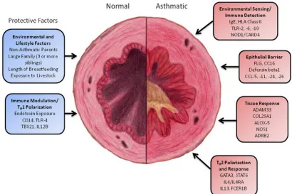
2 Pathogenesis of asthma 13
3 Neural networks in asthma 19
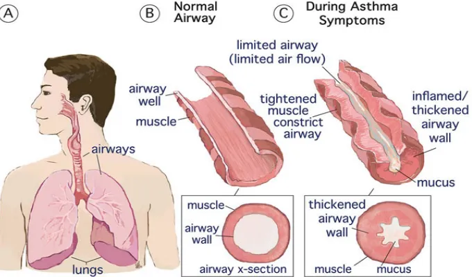
4 Airway remodeling 20
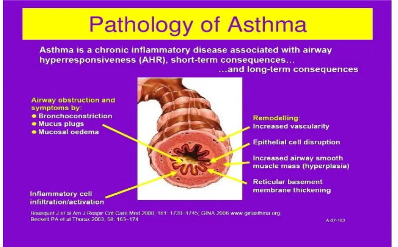
5 Pathology of asthma 22
6 Mucosa of the airway in asthmatics 23
7 Flow-chart for diagnosis of bronchial asthma 28
8 Control based asthma management cycle 39
9 Step wise approach to control asthma symptoms and
minimize future risk 41
10 Structure of methylxanthine and theophylline 42
11 Reversal of steroid resistance - activation of hdac by
theophylline 44
12 Cellular effects of theophylline 45
13 Structure of doxofylline 50
14 Structure of deriphylline 51
15 Gender distribution 63
16 Asthma control test questionnaire score 66
17 Subjective rating of asthma control 67
18 Comparison of forced vital capacity 68
19 Forced expiratory volume at the end of 1 second 70
20 Peak expiratory flow rate 71
21 Comparison of FEV1/FVC 72
22 Deriphylline group - adverse reactions among the
participants 74
23 Doxofylline group - adverse reactions among the
participants 74
24 Treatment cost 76
LIST OF TABLES
S:No Title Page
number
1 Characteristics of asthma, COPD and ACOS 27
2 Classification of bronchial asthma 29
3 Gender distribution 63
4 Comparison of age, BMI, family history, aggravating
factor 64
5 Asthma control test questionnaire score 66
6 ACT score - improvement from baseline 66
7 Subjective rating of asthma control 67
8 Subjective rating of asthma control- improvement from
baseline 68
9 Comparison of forced vital capacity 68
10 Comparison of forced vital capacity- improvement from
baseline 69
11 Forced expiratory volume at the end of 1 second 69
12 Forced expiratory volume at the end of 1 second-
improvement from baseline 70
13 Peak expiratory flow rate 71
14 Peak expiratory flow rate- improvement from baseline 71
15 Comparison of FEV1/FVC 72
16 Comparison of FEV1/FVC - improvement from baseline 73
17 Adverse reactions encountered among the participants 73
ABBREVIATIONS
Ach - Acetylcholine
ACOS - Asthma – COPD Overlap Syndrome
AUC - Area Under the Curve
cAMP - Cyclic Adenosine Mono Phosphate
CCR - Chemokine receptor
CGRP - Calcitonin Gene Related Peptide
COPD - Chronic Obstructive Pulmonary Disease
CTZ - Chemoreceptor Trigger Zone
Cyp450 - Cytochrome P 450
FEV1 - Forced Expiratory Volume in one second
FGF - Fibroblast Growth Factor
FVC - Forced Vital Capacity
GM – CSF - Granulocyte, Monocyte Colony Stimulating Factor
HDAC - Histone deacetylase
HLA - Human Leukocyte Antigen
ICS - Inhaled Corticosteroid
IgE - Immunoglobulin E
IGF - Insulin like Growth Factors
IL - Interleukin
KGF - Keratinocyte Growth Factor
LABA - Long Acting Bronchodilator
MMEF - Maximum Mid Expiratory Flow Rate
NANC - Nonadrenergic, noncholinergic nerves
NF-KB - Nuclear factor K Beta
NK - Neurokinin
PAF - Platelet activating factor
PDGF - Platelet Derived Growth Factor
PEFR - Peak Expiratory Flow Rate
PI3K - Phospo Inositol 3 Kinase
TGF - Transforming Growth Factors
ABSTRACT
AIM :
To compare the efficacy, safety and cost effectiveness of oral
Doxofylline and Theophylline for mild to moderate persistent bronchial asthma
patients.
MATERIALS AND METHODS:
A Randomized prospective, open labeled comparative study of 1 year
(Jul2014-Jun2015) duration was conducted in 186 patients who were attending
the thoracic medicine outpatient department of Chengalpattu Medical College
satisfying the inclusion and exclusion criteria after obtaining ethical clearance.
METHODOLOGY:
The study subjects were randomly allocated into two groups. Group 1
patients were treated with Doxofylline 400mg once daily and group 2 patients
were treated with Theophylline twice daily. Demographic data, history,
clinical examination and details of drug prescription by the treating physician
were recorded in the study proforma. Relevant lab investigations were done at
the beginning and at the end of the study. The patients were followed up for 12
weeks. The schedule of patient visit is as follows Visit 1 for initial or baseline
assessment and follow-up at 4, 8 & 12 weeks.
STATISTICAL ANALYSIS:
The data collected were analyzed using Student t test (two tailed,
independent) to find the significance of study parameters on continuous scale
between two groups. Chi-square/ Fisher Exact test was used to find the
significance of study parameters on categorical scale between two or more
RESULT:
Doxofylline was better than deriphylline in subjective parameters of
asthma control test questionnaire and subjective rating of asthma control.
Doxofylline had equal efficacy as that of deriphylline in spirometric parameters
(p ≤ 0.001). Doxofylline was significantly safe compared to deriphylline as
inferred from lesser incidence of adverse drug reactions. Adverse reactions are
encountered in 10% of doxofylline and 22% of deriphylline group.
Deriphylline was the cheaper and cost effective methylxanthene for the
treatment of bronchial asthma in developing countries at population level.
Doxofylline even though costlier had better safety profile with less adverse
reactions compared to deriphylline. It can be used as an individual based
approach in asthma management.
CONCLUSION:
Doxofylline is a newer methylxanthine with few adverse effects and
equal efficacy as compared with deriphylline. It is a better alternative in the
management of bronchial asthma.
KEY WORDS:
Bronchial Asthma, Methylxanthene, Doxofylline, Deriphylline,
1
INTRODUCTION
Asthma is a heterogeneous disease usually characterized by chronic
airway inflammation .It is defined by the history of respiratory symptoms such
as wheeze, shortness of breath, chest tightness and cough that vary over in
time and intensity, together with variable respiratory airflow limitation.1, 2
The prevalence of asthma is 1-18%1 in the world. It contributes 1% of the
total disease burden of the world3. Genetic factors, allergens, infection,
occupational exposure, smoking and air pollution trigger the development
of bronchial asthma.
Treatment of asthma is directed towards the control of symptoms and
bringing down the frequency of acute attacks with fewer side effects. It
also aims to minimize remodeling of airways.
The drugs available for asthma are categorized as controllers,
relievers and add on therapies. Controllers are used to bring down
inflammation in airways, decrease the symptoms and minimize acute attacks.
These include steroids by both inhalational and systemic route, leukotriene
receptor antagonist, methylxanthines and anti IgE antibody. Relievers are used to
relieve symptoms whenever needed. Relievers also play a role in preventing
exercise induced asthma. Short acting β2 agonists, anticholinergics in inhaled route, and theophylline are used as reliever medications. When the patient’s
2
used. Tiotropium, anti IgE antibody , Leukotriene receptor antagonist are
given as add on therapy.1, 2
Methylxanthines are phosphodiesterase inhibitors. Theophylline is a
widely used drug belonging to this group. It has bronchodilatory,
anti-inflammatory, mucoregulatory, immune modulatory steroid sparing properties. It
has a narrow therapeutic index with cardiac, gastrointestinal and CNS side
effects which contribute non adherence to treatment6, 7.
Doxofylline , another methylxanthine has fewer side effects and
bronchodilation comparable to theopylline. They provide better asthmatic
control and decrease the frequency of acute attacks7. As the prevention
and management of asthma depends more on pharmacotherapy, there is a
quest for bronchodilatory medications with few adverse effects.
Methylxanthines are the first line of drugs for management of mild to
moderate bronchial asthma as per Cochrane group7.
For choosing among the asthma treatment options at
population-level, pharmaco- economics plays an absolute role. Cost effectiveness
analysis considers the cost of medications, effectiveness of the treatment,
safety data and serves to adapt the best choice for the patients. This
study is focused to compare the efficacy, safety and cost effectiveness of
Doxofylline over Theophylline in mild to moderate persistent bronchial
Aims
Aims
Aims
Aims
and
and
and
and
Objectives
Objectives
Objectives
Objectives
3
AIM & OBJECTIVES
• To compare the efficacy of oral Doxofylline and Theophylline for
mild to moderate persistent bronchial asthma patients.
• To understand the safety profile of Doxofylline and Theophylline.
Review
Review
Review
Review
Of Literature
Of Literature
Of Literature
Of Literature
4
REVIEW OF LITERATURE
HISTORY:
The word "ASTHMA" is derived from Greek which means a condition
involving difficulty in breathing. British adapted "ASMA" from Greek which
means "ASTMA"and then the final form "ASTHMA" evolved.
It was Hippocrates (460-370Bc) who noticed the constructive effect of
"ASTHMA". According to him, the various causes for the onset of this disease
were climate, level of moisture and occupation. He compared it with epilepsy
and distinguished asthma by its nature of occurrence due to external influence.
Later Areatus (2-3 century) explained the two forms of asthma as
a breathing difficulty caused by activities such as running
a constructive breathing difficulty due to humidity and low temperature8.
During the 12th century, the Egyptian physicians recommended the
details of the patient’s physical status, diet, hygiene, environment, personal
attitude and behaviour pattern had to be known for better management of the
disease. It was strongly believed that the mental condition of the patient will
influence his physical well being9.
More prime facts were unfolded in further studies. JEAN BAPTISTE
VAN HELMONT (1577-1644) pointed out bronchi as the major throne of
5
fish intake8. Henry Hyde Salt, in his work, "On Asthma, its Pathology and
Treatment"(1860), discriminated asthma from dyspnoea. He described the
childhood asthma as a different form. The inhalation of dust, fur, hay etc. are
other contributors towards the disease9.
The definition of bronchial asthma given by the American Thoracic
Society, New York in 1862 states that asthma is a hyper responsiveness of
trachea and bronchi to multiple factors which result in airway narrowing of
variable severity either spontaneously or due to therapy.
It was in the year 1900, adrenaline was identified as the drug for asthma.
It relaxes smooth muscles of the respiratory tract. Regular use of adrenaline for
asthma management came into practice. During 1906, the view of etiology of
asthma as anaphylaxis and allergy was put forward.
From 1930, theophylline was used for asthma treatment. From 1967, the
management included short acting β 2 agonist. In 1968, SIR JOHN FLOYER,
through his work, "A TREATIES OF ASTHMA", brought forward the fact of
bronchoconstriction. He pointed out different types of asthma like continuous,
periodic and convulsive. He also stated that environmental factors provoke
asthma.
The discovery of sodium chromoglycate in 1968 made a new path in this
disease. Using inhaled steroids, anti-inflammatory effect was brought in the
6
substance, which played a role in mediating asthma. The discovery of
Leukotriene receptor antagonist in 1990 helped in treating chronic asthma9.
Omalizumab, anti-IgE antibody is a recombinant humanized monoclonal
antibody10. It interrupts the reaction between IgE and inflammatory mediators,
7
EPIDEMIOLOGY
MAGNITUDE OF THE DISEASE:
Asthma is a major health hazard which affects the people of all age
groups. The current asthmatic population is around 300 million worldwide3. In
some geographical region, the prevalence exceeds 10% in adults and 30% in
children. India harbours about 15-20 million asthmatics. The estimated
prevalence of asthma in India is around 4% - 5% in adults and 10%-15% among
pediatric population12, 13 .In Tamilnadu, the prevalence is around 4.84%13, 14
THE SOCIO-ECONOMIC BURDEN:
As the global prevalence of asthma has an increasing trend, the
socio-economic burden due to this illness is severe. It imparts a heavy blow on the
society by decreasing in productivity, absenteeism in the work place, increase in
hospitalisation, decrease in the longitivity of lives15. The direct and indirect costs
for asthma care is evaluated and found to be more than that of TB/HIV AIDS
management.
AETIOLOGY:
Asthma is multifunctional in origin .It arises from complex interaction of
genetic and environmental factors. The airway inflammation seems to occur
when genetically susceptible individuals are exposed to certain environmental
factors .Additional environmental determinants are the concurrent exposure to
8
THE FOLLOWING ARE THE RISK FACTORS:
GEOGRAPHICAL DISTRIBUTION:
Asthma is more widely distributed in the West compared to the other
parts of the world.
AGE AND SEX:
Asthma is more commonly diagnosed during infancy. Childhood asthma
occurs frequently in boys than girls; but in adults, the incidence reverses.
ETHINICITY:
The blacks have higher morbidity and mortality than whites due to
asthma.
SMOKING:
Asthma occurs more often in children born to mothers who have smoking
habit.
AIR POLLUTION:
Global warming increased concentration of harmful gases in air.
Occupational exposure contributes to the rise in asthma prevalence.
ATOPY AND ALLERGENS:
Atopy is a major risk factor .Exposure to allergens in the early age of life
is associated with higher incidence of asthma. Family history of any form
9
INFECTIONS AND INFESTATIONS:
Few bacteria, viruses and parasites impart an increase in serum IgE level.
(e.g.) Respiratory syncytial virus, Measles virus, Vaccine against
pertussis21.
DIET
Occurrence of allergy in the early life is reduced when the baby is
breastfed. Obese individuals are more prone for asthma. Omega 3 fatty
acid in the diet and antioxidants reduce the risk of asthma.
Lot of stimuli, both constitutional and environmental are involved in
bronchial asthma triggering.
The common triggers are the following:
Allergens
Drugs
Occupational exposure
Smoking
Infections
Stress
10
Figure 1: Triggers Of Asthma
ALLERGENS:
The following are the array of allergens which trigger the asthmatic
attack: house dust mites, are found in high concentration in carpets, soft
furnishings and bedding. Pet derived allergens are widespread in homes where
dogs, cats or cattle are kept. Feathers also induce a kind of allergen. Fungal
spores and antigens from cockroaches play an immense role in spreading
allergens19.
DRUGS:
Beta blocking drugs can induce bronchoconstriction in asthmatics. A
11
salicylates or non-steroidal anti-inflammatory drugs. These drugs block
arachidonic acid metabolism down the prostaglandin pathway diverting it to the
leukotriene pathway. Verapamil, piperazine, cimetidine also trigger asthma.
OCCUPATIONAL EXPOSURE:
Many agents encountered in the work place may induce asthma. The
compounds include isocyanates, epoxyresins, persulphates, hardwood dusts,
grain dust etc,.
ENVIRONMENTAL AND AIR POLLUTION:
Increasing prevalence of asthma is attributable to atmospheric pollution
which plays a role in triggering exacerbations of pre-existing of asthma.
Nitrogen dioxide, ozone, sulphur dioxide and airborne particulates have acute
adverse effects on asthma during air pollution episodes20. Motor vehicle
emissions, power stations, fuel burning industries, gas cookers, kerosene heaters,
burning of fossil fuels, domestic coal burning, block smoke, pollens, allergens
are particularly associated with asthma.
SMOKING:
Cigarette smoking is associated with increased levels of Ig E and with
increased sensitization to certain occupational allergens in particular. Maternal
smoking during pregnancy increases the risk of developing atopic disease in
infancy. Passive exposure to cigarette smoke at home has an adverse effect on
12
INFECTIONS:
Many respiratory infections like influenzaA, mycoplasma pneumonia,
Chlamydia pneumonia, respiratory syncytial virus etc. provide a transient
increase in airway responsiveness in normal individuals and in asthmatics21, 22.
EXCERSIE INDUCED:
Hyperventilation during exercise along with respiratory heat exchange
and water loss provoke the occurrence of bronchoconstriction and bronchial
asthma. The bronchoconstriction due to exercise depends on the strength of the
stimulus and also on the quality of nonspecific airway responsiveness23.
STRESS:
The emotional and cognitive factors induce the T helper cytokines. When
the individual is provoked by a stimuli, inflammatory response is generated
resulting in prolonged duration and more frequent severe asthmatic attack. The
other mechanism suggested is vagal mediated response promoting
bronchoconstriction.
CO-EXISTING DISORDERS:
There are few conditions which occur along with asthma. Management of
these disorders are essential for better control of asthma.
1. RHINITIS AND SINUSITIS:
More than 50% of the asthmatics have co-existing rhinitis and sinusitis.
13
2. GASTRO-OESOPHAGEAL REFLUX DISEASE:
GERD occurs in about 75% of asthma patients. The mechanism
implicated are bronchoconstriction as a result of lower esophageal sensory
stimulation and micro aspiration27. Treatment of GERD is essential.
3. OBESITY:
Obesity triggers the expression of inflammatory mediators responsible for
airway constriction. Obesity is directly proportional to the rate of asthma
occurrence28.
[image:30.595.106.516.420.693.2]PATHOGENESIS OFASTHMA:
14
Asthma is a spectrum of disorders with airway inflammation along with
hyper responsiveness resulting in airway obstruction. Proper understanding of its
mechanism of development is necessary for its management.
GENETICS OF ATOPY AND ASTHMA:
There is strong evidence for the hereditary contribution to the etiology of
asthma24. Asthma and atopy run in families. First degree relatives of asthmatics
have a significantly higher prevalence of asthma than the relatives of non
asthmatic patients.
Atopy is a constitutional tendency to produce significant amount of IgE
on exposure to small antigens. Atopic individuals demonstrate positive reactions
to antigens on skin prick test and have a high prevalence of asthma, allergic
rhinitis, utricaria and eczema.
The genetic contribution to asthma is complex involving polygenic
inheritance and genetic heterogeneity.
Asthma has an autosomal recessive inheritance with dominant and
co-dominant expression. The below mentioned are some suggested polymorphisms
seen in the genes of asthma patients: chromosome 5 has genes representing IgE,
Interleukin 3, interleukin 4, interleukin 5, interleukin 9, interleukin 13 and
15
Chromosome 2 encodes the atopy gene which is represented by FcER 1
region. FcER 1 is present on basophils, mast cells and dendritic cells. Its
function is to increase the uptake of antigen and its expression.
Chromosome 12 provides a connection between asthma and atopy by the
way of gamma interferon and nitric oxide production.
Chromosome 6 harbours the gene encoding HLA class II and cytokine
TNF α. These are involved in the susceptibility and severity of asthma.
Chromosome 14 and 7 encodes the T cell receptor proteins which
influence the IgE function18.
BRONCHIAL HYPERRESONSIVENESS:
Most of the symptoms and signs of asthma are due to bronchial hyper
responsiveness. There is an altered response in asthma patients compared with
normal subjects to the external allergens. This response can be measured with
the help of bronchial provocation test. There is a direct link among the severity
of the disease, the need of the drugs and provocation concentration. This test
measures the only one important component of asthma and serves as a tool for
16
INFLAMATORY CELLS EXPRESSED IN ASTHMA:
The range of cells associated with asthma include not only mast cells,
eosinophils, cytokines, T – lymphocytes but also structural cells like epithelial
cells, endothelial cells, smooth muscle cells and fibroblasts32.
MAST CELLS:
Mast cells are seen on entire respiratory tract. They are clustered in the
sub mucosal and epithelial surface of the bronchi. Mast cells release a cascade
of preformed mediators as well as produce new mediators in a time interval of
30 minutes. Histamine, mast cells proteases particularly tryptase, newly formed
mediators like prostaglandin D2, leukotriens, cytokines IL 4, IL5, IL6 and
TNF α which are contributed by mast cells form the central mechanism34.
BASOPHILS:
The only other cells to secrete histamine are basophils. Basophils can be
triggered not only by Ig E mediated factors but also other substances such as IL
1, IL -3, IL – 8, RANTES, PAF etc. Their sensitivity to anti IgE is 100 times
higher than that of mast cells. Basophils produce LTC 4, and they contribute
more to the allergies occurring in skin32.
EOSINOPHILS:
Eosinophilia particularly in the airway mucosa is linked with the
diagnosis of asthma. Their count correlates with the disease activity. The
granules present in the eosinophils such as eosinophil cationic protein, major
17
the cytotoxic action of eosinophils. Eosinophils are the generators of oxygen
free radicals, sulphidopentide, leukotrienes lineage of cytokines which induce
bronchospasms and inflammation36.
NEUTROPHILS:
Infiltration of neutrophils in the airways are observed in patients having
severe asthma and those exposed to ozone and sulphur dioxide and also smokers.
Their role in causing asthma or contributing to the disease severity is not clear19.
MONOCYTES AND MACROPHAGES
These are the major cell types present in bronchial lumen. They have low
affinity IgE Fc receptors which contribute to the release of mediators. They
have significant role in atopic asthma.38
DENDRITIC CELLS
Dendritic cells have MHC class II molecules and they serve as prime
antigen presenting cells in the airways. They induce T- Cell differentiation and
Th 2 cytokine formation and the cascade of inflammatory events occur.37
LYMPHOCYTES
There are more number of T lymphocytes in asthma than B lymphocytes.
Ig E mediated cellular activities in asthma rely upon T – cell activation. The
activated T cells also regulate mast cells, eosinophils, epithelial cells and
fibroblasts. The T lymphocytes are divided into CD4+ CD8+ Cells. The CD 4 +
18
The Th 2 cells play a vital role in triggering IgE mediated inflammatory
reactions, while Th1 inhibit IgE and have an opposite effect.33
AIRWAY SMOOTH MUSCLE CELLS
The smooth muscle cells of air passages are contracted in response to
inflammatory mediators. The other events that occur as a consequence of
inflammation are activation, proliferation, hypertrophy of the smooth muscle
cells.34
EPITHELIAL CELLS
The epithelial cells participate in the inflammatory process and this
induces damage to the epithelium. The repair of this event which is abnormal in
the most of the asthmatic individuals, further contribute to the obstruction of
airways.39
NEURAL MECHANISM OF ASTHMA
There exists an inter relationship between the inflammatory pathways
and the neuronal aspects of airway function. The mediators of inflammation
modulate the effect of neural tissues. On the other hand, the neuro transmitters
19
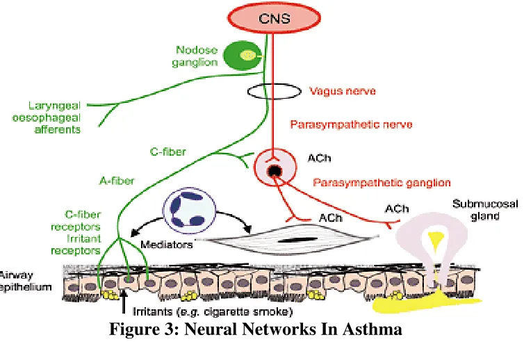
Figure 3: Neural Networks In Asthma
Cholinergic, adrenergic, NANC neural networks exert their control over airways.
The cholinergic control of respiration is exerted through:
1. Increased vagal tone
2. Triggering of airway sensory receptors resulting in reflex
bronchoconstriction.
3. Enhanced acetylcholine release from parasympathetic ganglia as well as
post ganglionic neurons.
Adrenergic system exerts its effects through sympathetic nerves via α and
β receptors. These nerves indirectly control cholinergic transmission through the
prejunctional α or β receptors. Adrenergic influence is vital in exercise induced
asthma and when asthma is triggered by cold air. The non adrenergic, non
cholinergic nerves (NANC) are believed to release vasoactive intestinal peptide
and nitric oxide. These neuro transmitters counteract the bronchial smooth
20
inflammatory cells release superoxide which degrade VIP. This contributes to
increased bronchial contraction.29, 31
AIRWAY NEUROPEPTIDES:
The sensory nerve fibres (C) which are unmyelinated are present in the
airway.
They harbour various neuropeptides like substance P, neurokinin (NK),
calcitonin gene related peptide (CGRP)
Due to the damage of the airway epithelium in asthma, the sensory nerve
endings are exposed which may trigger the release of this neuro transmitters and
result in the airway inflammation.32
AIRWAY REMODELLING:
Due to chronic inflammation and structural changes occur in the airway
[image:37.595.150.490.527.727.2]of asthmatics, these changes are referred as airway remodelling.
21
GROWTH FACTORS IMPLICATED IN AIRWAY REMODELLING:
The damaged airway epithelial cells produce many growth factors such as
transforming growth factors (TGF) α, β endothelin (ET). Insulin like growth
factor (IGF) basic fibroblast growth factor b (FGF), platelet derived growth
factor (PDGF).The activated fibroblast release connective tissue growth factor
(CTDGF), vascular endothelial growth factor (VEGF), Keratinocyte growth
factor (KGF) etc. which play a role in airway remodelling.31
CONSEQUENCE OF AIRWAY REMODELLING:
Airway remodelling, which involves all the layers of airway wall results
in the following modifications:
1. Increasing in sloughing of the epithelium, leading to loss of integrity.
2. Exposure of the nerve endings.
3. Hypertrophy and hyperplasia of airway smooth muscles.
4. Destruction of Elastic tissues.
5. Increase in the number of blood vessels in the mucosa.
6. Micro vascular leakage.
7. Sub mucosal edema of the airways.
22
PATHOLOGY
The main pathology involved in asthma is airway thickening and
remodelling which leads to narrowing of the airway lumen.30 Apart from
constriction of airways, bronchial congestion and edema are seen in the
asthmatic. In chronic asthma there is inflammatory cells infiltration.
Eosinophilia is consistently seen in asthmatic which is triggered by T – helper
cells (CD4+). The role of mast cells are evident in immediate type of
[image:39.595.116.515.326.574.2]hypersensitivity reaction in asthmatics.34
Figure 5: Pathology Of Asthma.
SPUTUM & BRONCHIO ALVEOLAR LAVAGE
The sputum and BAL in the asthma patient have the following features:
1. Cork screw shaped twists of condensed mucus ( Curschmann’s spirals)
23
3. Eosinophils, metachromatic cells admixed with granule membrane
lysophospholipase (Charcot – Leyden crystals)
4. Epithelial cells sloughing, eosinophilic cationic protein, major basic
protein.30
AIRWAY PLUGGING
These are the result of mucus production by enlarged sub mucosal glands,
mixed with inflammatory exudate, arranged in concentric lamellae. These plugs
block the lumen of airways in asthmatics.
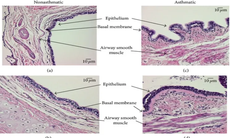
HISTOLOGICAL APPEARANCE
The mucosa of the airway in asthmatics show loss of integrity of
epithelial surface, thickening of the basement membrane, mucosal infiltration
with eosinophils, lymphocytes, smooth muscle hypertrophy. There is also
[image:40.595.133.505.516.739.2]dilatation of bronchial mucosal blood vessels as well as angiogenesis.32
24
DIAGNOSIS
HISTORY
• Abrupt onset of symptoms • Variable in time and intensity • More at Night
• Exaggerated by laughing, walking, allergens and low temperature • Exacerbate with respiratory infection
• Changes in symptoms according to seasons • Family history asthma, allergy or atopy.
SYMPTOMS
The symptoms vary with time, intensity or disappear during symptom free
intervals.
• Wheeze
• Chest tightness
• Cough
• Shortness of breath.
FINDINGS ON PHYSICAL EXAMINATIONS
• Breath sounds with prolonged expiration • Increased respiratory rate
• Orthopnoea • Chest constriction
25
PULMONARY FUNCTION TEST
1. SPIROMETRY
Spirometry is the measure of airflow and lung volumes during a forced
expiratory manoeuvre from full inspiration. It is fundamental to the diagnosis
and assessment of airway disease. The measurements made during spirometry
are referred as `dynamic lung volumes’. Spirometry provides three basic
measurements.
• Forced vital capacity
• The forced expiratory volume in one second. • The ratio of FEV1/FVC.
Airway obstruction is established by a decrease in FEV, out of proportion
to the decrease in vital capacity (VC).This imparts a reduction in FEV1/FVC
ratio. The maximum mid expiratory flow (MMEF) represents the airflow in the
medium and small airways (FEF 25-75%).It may indicate mild airway disease.
Confirmation of asthma in spirometry is by establishing
bronchoconstriction as evidenced by FEV1/FVC<70% and an improvement of
>15% or 200ml in FEV1, 15 min after giving short acting beta 2 agonist by
26
PEAK EXPIRATORY FLOW
This can be measured during spirometry or independently using a peak
flow meter. Peak flow meter indicates only the greatest expiratory flow. It is
used for monitoring the treatment. The two largest repeated measurements with
variation within 5% is acceptable. The PEFR measurements are evaluated by
comparing with the same person’s best value, which is obtained during symptom
free period. It has diurnal variation, being lowest in early morning.
The meters used by the patients at home have a zonal system with colour
code for better interpretation. The PEFR of 80%-100% of the patient’s best
value is indicated by green zone, 60%-80% by yellow zone, below 60% by red
zone. Red zone warns immediate medical care.1
LUNG VOLUMES
The changes occuring in lung volumes and capacities of the asthmatic
patients are:
1. Increased total lung volume
2. Increased functional residual capacity
BRONCHIAL PROVOCATION TEST:
This is used in adult patients who do not have limitation of airflow at the
time of assessment. Airway hyper responsiveness in such patients is
demonstrated by bronchial provocation with methacholine or histamine. A
27
specificity of this test is low as it can give false positive results in patients with
other diseases.40,42
TYPICAL CHARACTERISTICS OF ASTHMA, COPD, AND ACOS
(Asthma-COPD overlap syndrome) 1
FEATURE ASTHMA COPD ACOS
Age Any age Early onset Rare < 35 yrs.
Usually >40 year History of symptoms
present at early age.
Occurrence Variable Aggravating factors present Persistent symptoms Prolonged course Persistent symptoms Prominent variability Lung function Variable airflow limitation, normal between symptoms Post bronchodilator
FEV1/FVC < 0.7 Not fully reversible
History Mostly family/ personal history of allergy/ asthma present History of exposure to noxious substance present
History of asthma, exposure to noxious
substance present
Disease course
Improve after
treatment Progressive
Improve after treatment, Progressive
Chest X ray Usually within normal limits
Changes suggestive
of COPD present Same as COPD
Airway pathology Presence of eosinophils and/or neutrophils Neutrophils in sputum, may have
systemic inflammation
[image:44.595.96.533.204.690.2]Eosinophils and/or neutrophils in sputum
FLOW-CHART FOR DIAGNOSIS OF BRONCHIAL ASTHMA
Figure:7 Flow
Patient with following symptoms either intermittent or persistant:
Elaborate history and thorough physical examination
Do spirometry/PEFR with reversibility test
FEV1≤80% of
normal
FEV1/VC≥70%
Suspect
restrictive lung
disease
Specific
diagnostic
testing
28CHART FOR DIAGNOSIS OF BRONCHIAL ASTHMA
Flow-chart For Diagnosis of Bronchial A
Patient with following symptoms either intermittent or persistant:
1. Shortness of breath
2. Wheeze
3. Chest tightness
4. Cough
Elaborate history and thorough physical examination
Do spirometry/PEFR with reversibility test
FEV1≥15%
Reversibility test
FEV1/VC<70%
FEV1>80%of
FEV1/VC
Bronchial
hyperactivity and
or peak flow
variability
ASTHMA
CHART FOR DIAGNOSIS OF BRONCHIAL ASTHMA42, 1Asthma
Patient with following symptoms either intermittent or persistant:
Elaborate history and thorough physical examination
FEV1>80%of
normal
FEV1/VC≥70%
Bronchial
hyperactivity and
or peak flow
variability
29
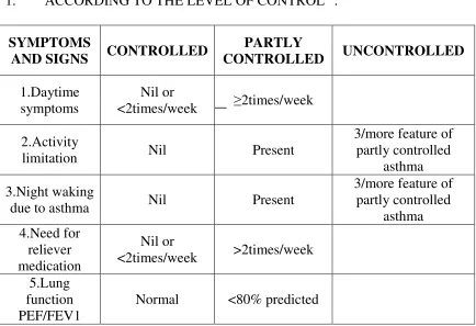
CLASSIFICATION OF BRONCHIAL ASTHMA:
1. ACCORDING TO THE LEVEL OF CONTROL42:
SYMPTOMS
AND SIGNS CONTROLLED
PARTLY
CONTROLLED UNCONTROLLED
1.Daytime symptoms
Nil or
<2times/week ≥2times/week
2.Activity
limitation Nil Present
3/more feature of partly controlled
asthma
3.Night waking
due to asthma Nil Present
3/more feature of partly controlled asthma 4.Need for reliever medication Nil or
<2times/week >2times/week
5.Lung function PEF/FEV1
[image:46.595.96.530.109.406.2]Normal <80% predicted
Table 2: Classification Of Bronchial Asthma
2. IN ACCORDANCE WITH THE CLINICAL SEVERITY:
(i). MILD ASTHMA
It is a well controlled asthma. It is treated with reliever medications or
with low intensity controller medications as and when needed basis. The
spirometry value of FEV1 is 60-80% of the predicted value.
(ii) MODERATE ASTHMA
These patients also have well controlled symptoms but with low dose
ICS/LABA. Moderate asthma correlates with the FEV1 of 40-60% of the
30
(iii) SEVERE ASTHMA
Patients in this state, need high dose ICS/LABA to control the symptoms.
It may remain uncontrolled even with this treatment.
In severe obstruction, the FEV1 value will be <40% of the predicted.40
PRIMARY PREVENTION
Focus is laid on environmental triggers of asthma and its evidence.
Priority to the reduction of exposure to occupational allergens helps in
decreasing the incidence of asthma. The main of aim primary prevention is to
protect the children from developing asthma. Few ways of achieving this are
avoidance of maternal smoking, promoting breast feeding and avoidance of
allergens. For obese individuals weight loss is advised.41, 43
ASTHMA - INFORMATION EDUCATION AND COMMUNICATION:
Effective asthma management depends on the patients and parents of
paediatric patients’ knowledge and understanding of asthma. Health education
should begin as early as the diagnosis is made and should continue at each visit
to hospital.
Healthcare providers should impart knowledge about the multifactorial
nature of the disease and teach the patients about the practical skills needed for
31
Importance has to be laid on the identification and avoidance of precipitating
factors, adherence to treatment and understanding the difference between
reliever and preventer drugs.
Special attention is given for the technique of using inhaler devices and
peak flow meter. They are educated to appreciate the day to day variability in
peak expiratory flow rate and its role in recognising the influence of
precipitating factors, their effect on disease process and in monitoring the
treatment.
The healthcare provider should have good communication skill. Then
only the needed information can reach to the patient. For this they should have a
friendly nature and allow the patients to express their problems, beliefs and
concerns. Besides being attentive, they have to impart appropriate information,
reassure and encourage the patients. Hence good communication has a great
impact on health literacy. The information should be in simple words with
illustrations, pictures, drawing, etc. Patient’s understanding can be confirmed by
asking to repeat the important points. It means the patients clear understanding
and the freedom to put forward their doubt.1,42
AVOIDANCE OF PROVOKING FACTORS:
Indoor allergens like house dust mite prevalence can be reduced by
32
Exposure to pet allergens should be avoided. Cigarette smoking both
active and passive has to be abstained. Occupations and hobbies that provoke
asthma have to be evaded as well. Ingested food additives, food allergens which
trigger asthma have also to be refrained. Furthermore, medications which cause
bronchoconstriction like beta blockers, aspirin etc. are to be avoided.1, 42
PHARMACOTHERAPY OF BRONCHIAL ASTHMA
Various means of bronchial asthma treatment are:
1. To decrease airway inflammation and hyper responsiveness.
2. To prevent the bronchoconstriction effect of vagal nerve
• Anti cholinergics
3. Dilatation of airway smooth muscles-
• Sympathomimetics • Methylxanthines
4. Blocking the release of anti-inflammatory mediators-
• Mast cell stabilizers
5. Antagonising the effect of already released mediators of inflammation
• Leukotriene antagonists • Antihistamines
• PAF antagonists
6. Blocking the circulating IgE-
33
ANTIASTHMATIC DRUG CLASSIFICATION
1. BRONCHODILATORS
• Agonists of beta2 adrenergic receptors • Anticholinergics
• Methylxanthines
2.LEUKOTRIENE RECEPTOR ANTAGONISTS
• Montelukast • Zafirlukast
• Lipoxygenase inhibitor-zileuton
3. PREVENT DEGRANULATION OF MAST CELLS
• Sodium chromoglycate • Ketotifen
4. CORTICOSTEROIDS
• Inhaled route: beclomethasone, dipropionate, budesonide,
fluticasone, Propionate, flunisolide, ciclesonide
• Systemic route: Hydrocortisone, Prednisolone etc. (42)
The use of above mentioned drugs fall under two categories:
The ‘relievers’ and the ‘controllers’. The relievers are bronchodilator
drugs used on need basis. Controllers are anti-inflammatory drugs taken in daily
34
BETA 2 ADRENORECEPTOR AGONISTS:
These act on the beta2 receptors on bronchial smooth muscle resulting in
bronchodilation. Beta2 agonists stimulate the G protein coupled receptors that
result in activation of adenylcyclase and finally increase cAMP which cause
smooth muscle relaxation. cAMP also decreases mediator release from mast
cells. These agents inhibit microvascular leakage and increase mucociliary
transport by increasing ciliary activity. 44
Selective beta2 agonists are preferred agents for bronchial asthma. By
inhalation route they are the fastest acting drug.
Salbutamol, pirbuterol and terbutaline are faster acting drugs by
inhalational route. So they are used for aborting an attack of acute asthma.
Owing to their short duration of action, they are not suitable for prophylaxis.
Salmeterol, formoterol, carmoterol, indacterol are long acting beta2
agonists. They are useful for prophylaxis of bronchial asthma.
Adverse reactions:
Prolonged use may lead to down regulation of beta receptors contributing
to tolerance. Tremors, palpitation, decrease serum potassium concentration,
35
METHYLXANTHINES:
This group includes caffeine, theophylline and theobromine.
Methylxanthines act by blocking the adenosine receptor, increase cAMP,
inhibition of phosphodiesterase enzyme. At higher doses, these drugs release
calcium from sarcoplasmic reticulum in skeletal and cardiac muscles.
When given orally they have rapid absorption. Metabolism is via cyp450
isoenzymes in liver. This paves way for a multitude of drug interactions.6
Side effects:
As these drugs are CNS stimulant at toxic doses they result in tremors,
delirium and convulsions. Vomiting occurs due to gastric irritation and CTZ
stimulation. More over theophylline has a narrow therapeutic index which plays
a central role in causing toxic symptoms. 6
ANTICHOLINERGICS:
Cholinergic blockers are effective only if bronchoconstriction is due to
cholinergic activity. These drugs cause mainly dilatation of large airways. These
are less efficacious and slower acting bronchodilators than the
sympathomimetics. 6These drugs are more effective for COPD than bronchial
asthma.
Ipratropium and tiotropium act as competitive antagonists of Ach
receptors on bronchial smooth muscle. They act through M3 receptors which
36
duration of action of Ipratropium is 4-6hrs while that of tiotropium is 24hrs.Both
are ionic drugs that are poorly absorbed.6, 29
Side effects:
Dryness of mouth, nervousness, pharyngitis, headache, palpitation.
CORTICOSTEROIDS:
These drugs have potent anti-inflammatory and also decrease bronchial
hyperactivity and mucosal edema. Anti-inflammatory action is due to decreased
recruitment of inflammatory cells as well as decreased production of PG and LT.
They block the mediators generated by arachidonic acid pathway.
Glucocorticoid bind to cytoplasmic receptors and the complex is
transferred to the nucleus. These GR-drug complex binds to the nuclear response
element present in the DNA. This results in up regulation transcription of
anti-inflammatory factors. 45
The primary indication of inhaled steroids is to decrease the inflammatory
process. These drugs are used as maintenance therapy of chronic persistent
asthma. Beclomethasone, dipropionate, Triamcinolone, Fluticasone, Budesonide,
Mometasone are some of the agents used in inhalational route. Inhaled steroids
are combined with long acting beta2 agonists and used as controller medication
37
Side effects:
The systemic therapy with glucocorticoids cause abnormalities in
metabolism of carbohydrates, lipids, cushingoid features with salt and water
retention, alteration in mood, osteoporosis, hypertension, cataract,
immunosuppression etc. 46 Local effects like oropharyngeal candidiasis,
dysphonia, pharyngitis are seen with inhaled steroid use.47
CROMOLYN LIKE DRUGS:
Sodium chromoglycate and nedocromil are the most common
nonsteroidal antiasthmatic drugs used. They prevent degranulation of mast cells
in response to allergic-non allergic stimuli .They are indicated only for
prophylaxis of asthma. When used as inhalational agents they prevent
bronchospasm and the cascade of further mediator release. Ketotifen has anti
histaminic action apart from mast cell stabilizing property and is specially
indicated for patients with multiple disorders like atopic dermatitis, perennial
rhinitis, conjunctivitis etc. chromoglycan and nedocromil are often used in
pediatric population as alternatives to inhale corticosteroids because of their
safety profiles.45
Adverse reactions:
Local reactions like pharyngeal irritation, cough, reflex bronchospasm are
38
LIPOOXYGENASE INHIBITOR:
Zileuton inhibits the synthesis of LTB4, LTC4, LTD4 from arachidonic
acid. This results in blocking of chemotaxis and bronchoconstriction. This drug
has short duration of action. It is used as controller in the management of
bronchial asthma. 50
Adverse reaction:
Nonspecific pain, headache, acid peptic disorder, elevated liver enzymes
and hepatotoxicity.51
LEUKOTRIENE RECEPTOR ANTAGONIST:
Montelukast and zafirlukast inhibit the bronchoconstricting action of
LTs at cys LT1 receptor.51They are selective antagonists of leukotriene receptors
LTD4 and LTE4. By this action they inhibit bronchoconstriction, mucus
secretion, vascular permeability and plasma exudation into the air way.
Antileukotrienes are indicated in prevention of exercise, cold air, allergens and
aspirin induced asthma.51
Adverse reactions:
Headache, nausea, abdominal pain, occasional elevation of liver enzymes
are reported. Few cases of Churg Strauss syndrome is associated with their
39
MONOCLONAL ANTIBODY-OMALIZUMAB:
Omalizumab is a monoclonal antibody against IgE. It blocks the reaction
between IgE and mast cell / basophils .This inhibits the release of inflammatory
mediators. It is indicated to prevent the attack of bronchial asthma in patients not
responding to combination of long acting beta 2 agonist and a high dose of
inhalational steroid. It is administered by subcutaneous route.
Adverse reactions:
Injection site reactions, viral, respiratory tract infections, headache,
sinusitis and pharyngitis. 52
MANAGEMENT OF ASTHMA
[image:56.595.224.391.439.602.2]BASED ON PATIENT’S CONTROL OF ASTHMA SYMPTOMS:
Figure 8: Control based asthma management cycle
Symptoms control serves as a tool for planning asthma management. The
exceptions are severe asthmatics whose symptoms do not correlate with
Assess
Adjust
treatment
review
40
responses or exacerbations .This approach of asthma management takes into
account not only symptom control but also the future risk reduction. 42
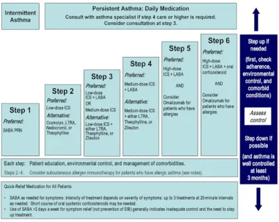
STEPPING DOWN AND STEPPING UP ASTHMA TREATMENT
Steps in asthma management
STEP1: Using reliever medication as and when needed basis.
STEP2: Continuous use of low dose controller medication added to step 1.
STEP3: Use of one or two controller medications along with step 1.
STEP4: Use two or more controller medications along with step 1.
STEP5: Tertiary care and / or add on medications.
Asthma patients should be followed up regularly and assessed for asthma
control, adherence to medications, inhaler techniques etc.
If they do not respond to treatment then these step up treatment has to be
given. If the asthma symptoms are well controlled and the lung function is stable
41
Figure 9: Step wise approach to control asthma symptoms and minimize future risk
DRUGS USED IN PRESENT STUDY
METHYLXANTHINE GROUP OF DRUGS:
Methylxanthine is present in beverages like tea, coffee, cocoa. The CNS
stimulant effect of these are attributed to methylxanthines. The major chemicals
present are theophylline, theobromine and caffeine.
CHEMICAL STRUCTURE:
Theophylline is 1,3-dimethylxanthine
Caffeine is 1, 3, 7-trimethylxanthine
[image:59.595.168.474.99.250.2]1-Methylxanthine
Figure 10: Structure Of Methylxanthine And Theophylline
MOLECULAR BASIS
1. Phosphodiesterase inhibition
2. Antagonism at adenosine A
3. Inhibition of nuclear factor K
4. Inhibition of phospho
5. Increased interleukin 10 secretion
6. Enhanced apoptosis of inflammatory cells
7. Decreased expression of ADP
death.
8. Increased histone deacetylase activity
PDE INHIBITION:
Theophylline is a nonselective inhibitor of phosphodiesterase. It acts on
cyclic nucleotides, increases intracellular ade
cyclic 3’5’ guanosine monophosphate (GMP) levels. Higher expression of
42
trimethylxanthine
Methylxanthine Theophylline
Structure Of Methylxanthine And Theophylline
MOLECULAR BASIS OF THEOPHYLLINE ACTION:
Phosphodiesterase inhibition.
Antagonism at adenosine A, A2A, A2B receptors.
Inhibition of nuclear factor K-β.
phospho-inositol-3 kinase δ.
Increased interleukin 10 secretion.
Enhanced apoptosis of inflammatory cells.
Decreased expression of ADP-ribose polymerase 1 which inhibits cell
ed histone deacetylase activity.
Theophylline is a nonselective inhibitor of phosphodiesterase. It acts on
cyclic nucleotides, increases intracellular adenosine monophosphate (AMP) and
cyclic 3’5’ guanosine monophosphate (GMP) levels. Higher expression of Theophylline
Structure Of Methylxanthine And Theophylline
ribose polymerase 1 which inhibits cell
Theophylline is a nonselective inhibitor of phosphodiesterase. It acts on
nosine monophosphate (AMP) and
43
isoenzymes of PDE are encountered in bronchial asthma patients owing to its
chronic inflammatory process or as a treatment outcome.54
ADENOSINE ANTAGONIST:
Theophylline inhibits A1 and A2 subtypes of adenosine receptors and is
less potent against A3 receptors. It antagonises the bronchoconstricting effects of
adenosine. 55
INTERLEUKIN 10 RELEASE:
Interleukin 10 plays major role in anti-inflammatory actions. IL-10
concentrations are low in diseased states like asthma and COPD. IL-10 secretion
enhanced by theophylline .56
EFFECTS OF GENE TRANSCRIPTION:
It decreases the expression of genes responsible for inflammation in
asthma. This effect is mediated by blocking the translocation of nuclear factor
KB(NF-KB)which is a proinflammatory transcription factor into the nucleus.57
EFFECTS ON KINASES:
Theophylline has direct inhibitory action on phosphoinositol 3 kinase,
particularly the subtype P13K (P110) δ. This enzyme takes part in oxidative
stress response. This property contributes to the theophylline’s reversal action of
corticosteroid resistance. This plays a major role in the management of severe
44
EFFECTS ON APOPTOSIS:
Theophylline has the property of reducing apoptosis by virtue of its action
on Bcl-2, an antiapoptic protein. This contributes to the inhibition of apoptosis in
neutrophils.59
HISTONE DEACETYLASE ACTIVATION:
Acetylated and deacetylated state of histone determines the inflammatory
gene expression. Bronchial asthma patients have increased expression of
inflammatory genes as a result of histone acetylation and transcription.
This phenomenon is prevented by histone deacetylases (HDAC) which
acts on the promoter site present in the nucleus. Corticosteroids inhibit the
inflammatory process by the activation of histone deacetylase, thus suppressing
the expression of inflammatory genes. (61) This process is defective in COPD
patients causing reduced expression of HDAC2 and contributing to steroid
resistance in COPD. This defect is also encountered in severe asthmatic patients
[image:61.595.124.503.549.714.2]who have the habit of smoking. 62
45
Theophylline enhances the action of HDAC5 and improve the
anti-inflammatory effect of corticosteroids. It also serves to reduce the resistance to
steroids in COPD patients. 63 This is encountered at low blood levels and during
oxidative stress and stress due to nitrogen and its metabolites. In bronchial
asthma theophylline decreases the formation of peroxy nitrite and increases the
function of HDAC2. 64
IMMUNOMODULATORY EFFECT:
Theophylline improves the function of CD8+ T-lymphocytes and
decreases the chronic inflammation of the airways .It also reduces IL-4 and IL-5
levels in asthmatics. 65
EXTRA PULMONARY EFFECTS OF METHYLXANTHINES:
Aminophylline enhances the contraction of diaphragm, thus reducing
[image:62.595.121.510.520.716.2]diaphragmatic fatigue. 66
46
CELLULAR EFFECT:
At cellular level theophylline has multiple actions which pave way in the
improvement of bronchial asthma.
PHARMACODYNAMICS OF METHYLXANTHINES:
The effects of methylxanthine are exerted in various organ systems of our
body like CNS, kidney, cardiac, skeletal muscles and smooth muscles. Various
methylxanthines prefer different tissues for their action .Theophylline has
preference for smooth muscles while caffeine acts on CNS.
EFFECTS ON CENTRAL NERVOUS SYSTEM:
These drugs cause cortical stimulation and improve the alertness while
decreasing fatigue. They cause nervousness and tremor in few individuals.
Higher doses lead to stimulation of medulla, convulsion and death.67
CARDIOVASCULAR EFFECTS:
Methylxanthines block the presynaptic adenosine receptors enhancing the
concentration of catecholamine in synaptic cleft. This results in positive
chronotropic as well as ionotropic effects on heart. Methylxanthine renders the
blood less viscous leading to increased blood flow which helps in treating
intermittent claudication .Pentoxifylline is used for this purpose.67
EFFECTS ON GI TRACT:
Methylxanthine promotes the secretion of gastric acid and other digestive
47
EFFECTS ON KIDNEY:
Methyxanthine cause mild diuresis as a result of enhanced glomerular
filtration as well as decreased tubular reabsorption. But this is a meagre response
which could not be utilized therapeutically. 67
EFFECTS ON SMOOTH MUSCLE:
This relaxation effect is utilised for management of bronchial asthma. No
tolerance is encountered for this action.
EFFECTS ON SKELETAL MUSCLE:
Methylxanthine enhance the magnitude of skeletal muscle contraction. It
improves the function of diaphragm and leads to better response to hypoxia and
decrease dyspnea.
CLINICAL USES
1. ACUTE SEVERE ASTHMA:
Aminophylline was used earlier as an intravenous agent for acute
management of severe asthma. Due to its enhanced spectrum of adverse effects,
nebulised beta2 agonists are now used in this situation. Aminophylline use is
now restricted to non responding individuals.68
2. CHRONIC ASTHMA:
In patients who are already on inhaled steroids for asthma control,
48
As sustained release formulations, methylxanthines are useful in the
management of nocturnal asthma. They provide overnight control symptoms.70
3. ADD ON THERAPY:
Studies suggest that including low dose theophylline as add on therapy
for patients not responding to inhaled steroids provide better relief than doubling
the inhaled steroid dose.71, 72
4. COPD:
Theophylline provides increased exercise tolerance, reduces air trapping,
stress due to nitrogen in COPD patients.73
ADVERSE EFFECTS:
Increased incidence of adverse events limit the use of theophylline. The
adverse effects mediated by adenosine receptor inhibition like central nervous
system stimulation, gastric acid production, diuretic effect, cardiovascular
effects like arrhythmias may be minimised by the use of drugs that inhibit
phosphodiesterase or doxofylline.75
The unwanted effects mediated through PDE inhibition are nausea,
vomiting, palpitations, arrhythmias, headaches 76.
The adverse effects of theophylline correlates with its serum level. It
occurs most commonly at the concentrations more than 20mg/l .Few individuals
49
effects, very high concentration may lead to convulsions & cardiac
arrhythmias.6
CLEARANCE OF METHYLXANTHINES:
The following situations increase the clearance of methylxanthines:
1. Enzyme induction (CYP1A2) by other drugs used simultaneously like
rifampicin, barbiturates, ethanol and others.
2. Smoking induces the enzyme CYP1A2 causing increased clearance.
3. A diet rich in protein and low in carbohydrates.
4. Barbecued meat.
The conditions which decrease the clearance of methylxanthine and increase the
serum concentration are:
1. Inhibition of cytochrome P450 isoenzyme by other drugs like cimetidine,
erythromycin, ciprofloxacin, allopurinol, zileuton, zafirlukast.
2. Congestive cardiac failure
3. Hepatic disease
4. Pneumonia
5. Viral infection
6. Carbohydrate rich diet
50
[image:67.595.216.413.122.247.2]