The regulation of insulin-like growth factors in barramundi, Lates calcarifer
238
0
0
Full text
(2) THE REGULATION OF INSULIN-LIKE GROWTH FACTORS IN BARRAMUNDI, Lates calcarifer.. Thesis submitted by Sue Janet MATTHEWS BSc (Hons.) (Deakin University) October 1997. for the degree of Doctor of Philosophy in the School of Biological Sciences at. M0/41 L. reit(. James Cook University of North Queensland.
(3) STATEMENT OF ACCESS. I, the undersigned, the author of this thesis, understand that James Cook University of North Queensland will make it available for use within the University Library and, by microfilm or other means, allow access to users in other approved libraries. All users consulting this thesis will have to sign the following statement: In consulting this thesis I agree not to copy or closely paraphrase it in whole or in part without the written consent of the author; or make proper public written acknowledgment for any assistance which I have obtained from it. Beyond this, I do not wish to place any restrictions on access to this thesis.. /0 10 -. (Sue Janet Matthews). (Date). - /?. 7. ?..
(4) STATEMENT ON SOURCES DECLARATION. I declare that this thesis is my own work and has not been submitted in any form for another degree or diploma at any university or other institution of tertiary education. Information derived from the published or unpublished work of others has been acknowledged in the text and a list of references is given.. io Ao 9. (Sue Janet Matthews). (Date). 57.
(5) All research work involving experimental animals described in this thesis was conducted within the guidelines of "The Australian Code of Practice for the care and use of Animals for Scientific Purposes". The research project undertaken received ethical clearance from the James Cook University of North Queensland Animal Ethics Committee (Approval number A250 )..
(6) To my parents, Paul and Janet Matthews, for always believing in my dreams and for their encouragement and support, enabling me to fulfill them..
(7) 1. ACKNOWLEDGEMENTS I am indebted to all who contributed their time and effort to this project and to those who helped me to complete this thesis. In particular, I would like to thank: Dr Trevor Anderson, my supervisor, for introducing me to IGFs and for his encouragement and advice throughout this project. I am particularly grateful for his belief in students with el fuelgo, despite their tendency to break his 3 major rules! Professor Vicki Sara, Queensland University of Technology (QUT), Brisbane, for her scientific expertise which broadened my knowledge of the IGF field and for her enthusiasm in supporting up and coming scientists. Dr Peter Hoeben for supporting collaborative work between James Cook University (JCU) and the IGF phylogeny group at the Centre for Molecular Biotechnology (CMB), QUT and for his continual encouragement and advice from the planning stages to the completion of the manuscript * The Australian Postgraduate Award Scheme for providing a generous stipend for the entire duration of the project. * The School of Biological Sciences at JCU for providing me with the opportunity to undertake this degree and for supporting the project financially with a combined Merit Research Grant (1995) and annual Internal Research Awards (1994-1996). * The Kirwan Women's Hospital, Townsville, and the Molecular Science Division at JCU for supplying the biological tissues required for receptor purification. * Dr Anne Kinhult and Gillian Walker for introducing me to molecular biology, answering endless questions and for their sincere friendship. Special thanks to Anne for the provision of anti-sense probes used in Chapter 4. My colleagues at QUT, Kharen Doyle, Dr Neil Richardson, Bronwyn O'Brien, Andrea McCracken, Dr Axel Stahlbom, Dr Chris Collet and others in the CMB for their welcoming friendships and assistance. Thanks also extend to Kharen Doyle for her assistance with rt-PCR in Chapter 4 and also to the Plaisted family for providing a 'home away from home' during my stay. My colleagues at JCU, but particularly, Dr Adrian Collins, Dr Jens Knauer, Shaun Smith and Kevin Kane, for their sense of humour and for sharing the office, lab or other day to day experiences of my PhD. Thanks extend to Kevin for measuring the crude energy of the five experimental diets used in Chapter 5..
(8) U * Dr Jens Knauer and Don Booth for their technical assistance in establishing a functional aquarium system and Warren Haydon for his practical experience which improved and kept the facilities operational. * My parents, Janet and Paul Matthews for their patience and care in supporting my goals - no matter what, why, when, or where they may be - and for their willingness to feed fish on holidays in Townsville. * My entire family and extended family friends in Victoria for their continual support and kindness and for reminding me that even when the experiments weren't working, Melbourne was still wet and cold. * All my friends, but particularly Lorrine Finlay, Janet McLeod, Paula Stiles, Phil Hocking, Courtney Goldsmith, Andrew Bond, Graham Murdoch and Yvette Williams for their encouragement and support both academically and personally over the past years and for always listening over a fine beverage or expensive phone call. * And finally, my special thanks to Dr Peter Appleford for his guidance, support and patience throughout my academic pursuits. And lastly, but most importantly and for sharing and loving the 'non-scientific' side of life with Shaka, Zana and I..
(9) 111 ABSTRACT A knowledge of the factors which regulate insulin-like growth factors (IGFs) in teleost fish is important for understanding the physiological process of growth and thus devising strategies to improve growth in culture systems. In mammals, two IGF molecules have been identified. Whilst IGF-I is predominantly regulated by growth hormone (GH) and the nutritional status of the animal, GH does not appear to regulate IGF-II which is thought to be important for fetal development. Although the IGF system has been well characterised in mammals, there is a paucity of data on IGFs in teleost fish. The detection of IGFs in teleost species has generally been determined using nonhomologous competitive binding assays, often without complete assay validation. In addition, there are few studies which investigate the mechanisms of IGF regulation. The aim of the present study was to determine the effect of nutritional status and water temperature on the growth and regulation of IGFs in juvenile barramundi, Latescakartfer. Following acidic size exclusion chromatography, circulating IGF-I and IGF-II were detected in the serum of juvenile barramundi using type I and type II radioreceptor (RRA) assays, respectively. Both RRA were rigorously validated using the recommended protocol for the measurement of IGFs in biological fluids (Bang etal., 1994). Peaks containing the IGF molecules were serially diluted and demonstrated parallelism to human IGF-I and IGF-II standard reference curves, in the type I and type II RRA, respectively. IGF binding proteins were identified as a false peak of immunoreactivity, ranging in molecular size from 12.3 - 66 kDa. The IGF binding proteins (IGFBPs) were further characterised using an IGF binding protein assay with subsequent neutral size exclusion chromatography and Western ligand blots. High quantitative recovery was demonstrated in the type I (99 ± 2.7 %) and type II (97 -± 3.4 %) RRA by the addition of unlabelled IGF-I or IGF-II respectively to the serum prior to analysis. Infra- and inter-coefficients of variation were within acceptable literature ranges being 1.7 ± 0.3 % and 8.2 ± 2.1 % respectively for the type I RRA and 3.9 ± 0.6 % and 8.3 ± 2.8 % respectively for the type II RRA. These findings satisfied the requirements of IGF assay validation and thus provided a method for detecting both IGF-I and IGF-II in the serum of juvenile barramundi. The present study demonstrated that ration size regulates the growth, level of circulating IGF-I and expression of hepatic IGF-I mRNA in juvenile barramundi. In contrast, the expression of IGF-I mRNA in the brain, the ratio of the alternatively spliced Ea-4 : Ea-2.
(10) iv IGF-I mRNA transcripts and the concentration of circulating IGF-II were not significantly affected by ration size. As hepatic IGF-I mRNA and circulating levels of IGF-I were reduced during starvation and there was an accompanying decreased growth, it is likely that systemic IGF-I of hepatic origin is important for somatic growth in this species. The response of IGF-I, both pre- (hepatic only) and post-translational, but not IGF-II, to ration size in juvenile barramundi is similar to findings in gilthead seabream, thereby providing further evidence for the general principle of regulation of the GH:IGF-I axis in fish by nutritional status. Dietary protein and energy regulated the growth and level of circulating IGF-I in juvenile barramundi, providing support for the theory of nutritional regulation of IGF-I, but not IGF-II, in this species. Since decreased dietary protein and energy caused a reduction in the concentration of circulating IGF-I, which was accompanied by decreased growth, it is likely that systemic IGF-I is affected by protein and energy restriction in this species. Although the mechanisms of this regulation remain unknown, results from studies conducted in other teleost fish demonstrate that protein and energy restriction result in an insensitivity of the liver to GH. Although the role of IGF-II remains somewhat uncertain in teleost fish, it is reasonable to suggest that IGF-II in fish, as in mammals, is not directly influenced by nutrition or GH. Although water temperature affected growth in both trials there was no clear effect of temperature on circulating IGF-I or IGF-II. The results of this study provide a technique for detecting changes in circulating IGF levels and indicate that environmental parameters, including ration size, dietary protein and energy content and water temperature affect the growth and the synthesis of insulin-like growth factor-I in juvenile barramundi, Lates calcarifer..
(11) V. Table of Contents Acknowledgements Abstract Table of Contents List of Tables List of Figures List of Equations List of Abbreviations Glossary of Common and Scientific Names. xi xii xiv. CHAPTER 1: GENERAL INTRODUCTION. 1. 1.1 Animal Growth: An Introduction 1.2 Environmental Regulation of Teleost Growth 1.2.1 Temperature 1.2.2 Food Availability 1.2.3 Diet Composition 1.3 Hormonal Regulation of Teleost Growth 1.4 Growth Factors 1.4.1 Insulin-like Growth Factors (IGFs) 1.4.2 Characterisation of IGFs 1.4.3 Structure of IGFs 1.4.4 IGF-I Gene 1.4.5 IGF Receptors 1.4.6 IGF Binding Proteins (IGFBPs) 1.4.7 Actions of IGFs 1.4.7.1 Skeletal Tissue 1.4.7.2 Larval Development 1.4.7.3 Osmoregulation 1.4.7.4 Reproduction 1.5 Nutritional Regulation of IGFs 1.6 Measuring IGFs 1.6.1 Bioassays 1.6.2 Radioreceptorassays for IGF-I and IGF-II 1.6.3 Radioimmunoassays for IGFs 1.7 Competitive Binding Assays for IGFs: Problems and Pitfalls 1.7.1 IGFBP Interference 1.7.2 IGF Heterogeneity 1.8 Barramundi, Latescalcanfer 1.9 Aims. CHAPTER 2: GENERAL MATERIALS AND METHODS 2.1 Introduction 2.2 Experimental Animals 2.2.1 Growth Parameters 2.2.2 Anaesthesia 2.2.3 Blood Sampling and Serum Preparation 2.3 Chromatography 2.3.1 Size Exclusion Chromatography of Serum 2.3.2 Calibration of Size Exclusion Chromatography Column. iii ix. 2 3 3 4 4 6 7 7 8 9 10 11 13 14 14 16 17 18 19 21 21 22 23 23 24 26 27 28 29 30 30 31 31 31 32 32 33.
(12) vi 2.3.2.1 Calibration With Pigmented Molecules 2.3.2.2 Calibration With Radioactive Peptides 23.2.3 Calibration With Ultraviolet Detection 2.4 Microsomal Membrane Preparation 2.41 Preparation of Human Placental Membranes 2.4 2 Preparation of Rat Liver Membranes 2.43 Microsomal Membrane Protein Determination 2.5 Preparation of Peptides and Radioligands 2.6 Radioreceptorassays 2.6.1 Type I RRA 2.6.2 Type II RRA. CHAPTER 3: DETECTION AND VALIDATION OF IGF ACTIVITY AND IGF-BINDING PROTEINS IN BARRAMUNDI.. 33 33 34 34 34 35 36 36 37 37 38 39. 3.1 INTRODUCTION 3.1.1 Measurement of Circulating IGFs 3.1.2 Measuring IGFs in Teleost Serum 3.1.3 Requirements for IGF Assay Validation 3.1.4 Assay Selection for Barramundi 3.1.5 Aims. 40 40 42 44 45 46. 3.2 MATERIALS AND METHODS 3.2.1 Fish, Sampling and Serum Preparation 3.2.2 Detection of IGF Activity 3.2.3 Cross-Reactivity 3.2.3.1 Scatchard Analysis 3.2.4 Parallelism 3.2.4.1 Statistical Testing of Parallelism 3.2.5 Quantitative Recovery 3.2.6 Storage 3.2.7 IGF Binding Protein (IGFBP) Characterisation 3.2.7.1 IGF Binding Protein Assay 3.2.7.2 Acid-Ethanol Extraction 3.2.8 Electrophoresis and Western Ligand Blots 3.2.8.1 SDS-PAGE 3.2.8.2 Western Ligand Blotting 3.2.8.3 Autoradiography. 47 47 47 48 48 48 49 49 49 51 51 51 52 52 53 54. 3.3 RESULTS 3.3.1 Detection of IGF Activity 3.3.2 Cross-Reactivity of Peptides 3.3.3 Scatchard Analysis 3.3.4 Parallelism 3.3.5 Quantitative Recovery 3.3.6 Storage 3.3.7 IGF Binding Protein Assay 3.3.8 SDS-PAGE and Western Ligand Blot. 55 55 55 57 57 57 61 61 64. 3.4 DISCUSSION 3.4.1 Membrane Specificity 3.4.2 Detection of IGF Activity 3.4.3 Validation 3.4.4 Conclusion. 67 67 68 72 73.
(13) vu. 74. 4.1 INTRODUCTION 4.1.1 Regulation of Energy Mobilisation During Food Deprivation in Teleosts 4.1.2 Ration size and Growth 4.1.2.1 Effect of Food Restriction on the GH:IGF-I Axis and IGF-II 4.2.2.2 Ration Size and IGF Levels in Fish 4.1.3 Aims. 76 77 78. 4.2 MATERIALS AND METHODS 4.2.1 Experimental Animals 4.2.2 Experimental Design 4.2.3 Measurement of Growth and Sampling 4.2.4 Circulating IGF Analysis 4.2.5 IGF-I Messenger RNA Analysis 4.2.5.1 Extraction of Total RNA 4.2.5.2 Estimation of Total RNA Concentration 4.2.5.3 RNA Gel Electrophoresis 4.2.6 Barramundi IGF-I Complementary DNA Templates 4.2.6.1 Plasmid Preparation and Extraction 4.2.6.2 Characterisation, Linearisation and Transcription 4.2.6.3 Purification of Antisense Riboprobe 4.2.6.4 Transcription of Sense RNA 4.2.7 RNA Protection Assay 4.2.8 Normalisation of Total RNA 4.2.9 Reverse Transcriptase Polymerase Chain Reaction 4.2.10 Statistics. 61'`4E"11). 888 3883eM22P-°- 8888. CHAPTER 4: THE EFFECT OF RATION SIZE AND WATER TEMPERATURE ON GROWTH AND PRE- AND POSTTRANSLATIONAL IGF LEVELS IN BARRAMUNDI.. 4.3 RESULTS 4.3.1 Growth 4.3.2 Circulating IGF-I and IGF-II Activities 4.3.3 Expression of IGF-I mRNA 4.3.3.1 Total IGF-I mRNA 4.3.3.2 Ea-2 and Ea-4 IGF-I mRNA Transcripts 4.3.3.3 Another Alternatively Spliced IGF-I mRNA Transcript. 97 97 100 103 103 111. 4.4 DISCUSSION 4.4.1 Food Deprivation, Growth and IGFs 4.4.2 Conclusion. 114 114 119. 75 75 76. 111. CHAPTER 5: THE EFFECT OF DIETARY PROTEIN AND ENERGY AND WATER TEMPERATURE ON GROWTH AND CIRCULATING IGFs IN BARRAMUNDI. 120 5.1 INTRODUCTION 5.1.1 Dietary Requirement for Growth 5.1.2 Effect of Dietary Protein on IGFs. 121 121 122.
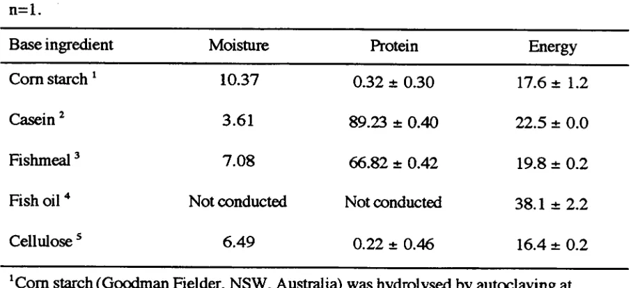
(14) yin 5.1.3 Dietary Protein, Water Temperature and the GH:IGF-I axis in Fish 5.1.4 Aims. 123 124. 5.2 MATERIALS AND METHODS 5.2.1 Experimental Animals 5.2.2 Base Ingredient Analysis and Diet Formulation 5.2.3 Experimental Design 5.2.4 Growth, Blood Sampling and Hepato-Somatic Index 5.2.5 Circulating IGF Analysis 5.2.6 Preparation of Fish Homogenates 5.2.7 Proximate Analysis 5.2.7.1 Moisture 5.2.7.2 Total Crude Protein 5.7.7.3 Total Lipid 5.2.7.4 Total Ash 5.2.7.5 Gross Energy 5.2.8 Statistical Analysis. 125 125 125 125 129 130 130 130 130 130 131 132 132 132. 5.3 RESULTS 5.3.1 Growth 5.3.2 Proximate Composition 5.3.3 Protein and Lipid Deposition 5.3.4 Circulating IGF-I and IGF-II Activities. 134 134 138 140 143. 5.4 DISCUSSION 5.4.1 Growth 5.4.2 Effects of Isonitrogenous Diets on Body Lipid and Protein Deposition 5.4.3 Effects of Isoenergetic Diets on Body Lipid and Protein Deposition 5.4.4 Response of Other Measures of Growth to Isonitrogenous or Isoenergetic Diets 5.4.5 Response of Circulating IGF-I and IGF-II to Isonitrogenous or Isoenergetic Diets 5.4.6 Conclusion. 149 149. CHAPTER 6: GENERAL DISCUSSION 6.1 Discussion 6.2 Conclusion. 149 151 152 153 155 156 157 163. REFERENCES. 164. APPENDIX 1 APPENDIX 2 APPENDIX 3. 182 185 187.
(15) ix LIST OF TABLES Table 3.1. 60. Table 3.2. 62. Table 4.1. 98. Table 4.2. 112. Table 5.1. 127. Table 5.2. 128. Table 5.3. 135. Table 5.4. 137. Table 5.5. 139. Table 5.6. 141.
(16) x LIST OF FIGURES Figure 3.1. 50. Figure 3.2. 56. Figure 3.3. 58. Figure 3.4. 59. Figure 3.5. 63. Figure 3.6. 65. Figure 3.7. 66. Figure 4.1. 85. Figure 4.2. 86. Figure 4.3. 92. Figure 4.4. 93. Figure 4.5. 101. Figure 4.6. 102. Figure 4.7. 104. Figure 4.8. 105. Figure 4.9. 106. Figure 4.10. 108. Figure 4.11. 109. Figure 4.12. 110. Figure 4.13. 113. Figure 5.1. 126. Figure 5.2. 144. Figure 5.3. 146. Figure 5.4. 147. Figure 5.5. 148.
(17) Xi. LIST OF EQUATIONS Equation 2.1. 31. Equation 2.2. 31. Equation 2.3. 31. Equation 2.4. 31. Equation 2.5. 33. Equation 2.6. 38. Equation 2.7. 38. Equation 4.1. 83. Equation 4.2. 83. Equation 4.3. 83. Equation 5.1. 129. Equation 5.2. 129. Equation 5.3. 130. Equation 5.4. 131. Equation 5.5. 131.
(18) xii LIST OF ABBREVIATIONS Abbreviation used ATP ANOVA B. by BPB BP Ins BSA BWI CA CAPS cDNA CF CHISAM Cpm CTP CYT DB DEPC DNA DTT EDTA EtBr FCR GTC GTP HM Ins HPLC IGF-I IGF-II I GFBP J kb kDa kJ LMWS MJ MOPS ng nm Nonidet® P40 NPU NSB OV PER PCR pg PSL RIA RES. Full name 2-Deoxy adenosine triphosphate Analysis of variance Binding capacity base pair Bromophenol blue Bovine Insulin from pancreas Bovine serum albumin Body weight increase Carbonic anhydrase 3-[Cyclohexlamino]-1-propanesulfonic acid Complementary DNA Condition factor Chloroform Isoamylalcohol buffer Counts per minute 2-Deoxy cystidine triphosphate Cytochrome C Dextran blue Diethylpyrocarbonate Deoxyribose nucleic acid Dithiothreitol Ethylenediamine tetraacetate Ethidium bromide Apparent food conversion ratio Guanidium thiosulphate 2-Deoxy guanosine triphosphate Human Monocomponent Insulin High performance liquid chromatography Insulin-like growth factor I Insulin-like growth factor II Insulin-like growth factor binding protein Joule Dissociation constant kilobase kilodalton kilojoule Low molecular weight standards millijoule 3-(N-Morpholino) propane sulphonic acid nanogram nanometer Aethylphenylpolyaethylenglycol Net protein utilisation Non-specific binding Ovalbumin Protein efficiency ratio Polymerase chain reaction picogram Phospho-stimulated luminescence Radioimmunoassay Restricted ration.
(19) RNA RPA RRA rt-PCR SAT SDS SDS-PAGE SGR STV TAE TB TBE TC TMA Tween-20 UV. Ribonucleic acid RNA protection assay Radioreceptor assay Reverse transcriptase PCR Satiety ration Sodium doecyl sulphate SDS Polacrylimide gel electrophoresis Specific growth rate Starved Tris-acetate EDTA Total binding Tris-borate EDTA Total counts Trimethylamine Polyoxyethylenesorbitan monolaurate Ultraviolet.
(20) xiv GLOSSARY OF COMMON AND SCIENTIFIC NAMES Common name American eel Atlantic bluefish Atlantic halibut Atlantic salmon Barramundi Brook trout Brown trout Channel catfish Chinook salmon Cichlid Coho salmon Common carp Couch's seabream European eel Flounder Gilthead seabream Goby Golden perch Goldfish Japanese eel Mossambique tilapia Nile tilapia Rabbitfish Rainbow trout Red Drum Red seabream Sheatfish Sockeye salmon Spiny dogfish shark Sting ray Striped bass Tilapia Tilapia. Scientific name Anguillarostrata Pomatomus salteris Hippoglossus hippoglossus Sal= salar Latescalcarifer Salvelinus fontinalis Salm° trutta Ictalurus punctatus Oncorhynchus tshawytscha Haplochromis burtoni Oncorhynchus kisutch Cyprinus carpio Pagrus pagrus Anguilla anguilla Parlichthys olivaceus Sparta aurata Gillichthys mirabilis Macquariaambigua Carrassius auratus Anguilla japonica Oreochromis mossambicus Oreochromis nilotus Siganus guttatus Oncorhynchus mykiss Scianepnops ocellatus Pagrus major Silurus glanis Oncorhynchus nerka Squalus acanthias Rajaclavata Morone saxatilis Tilapiaandersoni Tilapiaaurea.
(21) CHAPTER 1: GENERAL INTRODUCTION.
(22) 2 1.1 Animal Growth : An Introduction The term growth is most simply defined as an increase in size by natural development (Atkinson, 1991). The growth of an animal represents a series of defined events that begin with food intake and terminate in the deposition of proteins or alternative substances. Between these events, the rates of digestion, absorption, assimilation, synthesis and degradation, energy expenditure and excretion interact to regulate the phenomenon of growth. Growth can be analysed quantitatively by monitoring the expansion of physical dimensions of the organism representing the overall manifestations of the growth process. Such measurements are usually expressed as simple length-weight relationships, relative growth or as growth rates. The perceived ease and readiness with which growth can be observed and measured often implies that growth is a simple, regular process. However, when the variables associated with the regulation of growth are considered, growth is viewed as a more complex, irregular event (Brett, 1979). Animal growth is influenced by a wide range of extrinsic and intrinsic parameters (Brett, 1979; Donaldson etal., 1979). Such parameters, broadly classified as environmental, genetic and hormonal factors, play significant and often interactive roles in regulating growth. Accordingly, disturbances in one or more growth determinants may result in dramatic changes to normal body growth (McLean and Donaldson, 1993). Environmental factors such as food availability, diet composition and seasonal water temperature all influence the metabolic rate and availability of nutrients for growth (Brett, 1979). The nutritional status of the animal, as well as other environmental parameters, including photoperiod and salinity, affect the synthesis of growth-promoting hormones such as thyroid hormone, growth hormone (GH), insulin and insulin-like growth factors (IGFs) (Donaldson etal., 1979; Sumpter, 1992). In addition, social factors such as predation and competition, and intrinsic genetic traits may also influence the scope for growth. When the opportunity for interactions among these factors is considered, it is evident that the mechanisms.
(23) 3 of growth regulation are extremely complex. It is therefore difficult to discuss all factors regulating the growth process. As such, this chapter will briefly discuss growth regulation in teleosts, with emphasis on food availability, diet type, water temperature and the physiological actions of the growth-promoting hormones, namely GH and the IGFs. 1.2 Environmental Regulation of Teleost Growth 1.2.1 Temperature The environment, both natural or captive, has the potential to influence the growth of an animal in many ways. Of particular influence on the growth of teleost fish is the external temperature, since the body temperature of these animals is reliant upon the temperature of the external environment (Brett, 1979; Sumpter, 1992; Jobling, 1994). In teleosts, fluctuations in water temperature affect growth by changing the metabolic rate and thus the mechanisms of energy partitioning within the animal (Elliot, 1979; Forster and Wieser, 1990; Sumpter, 1992; Jobling, 1994). During exposure to higher water temperatures, the amount of energy required to maintain the standard metabolic rate increases. Under such conditions, the excess energy which was available at a lower temperature for processes such as growth is now required to fulfill the basal metabolic energy requirements (Elliot, 1979; Sumpter, 1992; Jobling, 1994). Therefore, in circumstances where the availability of food remains constant but water temperature increases, the growth rate of teleosts will decrease (Jobling, 1981; Sumpter, 1992). The fluctuations in water temperature in natural environments are usually associated with seasonal transition, and providing the temperature remains within the tolerance level of the species, often the animal will slowly acclimatise and the effect of the energy consumed by the metabolic rate is minimal (Bosclair and Tang, 1993). In artificial conditions, such as those used in aquaculture practices, the environmental temperature may vary significantly over much shorter periods of time causing the effect on the metabolic rate to be quite significant (Jobling, 1994). A more detailed discussion of the changes in energy partitioning during temperature changes has been compiled by Jobling (1994)..
(24) 4 1.2.2 Food Availability The availability of food may also significantly affect teleost growth by altering energy partitioning (Brett, 1979; Storebakken et al., 1991). The relationship between ration and growth is curvilinear (Brett, 1979; De Silva and Anderson, 1995). Growth is negative until the energy supplied by the diet equals the energy required to maintain the standard metabolic energy and the then animal no longer loses weight (Brett, 1979; Jobling, 1994; De Silva and Anderson, 1995). The amount of food required to satisfy the basal metabolic rate is often referred to as the maintenance ration, since at this point, the animal will not lose or gain weight (De Silva and Anderson, 1995). From this point, the growth rate continues to increase gradually with increasing ration size, until the maximal feeding rate is achieved and growth plateaus (Brett, 1979; De Silva and Anderson, 1995). The identification of the optimal feeding rate is therefore of great benefit for commercially farmed animals. Brett (1979) provides a more detailed review of food availability and growth of teleost fish. 1.2.3 Diet Composition The growth of teleost fish is reliant upon the synthesis and accretion of various tissue types, predominantly muscle and fat, but also including connective and epithelial tissue. The proportion of protein that can be synthesised or fat deposited is highly dependent upon the diet composition. Fish require relatively high levels of dietary protein, compared to other commercially reared animals (Millikin, 1982), thus many studies of teleosts have aimed to identify the amount of dietary protein required for maximum growth. An early study defined the minimum dietary protein level producing optimal weight gain in chinook salmon, Onchorhynchus tshawtscha (Delong et al., 1958). Subsequently, the determination of optimal dietary protein has been investigated in many fish species including rainbow trout, Oncorhynchus mykiss • (Lee and Putnam, 1973), channel catfish, ktalurus punctatus (Garling and Wilson, 1976), striped bass, Morone saxatilis (Millikin, 1983), red drum, Scianenops ocellatus (Daniels and Robinson, 1986), tilapia, Tilapiaaurea (Winfree and Stickney, 1981), hybrid tilapia, Oreochromis niloticus x 0. aurea (Shia and Huang, 1989) and.
(25) 5 rabbitfish, Siganus guttatus (Parazo, 1990). The growth produced by the dietary protein is reliant upon factors other than crude protein level. Firstly the amino acid composition of the protein source must be considered. Fish require certain essential amino acids (Millikin, 1982) and limitation in one of these essential amino acids results in the deamination of the remaining dietary amino acids and fat deposition (Jobling, 1994; De Silva and Anderson, 1995). Secondly, the amount of growth produced is not only dependent on the provision of the essential amino acids, but also upon the amount of non-protein energy, since the process of protein synthesis requires energy which is additional to that of the basal metabolic rate (Millikin, 1982). Ideally, this energy is provided in the diet in a non-protein form, such that the fish can optimise the dietary protein for tissue accretion (Jobling, 1994; De Silva and Anderson, 1995). If a non-protein source is not supplied, or is not adequate, the fish will deaminate dietary amino acids to produce energy, thus limiting the amount of protein available for growth (Jobling, 1994; De Silva and Anderson, 1995). Increasing the non-protein energy content of a diet can, therefore, result in a greater proportion of the dietary protein being used for protein deposition than for energy. This process is known as protein-sparing and has been reported in several species of fish (De Silva and Anderson, 1995). Both carbohydrate and lipid can be included in the diet as alternative energy sources. Lipid is extremely useful as an alternative energy source in fish diets as it contains high levels of energy, can be effectively utilised by fish, often acts as a binding agent and increases the palatability of the diet (Jobling, 1994; De Silva and Anderson, 1995). Unlike the situation in mammals, carbohydrates are less efficiently used by fish as an energy source (Jobling, 1994; De Silva and Anderson, 1995). However, since carbohydrates are considerably cheaper than lipids, they are often used in fish diets. To optimise the growth of fish, the diet should be formulated to meet the optimal protein-energy ratio. Optimal protein-to-energy ratios vary significantly among fish species and are significantly affected by water temperature and other parameters affecting energy partitioning (Jobling, 1994)..
(26) 6 1.3 Hormonal Regulation of Teleost Growth Although environmental parameters affect the growth of fish, many hormonal systems also have direct and important roles in the regulation of growth (Donaldson etal. , 1979; Sumpter, 1992). The endogenous control of growth in fish is very complex and involves a considerable degree of interaction between a variety of hormones such as GH, thyroid hormones, insulin, IGFs and sex steroids (Donaldson etal., 1979; Sumpter, 1992). In mammals, GH plays a significant role in growth regulation, a fact adequately illustrated by growth cessation in many animals following removal of the pituitary gland (Sara and Hall, 1990). These growth-promoting actions of GH are mediated by insulin-like growth factor-I (IGF-I), a small polypeptide hormone with potent mitogenic effects (Sara and Hall, 1990). Structurally related, insulin-like growth factor-II (IGF-II) also plays a role in regulating growth, but in earlier stages of development and it is not as stringently regulated by GH as IGF-I (Sara and Hall, 1990). Growth hormone and IGFs also play roles in the regulation of growth of teleost fish (Donaldson etal., 1979; Bern etal., 1991; Sumpter, 1992; Siharath and Bern, 1993). The pronounced role that GH has in controlling growth in fish has been clearly demonstrated by growth cessation following hypophysectomy (Ball, 1969; Donaldson etal., 1979) and the increase in growth following GH administration (Sumpter, 1992). The detection of IGF molecules in teleosts is more recent (Bern etal., 1991; Siharath and Bern, 1993), hence our understanding of these molecules remains relatively limited in comparison with mammals. Therefore, where necessary, this review will refer to mammalian IGF literature to enable a complete discussion of the GH:IGF-I axis. Although this chapter focuses on IGFs and the GH:IGF-I axis, the importance of other growth regulatory hormones or the synergistic actions of hormone combinations on body growth of teleosts should not be readily dismissed. Reviews by Donaldson et al. (1979), Weatherley and Gill (1987), Sumpter (1992) and Peter and Marchant (1995) provide detailed information regarding hormonal regulators of growth in bony fish..
(27) 7 1.4 Growth Factors Growth factors are molecules produced by a cell or isolated from a tissue, which display growth-regulatory actions upon other cell types. Early attempts to elucidate the regulatory processes of growth in higher vertebrates resulted in the discovery of many growth factors with mitogenic activity (Sara and Hall, 1990). However, in many cases specialised glands which synthesize and release these growth factors were not identified, thus making the classic endocrine ablation experiments impossible to perform. For this reason, the application of in vitro bioassays, performed on cell cultures or tissue slices were primarily used to determine the biological activity of these factors. 1.4.1 Insulin-like Growth Factors (IGFs) The separate findings of three types of biological activity in serum led to the initial discovery of the IGFs. Salmon and Daughaday (1957) found that hypophysectomised rats had a defect in the synthesis of cartilage matrix protein, chondroitin sulphate, which was rapidly corrected by the administration of GH in vivo. This impairment could not be restored by the administration of GH to cartilage segments in primary tissue culture (Salmon and Daughaday, 1957). In addition, serum from intact rats stimulated in vitro [35S]sulphate uptake into cartilage segments, whereas serum from hypophysectomised rats provided no such stimulation (Salmon and Daughaday, 1957). These findings led to the development of the classic 'somatomedin hypothesis' which proposed that the growth-promoting action of GH was mediated by a serum factor defined as 'sulphation activity' (Salmon and Daughaday, 1957). Subsequent to the investigations of Salmon and Daughaday (1957), serum factors with insulin-like metabolic actions were identified in rat and human serum (Froesch et al. , 1963). Approximately 90% of the insulin-like activity in the serum was not suppressed by anti-insulin serum from guinea pigs and it was therefore termed nonsuppressible insulin-like activity (NSILA). NSILA detected in both rat and human.
(28) 8 serum (Froesch etal., 1963) demonstrated approximately forty fold higher basal levels than that of insulin (Froesch eta, 1967; Gliemann, 1968; Poffenbarger etal., 1968). As NSILA in serum was not immunologically identical with insulin, the identification of the molecule(s) exhibiting this activity was sought. The search for serum constituents necessary to sustain cell growth in culture was the basis for the third line of research. Studies by Dulak and Temin (1973) demonstrated that cell proliferation in certain cell lines was dependent upon the presence of specific factors in serum, whereas other cell lines were capable of producing their own growthpromoting substances. One such activity, termed multiplication-stimulating activity (MSA), was purified from serum-free medium conditioned by a Buffalo rat cell line (BRL-3A). This activity was observed to stimulate deoxyribose nucleic acid (DNA) synthesis and multiplication of normal fibroblasts in vitro. As further work was completed, the similarities between these activities suggested that all three activities represented a similar, if not identical group of molecules, with a much broader range of biological activity than originally anticipated. In 1972, the term 'somatomedin' (mediating the actions of somatotropin) was introduced to describe serum factors which stimulated sulphate uptake into cartilage, had non-suppressible insulin-like activity in adipose and diaphragm tissue, and caused increased thymidine incorporation into DNA in various tissues (Daughaday etal., 1972). 1.4.2 Characterisation of IGFs Following the pioneering work through which these growth-promoting, insulin-like substances were identified, these molecules were purified and characterised. The isolation of the somatomedins proved difficult as their concentration was not particularly high in any particular tissue. Using a method including acid/ethanol extraction, total NSILA (20010a) was found to be easily removed from native serum Poffenbarger etal. (1968). Three different forms of NSILA were subsequently discovered; a soluble 7.5 (kDa) portion (NSILA-s), a larger portion (NSILA-P) which was found to denature and remain within the precipitate and a protein (NSILP) which was detected following Dowex-chromatography (Poffenbarger etal., 1968). Of the.
(29) 9 isolated forms, NSILA-s was observed to exhibit potent growth-promoting effects in chick embryo and other fibroblast cells and to stimulate the sulphation of cartilage in various animals (Zing and Froesch, 1973). The growth-promoting action, molecular size and the high abundance of NSILA-s in serum compared to other tissues, raised the possibility of this substance being a circulating hormone. Two biologically active peptides showing similar growth-promoting properties to NSILA-s were purified from human blood products (Rinderknect and Humbel, 1978a; 1978b). The amino acid sequence of these activities was found to be 48% homologous with human pro-insulin and this peptide was renamed insulin-like growth factor-I (IGF-I) (Rinderknect and Humbel, 1978a). The second bioactive peptide was structurally similar, but not identical to IGF-I, and was named IGF-II (Rinderknect and Humbel, 1978b). The subsequent sequencing of somatomedin-C (Klapper etal. , 1983) and somatomedin-A (Endberg etal., 1984) also showed that they were structurally identical to IGF-I, and the IGF-I and IGF-II nomenclature became universal. Subsequently, IGF-I and IGF-II have been extensively studied in mammals and information on the structure, regulation, function and actions of these molecules has been well documented. 1.4.3 Structure of IGFs Insulin-like growth factor-I and IGF-II are single chained polypeptide molecules comprised of 70 (7646 kDa) and 67 (7471 kDa) amino acids, respectively and each contains three intra-chain disulphide bridges (Rinderknect and Humbel, 1978a; 1978b). The structural homology between these molecules in humans is 62%, suggesting that the genes coding for these molecules were derived from a common ancestral gene. The structural characteristics of IGFs are similar to pro-insulin, demonstrating 50% homology. Insulin-like growth factors are comprised of an amino-terminal B domain which is separated from the A domain by a C domain which consists of 12 amino acids. In contrast to pro-insulin, the IGF molecules also contain a D domain extension peptide at the carboxy terminus and an E domain. Mature IGF peptides are synthesised by proteolytic cleavage of the E domain, which contrasts to.
(30) 10 the synthesis of insulin which is produced by cleavage of the signal peptide and removal of the C domain (Sara and Hall, 1990). The structure of the IGF molecules appears to be highly conserved throughout vertebrate evolution. Cao etal. (1989) cloned IGF-I mRNA from coho salmon, Oncorhynchus kisutsch, and reported sequence homology with human IGF-I of approximately 80%. Subsequently, recombinant DNA techniques have been used to identify the nucleotide sequences of the IGF prohormones in Atlantic salmon, Salmo salar (Duguay et al., 1992), rainbow trout (Shamblott and Chen, 1992) and chinook salmon (Wallis and Devlin, 1993), and it appears that the structure of the mature IGF-I molecule in these species is identical to that of coho salmon (Cao et al. , 1989). The presence of cDNA coding for an IGF molecule similar in structure to mammalian IGF-II has been reported in,rainbow trout (Shamblott and Chen, 1992). In addition, findings of distinct IGF-II-like peptides in the liver of spiny dogfish shark, Squalus acanthias (Duguay et al., 1995) and in the endocrine pancreas of the sting-ray, Raja clavata (Reineke et al., 1994) suggest that the prototypical IGF molecule duplicated and diverged in an ancestor of the extant gnathostomes (Reineke etal., 1994), thus providing support for the existence of structurally distinct IGF-I and IGF-II molecules in teleost fish. 1.4.4 IGF-I Gene The IGF-I gene in rats and humans consists of six exons and is distributed over more than 100 kilobases (kb) of genomic DNA (Sussenbach et al., 1992). The exons have been shown to contain the same coding information in both species. Exon 1 and 2 contain multiple sites of transcription initiation, with either exon 1 or 2 being spliced to exon 3. Exons 3 and 4 encode the mature peptide, whilst exons 5 and 6 encode alternate E peptides and 3' untranslated regions (UTRs) (Sara and Hall, 1990). The complexity of the mammalian genes analysed so far explains the presence of multiple mRNA transcripts, which range from 0.8 to 7.5 kb in length. Two cDNAs for IGF-I, known as IGF-Ia and IGF-Ib, have been detected in humans and consist of 153 and 195 amino acids, respectively (Jansen et al., 1983; Rotwein et al., 1986). Upon.
(31) 11 characterisation, the first 134 amino acids are identical, with the difference in structure relating to the carboxyl-terminal E domain (Etherton, 1993). The mRNAs which encode for these proteins are the result of alternative splicing of the single primary RNA transcript. Similarly, two IGF-I genes which produce four alternatively spliced IGF-I mRNA transcripts, known as Ea-1, Ea-2, Ea-3 and Ea-4 have been characterised in chinook and coho salmon (Duguay etal., 1994). Using an RNA protection assay to determine the expression level, the Ea-1 and Ea-3 transcripts were found in abundance in the liver, but were barely detectable in other non-hepatic tissues. Conversely, the Ea-4 transcript was present in both hepatic and non-hepatic tissues (Duguay etal., 1993). Recently, two alternatively spliced transcripts, corresponding in structure to salmonid Ea-2 and Ea-4 were reported in barramundi, Latescalcarifer (Kinhult, 1997). The Ea-2 and Ea-4 transcripts appeared to be widely distributed with detection in the liver, kidney, gill, brain, heart and spleen (Kinhult, 1997). Although the function of the multiple IGF-I transcripts is not understood, the expression of alternatively spliced transcripts in rats is associated with development and tissue distribution. A similar role has therefore been suggested for these transcripts in teleost fish (Wallis and Devlin, 1993). Little is known of the IGF-II gene in bony fish. 1.4.5 IGF Receptors In mammals there are two types of specific IGF receptors. The type I (IGF-I) receptor is structurally similar to the insulin receptor and has been reported as the only IGF receptor to have definite IGF-mediated signaling functions (Jones and Clemmons, 1995). Type I IGF receptors and insulin receptors are homologous in structure and both are down-regulated by IGFs and insulin (Rechler and Nissley, 1985). The type I receptor has a molecular weight of > 300,000 and is composed of disulphide linked 130,00 Da alpha (a) and 90,000 beta (13) subunits (Rechler and Nissley, 1985). The a subunit binds the hormone and the fi subunit appears to have intrinsic tyrosine.
(32) 12 kinase activity and to be autophosphorylated. Type I receptors bind IGF-I and IGF-II with equal affinity and insulin weakly (Daughaday etal., 1981). The type II (IGF-II) receptor is different from the type I and insulin receptor in both structure and function (Rechler and Nissley, 1985). Comprised of a 250,00 Da protein that contains internal disulphide bonds but is not linked to other membrane components, the type II receptor demonstrates structural homology with the cationindependent mannose 6-phosphate receptor and is thought to function as a 'clearance' receptor, moving lysosomal enzymes (Jones and Clemmons, 1995). Type II receptors bind IGF-II with 100-fold greater affinity than IGF-I, but do not interact with insulin, even at high concentrations (Scott and Baxter, 1987). Furthermore, they are not down-regulated and although type II receptors appear phosphorylated in intact cells, they do not possess intrinsic protein-kinase activity (Rechler and Nissley, 1985). In addition to binding with their own receptors, the IGFs also bind to the insulin receptor, but with low affinity. Furthermore, receptor hybrids, composed of insulin and IGF-I subunits have been reported in NIH-3T3 and HepG2 cell lines (Moxham et al., 1989). The hybrid receptors bind the IGFs and reportedly signal cytoplasmically in vitro (Jones and Clemmons, 1995). Receptors for IGF-I have recently been identified in teleost fish. Using wheat germ agglutination-agarose affinity chromatography, IGF-I receptors were detected in the ovary of carp, Cyprinus carpio, (Gutierrez etal., 1993), cardiac and skeletal muscle of brown trout, Salmo trutta, coho salmon and carp (Gutierrez etal., 1995) and brain of carp and trout (Leibush etal., 1996). A recent study also found IGF-I receptors in the testis of trout (Le Gac etal., 1996). These receptors are presumably type I-like. The specific binding of IGF-I in the skeletal and cardiac muscle of the various teleost species differed from patterns observed in higher vertebrates. In the skeletal muscle, specific IGF-I binding was 2-6 fold higher than insulin binding, contrasting with the prevalent nature of insulin receptors in mammals (Gutierrez et al., 1995). In addition, the specific binding of IGF-I to purified cardiac muscle IGF-I receptors was 20% higher (44 - 68%) in fish heart when compared to rat heart (Gutierrez etal., 1995)..
(33) 13 These findings suggest the possibility of functional and structural differences between the IGF-I receptors of teleost fish and mammals. Furthermore, the presence of IGF-I receptors in the reproductive tissues of teleosts may provide support for the role of IGF-I in teleost reproduction (Section 1.4.7.4). 1.4.6 IGF Binding Proteins (IGFBPs) The likely existence of insulin-like growth factor binding proteins (IGFBPs) was recognised during early attempts to extract IGFs (NSILA) from serum. During attempts to purify IGFs, discrepancies in the molecular size of the whole serum NSILA activity (200 kDa; Poffenbarger etal., 1968) and that of the soluble extract (7.5 kDa; Froesch etal., 1967) led to the proposal that specific carrier proteins existed. Although acidic treatment of the NSILA did not yield the soluble NSILA-s extract, binding proteins which demonstrated high affinity for the IGFs were later characterised. Subsequently, six different IGFBPs have been cloned and sequenced in mammals (Rechler, 1993). As much as 90% of the circulating IGFs in mammals are bound with IGFBP-3 and an acid-labile subunit (ALS), forming the 150 kDa IGF complex (Rechler, 1993). The remaining IGFs either bind with IGFBP-1, -2 or -4 though a small amount (5%) circulates as free peptide (Ketelslegers etal., 1996). The 150 kDa complex stabilises the IGF-I in circulation, preventing the movement to the extravascular compartment and prolonging the half life of the molecule (Clemmons and Underwood, 1991). The IGFBPs are also thought to aid in targeting tissues, since they regulate the transport of IGFs from the vascular to the extracellular compartment and regulate their interaction with the IGF receptors (Clemmons and Underwood, 1991). Details of the regulation and specific functions of the IGFBPs have been detailed by Clemmons and Underwood (1991). Multiple forms of IGFBPs have recently been reported in teleost serum, however information on their regulation and functional significance remains limited. Three major IGFBPs were detected using Western ligand blotting in the serum of coho salmon, striped bass, the goby, Gillichthys mirabilis and Mossambique tilapia, Oreochromis mossambicus (Kelley etal., 1992; Siharath etal., 1992; Siharath and.
(34) 14 Bern, 1993). Based on the molecular weights of these molecules, the three bands were characterised as fish IGFBP-1 (29 kDa), IGFBP-2 (31 lcDa) and IGFBP-3 (4050 kDa) (Kelley et al., 1992). IGFBPs have also been reported in rainbow trout (Niu and Le Bail, 1993), channel catfish (Delahunty etal., 1993) and golden perch,. Macquariaambigua (Anderson et al., 1993). The regulation of the IGFBPs in teleosts has recently been reviewed in Siharath and Bern (1993), and generally appears to parallel some of the regulatory mechanisms reported in mammals. 1.4.7 Actions of IGFs The actions of the IGFs in mammals have been reviewed in detail (Daughaday and Rotwein, 1989; Sara and Hall, 1990) and generally include mitogenic, metabolic and growth-related actions in a variety of cell and tissue types. Insulin-like growth factors have also been shown to be important for skeletal growth, larval development, osmoregulation and reproduction in teleosts (Bern et al., 1991; Siharath and Bern, (1993). 1.4.7.1 Skeletal Tissue Although many tissues are known to be responsive to the mitogenic actions of IGFs, the hormonal regulation of skeletal tissues is of particular significance in understanding entire body growth. The effect of GH was earlier thought to be mediated by circulating IGF-I of hepatic origin (Daughaday and Rotwein, 1989), however there is recent evidence suggesting that GH also stimulated paracrine and autocrine production of IGF-I in both cartilage and skeletal muscle in mammals (Holly and CwyfanHughes, 1994). This theory is referred to in the current literature as the dual (Green et. al., 1985) or multiple (Siharath and Bern, 1993) effector theory of GH action. The effects of GH on the expression of IGF-I mRNA have also been investigated. Hepatic IGF-I mRNA was observed to increase in a dose-dependent manner following intraperitoneal GH injections in coho salmon (Duan et al., 1994). In salmon, the expression of the alternatively spliced, Ea-1 and Ea-3, IGF-I transcripts in the liver was also found to be GH responsive (Duguay et al., 1994). In contrast, the.
(35) 15 expression of the alternatively spliced, Ea-2 and Ea-4, transcripts was not regulated by GH in salmonids (Duguay etal., 1994) or barramundi (Kinhult, 1997). The effect of IGF-II was investigated in only one study, with rat IGF-II reportably 50 times less potent than IGF-I in stimulating sulphate uptake in intact eels (Duan and Hirano, 1990). Substantial evidence supports coordinated roles of GH and IGF-I in the regulation of body growth of teleost fish. Ash (1977) reported bovine GH directly increased sulphate incorporation in branchial cartilages of rainbow trout and tilapia (Tdapia. andersom). In contrast, porcine GH, thyroxine and somatotropin, alone, or in combination had no effect on rainbow trout gill arch bone, unless liver slices were added (Komourdjian and Idler, 1978). The conflicting results were attributed to the use of cartilage versus bone, with the suggestion of local IGF-I production in cartilage (Siharath and Bern, 1993). Subsequently, Duan and Inui (1990a) found sulphate uptake was restored in hypophysectomised Japanese eel, Anguillajaponica, injected with eel GH. The stimulation of sulphate uptake in cartilage by IGF-I has now been reported in coho salmon (McCormick, etal., 1992; Tsai, 1993), Japanese eel (Duan and Hirano, 1990; Duan and Inui, 1990b), the goby (Gray and Kelley, 1991; Kelley, 1993) and the goldfish, Carrassiusauratus (Marchant and Moroz, 1993). The results from these studies provide clear evidence that the stimulation of cartilage matrix protein synthesis is one mechanism by which GH and IGF-I promote skeletal tissue growth in teleosts. The administration of GH has been shown to increase the growth rate of teleost fish (Bern etal., 1991). Juvenile brook trout, Salvelinus fontinalis, treated with human GH were significantly heavier and longer at three weeks than controls (Skyrud etal. , 1989). As in mammalian studies, the treatment of teleost fish with IGF-I in vivo has produced variable growth effects. Recombinant human IGF-I administered weekly for three weeks to juvenile brook trout did not stimulate growth, and the higher doses, 1.0 and 3.0 ggig body weight, resulted in decreased growth and profound insulin-like effects,.
(36) 16 causing hypoglycemia and hypoaminoacidemia (Skyrud et al., 1989) In contrast, McCormick etal. (1992) observed an increased growth rate in coho salmon which were treated using bovine IGF-I via mini-pumps and fed a limited ration, however, increased growth rate was not observed in fish given an unlimited food supply. The discrepancy between the two studies has recently been attributed to the method of IGFI administration (Siharath and Bern, 1993) since IGF-I infusion but not injection stimulates growth in adult rats (Lowe, 1991). A comparison between the studies is difficult since nutritional status, which varied between studies, has profound effects on the in vivo effects of IGF-I and may result in differing responses to IGF-I administration. 1.4.7.2 Larval Development. _. Recent studies have indicated that IGF-I is present during differentiation of juvenile fish. In salmonid species, IGF-I is expressed during embryonic development (Duan et. al., 1994; Funkenstein etal., in press) and all tissues were reported to produce IGF-I mRNA transcripts (Duguay etal., 1992; Duan etal., 1994). Funkenstein etal. (in press) found IGF-I and IGF-I receptor mRNA in gilthead seabream, Sparus aurata, larvae with high expression in the gill arch chondrocytes, skeletal muscle, brain, pancreas and in the epithelial cells surrounding the lens. In barramundi, IGF-I immunoreactivity was found to be age and tissue specific with IGF-I immunoreactive cells found in the retina (42 hr), pectoral fin musculature (57 hr), renal tubule epithelia (9 days), gill cytoplasmic structures (13-28 days), endocrine pancreas (14 days), brain (22 days) and perikaraya (13-28 days) (Richardson etal., 1995). These results indicate that IGF-I plays an important role in regulating cell growth and differentiation of juvenile fish in a variety of tissues. The presence of IGF-I immunoreactive cells associated within the eye indicates IGF-I may play a role in its development (Richardson etal., 1995; Funkenstein etal., in press). The stimulation of rod-precursor cell proliferation in retinal slices prepared from eyes of 12-15 day juvenile cichlid fish, Haplochromis burtoni by both bovine.
(37) 17. IGF-I (20 ng/ml) and bovine insulin (5 µg/m1) supported this hypothesis of a role of IGF-I in eye development (Mack and Fernald, 1993). 1.4.7.3 Osmoregulation Osmoregulation in mammals is achieved largely through the kidney, however in fish the gill also plays a major role. The importance of GH and IGF-I in regulating kidney function in mammals was originally indicated by observations of renal hypertrophy in human patients with acromegaly, and low serum IGF-I in patients with chronic renal failure (Takano etal., 1979). In hypophysectomised rats, GH treatment increases both the kidney weight and the level of IGF-I mRNA expression in the kidney (Rotwein etal., 1989). Similarly, the direct infusion of IGF-I increases kidney weight in rats (Flyvberg etal., 1991; Martin etal., 1991). In humans, both GH and IGF-I stimulate renal plasma flow and glomerular filtration rate and reduce total renal vascular resistance (Guler etal., 1989). These results support the role of GH and IGF-I as regulators of renal function in mammals (Cohick and Clemmons, 1993). There is evidence that GH and IGF-I also play a role in osmoregulation in teleost fish. Growth hormone increased the hypoosmoregulatory ability of juvenile salmonids. One day after transfer to seawater immature rainbow trout GH levels were significantly elevated (Sakamoto etal., 1993). Similarly, IGF-I mRNA expression increased in the gill and kidney of immature rainbow trout following transfer to 80% seawater. Increased levels of IGF-I mRNA were also observed in the liver and gill of coho salmon maintained in freshwater during smoltification. The administration of IGF-I was also found to effectively enhance saltwater adaptation. Rainbow trout treated with a single dose of bovine IGF-I (0.01, 0.05, 0.2 µg/g) demonstrated a greater ability to maintain plasma osmolarity and sodium following transfer to 80% seawater (McCormick eta/., 1991). In a later study, single injections of GH or IGF-I improved ion regulation in Atlantic salmon transferred from 12 ppt to higher salinities (McCormick, 1995). Interestingly, the administration of GH or IGF-I did not effect ion regulation in Atlantic salmon kept in fresh water.
(38) 18 (McCormick, 1996). The presence of high levels of circulating prolactin was suggested to cause the inefficiency of GH and IGF-I enhancing freshwater adaptation (McCormick, 1996). Antagonism between prolactin and GH effects on salinity tolerance of rainbow trout and brown trout supports this hypothesis (Madsen and Bern, 1992). Although the exact mechanisms of GH and IGF-I in seawater adaptation in teleosts are not understood, there is some evidence to suggest that these hormones affect chloride cell and gill Nat, 1C+-ATPase production. The administration of growth hormone to juvenile Nile tilapia, using single (0.025 or 2.5 µg/g) or repetitive (3 x 0.25 pg/g) doses, enhanced seawater adaptability and induced the differentiation of chloride cells towards seawater adaptation (Xu et al., 1996). In brown trout, both GH and cortisol increased gill Nat, IC-ATPase, chloride cell numbers and salinity tolerance; the actions of the hormones were synergistic (Madsen, 1990). Growth hormone and cortisol, both alone and in combination, stimulated gill Nat, Kt-ATPase in Atlantic salmon (McCormick, 1996). An increase in gill cortisol receptors in response to GH in coho salmon (Shrimpton et al., 1995), provides an explanation for the actions of GH alone and in combination with cortisol. Following in vivo GH treatment, IGF-I was found to stimulate in vitro gill Nat, IC-ATPase activity of coho salmon (Madsen and Bern, 1993) suggesting that the effects of GH on gill Nat, K"-ATPase are mediated by IGFI. McCormick (1995) demonstrated that IGF-I and cortisol, in combination, stimulated Nat, IC-ATPase in Atlantic salmon, although the effect was less stimulatory than GH and cortisol. In contrast, IGF-I alone did not stimulate Nat, ICATPase in striped bass (Madsen and Bern, 1993) or in coho salmon gill filaments in. vitro (McCormick, 1995) with the lack of response attributed to an inappropriate dose of IGF-I and the interference by IGFBPs. The effect of IGF-I on Nat, r-ATPase in teleosts thus remains unclear. 1.4.7.4 Reproduction IGF-I and IGF-II are potent in vivo stimulators of mammalian oocyte maturation (Feng etal., 1988) and evidence is emerging to suggest a similar role in teleosts..
(39) 19 IGF-I (10 mM) induced germinal vesicle breakdown (GVBD) in red seabream, Pagrus. major (Kagawa etal., 1994), appearing to act directly on oocytes rather than via maturation-inducing hormones. IGF-II (13 mM) also induced GVBD (Kagawa etal. , 1994). IGF-I mRNA is expressed in the ovaries of coho salmon (Duan and Plisetskaya, 1993) and IGF-I immunoreactvity is localised in ovaries of red seabream (Kagawa et. al., 1994). Intense IGF-I immunoreactivity appears in the peripheral region of the ooplasm at the primary vitellogenic stage, in the granulosa cells at the lipid stage and the outer layer of the zona radiata, suggesting that the granulosa cell layer is the major site of IGF-I production. 1.5 Nutritional Regulation of IGFs In mammals, nutritional status causes an insensitivity of the liver to GH thereby altering the normal production of IGF-I, which is mainly derived from the liver in response to GH (Clemmons and Underwood, 1991). Accordingly, low serum IGF-I bioactivity is observed in children with protein-calorie malnutrition, despite elevated GH concentrations (Grant et al., 1973). A significant reduction in IGF-I bioactivity was found in 72 hour fasted rats, compared to fed rats (Phillips and Young, 1976). IGF-I bioactivity was restored by feeding (Phillips and Young, 1976). Similar changes were observed in a variety of species using more definitive IGF-I assay techniques. During periods of nutrient deprivation, IGF-I plasma concentrations were found to be significantly decreased, with respect to fed controls, in man (Musey et al., 1993; Kiddy et al., 1989), growing and pre-weaning rats (Maiter et al., 1988; Phillips. et al., 1989; Donovan et al., 1990), sheep (Hua et al., 1993), weaning calves (Breier et al., 1988), steers (Breier et al., 1986), chickens (Morishita et al., 1993), maternal and fetal guinea pigs (Dwyer and Strickland, 1992), dogs (Eigenmann et al., 1985) and teleost fish (Perez-Sanchez et al., 1994; Duan and Plisetskaya, 1993). The mechanisms by which nutrient deprivation decreases IGF-I is poorly understood but appears rapid and complex. Following 12 and 24 hours of protein restriction,.
(40) 20 IGF-I levels were reduced by 58 and 66% respectively in rats (Maiter et al., 1988). Serum GH was not affected by low protein intake and injections of 5 to 100 pg rat GH failed to raise to IGF-I serum concentrations in the protein deficient animals. A 15 to 20% reduction in the number of GH binding sites was observed indicating regulation occurs through post-GH receptor processes. However, regulation occurs upstream of gene expression as reduced levels of hepatic IGF-I mRNA expression (Emler and Schalch, 1987; Straus and Takemoto, 1990) and hypoinsulinemia (Merimee et al., 1982; Cheetham et al., 1994) during fasting, protein and energy deficiency have been reported. In mammalian systems, IGF-II appears to be less closely regulated by GH and concentrations of IGF-II in serum have been reported to be unchanged by short-term fasting (Davenport et al., 1988) or during restricted feeding (Dwyer and Strickland, 1992). However, due to the limited information, it remains difficult to propose a role for IGF-II in mediating metabolic responses to changes in nutrient intake. The general principles described above also apply to teleost fish. Short or long-term fasting was found to significantly increase GH levels in tilapia (Rodgers et al., 1992), rainbow trout (Sumpter et al., 1991), juvenile coho salmon (Duan and Plisetskaya, 1993) and gilthead sea bream (Perez-Sanchez et al., 1995). In gilthead seabream fed protein deficient diets for 6 weeks, GH levels increased above those of fish fed diets of higher protein content (Perez-Sanchez et al., 1995). Despite elevated GH levels, total GH binding in gilthead sea bream (Perez-Sanchez etal, 1995) and hepatic GH binding in coho salmon (Gray et al., 1992) decreased during periods of food restriction. Serum IGF-I bioactivity, measured by sulphate incorporation in bone or oral cartilage, was significantly decreased by short-term starvation in coho salmon (Komourdjian and Idler, 1978; McCormick et al., 1992). Impairment of sulphate incorporation into cartilage by liver slices from Japanese eel starved for 2 weeks suggests that the liver is.
(41) 21 important in regulating systemic IGF activity (Duan and Inui, 1990b). In addition, serum IGF-I immunoreactivity was significantly reduced by feeding limited ration sizes and restricted protein diets to gilthead sea bream for 6 weeks (Perez-Sanchez et. a/., 1995). In contrast, Drakenberg et al. (1989) found no effect of starvation on IGF-I receptor binding activity in whole serum of tilapia, however this may be attributed to the relatively short period of fasting. Available information suggests nutritional factors affect IGF-I synthesis at least at the translational level in teleosts. Hepatic IGF-I mRNA, 4 kb, decreases following two weeks starvation in the Japanese eel (Duan etal., 1994). In coho salmon, a reduction in the levels of the hepatic, alternatively spliced, IGF-I mRNA transcripts Ea-1 and Ea3 was found following two weeks starvation with re-feeding restoring levels-to control values (Duan etal., 1994). However, fasting did not affect the levels of the universally distributed Ea-4 transcript (Duan and Duguay, 1994). 1.6 Measuring IGFs In view of the complexity of the IGF system in mammals with two peptides, two receptors- one of which shows cross-reactivity in binding between the two peptides, and at least six different binding proteins described, measurement of IGFs requires considerable care. Care must also be shown in the interpretation of data generated by the assays. A number of methods have been employed to measure IGFs, developing as the knowledge of the molecules has expanded. 1.6.1 Bioassays Originally, bioassays were used to detect the IGFs. These were based on sulfate or thymidine incorporation into cartilage (Daughaday etal., 1975; Almquist, 1961), insulin-like activity in non-skeletal tissues (Labhart, 1963) or growth-promoting activity in cultured cells (Frykland etal., 1974). As the bioactivity of IGF in the tissues results from the net interactions between IGF, IGFBPs and other components such as proteases (Holly and Cwyfan-Hughes, 1994) the interpretation of the bioassay results was complex and this was recognised as the major disadvantage of these.
(42) 22 assays. It is also difficult to mimic the IGF system in vitro, since other serum constituents may be foreign to the target cells in vivo and cultured cells can be made to produce abnormal patterns of IGFBPs and proteases (Holly and Cwyfan-Hughes, 1994). The problems with the interpretation of IGF bioassays and the characterisation and production of purified mammalian IGFs led to the development of competitive binding assays, such as radioreceptor (RRA) and radioimmunoassays (RIA). 1.13.2 Radioreceptorassays for IGF-I and IGF-II The discovery of distinct type I and type II IGF receptors in various mammalian tissues and cell types enabled the development of specific radioreceptor assays (RRA) for IGF-I and IGF-II (Marshall etal., 1974; Megeysi etal., 1975). The RRAs allowed routine and precise measurements of the IGF molecules, overcoming the problems associated with bioassays. Human placenta was the first tissue shown to have receptors for IGFs (Marshall etal. , 1974). Two structurally dissimilar types of IGF binding sites were found in particulate human placental preparations (Kasuga etal., 1981). The type I receptor bound IGF-I preferentially and was thought to resemble the insulin receptor structurally. Studies using affinity labelling and SDS-PAGE demonstrated that the type I receptor, like the insulin receptor, was composed of two a and two. p subunits. linked together by disulphide bonds. The type I receptor from human placenta bound IGF-I and IGF-II with equal affinity (Daughaday etal., 1981). The second receptor found in human placental membranes, the type II receptor, differed in structure and function from the type I and insulin receptor (Daughaday etal., 1981; Kasuga etal. , 1981). The human placenta type II receptor consisted of a single polypeptide chain containing interlinking disulphide bonds. Functionally, the type II receptor bound IGF-II preferentially and exhibited low affinity for IGF-I, approximately 10 % of that of IGF-II. Insulin did not bind the type II receptor at any concentration. Type II receptors were also found in rat placental and rat liver membranes, however their abundance differed in comparison with human placental membrane preparations.
(43) 23 (Daughaday etal., 1981; Pilistine etal., 1984; Scott and Baxter, 1987). Using competitive binding techniques, Daughaday etal. (1981) reported that the specific binding of IGF-I in rat placental membranes did not exceed 3.3%, but displayed a high abundance of type II receptors. Similarly, Scott and Baxter (1987) found rat liver microsomal membranes to be a very abundant source of high affinity IGF-II receptors. The type II receptors found in rat placental and rat liver membranes displayed high specificity for IGF-II, a lower affinity for IGF-I and no affinity for insulin. The detection and characterisation of IGF receptors in various membranes preparations was integral to the development of RRA. Daughaday etal. (1981) used human placental membrane preparations as a matrix in RRA by developing the first RRA used to measure IGFs. Since the ligand [ 125I]hIGF-I was displaced by both IGF-I and IGF-II, this assay was considered capable of detecting both IGF-I and IGF-II in serum samples. The high abundance of the type II receptors in different rat tissues allowed the development of RRA specific to IGF-II. Using [ 125I]hIGF-II as a ligand and type II receptors from rat placental membranes as a matrix, a RRA specific for IGF II activity was developed (Daughaday etal., 1981). In this assay IGF-I was less than 1% effective in displacing [ 125I]hIGF-II. Similarly, a specific IGF-II RRA was also developed using rat liver membranes as the matrix (Scott and Baxter, 1987). 1.6.3 Radioimmunoassays for IGFs Following purification of IGFs in sufficient amounts, radioimmunoassays for measuring IGFs were developed. The development and validation of radioimmunoassays for IGF-I have been reviewed by Breier etal. (1991) and Blum and Breier (1994). 1.7 Competitive Binding Assays for IGFs - Problems and Pitfalls A number of problems resulting from the complexity of the IGF system require considerable care to avoid artifacts in IGF assays. Two problems warranting attention in this review are IGFBP interference and heterogeneity..
(44) 24 1.7.1 IGFBP Interference Inaccurate measurement of IGFs by competitive binding assays occurs when IGFBPs are present in samples. Binding proteins bind IGFs with high affinity (Kd 10 -10 to 1041 mo1/1) and, if present during the assay, cause severe interference by competing for the tracer with the antibody or receptor. Dissociation of the endogenous IGF from the IGFBPs and removal of the IGFBPs from the sample are therefore considered essential for pre-assay preparation (Bang etal., 1994). Parallel displacement curves are not, in themselves, satisfactory evidence of the lack of interference by IGFBPs. Parallel displacement curves were obtained for extracted and non-extracted serum in both the IGF-I (Moriyama etal., 1994) and IGF-II RIA (Gentil etal., 1996). These results suggest that the presence of IGFBPs, under the conditions in which the fish were held, did not interfere in the assay systems or interfered only weakly, and IGFBP extraction was not required. These authors concluded that the antibody used bound to a part of the IGF which was different from that which bound IGFBPs. An alternative explanation may be that the conditions of the fish from which the samples were drawn resulted in an absence (or substantial decline) of IGFBPs. It is well documented that the IGF:IGFBP ratio changes under different physiological and pathological conditions, causing the interference by IGFBPs in the RIA to also differ (Breier etal., 1991; Bang etal., 1994; Blum and Breier, 1994). Whilst Gentil et d. (1996) reported significant differences between IGF-II levels in extracted and unextracted serum from fasted rainbow trout, extraction had no effect on IGF-I concentrations from serum taken from parr, smolt and adult coho salmon (Moriyama et al., 1994). Further work is therefore required to fully determine the effect of IGFBPs under different physiological states in homologous RIA systems. Interference by IGFBPs is dependent on the amount, type, concentration and affinity of the IGFBPs with respect to the free IGF-I and the amount and affinity of the antiserum or receptor being used (Holly and Cwyfan-Hughes, 1994). Since these parameters vary markedly with physiological conditions, species and type of body.
(45) 25 fluid or conditioned cell media from which the sample was obtained, it is considered essential to perform assay validation with the actual experimental samples and the particular assay procedure involved (Bang etal., 1994; Blum and Breier, 1994; Holly and Cwyfan-Hughes, 1994). Techniques to reduce the interference of IGFBPs in competitive binding assays were based upon the discovery that the IGF:IGFBP interaction is dissociable under acidic conditions (Hintz and Liu, 1977). Extraction therefore relies upon IGFBPs being dissociated from the IGFs under acidic conditions and then being physically separated from each other prior to assay. The most commonly reported methods are size exclusion chromatography under acidic conditions (Zapf etal., 1981; Powell etal. , 1986), solid-phase extraction including SEP-PAK columns (Daughaday etal., 1987), acid ethanol extraction (Daughaday etal., 1980), acid-ethanol cryo-precipitation (Breier etal., 1991), formic acid extraction (Bowsher etal., 1991) and sephacryl extraction (Moore etal., 1993). Of these methods, size exclusion chromatography under acidic conditions has been accepted as the most reliable and effective extraction procedure. However, the time-consuming and laborious nature of the extraction and the cost of an effective column are considered to make it unsuitable for routine assays (Blum and Breier, 1994; Holly and Cwyfan-Hughes, 1994). Alternative methods which are based primarily on the use of organic solvents to precipitate the IGFBPs have been developed to allow multiple samples to be extracted easily and quickly. The acid-ethanol procedure reported by Franklin etal. (1976) and validated for use in human sera by Daughaday etal. (1980) utilised ethanol to precipitate the IGFBPs. Other methods which relied upon other combinations of acid and solvent were also adopted and reportedly improved the original IGFBP removal technique (Kaufmann et al., 1979; Mesiano etal., 1988; Moore and Mylek, 1993). Unfortunately, coprecipitation of IGF-I with the IGFBPs resulted in underestimation of IGF concentrations (Blum and Breier, 1994). In addition, acid-ethanol and/or formic acid extraction left considerable amounts of residual IGFBPs in sera obtained from different pathological states, biological fluids, species or developmental stages.
(46) 26 (Mesiano etal., 1988; Blum and Breier, 1994; Holly and Cwyfan Hughes, 1994). Acid gel chromatography is therefore considered the most rigorous extraction method. 1.7.2 IGF Heterogeneity As IGFs have species specific structure, competitive binding assays for IGFs are also complicated when a heterologous competitive binding assay is used. Heterologous IGF-I RIAs have been used to measure IGF-I in a wide range of species (Dawe etal. , 1988; Hansson etal., 1988; Ballard etal., 1990; Bautista etal., 1990; McGuiness and Cogburn, 1990; Bern etal., 1991; Harding etal., 1994). The cross-reactivity in these heterologous assay systems was found to be variable for different species. Furlanetto. etal. (1977) measured circulating IGF-I in rats using a human IGF-I RIA but chicken IGF-I cross-reacted by only 50% in a similar human IGF-I RIA (Dawe etal., 1988; Ballard etal., 1990). The discrepancy in the cross reactivity was attributed to the heterogeneity of the IGF-I molecule, since rat and chicken IGF-I differ from human IGF-I by 3 (4.3%) and 8 (14.3%) amino acids, respectively. The influence of low immunological cross-reactivity of IGF-I has also been attributed to sequence differences in particular domains of the IGF molecule (Wilson and Hintz, 1982). Variability in assay response using heterologous IGF assays for measurement in teleost plasma confounded early studies. Using mammalian RRA or RIA, circulating IGF-I-like activities were detected in tilapia (RRA only) (Drakenberg etal., 1989), gilthead seabream (Funkenstein etal., 1989), Atlantic salmon (Lindahl etal., 1985), rainbow trout and golden perch (Anderson etal., 1993). However using a similar RIA protocol, but different antibodies, no significant activity was detected in the serum of Atlantic bluefish, Pomatomus salteris (Furlanetto et al., 1977) or common carp (Wilson and Hintz, 1982). IGF-II like activities were detected in rainbow trout using a human IGF-II RIA (Daughaday et al., 1985; Bautista et al., 1990), but not in golden perch (Anderson et al., 1993) using similar methodology. It would appear that structural differences result in lowered cross-reactivity and, in extreme cases, a lack of.
Figure


+7


Related documents
Therefore, this study intends to investigate the phenomenon of how senior teachers are supported by the head teachers and explore strategies that are in place
Velocity contour profiles comparisons for annular fans with different aperture opening ratio outer. rings: (a) i =8.4% (b) i =4.8% and (c)
Figure 4.1 Comparison of the change in Hg concentrations with age among the three estuarine species in this study (Kingfish, Croaker, and Weakfish), two freshwater species from
In contrast, membrane binding of GagLZ constructs that contain human T-lymphotropic virus type 1 (HTLV-1) MA, murine leukemia virus (MLV) MA, and human endogenous retrovirus K
The overall prevalence of chlamydial infection in this study Le_ among asymptomatic infertile women was 33.3%_ Sixteen percent had a significant level of antibody
One mechanism is mediated through the interaction between the N-terminal region of CXCL10 and bacterial FtsX and does not require the C-terminal ␣ -helix (Fig. 3A); and the
From the consumer's point of view, the constitutional amendment does in fact limit the amount and terms of a home equity loan. Consumers who wish to borrow against
A tax based on adjusted book income would motivate some corpora- tions to report lower earnings simply to reduce their tax bill. Thus, the tax might have the
