Copyright © 2001, American Society for Microbiology. All Rights Reserved.
Posttranslational Processing of Infected Cell Proteins 0 and 4
of Herpes Simplex Virus 1 Is Sequential and Reflects the
Subcellular Compartment in Which the Proteins Localize
SUNIL J. ADVANI,1,2RYAN HAGGLUND,1RALPH R. WEICHSELBAUM,2ANDBERNARD ROIZMAN1*
The Marjorie B. Kovler Viral Oncology Laboratories1and Department of Radiation and
Cellular Oncology,2The University of Chicago, Chicago, Illinois 60637
Received 21 March 2001/Accepted 18 May 2001
The herpes simplex virus 1 (HSV-1) infected cell proteins 0 and 4 (ICP0 and ICP4) are multifunctional proteins extensively posttranscriptionally processed by both cellular and viral enzymes. We examined by two-dimensional separations the posttranslational forms of ICP0 and ICP4 in HEp-2 cells and in human embryonic lung (HEL) fibroblasts infected with wild-type virus, mutant R325, lacking the sequences encoding
the US1.5 protein and the overlapping carboxyl-terminal domain of ICP22, or R7914, in which the aspartic acid
199 of ICP0 was replaced by alanine. We report the following (i) Both ICP0 and ICP4 were sequentially posttranslationally modified at least until 12 h after infection. In HEL fibroblasts, the processing of ICP0
shifted from AⴙB forms at 4 h to DⴙG forms at 8 h and finally to G, E, and F forms at 12 h. The ICP4
progression was from the Aⴕform noted at 2 h to Bⴕand Cⴕforms noted at 4 h to the additional Dⴕand Eⴕforms
noted at 12 h. The progression tended to be toward more highly charged forms of the proteins. (ii) Although the overall patterns were similar, the mobility of proteins made in HEp-2 cells differed from those made in HEL fibroblasts. (iii) The processing of ICP0 forms E and F was blocked in HEL fibroblasts infected with R325 or with wild-type virus and treated with roscovitine, a specific inhibitor of cell cycle-dependent kinases cdc2, cdk2,
and cdk5. R325-infected HEp-2 cells lacked the Dⴕform of ICP4, and roscovitine blocked the appearance of the
most highly charged Eⴕform of ICP4. (iv) A characteristic of ICP0 is that it is translocated into the cytoplasm
of HEL fibroblasts between 5 and 9 h after infection. Addition of MG132 to the cultures late in infection resulted in rapid relocation of cytoplasmic ICP0 back into the nucleus. Exposure of HEL fibroblasts to MG132 late in infection resulted in the disappearance of the highly charged ICP0 G isoform. The G form of ICP0 was also absent in cells infected with R7914 mutant. In cells infected with this mutant, ICP0 is not translocated to the cytoplasm. (v) Last, cdc2 was active in infected cells, and this activity was inhibited by roscovitine. In contrast, the activity of cdk2 exhibited by immunoprecipitated protein was reduced and resistant to roscovitine and may represent a contaminating kinase activity. We conclude from these results that the ICP0 G isoform is the cytoplasmic form, that it may be phosphorylated by cdc2, consistent with evidence published earlier (S. J., Advani, R. R. Weichselbaum, and B. Roizman, Proc. Natl. Acad. Sci. USA 96:10996–11001, 2000), and that the processing is reversed upon relocation of the G isoform from the cytoplasm into the nucleus. The processing of ICP4 is also affected by R325 and roscovitine. The latter result suggests that ICP4 may also be a substrate of cdc2 late in infection. Last, additional modifications are superimposed by cell-type-specific enzymes.
Herpes simplex virus 1 (HSV-1) gene expression is sequen-tially ordered in a cascade fashion (33). The␣genes are the first set of genes to be transcribed. The products, the infected cell polypeptide 0 (ICP0), ICP4, ICP22/US 1.5, ICP27, and
ICP47, play a prominent role in regulating viral replication and the environment of the infected cell to ensure an orderly ex-pression of viral genes and evasion of cellular responses to infection. To attain these objectives, the ␣proteins with the possible exception of ICP47 express multiple functions. Re-lated to the multifunctionality of these proteins is the extensive posttranslational processing to which they are subjected throughout the replicative cycle of the virus. The posttransla-tional processing includes poly(ADP-ribosyl)ation (ICP4), nucleotidylylation by casein kinase II (ICP0, ICP4, ICP22, and
ICP27), and phosphorylation by both viral and cellular kinases (4, 5, 25, 26, 31, 41–43, 45). While earlier reports have focused on the viral kinases (US3 and UL13) and certain cellular
ki-nases (protein kiki-nases A and C and casein kinase II) and their role in modifications of HSV-1␣proteins, recent reports have suggested a possible involvement of JNK1 and of the cyclin-dependent kinase cdc2 in the regulation of viral gene expres-sion (2, 21). The focus of this report is on posttranslational modifications of two␣proteins, ICP4 and ICP0.
ICP4, a DNA-binding nuclear phosphoprotein, is the major regulatory protein encoded by HSV-1. The effect of its many and not fully characterized functions is to regulate viral gene expression both positively and negatively. Negative regulation is achieved by binding to high-affinity response elements situ-ated at the transcription initiation sites (18, 33), whereas pos-itive regulation of transcription is associated with low-affinity, nonconsensus sites scattered throughout the genome (23, 24). The protein contains consensus phosphorylation sites for cel-lular protein kinases A and C and casein kinase II (42, 43). The
* Corresponding author. Mailing address: The Marjorie B. Kovler Viral Oncology Laboratories, The University of Chicago, 910 E. 58th St., Chicago, IL 60637. Phone: (773) 702-1898. Fax: (773) 702-1631. E-mail: [email protected].
7904
on November 9, 2019 by guest
http://jvi.asm.org/
state of phosphorylation of ICP4 has been reported to differ-entially regulate its ability to bind to HSV-1 viral promoters of different kinetic gene classes (28). Whereas unphosphorylated ICP4 can retain its ability to bind to␣ promoter elements, phosphorylation of ICP4 is needed to bind to promoter ele-ments ofand␥genes.
Early in infection, ICP0 localizes to the nucleus. In some cells, and particularly in primary human embryonic lung (HEL) fibroblasts, ICP0 is translocated to the cytoplasm be-tween 5 and 9 h after infection (14, 40). The apparent pheno-type of ICP0 is that of a promiscuous transactivator. Biochem-ical studies indicate that ICP0 interacts with several viral and cellular proteins (14–16, 39, 44). Consistent with the presence of a ring finger structure, ICP0 is involved in the ubiquitin-proteasomal degradation pathway (8–11, 29). In addition to the posttranslational modifications described above, ICP0 is also phosphorylated by both US3 and UL13 viral protein
ki-nases (26, 31).
Both ICP0 and ICP4 form multiple bands on electrophoresis in denaturing polyacrylamide gels. In early studies, each of these proteins has been shown to form multiple spots on two-dimensional separations (1). In order to relate the posttrans-lational modifications of these two proteins to stages of viral replication and, in the case of ICP0, to the translocation from nucleus to the cytoplasm, we have used two-dimensional gel electrophoresis to discern specific forms of ICP0 and ICP4 that accumulate during a 12-h time course of infection. Two-dimen-sional gel electrophoresis analysis allows resolution of differ-entially charged protein isoforms that migrate at similar ap-parent molecular weights on one-dimensional separations in denaturing polyacrylamide gels. Relevant to these studies are the following observations.
(i) Cdc2 kinase activity increases in HSV-1-infected cell ly-sates between 8 to 12 h after infection (2) even though its partners, cyclins A an B, are degraded and virtually undetect-able at that time after infection. Cdc2 is a proline-directed serine/threonine kinase normally active during the G2/M phase
of the cell cycle, and its consensus phosphorylation site has been defined as (S/T)PX(K/H/R) (17, 20). This consensus phosphorylation site is present in many HSV-1-encoded pro-teins (3). Of particular interest are the ␣ proteins ICP0 and ICP4, which contain multiple potential cdc2 kinase phosphor-ylation sites. In HSV-1-infected cells treated with roscovitine, an inhibitor of cdc2, cdk2, and cdk5, viral replication and transcription are inhibited (13, 34–36).
(ii) The activity of cdc2 is regulated by both ICP22 and UL13
(2). Inhibition of cellular cdc2 kinase by overexpression of a dominant negative form results in diminished accumulation of US11 (a␥2protein) (3). ICP22 and UL13 expression are
es-sential for wild-type-level expression of␥2genes (27, 32).
(iii) ICP0 binds to and stabilizes cyclin D3. A single amino acid substitution (D199A) abolishes this function of ICP0. In addition, ICP0 of the D199A substitution mutant (R7914) is not translocated from the nucleus to the cytoplasm, and the mutant virus exhibits reduced replication in quiescent HEL fibroblasts and reduced neuroinvasiveness in mice infected at a site peripheral to the central nervous system (39). Published evidence suggests that the translocation of ICP0 to the cyto-plasmic compartment of HEL fibroblasts correlates with bind-ing of cyclin D3 and involvement of proteasomal components.
The latter association is based on the observation that protea-somal degradation inhibitor MG132 administered late in in-fection causes the relocation of ICP0 from the cytoplasm to the nucleus (19).
The central hypotheses tested in this study are that the posttranslational modifications of ICP4 and ICP0 are sequen-tial and that they are associated with specific locations of the proteins within cellular compartments. We report that this is indeed the case.
MATERIALS AND METHODS
Cells and viruses.HEp-2 cells were obtained from the American Type Culture Collection and maintained in Dulbecco’s modified Eagle’s medium supple-mented with 10% serum. HEL fibroblasts were initially obtained from Aviron (Mountain View, Calif.) and maintained in 10% newborn calf serum. HSV-1(F) is the prototype wild-type HSV-1 strain used in our laboratory (7). R325, lacking the carboxyl-terminal domain of ICP22, and R7914, in which the aspartic acid 199 of ICP0 was replaced by alanine, have been previously described (30, 39).
Cell Infection.HEp-2 cells were harvested and reseeded on 25-cm2flasks.
Cells were allowed to adhere for 1 h, after which unattached cells were aspirated. The adhered cells were exposed to 2⫻107PFU of appropriate virus in 1 ml of
199V (mixture 199 supplemented with 1% calf serum) on a rotary shaker at 37°C. After 2 h, the inoculum was replaced with 5 ml of fresh Dulbecco’s Modified Eagle’s medium supplemented with 10% calf serum. Flasks were incubated at 37°C until the cells were harvested. HEL fibroblasts grown to 100% confluency in 150-cm2 flasks were maintained for 1 week prior to infection. They were
exposed to virus in 199V for 2 h and then maintained in the spent growth medium. The proteasome inhibitor, MG132, was added to infected HEL fibro-blast cultures at 8 h after infection at a final concentration of 0.5 mg/ml (Cal-biochem).
Two-dimensional gel electrophoresis.Cells were harvested for two-dimen-sional electrophoresis at time points indicated in Results as follows. The medium was aspirated from flasks, and the cells were rinsed with phosphate-buffered saline, scraped into 5 ml of phosphate-buffered saline, pelleted by centrifugation, and suspended in 80l of lysis solution (8 M urea, 4% CHAPS {3-[(3-cholami-dopropyl)-dimethylammonio]-1-propanesulfonate}, 40 mM Tris base). The ex-tract was kept on ice for 1 h and sonicated, and the insoluble material was pelleted by centrifugation. The soluble fraction was transferred to a new tube, 170l of rehydration stock solution (8 M urea, 2% CHAPS, 20 mM dithiothre-itol [DTT], bromophenol blue) was added to the sample for a total volume of 250
l, and 0.5% of pH 3 to 10 IPG buffer (Amersham-Pharmacia Biotech) was added. First-dimension isoelectric focusing was done in an IGPhor electrophore-sis unit (Amersham-Pharmacia Biotech). Immobilized pH 3 to 10 linear gradient strips (13 cm in length) were rehydrated with the sample solution for 12 h and then subjected to the following procedures: 500 V for 1 h, 1,000 V for 1 h, and 8,000 V for 2 h, for a total of 17,500 V-h. The immobilized strips were then equilibrated in 10 ml of sodium dodecyl sulfate (SDS) buffer (50 mM Tris [pH 8.8], 6 M urea, 30% glycerol, 2% SDS, 70 mM DTT, bromophenol blue) for 15 min. The immobilized strips were then overlaid onto 6% bisacrylamide gels and sealed with 0.5% agarose in SDS-gel running buffer containing bromophenol blue. Molecular weight markers were placed adjacent to the pH 10 side of the immobilized strips. Gels were subjected to 20 mA for the first hour followed by 30 mA. The electrophoretically separated proteins in the second dimension were then transferred to a nitrocellulose sheet and probed with antibodies to ICP0 and ICP4 as described elsewhere (1).
32P labeling and roscovitine treatment.HEp-2 cells were seeded and infected
as above. At 5 h after infection, the cells were incubated in phosphate-depleted Eagle’s minimal essential medium (EMEM) containing dimethyl sulfoxide (DMSO) or 100M roscovitine (Calbiotech) in DMSO. The cells were labeled from 6 to 10 h after infection with 200Ci (per 25-cm2flask) of32
P-orthophos-phate (Amersham) in EMEM (1% serum) containing DMSO or roscovitine and DMSO and harvested as above for two-dimensional gel electrophoresis. After transfer to nitrocellulose, membranes were analyzed with the aid of a Molecular Dynamics Storm 860 PhosphorImager and autoradiography and then immuno-blotted with antibodies to ICP0 and ICP4.
In vitro kinase assays.HEp-2 cells were mock or HSV-1(F) infected as above. Five hours postinfection, roscovitine (10 or 100M) or an equivalent volume of vehicle (DMSO) was added to the cells. Cells were harvested 12 h after infection in high-salt lysis buffer (20 mM Tris [pH 8.0], 1 mM EDTA, 0.5% NP-40, 400 mM NaCl, 2 mM DTT, 0.1 mM sodium orthovanadate, 10 mM NaF 100g each of
on November 9, 2019 by guest
http://jvi.asm.org/
phenylmethylsulfonyl fluoride and tolylsulfonyl phenylalanyl chloromethyl ke-tone per ml, 2g each of aprotonin and leupeptin per ml). Equivalent protein concentrations of cell lysates were initially precleared with preimmune sera followed by the addition of 50l of a 50% protein A slurry (Sigma). Cdk2 and cdc2 were then immunoprecipitated with their specific antibodies and collected by addition of 20l of a 50% protein A slurry. The immunoprecipitates were then washed twice in high-salt lysis buffer, twice in low-salt lysis buffer (20 mM Tris [pH 8.0], 1 mM EDTA, 0.5% NP-40, 1 mM NaCl, 2 mM DTT), and twice in incomplete kinase buffer (50 mM Tris [pH 7.4], 10 mM MgCl2, 5 mM DTT).
In vitro kinase reactions were carried out using histone H1 (Boehringer Mann-heim) as the substrate. Forty microliters of complete kinase buffer (50 mM Tris [pH 7.4], 10 mM MgCl2, 5 mM DTT, 10M ATP, 20Ci of [␥-32P]ATP, 2g
of histone H1) was added to immunoprecipitated cdk2 or cdc2. The samples were reacted at 30°C for 20 min, and the reaction was terminated by the addition of SDS-gel loading buffer (2% SDS, 5%-mercaptoethanol, 50 mM Tris [pH 6.8], 2.75% sucrose) and heated to 95°C for 5 min. The samples were resolved by polyacrylamide gel electrophoresis, transferred to a nitrocellulose sheet, and analyzed by autoradiography. Quantification of32P phosphorylation of the
his-tone H1 was done with the aid of a PhosphorImager (Storm 860; Molecular Dynamics).
RESULTS
ICP0 and ICP4 are progressively modified over the course of
HSV-1 infection. The purpose of these experiments was to
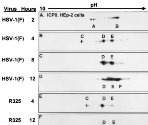
determine the sequence of accumulation of modified forms of ICP0 and ICP4 in the course of HSV-1 infection. Infected cells were harvested at 2, 4, 8, and 12 h after infection for HEp-2 cells and at 4, 8, and 12 h after infection of HEL fibroblasts. The cell lysates were then subjected to two-dimensional elec-trophoresis as described in Materials and Methods. The two-dimensionally separated polypeptides were reacted with anti-bodies to ICP0 and ICP4. Over the course of the first 12 h of infection, both ICP0 and ICP4 were continuously posttransla-tionally modified. Figure 1 shows the forms of ICP0 that were detected in HEp-2 cells. At 2 h after infection, two sets of
species, designated A and B, were present. At 4 h after infec-tion, A and B were replaced by three new species, C, D, and E. By 8 h after infection, species C disappeared, whereas species D and E accumulated in higher quantities. Finally at 12 h after infection, a reduction in species D occurred and there was a trend toward more negatively charged forms (E and F).
[image:3.612.56.292.73.273.2]The patterns of posttranslational modifications of ICP0 pro-cessing in infected HEL fibroblasts are shown in Fig. 2. At 4 h after HSV-1 infection, the prominent species of ICP0 present were designated A and B. By 8 h after infection, these forms were replaced by a faster, less negatively charged D form and a more abundant, slower-migrating, more negatively charged G species of ICP0. By 12 h after infection, the three major species of ICP0 were E and F in addition to the G form. The C isoform of ICP0 was not detected in lysates of infected HEL fibroblasts, suggesting that the accumulation of this isoform is cell type dependent.
Figure 3 shows the pattern of ICP4 forms that accumulate over the first 12 h after infection in HEp-2 cells. At 2 h after infection, we identified a species designated A⬘. At 4 h after infection, ICP4 was present as a major species (B⬘) with a less negative charge and a more highly negatively charged, C⬘ spe-cies. By 8 h after infection, the majority of ICP4 migrated around C⬘. Finally, at 12 h after infection, as was the case for ICP0, the accumulating ICP4 formed two new, more highly negatively charged species designated D⬘and E⬘in addition to the preexisting C⬘species.
Phosphorylation of ICP0 and ICP4 is sensitive to
roscovi-tine.Roscovitine has been reported to be a relatively specific
inhibitor of certain cyclin-dependent kinases (cdk2, cdk5, and cdc2) (22). In addition, roscovitine was reported to adversely affect HSV-1 transcription and overall replication (13, 34–36). Elsewhere, this laboratory reported that cdc2 kinase activity is upregulated in HSV-1-infected cells between 8 and 12 h after infection (2). Also, ICP0 and ICP4 both contain multiple
[image:3.612.315.547.486.649.2]con-FIG. 1. Immunoblots of ICP0 in two-dimensionally separated HSV-1- or R325-infected HEp-2 cell lysates. Infected cells were har-vested at the indicated times postinfection. Time zero was defined as the time the viral inoculum was added to the cells. Lysates were first separated by charge on linear pH 3 to 10 gradients and then separated by size on 6% bisacrylamide gels as described in Materials and Meth-ods. Blots were reacted with a monoclonal antibody to ICP0. The major isoforms that accumulated are given letter designations.
FIG. 2. Immunoblots of ICP0 in two-dimensionally separated HSV-1-infected HEL fibroblast cell lysates. HSV-1-infected cells were harvested at 4, 8, and 12 h postinfection (P.I.). In addition cells were also treated with MG132 or roscovitine. MG132 was given to HSV-1-infected cells 8 h after infection and harvested at 12 h. Roscovitine was added to HSV-1-infected cells at 5 h after infection, and cells were harvested at 12 h postinfection. Immunoblots were reacted with ICPO monoclonal antibody.
on November 9, 2019 by guest
http://jvi.asm.org/
sensus phosphorylation sites for cdc2, and a glutathione S -transferase fusion protein encoding ICP0 exon II was phos-phorylated by cdc2 kinase (3).
To determine whether roscovitine blocked the accumulation of any of the isoforms of ICP0 or ICP4, HEp-2 cells were infected with HSV-1(F). At 5 h after infection, the cells were treated with 100M roscovitine or an equivalent volume of DMSO (0.1%) in phosphate-depleted EMEM. At 6 h after infection, the medium was supplemented with32
P-orthophos-phate, and the exposure to roscovitine or DMSO was contin-ued until 10 h after infection. Cells were then harvested and lysed, and whole-cell lysates were subjected to two-dimensional gel electrophoresis. Autoradiograms and immunoblots for ICP0 and ICP4 were done. The results are shown in Fig. 4 and 5. At 10 h after infection, the lysates of cells exposed to DMSO contained two isoforms of ICP0 corresponding to isoforms D and E of Fig. 1 that were readily apparent by their reactivity with the anti-ICP0 antibody (Fig. 1 and 4A). These forms corresponded to phosphoforms as determined by autoradiog-raphy of the same blot (Fig. 4B). Exposure to roscovitine resulted in a decreased accumulation of the more negatively charged E species of ICP0 and greater accumulation of the less negatively charged D isoform (Fig. 4A and C). The results of autoradiography of the two-dimensionally separated polypep-tides from roscovitine-treated infected cell lysate yielded con-cordant decreased accumulation of the labeled E species com-pared to that of DMSO-treated controls (Fig. 4B and D).
The same blots were also probed with antibody against ICP4. At 10 h after infection, three species of ICP4 corre-sponding to the C⬘, D⬘, and E⬘ of Fig. 3 reacted with the anti-ICP4 antibody (Fig. 3D and 5A). All three forms were labeled with32P-orthophosphate (Fig. 5B). Roscovitine had a
similar effect on the accumulation of specific species of ICP4 phosphoproteins. Roscovitine treatment resulted in the loss of the most negatively charged ICP4 species (E⬘) (Fig. 5C). In lysates of roscovitine-treated cells, there was a greater
accu-mulation of less negatively charged phosphorylated species (C⬘ and D⬘). We conclude from this experiment that roscovitine blocked the accumulation of specific, highly negatively charged species of ICP0 (isoform E) and ICP4 (isoform E⬘) in HEp-2 cells infected with wild-type virus. The forms blocked by rosco-vitine were those that accumulated in significant amounts at or after 8 h after infection.
Specific forms of ICP0 in HEL fibroblasts were also sensitive
FIG. 3. Immunoblots of ICP4 in two-dimensionally separated HSV-1-⫹and R325-infected HEp-2 cell lysates. Immunoblots were prepared and reacted with the anti-ICP4 monoclonal antibody as de-scribed in the legend to Fig. 1.
FIG. 4. Immunoblots (A and C) and corresponding autoradio-grams (B and D) of ICPO in two-dimensionally separated32
P-or-thophosphate-labeled HSV-1-infected HEp-2 cell lysates to determine roscovitine-sensitive ICP0 phosphorylation. HSV-1-infected HEp-2 cells were treated with roscovitine or an equivalent volume of DMSO at 5 h after infection in phosphate-free medium. Infected cells were labeled with32P-orthophosphate from 6 to 10 h postinfection in the
presence of roscovitine. Cells were harvested at 10 h postinfection, and whole-cell lysates were subjected to two-dimensional electrophoresis. Membranes were developed by autoradiography and immunoblotted with an ICP0 antibody. Isoform designations are identical to those shown in Fig. 1.
FIG. 5. Immunoblots (A and C) and corresponding autoradio-grams (B and D) of ICP4 in two-dimensionally separated32
P-ortho-phosphate-labeled HSV-1-infected HEp-2 cell lysates to determine roscovitine-sensitive ICP4 phosphorylation. HSV-1-infected HEp-2 cells were treated with roscovitine or an equivalent volume of DMSO at 5 h after infection in phosphate-free medium. Infected cells were labeled with32P-orthophosphate from 6 to 10 h after infection in the
presence of roscovitine. Cells were harvested at 10 h postinfection, and whole-cell lysates were subjected to two-dimensional electrophoresis. Membranes were developed by autoradiography and immunoblotted with an ICP4 antibody. Isoform designations are idential to those shown in Fig. 3.
on November 9, 2019 by guest
http://jvi.asm.org/
to roscovitine treatment (Fig. 2B, C, and E). In this instance roscovitine blocked the accumulation of the E and F species, which normally accumulated between 8 and 12 h after infec-tion. Instead, roscovitine-treated HEL fibroblasts accumulated the less negatively charged D isoform of ICP0 at 12 h after infection that was present in wild-type virus-infected cells at 8 h after infection. The pattern of ICP0 accumulation at 12 h after 7 h of exposure to roscovitine was similar to that observed at 8 h after infection, suggesting that roscovitine blocked further processing of ICP0 in HEL fibroblasts so that forms E and F of ICP0 could not accumulate.
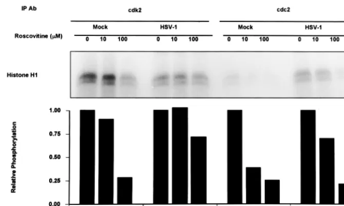
Exposure of HSV-1-infected cells to roscovitine inhibits cdc2 but not the background activity associated with
immunopre-cipitated cdk2 kinase.As stated above, roscovitine is a
rela-tively specific inhibitor of cyclin-dependent kinases cdc2, cdk2, and cdk5 (22). To determine if cdk2 or cdc2 is a target of roscovitine in HSV-1-infected cells, the following experiment was done. Mock- or HSV-1-infected cells were exposed to either roscovitine (10 and 100 M) or DMSO at 5 h after infection. Cdk2 and cdc2 kinase activities were then measured at 12 h after infection. The results are shown in Fig. 6. Mock-infected cells had elevated levels of cdk2 kinase activity, with significantly lower levels of cdc2 activity. In mock-infected cells, a dose response was seen upon addition of roscovitine when histone H1 was used as a substrate. At 100M concen-trations of roscovitine, both cdk2 and cdc2 resulted in a 75% reduction in phosphorylation of histone H1. In HSV-1-infected cells, cdk2 was not activated. Addition of up to 100 M roscovitine resulted in only a 25% reduction in histone H1 phosphorylation by cdk2-immunoprecipitated samples from HSV-1-infected lysates. Thus, the level of histone H1 phos-phorylation by immunoprecipitated cdk2 from lysates of mock-infected cells treated with 100M roscovitine was comparable to cdk2 activity in HSV-1-infected cell lysates with no roscovi-tine. These results argue that cdk2 activity is shut down in
HSV-1 infected cells and that the phosphorylation of histone H1 observed in HSV-1 lysates is background phosphorylation, consistent with the results previously reported for cdk2 kinase activity in HSV-1 infected cells (6, 40). In contrast, immuno-precipitated cdc2 kinase activity is greater in HSV-1-infected cell lysates than in mock-infected cell lysates, as has been previously reported (2). cdc2 kinase activity from HSV-1-in-fected cell lysates shows a roscovitine dose-dependent re-sponse. At 100M roscovitine, the cdc2 kinase activity from HSV-1-infected cell lysates was reduced by 80% compared to 0M roscovitine.
We conclude from these studies that roscovitine at the dose used (100 M) targets cdc2 kinase in HSV-1-infected cells. The effect on cdk2 is inapparent since this cyclin-dependent kinase is either inactive or rendered insusceptible to inhibition by roscovitine.
Involvement of ␣22/US1.5 in the accumulation of specific
forms of ICP0 and ICP4. Earlier studies have shown that in
cells infected with the R325 mutant lacking the carboxyl-ter-minal domain of ICP22 and also the domain encoding the overlapping US1.5 open reading frame, the expression of ␥2
genes was reduced (30, 37). Activation of cdc2 in HSV-1-infected cells is also dependent on ICP22, and overexpression of a dominant negative form of cdc2 resulted in the decreased accumulation of US11, a␥2protein (2, 3, 38). To examine the
effects of ␣22 deletion virus on ICP0 and ICP4 processing, two-dimensional electrophoresis was done on lysates of HEp-2 cells harvested at 4 and 12 h after infection with R325 deletion mutant. The results shown in Fig. 1 and 3 were as follows.
(i) As illustrated in Fig. 1, the electrophoretic mobility of ICP0 in lysates of R325 mutant-infected HEp-2 cells harvested at 4 h after infection was similar to that of ICP0 harvested from wild-type virus-infected cells harvested at the same time after infection. ICP0 present in lysates of cells harvested 12 h after infection with R325 mutant consisted primarily of the D
iso-FIG. 6. Autoradiograms of histone H1 phosphorylated by in vitro kinase assays using immunoprecipitated cdk2 or cdc2 from HSV-1- or mock-infected cells. HEp-2 cells were HSV-1 or mock infected; 5 h after infection, the medium was replaced with medium containing 0, 10, or 100 M roscovitine. Cells were harvested 12 h after infection, and cdk2 or cdc2 was immunoprecipitated (IP) with antibodie (Ab) as described in Materials and Methods. Kinase assays were done using histone H1 as the substrate. Phosphorylated histone H1 was quantitated by a Phosphor-Imager. Relative reduction in the phosphorylation of histone H1 due to roscovitine was determined relative to untreated samples.
on November 9, 2019 by guest
http://jvi.asm.org/
[image:5.612.135.470.70.271.2]form and a small amount of the E isoform. Absent were the highly negatively charged F species present in lysates of wild-type virus-infected cells. The electrophoretic mobility of ICP0 accumulated in R325 virus-infected cells at 12 h after infection corresponded to that of ICP0 in wild-type virus-infected cells at 8 h after infection.
(ii) As illustrated in Fig. 3, ICP4 present in HEp-2 cells harvested 4 h after infection with R325 mutant was primarily of the C⬘isoform, although we also detected trace amounts of the B⬘isoform. In contrast to ICP4 harvested from wild-type virus-infected cells, the B⬘species was absent (Fig. 3B and E). The posttranslational modification of ICP4 was arrested at this point since the protein present in cells harvested at 12 h after infection with R325 mutant formed primarily the C⬘ species (Fig. 3E and F). Absent were the highly negatively charged D⬘ and E⬘ species which accumulated in cells harvested at 12 h after infection with wild-type virus (Fig. 3D).
In HEL fibroblasts, R325 infection resulted in the accumu-lation of species of ICP0 that resembled those present in in-fected cells treated with roscovitine (Fig. 2E and 7B). At 12 h after infection, the G isoform of ICP0 was present in lysates of both wild-type virus- and R325 mutant-infected HEL fibro-blasts (Fig. 7). However, form E was not observed in R325-infected cell lysates; instead, a less negatively charged, D spe-cies was present. This spespe-cies was also present in lysates of cells infected with wild-type virus and treated with roscovitine (Fig. 2E).
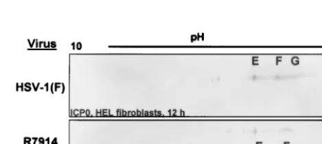
The pattern of accumulation of the ICP0 isoforms in cells infected with R7914 was similar to that of HEL fibroblasts
infected with wild-type virus and treated with MG132.Studies
reported elsewhere have shown that in HSV-1(F)-infected HEL fibroblasts, ICP0 localized in the nucleus early in infec-tion and in the cytoplasmic compartment at late times after infection (14, 40). Treatment of HSV-1-infected HEL fibro-blasts late in infection with the proteasome inhibitor MG132 resulted in the relocation of ICP0 to the nucleus (19). The goal of this set of studies was to determine if differential subcellular localization of ICP0 was in part dependent on its posttransla-tional modifications. To this end, two experiments were done. HEL fibroblasts were infected with R7914 or HSV-1 and then treated with MG132.
HEL fibroblasts infected with HSV-1 or R7914 were har-vested 12 h after infection. Two-dimensional gel electrophore-sis revealed that whereas isoforms E and F were found in lysates from both HSV-1- and R7914-infected cells, the highly
negatively charged species (G) was present only in HSV-1-infected cell lysates (Fig. 8). Treatment of HSV-1-HSV-1-infected HEL fibroblasts with MG132 at 8 h after infection also resulted in the loss of species G (Fig. 2D). MG132 had no appreciable effect on the accumulation of E and F forms of ICP0. These results suggest that the G species may represent cytoplasmic forms of ICP0 whereas the E and F species represent nuclear species of ICP0.
DISCUSSION
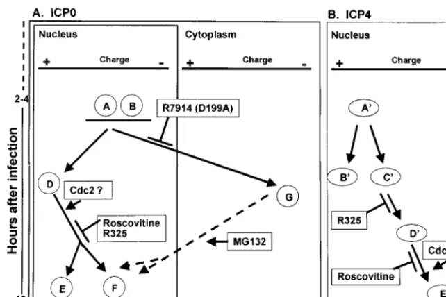
The fundamental hypothesis underlying these studies is that in the case of multifunctional HSV-1 proteins, posttransla-tional modifications determine the specific function performed by the protein at the specific time or cellular compartment in which it is localized. ICP4 and ICP0, the two ␣ regulatory proteins selected for this study, are posttranslationally modi-fied by multiple viral and cellular enzymes and play a role in viral replication throughout the viral replicative cycle. In this report we show that consistent with this hypothesis, both ICP4 and ICP0 are sequentially modified throughout at least 12 h after infection and that at least some of the modifications reflect the action of specific enzymes or correlate with local-ization in specific cellular compartments. The salient features of the data are schematically depicted in Fig. 9 and may be summarized as follows.
(i) Over the course of infection, ICP0 and ICP4 are sequen-tially processed. The general trend was the accumulation of more negatively charged forms of both ICP0 and ICP4 as infection proceeded. This was observed in both infected HEp-2 cells and HEL fibroblasts. Earlier reports indicated that hyper-phosphorylated forms of ICP4 are necessary to bind to pro-moter elements onand␥genes (28). Increasing accumula-tion of negatively charged forms of ICP0 and ICP4 is consistent with the requirements for post-␣gene expression and with the evidence that ICP4 is required throughout viral replication.
[image:6.612.57.291.73.155.2](ii) We have previously shown that cdc2 kinase activity is enhanced in HSV-1-infected cells (2). A central question re-lated to that observation is why cdc2 was activated so late in the replicative cycle. This question was solved to some extent in studies with an inactive, dominant negative mutant form of cdc2 (3, 38). Cells infected with wild-type virus and transfected
FIG. 7. Immunoblots of ICP0 in two-dimensionally separated ly-sates of HSV-1- or R325-infected HEL fibroblasts. Cells were har-vested at 12 h after infection and separated by two-dimensional elec-trophoresis. Immunoblots were reacted with ICP0 monoclonal antibody. Isoform designations are the same as in Fig. 2.
FIG. 8. Immunoblots of ICP0 in two-dimensionally separated ly-sates of HSV-1 or R7914 (alanine substituted for aspartic acid of ICP0)-infected HEL fibroblasts. The cells were harvested at 12 h after infection, lysed, and subjected to two-dimensional electrophoresis. Im-munoblots were reacted with anti ICP0 antibody. Isoform designations are the same as in Fig. 2.
on November 9, 2019 by guest
http://jvi.asm.org/
[image:6.612.317.552.563.668.2]with cdc2 dominant negative construct expressed representa-tive ␣, , and ␥1 genes but failed to accumulate significant
amounts of US11, a representative␥2protein. Optimal
expres-sion of␥2genes is in part dependent on ICP22 and UL13 (27,
32). A cascade can be envisioned involving UL13 and ICP22
activating cdc2, resulting in enhanced␥2gene expression. The
data in the present study add further weight to such a scenario. Forms of ICP0 and ICP4 that accumulated between 8 and 12 h after HSV-1 infection were sensitive to both roscovitine as well as infection by the recombinant R325, which lacks the domain encoding the US1.5 protein and the coterminal
carboxyl-ter-minal domain of ICP22.
Roscovitine treatment of HSV-1-infected cells resulted in a loss of specific phospholabeled forms of both ICP0 and ICP4 (Fig. 4, 5, and 9). The forms of ICP0 and ICP4 phospholabeled in the presence of roscovitine may reflect the effects of other kinases that phosphorylate ICP0 and ICP4. Roscovitine is a relatively selective inhibitor of cdk2, cdc2, and cdk5 (22). The effects of roscovitine on HSV-1 replication and gene transcrip-tion have been previously reported (13, 34–36). The major effect of roscovitine in HSV-1-infected cells was on cdc2; the cdk2-associated activity, the other target of roscovitine, either represented background kinase activity not associated with this enzyme or was shielded from the inhibitory effects of roscovi-tine.
The absence of the carboxyl-terminal domain of ICP22 and the corresponding absence of US1.5 protein had a drastic effect
on the posttranslational modification of ICP0 and ICP4. In contrast to wild-type virus-infected cells, cells infected with the R325 mutant virus accumulated at 12 h after infection the same forms of ICP0 and ICP4 as those that were present at 4 h
after infection with either wild-type or mutant virus (Fig. 9). The missing forms were those that were more negatively charged and that were sensitive to roscovitine. These observa-tions are consistent with the data showing that activation of cdc2 requires intact␣22/US1.5 gene products. In light of the
observation reported earlier that exon II of ICP0 is a substrate for cdc2, it is likely that ICP0 and ICP4 serve as bona fide in vivo substrates for cdc2. However, it appears that ICP22 may influence the activity of other kinases as well.
(iii) The results presented in this study indicate a correlation between the extent of posttranslational modifications of ICP0 and its localization in the infected cell. In HEL fibroblasts, ICP0 initially localizes to the nucleus (14, 40) and later, be-tween 5 and 9 h after infection, is translocated to the cyto-plasm. One mechanism for altering subcellular localization of proteins is through posttranslational processing (12). In lysates of HSV-1-infected HEL fibroblasts, a novel, highly negative charged form of ICP0 appeared between 4 and 8 h after in-fection (isoform G [Fig. 2]). Two lines of evidence support that this highly negatively charged form of ICP0 in HEL cells cor-relates with cytoplasmic localization. First, ICP0 encoded by the recombinant virus R7914 accumulates in the nucleus and is not translocated to the cytoplasm (40). In HEL fibroblasts infected with the R7914 mutant, the highly negatively charged G form did not accumulate (Fig. 8). Second, in cells infected with wild-type virus and treated with MG132 at late times after infection, ICP0 is translocated from the cytoplasm to the nu-cleus (again), the D form of ICP0 was not detected in HEL fibroblasts infected with HSV-1(F) and treated with MG132 late in infection (Fig. 2C and D). The latter observation is particularly significant since it suggests that the
posttransla-FIG. 9. Schematic representation of ICP0 and ICP4 isoforms observed after two-dimensional separation of lysates of HSV-1-infected HEL fibroblasts. (A) Absence of specific isoforms from lysates of HEL fibroblasts infected with HSV-1 mutants or with wild-type virus and treated with roscovitine. Isoforms E and F failed to accumulate in lysates of R325-infected or roscovitine-treated, HSV-1-infected cells. These isoforms are putative nuclear forms, and their processing is dependent on cdc2 kinase. HEL fibroblasts infected with R7914 or wild-type virus and treated with MG132 failed to accumulate the G isoform, and this isoform appears to be associated with ICP0 translocated into the cytoplasm. (B) Isoforms D⬘ and E⬘of ICP4 failed to accumulate in HEL fibroblasts infected with R325 mutant. The most negatively charged, E⬘isoform of ICP4 was not detected in significant amounts in cells infected with wild-type virus and treated with roscovitine. The accumulation of the ICP4 isoform E⬘may also be dependent on cdc2 kinase.
on November 9, 2019 by guest
http://jvi.asm.org/
[image:7.612.144.465.73.287.2]tional modification associated with the G form of ICP0 is reversible and specifically associated with the cytoplasmic lo-calization of the protein. Whereas the G form of ICP0 is cytoplasmic, the E and F isoforms may represent nuclear spe-cies.
The G form of ICP0 was present in cells infected with R325 and in cells infected with wild-type virus and exposed to inhib-itory concentrations of roscovitine. These results indicate that neither ICP22/US1.5 protein or cdc2 is required for
cytoplas-mic localization of ICP0.
(iv) Finally, we observed differences in the electrophoretic mobility of ICP0 and ICP4 derived from HEp-2 cells or HEL fibroblasts infected with wild-type or mutant virus. A notable example of these differences is the absence of the C isoform of ICP0 in lysates of wild-type virus-infected HEL fibroblasts. The differences most likely reflect posttranslational modifica-tions of the viral proteins effected by enzymes specific for each cell type. The significance of these observations stems from the observation that the level of accumulation of many HSV-1 proteins varies depending on the cell line. In this laboratory at least, several cell lines are screened to determine the level of accumulation of specific proteins. The significant aspect rele-vant to this study are deletion mutants in ␣22/US1.5. The
recombinant virus R325, for example, replicates almost as well as wild-type virus in HEp-2 cells but at much reduced levels in primary human cell lines or in cell lines of rodent or rabbit derivation (37). Cell infected with this mutant underproduce a subset of␥2proteins; the magnitude of the effect is cell type
dependent (27, 37). It is conceivable that cell type specificity reflects the mixture of enzymes available for posttranslational modifications of ICP0 and ICP4 and that host enzymes can compensate, at least in part, for the absence of appropriate viral regulatory proteins.
ACKNOWLEDGMENTS
We thank Charles Van Sant for invaluable advice, Alice P. W. Poon for a careful reading of the manuscript, and Vladimir Mogilner for photographic services.
These studies were aided by Public Health Service grants CA87761, CA83939, CA71933, and CA78766 U from the National Cancer Insti-tute. R.H. is a Howard Hughes Medical Institute predoctoral fellow.
REFERENCES
1.Ackermann, M., D. K. Braun, L. Pereira, and B. Roizman.1984. Character-ization of herpes simplex virus 1 alpha proteins 0, 4, and 27 with monoclonal antibodies. J. Virol.52:108–118.
2.Advani, S. J., R. Brandimarti, R. R. Weichselbaum, and B. Roizman.2000. The disappearance of cyclins A and B and the increase in activity of the G2/M phase cellular kinase cdc2 in herpes simplex virus 1-infected cells
requires the expression of␣22/US1.5 and UL13 viral genes. J. Virol.74:8–15.
3.Advani, S. J., R. R. Weichselbaum, and B. Roizman.2000. The role of cdc2 in the expression of herpes simplex virus genes. Proc. Natl. Acad. Sci. USA
97:10996–11001.
4.Blaho, J. A., and B. Roizman.1991. ICP4, the major regulatory protein of herpes simplex virus, shares features common to GTP-binding proteins and is adenylated and guanylated. J. Virol.65:3759–3769.
5.Blaho, J. A., C. Mitchell, and B. Roizman.1993. Guanylylation and adeny-lylation of the alpha regulatory proteins of herpes simplex virus require a viral beta or gamma function. J. Virol.67:3891–3900.
6.Ehmann, G. L., T. I. McLean, and S. L. Bachenheimer.2001. Herpes simplex virus type 1 infection imposes a G1/S block in asynchronously growing cells
and prevents G1entry in quiescent cells. Virology267:335–349.
7.Ejercito, P., E. D. Kieff, and B. Roizman.1968. Characterization of herpes simplex virus strains differing in their effects on social behavior of infected cells. J. Gen. Virol.2:357–364.
8.Everett, R. D., and G. G. Maul.1994. HSV-1 IE protein Vmw110 causes redistribution of PML. EMBO J.13:5062–5069.
9.Everett, R. D., M. Meredith, A. Orr, A. Cross, M. Kathoria, and J. Parkin-son.1997. A novel ubiquitin-specific protease is dynamically associated with the PML nuclear domain and binds to a herpesvirus regulatory protein. EMBO J.16:566–577.
10. Everett, R. D., W. C. Earnshaw, J. Findlay, and P. Lomonte.1999. Specific destruction of kinetochore protein CENP-C and disruption of cell division by herpes simplex virus immediate-early protein Vmw110. EMBO J.18:1526– 1538.
11. Everett, R. D.2000. ICP0 induces the accumulation of colocalizing conju-gated ubiquitin. J. Virol.74:9994–10005.
12. Hood, J. K., and P. A. Silver.1999. In or out? Regulating nuclear transport. Curr. Opin. Cell Biol.11:241–247.
13. Jordan, R., L. Schang, and P. A. Schaffer.1999. Transactivation of herpes simplex virus type 1 immediate-early gene expression by virion-associated factors is blocked by an inhibitor of cyclin-dependent protein kinases. J. Vi-rol.73:8843–8847.
14. Kawaguchi, Y., R. Bruni, and B. Roizman.1997. Interaction of herpes simplex virus 1 alpha regulatory protein ICP0 with elongation factor 1␦: ICP0 affects translational machinery. J. Virol.71:1019–1024.
15. Kawaguchi, Y., C. Van Sant, and B. Roizman.1997. Herpes simplex virus 1 alpha regulatory protein ICP0 interacts with and stabilizes the cell cycle regulator cyclin D3. J. Virol.71:7328–7336.
16. Kawaguchi, Y., M. Tanaka, A. Yokoymama, G. Matsuda, K. Kato, H. Ka-gawa, K. Hirai, and B. Roizman.2001. Herpes simplex virus 1 alpha regu-latory protein ICP0 functionally interacts with cellular transcription factor BMAL1. Proc. Natl. Acad. Sci. USA98:1877–1882.
17. King, R. W., P. K. Jackson, and M. W. Kirschner.1994. Mitosis in transition. Cell79:563–571.
18. Leopardi, R., N. Michael, and B. Roizman.1995. Repression of the herpes simplex virus 1 alpha 4 gene by its gene product (ICP4) within the context of the viral genome is conditioned by the distance and stereoaxial alignment of the ICP4 DNA binding site relative to the TATA box. J. Virol.69:3042–3048. 19. Lopez, P., C. Van Sant, and B. Roizman.2001. Requirements for the nucle-ar-cytoplasmic translocation of infected-cell protein 0 of herpes simplex virus 1. J. Virol.75:3832–3840.
20. Marin, O., F. Meggio, G. Draetta, and L. A. Pinna.1992. The consensus sequences for cdc2 kinase and for casein kinase-2 are mutually incompatible. A study with peptides derived from the beta-subunit of casein kinase-2. FEBS Lett.301:111–114.
21. McLean, T. I., and S. L. Bachenheimer.1999. Activation of cJUN N-terminal kinase by herpes simplex virus type 1 enhances viral replication. J. Virol.
73:8415–8426.
22. Meijer, L., A. Borgne, O. Mulner, J. P. Chong, J. J. Blow, N. Inagaki, M. Inagaki, J. G. Delcros, and J. P. Moulinoux.1997. Biochemical and cellular effects of roscovitine, a potent and selective inhibitor of the cyclin-dependent kinases cdc2, cdk2 and cdk5. Eur. J. Biochem.243:527–536.
23. Michael, N., D. Spector, P. Mavromara-Nazos, T. M. Kristie, and B. Roiz-man.1988. The DNA-binding properties of the major regulatory protein alpha 4 of herpes simplex viruses. Science239:1531–1534.
24. Michael, N., and B. Roizman.1989. Binding of the herpes simplex virus major regulatory protein to viral DNA. Proc. Natl. Acad. Sci. USA86:9808– 9812.
25. Mitchell, C. A., J. A. Blaho, A. L. McCormick, and B. Roizman.1997. The nucleotidylation of herpes simplex virus 1 regulatory protein␣22 by human casein kinase II. J. Biol. Chem.272:25394–25400.
26. Ogle, W. O., T. I. Ng, K. L. Carter, and B. Roizman.1997. The UL13 protein kinase and the infected cell type are determinants of post-translational processing modifications of ICP0. Virology235:406–413.
27. Ogle, W. O., and B. Roizman.1999. Functional anatomy of herpes simplex virus 1 overlapping genes encoding infected-cell protein 22 and US1.5 pro-tein. J. Virol.73:4305–4315.
28. Papavassiliou, A. G., K. W. Wilcox, and S. J. Silverstein.1991. The interac-tion of ICP4 with cell/infected-cell factors and its state of phosphorylainterac-tion modulate differential recognition of leader sequences in herpes simplex virus DNA. EMBO J.10:397–406.
29. Parkinson, J., S. P. Lees-Miller, and R. D. Everett.1999. Herpes simplex virus type 1 immediate-early protein Vmw110 induces the proteasome-de-pendent degradation of the catalytic subunit of DNA-deproteasome-de-pendent protein kinase. J. Virol.73:650–657.
30. Post, L. E., and B. Roizman.1981. A generalized technique for deletion of specific genes in large genomes: alpha gene 22 of herpes simplex virus 1 is not essential for growth. Cell25:227–232.
31. Purves, F. C., and B. Roizman.1992. The UL13 gene of herpes simplex virus 1 encodes the functions for posttranslational processing associated with phosphorylation of the regulatory protein alpha 22. Proc. Natl. Acad. Sci. USA89:7310–7314.
32. Purves, F. C., W. O. Ogle, and B. Roizman.1993. Processing of the herpes simplex virus regulatory protein alpha 22 mediated by the UL13 protein kinase determines the accumulation of a subset of alpha and gamma mRNAs and proteins in infected cells. Proc. Natl. Acad. Sci. USA90:6701–6705. 33. Roizman, B., and A. E. Sears.1996. Herpes simplex viruses and their
repli-cation, p. 2231–2296.InB. N. Fields, D. M. Knipe, and P. M. Howley (ed.),
on November 9, 2019 by guest
http://jvi.asm.org/
Fields virology, 3rd ed. Lippincott-Raven, Philadelphia, Pa.
34.Schang, L. M., J. Phillips, and P. A. Schaffer.1998. Requirement for cellular cyclin-dependent kinases in herpes simplex virus replication and transcrip-tion. J. Virol.72:5626–5637.
35. Schang, L. M., A. Rosenberg, and P. A. Schaffer.1999. Transcription of herpes simplex virus immediate-early and early genes is inhibited by rosco-vitine, an inhibitor specific for cellular cyclin-dependent kinases. J. Virol.
73:2161–2172.
36.Schang, L. M., A. Rosenberg, and P. A. Schaffer.2000. Roscovitine, a specific inhibitor of cellular cyclin-dependent kinases, inhibits herpes simplex virus DNA synthesis in the presence of viral early proteins. J. Virol.74:2107–2020. 37. Sears, A. E., I. W. Halliburton, B. Meignier, S. Silver, and B. Roizman.1985. Herpes simplex virus 1 mutant defective in the alpha 22 gene: growth and gene expression in permissive and restrictive cells and establishment of latency in mice. J. Virol.55:338–346.
38. Van den Heuvel, S., and E. Harlow.1993. Distinct roles for cyclin-dependent kinases in cell cycle control. Science262:2050–2054.
39. Van Sant, C., Y. Kawaguchi, and B. Roizman.1999. A single amino acid
substitution in the cyclin D binding domain of the infected cell protein no. 0 abrogates the neuroinvasiveness of herpes simplex virus without affecting its ability to replicate. Proc. Natl. Acad. Sci. USA96:8184–8189.
40. Van Sant, C., P. Lopez, S. J. Advani, and B. Roizman.2001. Role of cyclin D3 in the biology of herpes simplex virus ICP0. J. Virol.75:1888–1898. 41. Wilcox, K. W., A. Kohn, E. Sklyanaskaya, and B. Roizman.1980. Herpes
simplex virus phosphoproteins. I. Phosphate cycles on and off some viral polypeptides and can alter their affinity for DNA. J. Virol.33:167–182. 42. Xia, K., D. M. Knipe, and N. A. DeLuca.1996. Role of protein kinase A and
the serine-rich region of herpes simplex virus type 1 ICP4 in viral replication. J. Virol.70:1050–1060.
43. Xia, K., N. A. DeLuca, and D. M. Knipe.1996. Analysis of the phosphory-lation sites of herpes simplex virus type 1 ICP4. J. Virol.70:1061–1071. 44. Yao, F., and P. A. Schaffer.1994. Physical interaction between the herpes
simplex virus type 1 immediate-early regulatory proteins ICP0 and ICP4. J. Virol.68:8158–8168.
45. Zhi, Y., and R. M. Sandri-Goldin.1999. Analysis of the phosphorylation sites of herpes simplex virus 1 regulatory protein ICP27. J. Virol.73:3246–3257.