FORMULATION AND EVALUATION OF
REPACLINIDE TRANSDERMAL PATCH FOR AN
ANTI DIABETIC ACTIVITIES
A dissertation submitted to
THE TAMILNADU Dr.M.G.R MEDICAL UNIVERSITY
CHENNAI - 600 032.In partial fulfillment of the requirements for the award of Degree of
MASTER OF PHARMACY
IN
PHARMACEUTICS
SubmittedBy
Reg.No:
261311153
Under the guidance of
Dr.M.Murugan
,
M.Pharm.,Ph.D.,DEPARTMENT OF PHARMACEUTICS
EDAYATHANGUDY.G.S PILLAY COLLEGE OF PHARMACY
NAGAPATTINAM-611002
FORMULATION AND EVALUATION OF
REPACLINIDE TRANSDERMAL PATCH FOR AN
ANTI DIABETIC ACTIVITIES
A dissertation submitted to
THE TAMILNADU Dr.M.G.R MEDICAL UNIVERSITY
CHENNAI - 600 032.In partial fulfillment of the requirements for the award of Degree of
MASTER OF PHARMACY
IN
PHARMACEUTICS
SubmittedBy
G.PANCHACHARAM
(Reg. No:
261311153
Under the guidance of
Dr.M.Murugan
,
M.Pharm.,Ph.D .,DEPARTMENT OF PHARMACEUTICS
EDAYATHANGUDY.G.S PILLAY COLLEGE OF PHARMACY
NAGAPATTINAM-611002
Dr.M.Murugan
, M . Ph a r m., P h. D .,Director cum Professor,
Department of Pharmaceutics,
Edayathangudy.G.S.Pillay College of Pharmacy,
Nagapattinam – 611 002.
CERTIFICATE
This
is
to
certify
that
the
dissertation
ent itled
“FORMULATION AND EVALUATION OF REPACLINIDE
TRANSDERMAL
PATCH
FOR
AN
A NTI
DIABETIC
ACTIVITIES”
submitted
by
G.Panchacharam
(Reg.
No:
261311153
) in partial fulfillment for the award of degree of Master
of Pharmacy to the Tamilnadu Dr. M.G.R Medical Uni versity,
Chennai is an independent bonafide work of the candidate carried
out under my guidance in the Department of Pharmaceutics,
Edayathangudy.G.S Pillay College of Pharmacy during the academic
year 2014-2015.
Place: Nagapattinam
Dr.M.Murugan, M.Pharm., Ph.D.,
Prof.Dr.D.Babu Ananth,
M.Pharm., Ph.D. ,Principal,
Edayathangudy.G.S.Pillay College of Pharmacy,
Nagapattinam – 611 002.
CERTIFICATE
This is to certify that the dissertation
“FORMULATION AND
EVALUATION OF REPACLINIDE TRANSDERMAL PATCH
FOR
AN
ANTI
DIABETIC
ACTIVITIES ”
submitted
by
G.Panchacharam
(Reg. No:
261311153
) in partial fulfillment for
the award of degree of Master of Pharmacy to the Tamilnadu Dr.
M.G.R Medical University, Chennai is an independent bonafide
work of the candidate carried out under the guidance of
Dr.M.Murugan, M.Pharm., Ph.D.,
Director cum Professor,
Edayathangudy.G.S Pillay College of Pharm acy during the academic
year 2014-2015.
Place: Nagapattinam
Prof.Dr.D.Babu Ananth,
M.Pharm., Ph.D.,ACKNOWLEDGEMENT
I would like to express profound gratitude to
Mrs.Jothimani
G.S.Pillay,
Chairman, E.G.S.Pillay College of Pharmacy, and
Thiru. S.Paramesvaran,
M.Com., FCCA.,Secretary, E.G.S.Pillay
College of Pharmacy.
I express my sincere and deep sense of gratitude to my guide
Dr.M.Murugan, M.Pharm., Ph.D.,
Director cum Professor Head,
Department of Pharmaceutics E.G.S.Pillay College of Pharmacy, for
his invaluable and extreme support, encouragement, and co
-operation throughout the course of my work.
It is my privilege to express my heartfelt thanks to
Prof.
Dr.D.Babu Ananth, M.Pharm, Ph.D. ,
Principal, E.G.S.Pillay
College of Pharmacy, for providing me all facilities and
encouragement throughout the research work .
I express my sincere gratitude to
Prof.K.Shahul Hameed
Maraicar,
M.Pharm., (Ph.D).
, Director cum Professor, Department
of Pharmaceutics. E.G.S.Pillay College of Pharmacy, for his
encouragement throughout the course of my work.
I would like to extend my thanks to all the
Teaching Staff
and
Non Teaching Staff
, who are all supported me for the
successful completion of my project w ork.
Last but not least, I express my deep sense of gratitude to my
parents, my wife, family members and friends for their constant
INDEX
S.NO
CONTENTS
PAGE NO
1
INTRODUCTION
1
2
LITERATURE REVIEW
20
3
OBJECTIVE
28
4
DRUG PROFILE
29
5
MATERIALS & METHODS
39
6
RESULTS & DISCUSSION
56
7
SUMMARY
102
8
CONCLUSION
106
INTRODUCTION
The term drug delivery covers a very broad range of techniques
used to deliver therapeutic agents into the human body. The limitations
of the most obvious and trusted drug delivery techniques, those of the
ingested tablet and of the intraveno us (IV) / subcutaneous (SC) /
intramuscular (IM) injections, have been recognized for some time
now. The former delivers drug into the blood only through hepatic
system, and hence the amount in blood stream may be much lower than
the amount formulated into tablet (i.e., it has low bio -availability);
furthermore, liver damage is an unfortunate side effect of many soluble
tableted drugs. The injection mode of delivery can be used to deliver
any size of drug molecule and is versatile in this regard, but suffer s
from the obvious disadvantage of being invasive and painful, and the
less obvious disadvantage of shortness of duration (for drugs with short
half lives).
To overcome some of these limitations, other modes of delivery
of drugs into the body were investi gated, beginning in the early 1970s.
Transdermal (through intact skin), transmucosal, transalveolar
(inhalation, through lung tissue), implantable (subcutaneous and deeper
implants, delivery into surrounding tissue), and injectable (IM or SC)
modes of deli very have all been explored extensively over the last 25
The delivery of drugs using skin as the port of entry is known as
transdermal administration and the drug delivery systems are kn own as
transdermal therapeutic systems or transdermal drug delivery systems
or popularly known as transdermal patches.1
“Transdermal therapeutic systems are defined as self contained,
discrete dosage forms which, when applied to the intact skin, deliver
the drug(s), through the skin, at a controlled rate to the systemic
circulation.”
The success of this approach is evidenced by the fact that there
are currently more than 35 approved transdermal drug delivery
products for the treatment of a wide variety o f conditions including:
hypertension, angina, motion sicknesses, and recently contraception
and urinary incontinence. There are also several products in late -stage
development that will further expand transdermal drug delivery usage
into new therapeutic ar eas, including Parkinson‟s disease.1 , 2 , 3
The polymers selected for the study are Ethyl cellulose and
Hydroxypropylmethyl cellulose. The drug taken up for the study is
Repaglinide.
Repaglinide, an antidiabetic chemically
2-ethoxy-4-[{[(15}-3-methyl -1-[2-(piperdinyl) phenyl] butylcarbamoyl} 2-ethoxy-4-[{[(15}-3-methyl] benzolicacid
In the present study, transdermal patches of the Repaglinide were
prepared using polymers like H ydroxypropylmethy l cellulose and Ethyl
cell ulose, both in different combination . These transdermal patches
were characterized for their physicochemical properties including drug
release.
In recent years considerable attention has been focused on the
development of new drug delivery systems. The c onventional dosage
forms such as tablets, capsules, and parenterals with the one exception
of continuous intravenous infusion are inefficient because of several
reasons. These dosage forms release the drug at faster rate initially
leading to quick rise in blood levels and thereafter there is an
exponential fall until another dose is administered. This results in peak
and valley pattern of drug levels in blood. Thus, for most of the time
drug concentration is either above the therapeutic level leading to
adverse reaction with potent drug or below it leading to
ineffectiveness. The need to minimize blood level fluctuation of drug
has led to the development of controlled drug delivery system.
Recently, it is evident that the benefits of intravenous drug
infusion can closely be duplicated, without its disadvantages, by using
the skin as a port for drug administration, to provide continuous
Transdermal drug delivery systems (TDDS) are designed to
deliver the drug substances from the surface of the skin through its
various layers into the systemic circulation. TDDS is the delivery of
drugs through the skin to achieve systemic effects. Transdermal
patches control the delivery of drugs at controlled rates by employing
an appropriate combination of hydrophilic and lipophillic polymers.3 , 4
Advantages 3 , 4
Prevents the variation in the absorption and metabolism
associated with oral drug administration.
Prevents the risk and inconvenience of intravenous therapy.
Permits continuous zero -order drug administration and the use of
drugs with short biological half -lives.
Increases the bioavailability and efficacy of drugs, since it
bypasses hepatic first -pass elimination.
Provide a simple therapeutic regime, leading to good patient
compliance that can be easily terminated by simple removal of
the patch.
Transdermal medication delivers a steady infusion of a drug over
an extended period of time. Adverse effects or therapeutic
failures frequently associated with intermitte nt dosing can also
be avoided.
Limitations 4 , 5
The transdermal route of administration is unsuitable for drugs
that irritate or sensitize the skin.
Only potent drugs are suitable candidates for transdermal
delivery due to t he natural limits of drug entry imposed by the
skin permeability.
Technical difficulties are associated with the adhesion of the
systems to different skin types and under various environmental
conditions, and the development of rate controlling conditions.
Drugs requiring high blood levels to achieve an effect are
difficult to load into a transdermal system due to large physical
amount of material required.
Factors Affecting Transdermal Permeability
The principal transport mechanism across mammalian skin i s by
passive diffusion, which is primarily the trans -epidermal route at
steady state or through trans -appendageal route at initial non -steady
state.
The factors controlling transdermal permeability are as follows:
Partition Coefficient: Drugs possessing b oth lipid and water
solubility are favourably absorbed through the skin. Transdermal
permeability coefficient shows a linear dependency on partition
coefficient. A lipid/ water partition coefficient of one or greater is
The partition coefficient of a drug molecule may be altered by
chemical modification of its functional groups, which can be done
without affecting the pharmacological activity of the drug. It has been
established that membrane partit ion coefficient increases exponentially
as the length of the lipophilic alkyl chain increases. Varying the
vehicle may also alter the partition coefficient of a drug molecule.
pH condition: Solution whose pH values are very high or very
low can be destruc tive to the skin. With moderate pH values, the flux
of ionizable drugs can be affected by pH that alters the ratio of charged
and uncharged species and their transdermal permeability.
Penetrant concentration: Assuming membrane limited
transport, increasin g the concentration of dissolved drug causes a
proportional increase in flux. At concentration higher than the
solubility, excess solid drug acts as a reservoir and helps to maintain a
constant drug concentration for a prolonged period of time.
Transderma l Drug Penetration through Skin 6 , 7 , 8
Physiology of Skin
Although the skin is a large and logical target for drug delivery,
its basic functions limit its utility for this purpose. The skin functions
mainly to protect the body from external insults (e.g., h armful
substances and microorganisms) and to contain all body fluids. It must
skin serve as poor conductors of electricity and can protect us from
electrical current if the need arises. The skin also helps to regulate
body temperature.
There are two important layers to human skin: the epidermis and
the dermis. For transdermal delivery, drugs must pass through the two
sub layers of the epidermis to reach the microcirculation of the dermi s.
The stratum corneum (or the horny layer) is the top layer of the skin
and varies in thickness from approximately 10 microns to several
hundred microns, depending on the region of the body. It is composed
of layers of dead, flattened keratinocytes surrou nded by a lipid matrix,
which together act as a brick -and-mortar system that is difficult to
penetrate. The stratum corneum provides the most significant barrier to
diffusion. In fact, the stratum corneum is the barrier to approximately
90% of transdermal drug applications. However, nearly all molecules
penetrate it to some minimal degree. Below the stratum corneum lies
the viable epidermis. This layer is about ten times as thick as the
stratum corneum; however, diffusion is much faster here due to the
greater degree of hydration in the living cells of the viable epidermis.
It contains Langerhans' cells, which function as antigen presenting
cells to the immune system. These cells are the targets for transdermal
vaccine delivery. Melanocytes in the viable ep idermis provide skin
pigmentation. Below the epidermis lies the dermis, which is
approximately one millimeter thick and 100 times the thickness of the
drugs into the systemic circulation an d to regulate temperature, a
system known as the skin's microcirculation. The dermis also contains
sensory neurons and a lymphatic network. The lymphatic removal of
transdermally applied drugs has not been studied extensively.
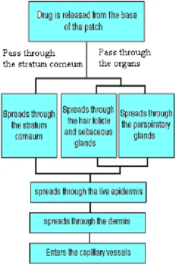
There are two main pathways by which the drugs cross the skin
and reach the systemic circulation. The most direct route is known as
the “transcellular pathway”. By this route, drugs cross the skin by
directly passing through both the phospholipid me mbranes and the
cytoplasm of the de ad keratinocytes that constitute the stratum
corneum. Although this is the path of shortest distance, the drugs
encounter significant resistance to permeation. This is because the
drugs must cross the lipophilic membrane of each cell, then the
hydrophilic cellular contents containing keratin, and then the
phospholipid bilayer of the cell one more time. This series of steps is
repeated number of times to traverse the full thickness of the stratum
corneum. A few drugs have the properties to cross via this met hod.
The more common pathway through the skin is via the
intercellular route. Drugs cross the skin by this route and pass through
the small spaces between the cells of the skin, making the route more
tortuous. Although the thickness of the stratum corneum is only about
20 µm, the actual diffusion path of most molecules crossing the skin is
on the order of 400 µm. The 20 -fold increase in the actual path of
A less important pathway of drug p enetration is the follicular
route. Hair follicles penetrate through the stratum corneum, allowing
more direct access to the dermal microcirculation. However, hair
follicles occupy only 1/1000 of the entire skin surface area.
[image:15.595.207.386.458.731.2]Consequently, very little drug crosses the skin via the follicular route.
Basic Components of Transdermal Drug Delivery Systems 6 , 7 , 8
Transdermal drug delivery systems are designed to support the
passage of drug substance from the surface of skin, through its various
layers, and into the systemic circulation. There are basic components
of transdermal dosing system; which are those that cont rol the rate of
drug delivery to the skin. The components of devices include;
The drug substance
Polymer matrix
Permeation enhancer
Adhesives
Backing membrane
1. The drug substance: Judicious choice of drug is critical in
the successful development of a transdermal product. The important
drug properties that affect its diffusion from device as well as across
the skin include molecular weight, solubility, physical properties and
melting point. The structure of the drug also affects the skin
penetration. Di ffusion of the drug in adequate amount to produce a
satisfactory therapeutic effect is of prime importance. Other parameters
such as skin irritation and clinical need should be considered before a
The following are some of the desirabl e properties of a drug for
transdermal delivery.
The drug should have molecular weight less than 1000 Daltons.
The drug should have affinity for both lipophilic and hydrophilic
phases.
The drug should have a low melting point.
The half-life of drug should be short.
The drug must not induce a cutaneous or allergic response.
The drugs, which degrade in gastrointestinal tract or inactivated
by hepatic first pass effect are suitable candidates for
transdermal drug delivery system.
2. Polymer matrix: The polyme rs play a major role in transdermal
drug delivery systems of drugs. The release of drug to the skin is
controlled by drug free film known as rate controlling membrane.
Polymers are also used in the matrix devices in which the drug is
embedded in polymer ma trix, which control the duration of release of
drugs.
The polymers should fulfil the following requirements:
Molecular weight, physical characteristics, and chemical
functionality of the polymer must allow the diffusion of the drug
substances at desirable rate.
The polymer should be chemically non -toxic, non reactive or it
The polymer must be easy to manufacture and fabricate into the
desired product. It should allow incorporation of large amount of
active agent.
The cost of the polymer should not be expensive.
The polymer controls the release of drug from the device. The
polymer used should be stable, non -reactive with the drug, should
allow the drug to diffuse properly and release through it. Some of the
useful polymers a re as follows:
Natural polymers : Cellulose derivatives, Zein, gelatin, gums and
their derivatives etc.
Synthetic elastomers : Polybutadiene, polysiloxane, acrylonitrile,
butyl rubber, neoprene etc.
Synthetic polymers : Polyvinyl alcohol, polyvinyl chloride,
polyethylene, polyacrylate, polymethylmethacrylate etc.
3. Penetration enhancers: Penetration enhancers are molecules, which
reversibly alter the barrier properties of the stratum corneum. They aid
in the systemic delivery of drugs by allowin g the drug to penetrate
more readily to viable tissues.
They can be incorporated in transdermal formulation to obtain
layers of the skin or to achieve a given therapeutic effect with a
reduced concentration of the active constituents.
Penetration enhancer should have the following properties;
The material should be pharmacologically inert.
It should be non -toxic, non-irritant, and have a low index of
sensitization.
The penetration enhanc ing action should be immediate and
should have suitable duration of effect.
The enhancer should be chemically and physically compatible
with a wide range of drugs and pharmaceutical adjuncts.
The material should spread well on the skin.
It should be odourless, tasteless, and colourless .
They can be classified into three categories as shown in the Table 1.
Table 1: Classification of penetration enhancers
Lipophilic
solvents
Surface active
agents
Two component system
Dimethyl
sulfoxide
Dimethyl
formamide
2- Pyrrolidine
Sodium lauryl
sulphate
Dodecylmethyl
sulfoxide
Propylene glycol, oleic
acid,
1-4-Butanediol
4. Adhesives
The fastening of all transdermal devices to the skin using a
pressure sensitive adhesive is necessary.
An adhesive sy stem should fulfil the following requirements.
It should not cause irritation, sensitization or imbalance in the
normal skin flora during its contact with skin.
It should adhere to the skin aggressively.
It should be easily removable without leaving an unw ashable
residue.
It should be physically and chemically compatible with the drug,
the excipients and enhancers.
It should not affect the permeation of the drug.
The adhesive property should not deteriorate as the drug,
enhancers and excipients permeate int o the adhesive.
The types of adhesives commonly used in transdermal drug
delivery system are:
Rubber based adhesives: Natural gum (Karaya gum), olylisoprene,
polybutene, and polyisobutelene.
Polyacrylic based : Ethyl acrylate, 2 -ethylhexylacrylate, iso -octyl
acrylate.
Polysiloxane based: Polydimethyl siloxane, polysilicate resins,
5. Backing membrane: It provides protection from external factors
during application period. The backing layer must be impermeable to
the drugs and enhancers , and as a result, it is usually impermeable to
water vapour . The most commonly used backing materials are
metalized polyester laminated with polyethylene, Alupoly, polyester.
Approaches Used in the Development of Transdermal Drug Delivery
Systems 1 , 3 , 5 , 6 , 8
Four different approaches have been utilized to obtain
transdermal drug delivery systems:
Membrane Permeation – Controlled Systems
In this type of system, the drug reservoir is totally encapsulated
in a shallow compartment moulded from a drug -imperme able metallic
plastic laminate and a rate controlling membrane, which may be micro
porous or non -porous. The drug molecules are permitted to release only
through the rate -controlling membrane. In the drug reservoir
compartment, the drug solids are either d ispersed in a solid polymer
matrix or suspended in an unleachable, viscous liquid medium such as
silicone fluid to form a paste like suspension.
A thin layer of drug compatible, adhesive polymer like silicone
or polyacrylate adhesive may be applied to the external surface of the
rate controlling membrane to achieve an intimate contact of the
type of system can be tailored by varying the polymer composition,
permeability coefficient and thickness of the rate limiting membrane,
and adhesive.
The major advantage of membrane permeation controlled
transdermal system is the constant release of drug. However, a rare risk
also exists when an accidental breakage of the rate controlling
me mbrane can result in dose dumping or a rapid release of the entire
drug content. A few examples are as follows;
1. Nitroglycerin -releasing transdermal system (Transderm
-Nitro/Ciba, USA) for once a day medication in angina pectoris.
2. Scopolamine -releasing transderm al system (Transderm
-Scop/Ciba, USA) for 72 h. prophylaxis of motion sickness.
Matrix Diffusion -Controlled Systems
In this approach, the drug reservoir is prepared by ho mogenously
dispersing drug particles in a hydrophilic or lipophilic polymer matrix.
The resultant medicated polymer is then moulded into a medicated disc
with a defined surface area and controlled thickness.
The drug reservoir can be formed by dissolving drug and
polymer in a common solvent followed by solvent evaporation in a
mould at an elevated temperature and/or vacuum. The drug reservoir
containing polymer disc is then pasted on to an occlusive base plate in
a compartment fabricated from a drug imper meable plastic backing.
The adhesive polymer is then spread along the circumference to form a
strip of adhesive rim around the medicated disc.
The advantage of this system is the absence of dose dumping
since polymer cannot rupture. One example of this t ype is
Nitroglycerin -releasing transdermal therapeutic systems (Nitro -Dur and
Figure 4: Matrix controlled transdermal patch system8
Adhesive Dispersion -Type Systems
This system is a simplified form of the membrane permeation
-controlled system. Here the drug reservoir is formulated by directly
dispersing the drug in an adhesive polymer e.g., poly (isobutylene) or
poly (acrylate) adhesive and then spreading the medicated adhesive, by
solvent cast ing or hot melt, on to a flat sheet of drug impermeable
metallic plastic backing to form a thin drug reservoir layer. On the top
of the drug reservoir layer, thin layers of non -medicated, rate
controlling adhesive polymer of a specific permeability and con stant
thickness are applied to produce an adhesive diffusion -controlled
delivery system.
For example, Isosorbide dinitrate releasing transdermal
Micro reservoir Type or Micro sealed Dissolution Controlled
Systems
This system is a combination of the reservoir and matrix
diffusion type drug delivery systems. The drug reservoir is formed by
first suspending the drug solids in an aqueous solution of a water
-soluble l iquid polymer and then dispersing the drug suspension
homogenously in a lipophilic polymer viz. silicone elastomers by high
-energy dispersion technique to form several discrete, unleachable
microscopic spheres of drug reservoirs.
The quick stabilization o f this thermodynamically unstable
dispersion is accomplished by immediately cross -linking the polymer
chains in-situ, which produces a medicated polymer disc with a
constant surface area and fixed thickness. Positioning the medicated
disc at the center and surrounding it with an adhesive produce a
transdermal therapeutic system.
Nitroglycerin releasing transdermal therapeutic system (Nitro
disc, Searle, USA) for once a day therapy of angina pectoris is a good
REVIEW OF LITERAT URE
Dandagi PM et al.1 8 prepared transdermal films of ketotifen
fumarate using Eudragit L 100, Hydroxypropyl methyl cellulose and
ethyl cellulose in combinations. Polyethylene glycol 400 was used as
plasticizer. Permeation enhancers like dimethyl sulfoxid e and
propylene glycol were used at different concentrations and in vitro
drug release studies were carried out. They concluded that as the
concentration of permeation enhancers increased, the drug release
increased.
Mirohal A et al.2 2 prepared transderma l formulations of
testosterone designed for biphasic delivery, containing a blend of
polymeric components polyvinyl alcohol and polyvinyl pyrrolidone in
isopropyl alcohol. They reported that the film initially showed burst
effect and then sustained release of testosterone.
Kulkarni RV et al.2 3 prepared and evaluated E udragit RS 100
films as rate controlling membrane for transdermal use and studied the
effect of various plasticizers on the permeability and mechanical
properties using verapamil hydrochlorid e as a model drug. They
reported that films plasticized with polyethylene glycol showed higher
permeability of verapamil hydrochloride as compared to Dibutyl
the concentration of dibutyl phthalate and polyethylene glycol
increased.
Panigrahi L et al.2 4 prepared pseudo -latex transdermal drug
delivery system of terbutaline sulphate using combination of Eudragit
RS 100 and eudraflex. They reported that the drug release profiles from
the pat ches followed apparent zero order patterns for a period of 12 h
and skin permeation took place at a steady rate over a period of 12 h
after which the rate was reduced.
Nagarajan M et al.2 5 prepared gelatin films of salbutamol
sulphate transdermal drug de livery system using ethyl cellulose film
as rate controlling me mbrane. They reported that controlled release of
salbutamol sulphate was observed for a period of 12 -24 h and the drug
release was found to depend on the proportion of gelatin used in drug
reservoir layer. They showed that drug release from gelatin films can
be controlled using ethyl cellulose film as rate controlling me mbrane.
Sankar V et al.2 6 prepared drug free polymeric films of ethyl
cellulose to explore their suitability for transdermal application as rate
controlling me mbrane using castor oil and glycerol as plasticizers. The
permeability of free films were studied using nifedipine as model drug
and reported that faster release was observed from ethyl cellulose
Narasimha MS et al.2 7 carried out drug release studies from
transdermal films of terbutaline sulphate using polymers such as
hydroxypropylmethylcellulose and sodium carboxy methyl cellulose
and reported that the hydroxypropylmethylcel lulose films showed a
greater rate of release compared to that of sodium carboxy methyl
cellulose across all the barriers used.
Mishra A et al.2 8 prepared transdermal formulations of
testosterone designed for biphasic delivery, containing a blend of
polymeric components polyvinyl alcohol and polyvinyl pyrrolidone in
isopropyl alcohol. They reported that the film initially showed burst
effect and then sustained release of testosterone.
Aqil M et al.2 9 prepared and evaluated of monolithic matrix type
transdermal drug delivery system of metoprolol tartarate using
different concentration ratios of eudragit RL100 and polyvinyl
pyrrolidone K -30. They reported that, the release of drug from the
matrix was controlled and the skin permeation rate followed zero or der
kinetics.
Gupta SP et al.3 0 prepared metoprolol tartarate transdermal drug
delivery system for controlled release of drug for extended period of
time using Eudragit RL and Hydroxypropyl methyl cellulose. They
constant drug plasma profile for 24 h as compared to oral
administration.
Jain GK et al.3 1 studied the effect of penetration enhancer (-
limonene) on poly vinyl alcohol and polyvinyl pyrrolidone films
containing verapamil hydrochloride as drug. They reported that the
drug permeation was enhanced by using - limonene as permeation
enhancer.
Narasimha MS et al.3 2 prepared terbutaline sulphate transdermal
films using various cellulose like sodium carboxy methyl cellulose,
cellulose acetate, and ethyl cellulose. They studied the drug release
from human cadaver skin and reported that, the films made from
hydrophilic polymers showed a greater rate of release than that of
hydrophobic polymers.
Das MK et al.3 3 studied the effec t of polymeric composition,
drug content and plasticizer on the permeation of trazodone
hydrochloride across the mouse epidermis for the development of
transdermal therapeutic system and carried out in vitro drug release
studies. They observed that, drug r elease increased with increase in
amount of Eudragit RL100 in the film. They demonstrated the potential
use of fabricated pseudolatex transdermal films for sustained release of
Jin K et al.3 4 carried out the feasibility studies of using EVA for
developing transdermal delivery of atenolol. They studied the effect of
drug concentration, temperature, and plasticizers on drug release from
the atenolol -EVA matrix. They reported that the drug release from the
matrix increased with increa se in temperature and drug loading.
Xiaoping Z et al.3 5 studied new type of copolymer me mbranes
through photosynthesis of mixtures of three different monomers 2
hydroxy 3phenoxy Propyl acrylate, 4 hydroxy butyl acrylate and sec
-butyl tiglate by using cl onidine as model drug. They reported that the
prepared me mbranes showed near zero order permeation rates.
Michael HQ et al.3 6 studied the release of permeation enhancers
from transdermal drug delivery system of drug -in-adhesive type using
known enhancers from eight types of adhesive polymers. They showed
that, enhancers released completely from the adhesive and the release
rate depended on the types of adhesives used especially among the
acrylic polymers. They also showed that acrylic adhesive and
polyisobutylene adhesive showed slower drug release rate than silicon
adhesive.
Kulkarni RV et al.3 7 prepared drug free polymeric films of
polyvinylpyrolidone (PVP), ethyl cellulose (EC), Eudragit RS 100
(Ed), and ethylene vinyl acetate (EVA) using verapamil HC l as model
they showed that, drug diffusion followed nearly zero order kinetics
and it is in the order of PVP > EC > Ed > EVA.
Arora P et al.3 8 prepared matrix type transdermal patches
containing diclofenac diethylamine using different ratios of PVP and
ethyl cellulose by solvent evaporation technique. The drug matrix films
were casted on a polyvinyl alcohol backing me mbrane and physical
studies (moisture content, moisture uptake, and fla tness), in vitro
release studies were studied. They concluded that diclofenac
diethylamine can be formulated into the transdermal matrix type
patches for sustaining its release characteristics and the polymeric
composition (PVP/EC, 1:2) was found to be the best among the
formulations studied.
Kusum D et al.3 9 developed and evaluated free films and
transdermal patches of ketorolac tromethamine using polymers and
pressure sensitive adhesives. PVP and PVA were used as polymer
matrix materials and acrylic and silicone based pressure sensitive
adhesives were used as adhesive matrix materials. They observed that
the permeation of drug with Span 80 was more when compared to
Tween 80, oleic acid, propylene glycol as enhancers.
Ramesh et al.4 0 developed transderm al reservoir patch of
naloxone and evaluated for in vivo studies, stability studies, and
penetration enhancers and showed developed transdermal system of
naloxone is efficacious, stable and safe upon single and multiple dose
application.
Table 02: Commercially available TDDS4 1 , 4 2
Therapeutic agent
Trade name M Manufacturer Strengths available
Clonidine Catapress-TTS Boeh-ringer
Inyelheim
2.5 mg, 5.0 mg,
7.5 mg
Estradiol Estraderm Ciba 25 µg, 50 µg, 100
µg
Fentanyl Duragesic Janssen 25 µg, 50 µg, 75
µg,
100 µg
Nicotine Habitrol
Nicoderm Nicotrol Prostep Basel Marion merrell Parke-davis Lederle 21 µg 21 µg 15 µg 22 µg
Nitroglycerin Deponit
Nitrodisc
Nitro-dur I I
Transderm nitro
Schwarz Pharm
Searle
Key
Ciba
0.4 mg, 0.2 mg
0.3 mg, 0.2 mg
0.1 mg, 0.2 mg,
0.6 mg
0.1 mg, 0.2 mg,
0.3 mg, 0.4 mg,
0.6 mg
Backing Drug Membrane Adhesive Liner/Skin
Matrix
Reservoir
Drug-in-Adhesive
Multi-Layer Drug-in-Adhesive
Single-Layer
Figure 5: Different transdermal drug delivery systems
Keeping in view of the scientific reports enumerated in this
chapter, suitable experimental methodology was adopted to obtain the
OBJECTIVES
The absorption of drugs through the transdermal route improves
bioavailability of drugs that might otherwise be metabolized by first
-pass effect (pre -systemic drug elimination) during their -passage
through the gastrointestinal tract. Drug absorption from the
transdermal route is mainly via passive diffusion through the lipoidal
me mbrane. Thus, transdermal route of drug delivery has attracted the
attention worldwide for optimizing the drug delivery.3
The objectives of the present investigation are:
1. To design suit able transdermal films of Repaglinide using
bioadhesive polymers.
2. To characterize developed films for their in vitro drug release /
permeation studies.
3. To formulate and evaluate the transdermal patches / films for the
following parameters;
Thickness unifor mity.
Weight uniformity.
Swelling index.
Tensile strength.
Folding endurance.
Water vapour transmission rate.
Drug content uniformity.
In vitro drug release studies.
4. To carry out short term stability studies on the most satisfactory
DRUG PROFILE
REPAGLINIDE 9 , 1 0 , 1 1 , 1 2
Structure: It is a racemic mixture with the following structure:
OH
NH
N
Molecular Formula : C2 7H3 6N2O4.
Molecular Weight : 452.5857
IUPACName:2-ethoxy-4-[{[(15}
-3-methyl-1-[2-(piperdinyl)phenyl]butylcarbamoyl}methyl]benzoli
cacid
Description: White to off -white crystalline powder; hygroscopic.
Solubility : It is freely soluble in methanol and acetone; sparingly
soluble in 95% ethanol and isopropanol; partially soluble in water.
Category : Repaglinide is a non sulphonyl urea oral hypoglycaemic
agent
Use : Oral hypoglycaemic agent
Melting Point : Melting point is in between 129 to 130 oC. O
O
Mechanism of action: Repaglinide is a new class of non
sulphonyl urea oral hypoglycaemic agent. It reduces the blood glucose
by stimulating the release of insulin from the pancreas, dist inct from
all other antidiabetic agent in their chemical structure, mechanism of
binding to target channels in beta -cells, and mode of elimination.
These agents have several desirable properties including a rapid onset
and short duration of action and meta bolism, and excretion by non
-renal routes. The bioavailability is approximately 45% and half life is
1 - 3 hours.
Pharmacokinetics: Repaglinide is well absorbed from
gastrointestinal tract but it subjected to high first pass metabolism in
liver; the absol ute bioavailability is about 56% Peak plasma
concentration occurs in 1 -3 hours after drug administration. It has high
lipid solubility. Repaglinide is effective in lowering fasting blood
glucose concentrations ( -13%), glycosolated haemoglobin (0.6-1.0%), 2
hour postprandial plasma glucose concentration -time profile.
Therapeutic Uses: It has antidiabetic activity on type two
diabetes mainly which occurs in youths and in old aged people and
with combination with drugs of antidiabetic class of type one is fo und
to be more effective than the plain drugs used in treatment of type one
diabetes. at higher doses calcium channel blocking activity may
Adverse Effects: Acute renal failure and renal abnormalities
have been reported in patients with heart failure who also suffered
from diffuse vascular disease and/or renal impairment.
Contraindications:
Therapy with repaglinide is contraindicated in patients with
1. Congestive heart failure requiring pharmacological treatment
2. Renal disease or renal dysfunc tion, which may also result
from conditions such as cardiovascular collapse, acute
myocardial infarction and septicaemia .
3. Known hypersensitivity to repaglinide.
4. Acute or chronic metabolic acidosis, including diabeti c
ketoacidosis with or without coma. Diab etic ketoacidosis
should be tre ated with insulin.
Storage: Protect from moisture, Stored in tight light -resistant
container.
Dose: 2, 4, 10, 20 and 25.0 mg.
POLYMER REVIEW
1)Hydroxy Propyl Methyl Cellulose: 1 3 , 1 4 , 1 5
Non Proprietary Name :
British pharm acopoeia: Hypromellose
United State Pharmacopoeia: Hydroxy Propyl Methyl cellulose
Synonyms : Methocel, HPMC
Empirical Formula : HPMC is a partially o -methylated and o -
(2-Hydroxy propylated)
Structural Formula :
O
OH OH
CH2OCH2COONa O
O
OH
CH2OCH2COONa OH
O
n
Molecular Weight : Approximate 10000 to 1500000.
Functional Category : Tablet binder, coating agent, and film former.
Pharmacopoeia : BP and USP.
Description : Odourless , tasteless, white or creamy white
fibrous or granular powder.
pH : 5.5 to 8.0.
Aqueous Viscosity : (1% w/v) HPMC K4M = 3000 to 5600 cp.
HPMC K15M = 11250 to 21000 cp.
Solubility : Soluble in cold water, insoluble in alcohol,
ether, and chloroform, but soluble in a
mixture of methylene chloride and
methanol.
Stability : Stable in dry condition from pH 3.0 to 11.0.
Storage Condition : It is hygroscopic in nature. Should be stored
in well-closed container, in a cool and dry
Incompatibilities : Incompatible with some oxidizing agents.
Since it‟s non-ionic , hydroxypropylmehyl
cellulose will not complex with metallic
effect.
Safety : It‟s generally regarded as a nontoxic and
non-irritant material although excessive oral
consumption may have a laxative effect.
Application : HPMC is widely used in oral and topical
pharmaceutical formulations. In oral
products, it primarily used tablet binder and
extended release matrix.
ETHYLCELLULOSE1 6
Synonym : Ethylcellulosu m
Functional category: Coating agent, tab let binder, viscosity -increasing
agent. Description: Ethyl cellulose is a tasteless, free -flowing, white
to light tan colour ed powder.
Solubility: Ethyl cellulose practically insoluble in glycerine ,
propylene glycol and water. Ethyl cellulose is soluble in chloroform,
methyl acetate, tetrahydrofuran, and in mixture of aromatic
hydrocarbons with ethanol (95%).
Stability and storage conditions: Ethyl cellulose is a stable, slightly,
hygroscopic materials. It is chemically stable to alkalis, both dilute
and co ncentrated, and to salt solutions, although it is more sensitive to
stored in a dry place, in a well closed container at a temperature
between 7-32 ºC.
Incompatibilities: Incompatible wit h paraffin wax and
microcrystalline wax.
Application: Ethyl cellulose is widely used in oral and topical
pharmaceutical formulations. The main use of ethyl cellulose in oral
formulations is as a hydrophobic coating agent for tablets and granules.
Ethyl cel lulose coatings are used to modify the release of drug. Ethyl
cellulose, dissolved in an organic solvent or solvent mixture, can be
used on its own to produce water -insoluble films. Higher viscosity
ethyl cellulose grades are used to produce stronger and t ougher films.
In topical formulations, ethyl cellulose is used as a thickening agent in
creams, lotions or gels, provided an appropriate solvent is used. Ethyl
cellulose is additionally used in cosmetics and food products.
PLASTICI ZER REVIEW
1) DIBUTYL PH THALATE 1 7
Synonym : Butyl phthalate; DBP
Chemical Name : Dibutyl benzene,1,2 -dicarboxylate
Structural Formula:
Molecular Weight : 278.3
Description : A clear, colourless or faintly yellow,
somewhat viscous.
Densit y : 1.045
Boiling Point : 330 oC
Refractive Index : 1.492 to 1.495
Solubility : Very soluble in ethanol, ether, aceto ne,
benzene, 1 in 2500 parts of water,
miscible with ethanol, ether and most
other organic solvents.
PERMEATION ENHENCHER REVIEW
1) DIMETHYL FORMAMIDE:1 7
Synonyms: DMF,Dimethylforma mide, N,N -dimethylformamide, DMFA
Description: Colourless liquid, miscible with water and the majority
of organic liquids.
Chemical Name: N,N-dimethylmethanamide
Structural Formula:
Molecular Formula : C3H7NO
Molecular Weight : 73.09 g/mol
Density : 0.944 g/cm3
Boiling Point : 153 °C
Viscosity: 0.92 CP at 20 °C
Solubility : Miscible with water, alcohol, ether; acetone, benzene.
Storage Condition : Stored in air tight glass containers, protected
from light.
(2) DIMETHYL SULFOXIDE: 1 8
Synonyms : Dimet hyl sulfoxide, DMSO, Methyl sulfoxide
Description : A colourless hygroscopic odourless liquid or crystals.
Structural Formula:
Molecular Formula : C2H6 OS
Specific Gravity : 1.100 to 1.104
Molecular Weight : 78.13
Solubility : Miscible with water, alcohol, ether, acetone,
benzene .
Storage condition : Store in air tight glass containers, protect
from light.
Application : DMSO is highly polar substance which has exceptional
solvent properties for both organic and inorganic chemicals and is
widely used as an industrial solven t. The principal use of DMSO is as a
vehicle for drug, it aids penetration of drug into the skin, mucus layer
SURFACTANT REVIEW
1) TWEEN 801 9
Synonyms : Polyoxyethylene 20 oleate, Tween 80,
Polysorbate 80.
Chemical Name : Polyoxyethylene 20 sorbitan mono oleate.
Empirical Formula : C6 4 H1 2 4 O2 6
Molecular Weight : 1310.
Functional Category : Emulsifying agent; non -ionic
surfactant; solubili zing agent, wetting
agent.
Description : Polysorbates have a characteristic
odour and somewhat bitter in taste.
Their colour and physical form at 25 oC
is yellow oily liquid.
2) POLYETHYLENE GLYCOL -4002 1
Synonyms: poly (ethylene oxide ) (PEO) or polyoxyethylene (POE),
polyether‟s
Structural Formula:
Description : A clear, colourless , viscous and
practically odourless liquid having a
sweet, slightly acrid taste resembling
glycerol.
Molecular Formula : C2 nH4 n + 2On + 1
Molecular Weight : 380-420 g / mol
Boiling Point : 182 - 287 °C
Viscosity : 90.0 cSt at 25 °C, 7.3 cSt at 99 °C
Solubility : PEG is soluble in water , methanol,
benzene, dichloromethane and insoluble
in diethyl ether and hexane .
Storage Condition : Store in air tight glass containers,
MATERIALS AND METHODS
Distilled wa ter was prepared in the laboratory using all glass
distillation apparatus.
Experimental Methods
Analytical methods of Repaglinide
Determination of λm a x of repaglinide in 0.1 N HCl solution
Determination of λm a x of repaglinide in pH 7.4 phosphate buffer
solution
Calibration curve of repaglinide 0.1N HCl solution
Calibration curve of repaglinide in pH 7.4 phosphate buffer
solution
Preformulation Studies
Determination of melting point
Determination of partition coefficient
Determination of drug -excipients compatibility studies
Evaluation of film / patch formulation
Thickness uniformity
Weight uniformity
Tensile strength
Swelling index
Folding endurance
Water vapour transmission rate
Drug content uniformity of films
In vitro drug release studies
Preparation of Solutions4 2
Phosphate buffer (pH 7.4) solution: Fifty ml of 0.2M potassium
dihydrogen phosphate was taken in 2 00 ml volumetric flask, to which
39.1 ml of 0.2 M sodium hydroxide solution was added and the volume
was made up to the mark with distilled water.
Potassium dihydrogen phosphate (0.2 M) solution: Potassium
dihydrogen phosphate (27.218) g was added to 1000 ml volumetric
flask containing distilled water and the volume was made up to the
mark with distilled water.
Sodium hydroxide (0.2 M) solution: Eight gram of sodium
hydroxide was taken in 1000 ml volumetric flask containing distilled
water and volume was ma de up to the mark with distilled water.
Analytical Methods
Determination of λm a x of repaglinide in Phosphate Buffer (pH 7.4)
Solution :
Preparation of Repaglinide standard stock solution (200 µg/ml) in
methanol: Standard stock solution of repaglinide was prepared by
dissolving accurately weighed 10 mg of repaglinide in the little
quantity of methanol in 50 ml volumetric flask. The volume was then
made up to 50 ml by using methanol to obtain the solution of 200
g/ml.
Scanning of repaglinide by UV-spectropho tometer in 0.1 N
HCl solution: From the standard stock solution, 1 ml was pipetted into
HCl solutions. The resulting solution containing 10 g/ml was scanned
between 200 - 400 nm. The m a x was found to be 237 nm.
Figure 6: UV spectrum of Repaglinide in 0.1 N HCl solutions
(10 g/ml).
Calibration curve of Repaglinide in 0.1 N HCl solution: From
the repaglinide standard stock solution (200 g/ml), 1 ml was pipetted
and diluted to 20 ml by using 0.1 N HCl solutions. From this solution,
appropriate aliquots were taken into different volumetric flasks and
made up to 10 ml with 0.1 N HCl solutions, so as to get drug
concentrations from 2.5g/ml to 20.0 g/ml. The absorbencies of these
drug s olutions were estimated at m a x 237 nm. This procedure was
performed in triplicate to validate the calibration curve. The data are
given in Table 03. A calibration curve was constructed as shown in
Figure 07. Calibration equation and regression value a re shown in
Table 3: Data for calibration curve of repaglinide 0.1 N HCl
solution at 237 nm
Sl.
No.
Concentration, g/ ml
* Absorbance at 237 nm
AM + SD
1 0 0.000 0.000
2 2.5 0.115 0.002
3 5.0 0.216 0.005
4 7.5 0.345 0.007
5 10.0 0.483 0.006
6 12.5 0.552 0.007
7 15.0 0.675 0.007
8 17.5 0.798 0.002
9 20.0 0.903 0.004
Figure 7:Standard plot of repaglinide in 0.1 N hydrochloric
acid solutions.
Scanning of Repaglinide by UV -spectrophotometer in
phosphate buffer (pH 7.4) solution: From the standard stock solution,
1 ml was diluted to 20 ml with phosphate buffer solution (pH 7.4). The
resulting solution containing 10 g/ml was scanned between 200 and
Figure 8: UV spectrum of Repaglinide in 7.4 pH phosphate buffer
solution (10 g/ml)
Calibration curve of Repaglinide in phosphate buffer solution
(pH 7.4): From the repaglinide standard stock solution (200 g/ml), 1
ml solution was diluted to 19 ml using 7.4 pH phosphate buffer
solutions. Appropriate aliquots were taken into different volumetric
flasks and made up to 10 ml with phosphate buffer solution (pH 7.4),
so as to get drug concentrations of 2.5 g/ml to 22.5 g/ml. The
absorbencies of these drug solutions were estimated at m a x 237 nm.
This procedure was performed in triplicate to validate the calibration
curve. The data are given in Table 04. A calibration curve was
constructed as shown in Figure 0 9. Calibration equation and regression
Table 4: Data for calibration curve of repaglinide phosphate buffer
solution (pH 7.4) at 237nm
Sl.
No.
Concentration, g/ ml
* Absorbance at 237
nm
AM + SD
1 0 0.000 0.000
2 2.5 0.117 0.002
3 5.0 0.218 0.004
4 7.5 0.347 0.008
5 10.0 0.487 0.007
6 12.5 0.557 0.004
7 15.0 0.675 0.003
8 17.5 0.798 0.004
9 20.0 0.907 0.005
10 22.5 1.105 0.006
* Each value was an average of three determinations
Interference of Polymers in the Estimations
Scanning of HPMC solutions (0.2%) in phosphate buffer
solution (7.4 pH): It was necessary to identify the incompatibility of
polymer and d rug for the analysis. Keeping in view of the
concentration of polymer, an empirical concentration was fixed for the
study of analysis. Solutions of polymers were prepared as per the
concentrations given in the Table 05. The solutions were scanned in
UV- region, 200-400 nm, using corresponding blank solutions. Figures
10- 12 represent the UV scans of polymeric solutions.
Figure 10:UV- spectrum of HPMC in 7.4 pH phosphate buffer solution
(0.2 % w/v)
241nm
Figure 11: UV- spectrum of Ethyl cellulose (0.1% w/v) in ethanol.
Table 5: Data for estimation of interference of polymers
Polymer Concentration %
w/v
Solvent Absorbance at 241
nm
HPMC K 100
Ethyle
cellulose
0.2
0.2
Buffer
Methanol
0.1043
0.1063
At the wavelength of 237 nm, the polymer solutions had not
shown appreciable absorbance. In other words; polymer does not
interfere with the absorption of drug.
Fourier Transform Infrared Spectroscopy
The pure drug ( Repaglinide) and polymers were subjected to IR
studies alone and in combination. Pure d rug/pure polymer/combination
of drug-polymer were mixed with 100 mg of potassium bromide.
were then placed in the sample holder of the instrument. These were
analyzed by FTIR to study the i nterference of polymer for drug
analysis.
Preparation of Transdermal Films / Patches of R epaglinide
Transdermal films containing repaglinide were prepared by the
solvent evaporation technique for the formulations shown in Table 06.
Solution of HPMC K -100 and Ethyl cellulose were prepared separately
in ethanol. The two polymeric solutions were mixed to which weighed
amount of repaglinide was added slowly. To the mixture, 4 drop
dibutyl phthalate (0.15 mg) . The drug-polymer solution was casted in a
glass mou ld of 15 cm2 (3 × 5 cm2). The mould was kept aside for
drying at room temperature for 24 h. Inverted plastic funnel was placed
over the mould to prevent the current of air. After drying the films
were peeled from glass mould, wrapped in aluminium foil, and
preserved in desiccator for further studies. In the literature, the terms
“patches” and “films” were used synonymously. In this thesis, the two
terms are carefully used to avoid confusion. The term “patch” refers to
1 x 1 cm2 size formulations and the term “film” refers to the
Table 6: Composition of different formulations containing Repaglinide
INGEDIENTS F1 F2 F3 F4 F5
Repaglinide 10mg 10mg 10mg 10mg 10mg
Ethyl
cellulose
400mg 300mg 200mg 100mg 0mg
HPMC K-100 0mg 100mg 200mg 300mg 400mg
DBP 0.015mg 0.015mg 0.015mg 0.015mg 0.015mg
Preformulation Studies
The following Preformulation studies were performed for
Repaglinide and polymers;
1. Determination of melting point.
2. Determination of partition coeffici ent.
3. Determination of drug -excipients compatibility studies.
1. Determination of melting point
Melting point of the drug was determined by taking small amount
of drug in a capillary tube closed at one end. The capillary tube was
placed in a melting point apparatus and the temperature at which drug
melts was recorded. This was performed thrice and average value was
noted.
2. Determination of partition coefficient4 3 , 4 4
The oil-water partition coefficient is a measure of lipophilicity
of a molecule, which c an be used to predict its capability to cross
biological membrane. One of the most common ways of measuring
Procedure: The Repaglinide was added little at once into 5 ml of
n-octanol until saturated solution was obtained. This solution was
filtered to get a clear solution. Three ml of the saturated solution was
mixed with 2 ml of fresh n-octanol. In total 5 ml of n-octanol
containing repaglinide was mixed with 15 ml of water. Then two
phases were allowed t o equilibrate at 37 oC for 24 h, on cryostatic
constant temperature shaker bath (Research and Test Equipments,
Bangalore, India). The concentration of the drug in the aqueous
phase and organic phase was determined by UV spectroscopic method
after necess ary dilution. The apparent partition coefficient (Kp) was
calculated as the ratio of drug concentration in each phase by the
following equation.
where, Co r g is concentration of drug in organic phase and Ca q is the
concentration of drug in aqueous phase .
3. Drug – excipients compatibility studies3 3
In the preparation of film formulation, drug and polymer ma y
interact as they are in close contact with each other, which could lead
to the instability of drug. preformulation studies regarding the drug
-polymer interaction are therefore very critical in selecting appropriate
polymers. FT -IR spectroscopy was employed to ascertain the
compatibility between repaglinide and the selected polymers. The pure
Procedure: Potassium bromide was mixed with drug and/or
polymer and the spectra were tak en. FT-IR spectrum of repaglinide was
compared with FT-IR spectra of repaglinide with poly mer.
Disappearance of repaglinide peaks or shifting of peak in any of the
spectra was studied.
Thickness Uniformity of Films2 2 , 2 3 , 2 4 , 2 6
The thickness of each film was measured by using screw gauze.
The thickness was measured at six different places on each film and the
average thickness of the film was taken as the thickness of the film .
Weight Uniformity of Patches 2 2 , 2 3 , 2 4 , 2 6
The patch of size 1x1 cm2 was cut and weight of each patch was
taken individually, the average weight of the patch was calculated.
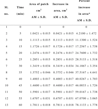
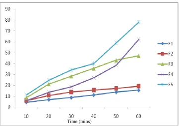
Swelling Index2 7
Weight increase due to swelling and area increase due to
swelling were studied.
a) Weight increase due to swelling : The drug -loaded patch of
size 1 x 1 cm2 was weighed on a pre-weighed cover slip. It was kept in
a petridish and 50 ml of phosphate buffer (pH 7.4) solution was added.
After every five min, the cover sli p was removed, wiped with tissue
paper, and weighed up to 30 min. The difference in the weights gives
the weight increase due to absorption of water and swelling of patch.
b) Area increase due to swelling: The drug loaded patch of size
1 x 1 cm2 was cut a nd placed in a petridish containing 50 ml of
phosphate buffer (pH 7.4) solution. A graph paper was placed beneath
of increase in the area. An increase in the length and breadth of the
patch was noted at five min intervals for 60 min and the area was
calculated. The percent swelling, % S was calculated using the
following equation:
Xt - Xo
%S = x 100 Xo
where Xt is the weight or area of the swollen patch after time t and Xo
is the original patch weight or area at zero time.
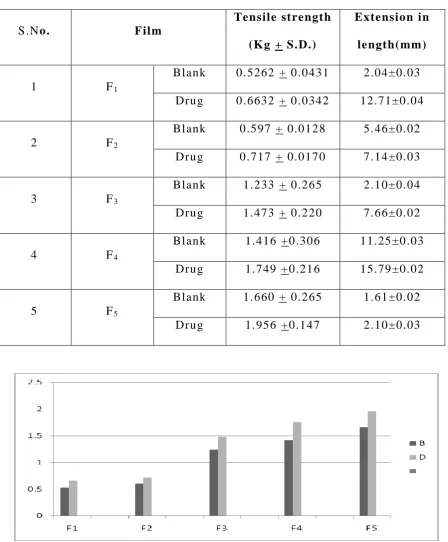
Tensile Strength of Films 2 8 , 2 9
Tensile strength of the film was determined with Universal
Strength Testing Machine (Hounsfield, Slinfold, Horsham, U.K.) as
shown in Figure 13 . The sensitivity of the machine was 1 gram. It
consisted of two load cell grips. The lower one was fixed
and upper one was movable. The test film of size (4 1 cm2) was fixed
between these cell grips and force was gradually applied till the film
broke. The tensile strength of the film was taken directly from the dial
reading in kg. Tensile strength is expressed as follows;
Tensile load at break Tensile strength =
Cross sectional area Folding Enduran