Copyright © 2003, American Society for Microbiology. All Rights Reserved.
Dephosphorylation of eIF-2
␣
Mediated by the
␥
1
34.5 Protein of
Herpes Simplex Virus Type 1 Is Required for Viral Response
to Interferon but Is Not Sufficient for Efficient
Viral Replication
Guofeng Cheng, Kui Yang, and Bin He*
Department of Microbiology and Immunology, College of Medicine, University of Illinois at Chicago, Chicago, Illinois 60612
Received 14 March 2003/Accepted 23 June 2003
The␥134.5 protein of herpes simplex virus type 1 (HSV-1) functions to block the shutoff of protein synthesis
involving double-stranded RNA-dependent protein kinase (PKR). In this process, the␥134.5 protein recruits
cellular protein phosphatase 1 (PP1) to form a high-molecular-weight complex that dephosphorylates eIF-2␣.
Here we show that the␥134.5 protein is capable of mediating eIF-2␣dephosphorylation without any other viral
proteins. While deletion of amino acids 1 to 52 from the␥134.5 protein has no effect on eIF-2␣
dephosphor-ylation, further truncations up to amino acid 146 dramatically reduce the activity of the␥134.5 protein. An
additional truncation up to amino acid 188 is deleterious, indicating that the carboxyl-terminal domain alone
is not functional. Like wild-type HSV-1, the␥134.5 mutant with a truncation of amino acids 1 to 52 is resistant
to interferon, and resistance to interferon is coupled to eIF-2␣dephosphorylation. Intriguingly, this mutant
exhibits a similar growth defect seen for the␥134.5 null mutant in infected cells. Restoration of the wild-type
␥134.5 gene in the recombinant completely reverses the phenotype. These results indicate that eIF-2␣
dephos-phorylation mediated by the␥134.5 protein is required for HSV response to interferon but is not sufficient for
viral replication. Additional functions or activities of the␥134.5 protein contribute to efficient viral infection.
The ␥134.5 gene of herpes simplex virus type 1 (HSV-1)
strain F encodes a protein of 263 amino acids consisting of a large amino-terminal domain, a linker region of triplet repeats (AlaThrPro), and a carboxyl-terminal domain (13). The triplet repeats are a constant feature of the␥134.5 protein, but the
number of repeats varies among different strains (13). Studies suggest that the number of triplet repeats in the␥134.5 protein
may affect the ability of HSV to invade the central nervous system from the peripheral tissue (2, 29). The carboxyl-termi-nal domain is essential to prevent the shutoff of protein syn-thesis in virus infection (12, 19, 20), but the role of the amino-terminal domain is unknown. It is well established that the
␥134.5 protein is essential for viral virulence. HSV mutants that
fail to express the␥134.5 protein are incapable of multiplying
and causing encephalitis in experimental animal models (10, 27, 35, 37).
Considerable evidence indicates that the ␥134.5 protein
functions, at least in part, to inhibit host interferon response mediated by the double-stranded RNA-dependent protein ki-nase (PKR) (7–9, 11, 12, 20, 21). Moreover, it has been dem-onstrated that the␥134.5 null mutant is virulent in PKR
knock-out mice but not in wild-type mice (10, 25). In contrast to the above observations, the␥134.5 null mutant, with an additional
deletion in the US11 promoter region, inhibits PKR activity but
nevertheless is avirulent in experimental mice (30). Similarly, the␥134.5 null mutant, with a secondary mutation outside the
US11 promoter region, only partially restored virulence.
Re-cent experiments showed that the ␥134.5 protein blocks the
surface expression of major histocompatibility complex class II molecules in HSV-1-infected cells, which is believed to impair the functions of CD4⫹ T cells (34). Interestingly, when
ex-pressed in mammalian cells, the␥134.5 protein is distributed
both in the nucleus and cytoplasm (6, 28). In fact, the␥134.5
protein bears nuclear import and export signals that direct its shuttling between the cytoplasm, nucleus, and nucleolus (6). This dynamic process is likely to be required for the different activities associated with the␥134.5 protein during viral
infec-tion (6).
The most extensively characterized function of the ␥134.5
protein is its ability to inhibit the antiviral action of PKR. In cells infected with HSV-1, viral DNA replication leads to the activation of PKR that phosphorylates the␣subunit of trans-lation initiation factor 2 (eIF-2␣) and thereby inhibits transla-tion initiatransla-tion (9, 11). As a countermeasure, the␥134.5 protein
is expressed by HSV to prevent the shutoff of protein synthesis (11). In doing so, the ␥134.5 protein interacts with cellular
protein phosphatase 1 (PP1) by its carboxyl-terminal domain, forming a high-molecular-weight complex that dephosphory-lates eIF-2␣(20, 21). Currently, it remains unknown whether additional viral or cellular proteins are present in this complex. Previous studies suggest that the carboxyl terminus of the
␥134.5 protein consists of a PP1 binding domain and an effector
domain, which is functionally interchangeable with the corre-sponding domain of cellular protein GADD34/MyD116 (8, 19, 20). GADD34/MyD116 belongs to a family of proteins induced under conditions of genotoxic stress, growth arrest, differenti-ation, and apoptosis (18, 23, 26, 31, 32, 39). Like the ␥134.5
* Corresponding author. Mailing address: Department of Microbi-ology and ImmunMicrobi-ology (M/C 790), College of Medicine, University of Illinois at Chicago, 835 S. Wolcott Ave., Chicago, IL 60612. Phone: (312) 996-2391. Fax: (312) 996-6415. E-mail: [email protected].
10154
on November 8, 2019 by guest
http://jvi.asm.org/
protein, GADD34/MyD116 associates with proliferating cell nuclear antigen (PCNA), a cellular protein required for DNA replication and cell cycle control (5). The PP1 binding domain of the␥134.5 protein contains 12 amino acids with a signature
sequence motif of (Arg/Lys)(Val/Ile)XaaPhe found in many PP1 binding proteins (14, 15, 20). These PP1 binding proteins perform diverse functions, such as cell division, mRNA splic-ing, glycogen metabolism, and neurotransmission (1, 14, 15, 22, 33, 36, 38). The effector domain of the␥134.5 protein consists
of 59 amino acids, but its precise role is not yet understood. It appears the concerted action of these two subdomains is crit-ical for eIF-2␣dephosphorylation (8, 20). However, the min-imal unit required for eIF-2␣ dephosphorylation has not yet been defined. The objective of this study was to further analyze the activities of the␥134.5 protein.
Defining the minimal functional module of the␥134.5
pro-tein that dephosphorylates eIF-2␣. In HSV-1-infected cells,
the ␥134.5 protein recruits PP1 to form a
high-molecular-weight complex that dephosphorylates eIF-2␣(20). However, it is not known whether the activity of this complex requires additional viral proteins. To address this issue, monolayers of HeLa cells (Tet-off) were transfected with a plasmid encoding the wild-type␥134.5 protein driven by a tetracycline-inducible
promoter. As a control, a plasmid encoding the mutant␥134.5
protein with Val193Glu and Phe195Leu substitutions was also
included in the experiment. Previous studies have shown that this mutant is unable to mediate eIF-2␣dephosphorylation in virus-infected cells (8, 20).
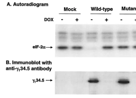
Cells were grown with or without doxycycline (1g/ml) for 36 h. Lysates prepared from the transfected cells were reacted with 32P-labeled eIF-2 and subjected to electrophoresis for
autoradiography. As shown in Fig. 1A, in cells transfected with the wild-type␥134.5 gene, eIF-2␣remained phosphorylated in
the presence of doxycycline, but it became dephosphorylated in the absence of doxycycline. Dephosphorylation of eIF-2␣
correlated with the expression of the wild-type␥134.5 protein
(Fig. 1B, lanes 3 and 4). In contrast, in cells transfected with the mutant␥134.5 gene, eIF-2␣remained phosphorylated
re-gardless of the expression of the␥134.5 protein (Fig. 1A, lanes
5 and 6). Similarly, eIF-2␣remained phosphorylated in mock-transfected cells (Fig. 1A, lanes 1 and 2). Western blot analysis with anti-␥134.5 serum showed that both the wild-type and
mutant␥134.5 proteins were expressed at similar levels in the
absence of doxycycline (Fig. 1B). We conclude from these experiments that when expressed alone in HeLa cells, wild-type but not the mutant␥134.5 protein mediates
dephosphor-ylation of eIF-2␣. This finding extends previous observations that eIF-2␣phosphatase is activated in cells infected with wild-type virus but not with the␥134.5 null mutants (20, 21). These
data suggest that the␥134.5 protein is a viral component
re-quired for the ␥134.5-PP1 complex, which functions in the
absence of other HSV proteins.
Prior studies have demonstrated that the carboxyl terminus of the␥134.5 protein is required for eIF-2␣dephosphorylation
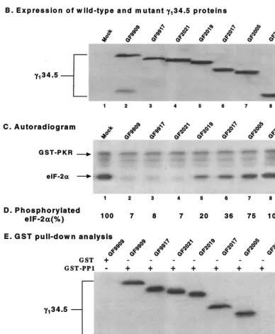
(8). To address whether the amino-terminal or central domain is involved in this process, a series of truncations were intro-duced into the␥134.5 protein. We took advantage of the
bac-ulovirus system in which the␥134.5 protein mediates efficient
eIF-2␣dephosphorylation (10). As shown in Fig. 2A, recom-binant baculoviruses were generated to express mutant forms
of the ␥134.5 protein with a series of nested deletions from
amino acids 1 to 188. Western blot analysis demonstrated that these mutants expressed the truncated forms of the ␥134.5
protein at levels comparable to that of the wild-type ␥134.5
protein (Fig. 2B).
We next examined the ability of these mutants to modulate the activity of eIF-2␣phosphatase. Aliquots of lysate from cells mock infected or infected with each virus were reacted with
32P-labeled eIF-2␣and processed for autoradiography (8). As
shown in Fig. 2C, lysates of cells mock infected exhibited no eIF-2␣phosphatase activity (lane 1), whereas lysates of cells infected with GF9909 (wild type), GF9917 (with amino acids 1 to 28 deleted [⌬1-28]), or GF2021 (⌬1-52) displayed a high level of eIF-2␣phosphatase activity (lanes 2, 3, and 4, respec-tively). There was no detectable difference between these mu-tants and the wild-type␥134.5 protein. However, lysates of cells
infected with GF2019 (⌬1-83), GF2017 (⌬1-116), or GF2005 (⌬1-146) exhibited a significant decrease in eIF-2␣ phospha-tase activities (lanes 5, 6, and 7, respectively). Lysates of cells infected with GF2112 (⌬1-187) completely lost eIF-2␣ phos-phatase activity (lane 8). Same results were observed when serially diluted cell lysates were used for eIF-2␣phosphatase assay (data not shown).
Phosphorimage analysis showed that detectable32P-labeled
eIF-2␣was less than 10% after reaction with lysates of cells infected with GF9909, GF9917 and GF2021. In contrast, the level of32P-labeled eIF-2␣remained between 20 and 75% for
GF2019, GF2017, and GF2005, respectively (Fig. 2D). These results indicated that deletions up to amino acids 52 in the amino terminus of the ␥134.5 protein did not affect eIF-2␣
dephosphorylation. However, additional truncations to amino FIG. 1. (A) eIF-2␣phosphatase activity in cells transfected with the
␥134.5 gene. HeLa cells (Tet-off) grown in Dulbecco modified Eagle medium with (⫹) or without (⫺) doxycycline (DOX) (1g/ml) were transfected with either pGF9912 expressing the wild-type␥134.5 pro-tein or pGF9913 in which Val193Glu and Phe195Leu substitutions were made in the ␥134.5 protein. Thirty-six hours after transfection, cells were harvested, and lysates were prepared. Aliquots of lysates were incubated with 32P-labeled eIF-2, subjected to electrophoresis, and processed for autoradiography (8). (B) Expression of the␥134.5 pro-tein. Cell extracts prepared from transfected cells were subjected to electrophoresis on a sodium dodecyl sulfate–12% polyacrylamide gel, transferred to a nitrocellulose membrane, and probed with anti-␥134.5 antibody (6).
VOL. 77, 2003 NOTES 10155
on November 8, 2019 by guest
http://jvi.asm.org/
[image:2.603.302.539.70.241.2]FIG. 2. (A) Schematic diagram of recombinant baculoviruses expressing wild-type and mutant forms of the␥134.5 protein. The virus desig-nations are indicated to the left of the␥134.5 protein constructs. The shaded bars at the top represent the domain structure of the␥134.5 protein. (ATP)10represents the triplet repeats of AlaThrPro, which connects the amino-terminal domain and the carboxyl-terminal domain. Thin lines indicate the coding regions retained in the wild-type␥134.5 protein or deletion mutants. The numbers denote the first and last amino acids in the
on November 8, 2019 by guest
http://jvi.asm.org/
[image:3.603.97.485.215.686.2]acid 146 in the␥134.5 protein reduced eIF-2␣
dephosphoryla-tion by 75%. A further deledephosphoryla-tion to amino acid 187 was dele-terious.
To examine whether deletions in the␥134.5 protein affected
its binding to PP1, we performed a glutathioneS-transferase (GST) pull-down experiment (8). GST-PP1 expressed from
Escherichia coli was incubated with lysates of HeLa cells in-fected with GF9909, GF9917, GF2021, GF2019, GF2017, GF2005, or GF2112. The protein complexes were electro-phoretically separated and processed for immunoblot analysis with anti-␥134.5 antibody. The results in Fig. 2E show that
GST-PP1, but not GST, bound to the wild-type␥134.5 protein.
Moreover, GST-PP1 bound to all mutants except GF2112. Our data indicated that GF2005, GF2019, and GF2017 were able to mediate eIF-2␣dephosphorylation, but they retained only a small fraction of the activity of the wild-type ␥134.5
protein. Although these mutants were capable of interacting with PP1, it is likely that truncations may cause improper assembly or arrangement of the␥134.5-PP1 complex with
re-duced activity. Thus, while it is not essential, the amino-termi-nal domain of the␥134.5 protein may facilitate eIF-2␣
dephos-phorylation. Early studies established that the carboxyl-terminal domain of the ␥134.5 protein is essential for eIF-2␣
dephos-phorylation, which is required to prevent the shutoff of protein synthesis (8, 12, 19, 20). It has been shown that removal of amino acids up to 257 from the carboxyl extreme had no effect on eIF-2␣dephosphorylation, whereas additional deletions of the carboxyl terminus were deleterious (8). Given that trunca-tion up to amino acid 52 from the amino terminus of the␥134.5
protein did not have any effect on eIF-2␣dephosphorylation and PP1 binding, it is reasonable to conclude that the domain containing amino acids 53 to 258 of the␥134.5 protein
consti-tutes a functional entity, which is capable of mediating efficient eIF-2␣dephosphorylation. It is also notable that truncation up to amino acid 188 from the animo terminus completely abol-ished eIF-2␣dephosphorylation. This indicates that deletions of both the amino terminus and the triplet repeats abolished the activity of the␥134.5 protein. This phenotype is attributed
to the disruption of the ␥134.5-PP1 interaction. These data
indicate that the carboxyl terminus of the ␥134.5 protein is
required but is not sufficient to mediate eIF-2␣ dephosphory-lation.
Analysis of the activity of␥134.5 mutants within the context
of the HSV genome.Data in Fig. 2C indicate that deletion of
amino acids 1 to 52 in the ␥134.5 protein had no effect on
dephosphorylation of eIF-2␣. In order to analyze the effect of this deletion in the context of the HSV genome, a recombinant virus KY0112 was constructed by using the bacterial artificial chromosome (BAC) system (24). As controls, this mutant was
further engineered to either restore or delete the full-length
␥134.5 gene, yielding KY0233 and KY0234, respectively (Fig.
3A). To verify the virus constructs, Southern blot analysis was performed afterNcoI andBspEI digestion of viral DNA (12). As seen in Fig. 3B, KY0112, which contains a deletion from amino acids 1 to 52 of the␥134.5 protein, gave rise to a 701-bp
fragment. Parental HSV-BAC and HSV-1(F) yielded an 857-bp fragment (16, 24). Similarly, KY0233, which contains the restored wild-type ␥134.5 gene, yielded an 857-bp
frag-ment. No bands were detected for KY0234 and R3616 due to deletion of the␥134.5 gene.
To examine expression of the␥134.5 protein, Western blot
analysis was performed with an anti-␥134.5 antibody (11).
Fig-ure 3C shows that in Vero cells, HSV-BAC, HSV-1(F), and KY0233 expressed the full-length ␥134.5 protein, and the
smaller bands likely result from proteolytic cleavage of the full-length ␥134.5 protein (lanes 1, 3, and 6). KY0112
ex-pressed a truncated␥134.5 protein with the expected size (lane
4). The␥134.5 protein was not detected in R3616 and KY0234.
Taken together, these experiments indicate that the recombi-nant viruses constructed contain the expected␥134.5 gene
de-rivatives.
Next we measured growth properties of the recombinant viruses. In this series of experiments, monolayers of cells were infected with HSV-1(F), R3616, HSV-BAC (wild-type␥134.5),
KY0112 (⌬1-52), KY0234 (⌬␥134.5), or KY0233 (restoration
of wild-type␥134.5) at 5 PFU per cell. At 24 h postinfection,
the cells were harvested, and virus yields were measured. As shown in Table 1, in mouse 10T1/2 cells, 1(F) and HSV-BAC replicated efficiently, as they expressed the wild-type
␥134.5 protein. Under this experimental condition, viral yield
reached up to 9.5⫻106PFU/ml. In contrast, both R3616 and
KY0234 replicated less efficiently, with titers reaching 1.4 ⫻
105and 4.2⫻105PFU/ml, respectively. This decrease in viral
replication is attributed to the deletion of the entire ␥134.5
gene. Interestingly, KY0112, which has a deletion of amino acids 1 to 52 in the␥134.5 protein, had a titer of only 1.8⫻105
PFU/ml. This mutant exhibited a growth defect similar to the defect observed for R3616 or KY0234. Notably, recombinant KY0233 displayed efficient replication similar to HSV-1(F) or HSV-BAC, as this virus has the restored wild-type␥134.5 gene.
As indicated in Table 1, the growth trend for these recombi-nants is similar in human neuroblastoma SK-N-SH cells. How-ever, it seems that KY0112 replicated better than KY0234 (sixfold) in SK-N-SH cells.
We also examined viral growth properties in Vero cells and HeLa cells. Although the general growth patterns were similar to those seen in mouse 10T1/2 or human SK-N-SH cells, there were smaller differences in the viral titers of wild-type and
constructs. (B) Expression of wild-type␥134.5 and mutant forms of the␥134.5 protein. Lysates of Sf9 cells which were mock infected or infected with the indicated viruses were subjected to electrophoresis on a denaturing 12% polyacrylamide gel, transferred to a nitrocellulose membrane, and reacted with anti-His tag antibody (Qiagen Inc.) (8). (C) Activity of the␥134.5 mutants in Sf9 cells. Sf9 cells were mock infected or infected with the indicated recombinant baculoviruses at 5 PFU per cell. At 48 h after infection, cells were harvested, and S10 fractions were prepared as described previously (8). Aliquots of each lysate were then processed for eIF-2␣dephosphorylation assay. (D) Quantitation of the phosphorylated eIF-2␣in panel C. The numbers indicate the percentages of phosphorylated eIF-2␣remaining after incubation with the cell lysates relative to that in mock-infected cell lysate. (E) Interaction of PP1 with the␥134.5 mutants. Sf9 cells infected with baculoviruses expressing wild-type␥134.5 or mutant forms of the␥134.5 protein were harvested at 48 h after infection. The lysates were prepared and then incubated with GST-PP1 (⫹) bound to beads at 4°C. After the beads were washed, the protein complexes were electrophoretically separated and processed for immunoblotting with anti-␥134.5 antibody (8).
VOL. 77, 2003 NOTES 10157
on November 8, 2019 by guest
http://jvi.asm.org/
FIG. 3. (A) Schematic representation of the genome structure of HSV-1 and its derivatives. HSV-1(F) is the prototype strain used in this laboratory (16). The location of the wild-type (Wt)␥134.5 gene is shown in the expanded portions of the inverted repeat sequences b and b⬘. The thick lines under the enlarged region of the␥134.5 gene denote wild-type HSV-BAC virus and the mutant viruses. Restriction sites and fragment sizes are indicated. The broken lines represent sequences deleted from the␥134.5 gene. Recombinant virus R3616 lacks 1,000 bp from the coding region of the HSV-1(F)␥134.5 gene (10). HSV-BAC is the parental virus used to construct mutant KY0112, in which the region coding amino acids 1 to 52 of the␥134.5 gene was deleted (24). KY0112 was used to construct KY0233 and KY0234, respectively. KY0233 has the restored wild-type
␥134.5 gene, whereas KY0234 has the deleted␥134.5 gene. (B) Autoradiographic image of recombinant and parental viral DNAs. Confluent Vero cells were infected with the indicated viruses at 10 PFU per cell. At 18 h postinfection, infected cells were harvested. Viral DNA was prepared and digested byNcoI andBspEI, electrophoretically separated on 0.8% agarose gels, and transferred to nitrocellulose sheets. The␥134.5 gene was detected by hybridization to electrophoretically separated digests of the viral DNA with a32P-labeledBstEII-BspEI fragment of the␥134.5 gene, followed by exposure to Kodak X-ray film (19). (C) Expression of the␥134.5 protein and its derivatives. Vero cells were either mock infected or infected with the indicated viruses at 10 PFU per cell. At 18 h after infection, the cells were harvested, solubilized, subjected to polyacrylamide gel electrophoresis, transferred to nitrocellulose sheets, and reacted with anti-␥134.5 antibody (6).
10158
on November 8, 2019 by guest
mutant viruses in these cell lines. As shown in Table 1, in Vero cells, all viruses expressing the wild-type␥134.5 grew to more
than 1.8⫻108PFU/ml, whereas the␥
134.5 deletion mutants
(R3616 and KY0234) grew to titers of 1.5⫻107to 1.9⫻107
PFU/ml. Nevertheless, in this cell line, growth of KY0112 was similar to that of R3616 or KY0234. There was generally a 10-fold decrease in viral yield. Similar results were also ob-tained in HeLa cells. Taken together, these experiments indi-cate that deletion of amino acids 1 to 52 decreased viral rep-lication, albeit to different extents, in infected cells.
It is interesting that the growth defect associated with the
␥134.5 mutant KY0112 (⌬1-52) varies in different cell lines. We
speculate that these cells may express different levels of cellu-lar inhibitors of viral infection. Conversely, they may express different levels of host factors required for viral infection. Con-sistent with these ideas, it has been reported that mutations in oncogenes that constitutively activate the Ras signaling path-way promote cell permissiveness to HSV infection (17). In addition, the growth phase of the cell may affect viral replica-tion. In mouse fibroblast 3T6 cells, replication of HSV that fails to express the␥134.5 protein is restricted in resting cells
but less so in actively dividing cells (3). In vitro studies showed that the␥134.5 protein interacts with PCNA, a cellular protein
required for DNA replication and cell cycle control (5). It is believed that the interaction of the␥134.5 protein with PCNA
may release cells from growth arrest and facilitate viral repli-cation in HSV-infected cells (5). At this point, the nature of the defect associated with the␥134.5 mutant KY0112 is not clear,
but it is conceivable that deletion of amino acids 1 to 52 in the
␥134.5 protein may alter the interactions between the␥134.5
mutant and host cells that are critical for viral replication. Since deletion of amino acids 1 to 52 in the␥134.5 protein
altered viral replication, next we assessed whether the deletion affected viral response to interferon. In this experiment, mono-layers of Vero cells were left untreated or pretreated with alpha interferon for 20 h and subsequently infected with a serial dilution of HSV-BAC, KY0112, KY0234, or KY0233. At 96 h after infection, the numbers of plaques were determined, and the results are presented as the ratio of the number of plaques without and with interferon treatment. The data sum-marized in Table 2 indicate that plaque formation for wild-type HSV-BAC (wild-type␥134.5) was reduced by alpha interferon
only slightly (threefold), exhibiting an interferon-resistant phe-notype. However, plaque formation for the ␥134.5 deletion
mutant KY0234 was reduced dramatically. This reduction is approximately 1,000-fold compared to the wild-type virus. Im-portantly, plaque formation for KY0112 (⌬1-52) was reduced only fourfold, which is the same as that of HSV-BAC or the repaired virus KY0233. These data demonstrate that deletion of amino acids 1 to 52 in the ␥134.5 protein has no effect on
viral response to interferon.
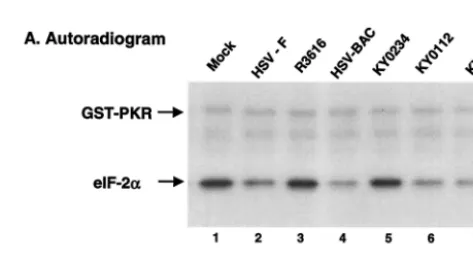
On the basis of the above analysis, we also examined eIF-2␣
phosphatase activity in vitro. Monolayers of HeLa cells were either mock infected or infected with wild-type or mutant vi-ruses at 20 PFU per cell. At 15 h after infection, cell lysates were prepared to analyze eIF-2␣ phosphatase activity. As shown in Fig. 4, lysates of cells infected with KY0112 displayed eIF-2␣phosphatase activity similar to that seen for wild-type HSV-1(F), HSV-BAC, or KY0233 in which the wild-type
␥134.5 gene was restored (Fig. 4, lanes 2, 4, 6, and 7). In
contrast, lysates of cells mock infected or infected with the
␥134.5 deletion virus R3616 or KY0234 displayed no eIF-2␣
phosphatase activity (Fig. 4, lanes 1, 3, and 5). Western blot
[image:6.603.301.542.90.147.2]FIG. 4. (A) eIF-2␣ phosphatase activity in HSV-infected cells. Confluent HeLa cells were mock infected or infected with the indi-cated viruses at 10 PFU per cell. At 16 h postinfection, cells were harvested, and S10 fractions were prepared (21). Aliquots of each lysate were then reacted with32P-labeled eIF-2 at 34°C. The reaction was stopped after 2 min of incubation. Samples were then separated electrophoretically on a denaturing 12% polyacrylamide gel and sub-jected to autoradiography (8). (B) Quantitation of the phosphorylated eIF-2␣. Phosphorylated eIF-2␣in each lane in panel A was quanti-tated after eIF-2␣phosphatase assays with the phosphorimage system (ImageQuant software). The numbers are the percentages of phos-phorylated eIF-2␣ remaining after incubation with the cell lysates relative to that of unreacted eIF-2␣. The data are averages from two independent experiments.
TABLE 1. Replication of the wild-type HSV-1(F) and␥134.5 mutantsa
Virus Virus replication (titer) in cell line:
10T1/2 SK-N-SH HeLa Vero
HSV-1(F) 9.5⫻106 3.3⫻107 3.1⫻107 1.8⫻108 R3616 1.4⫻105 4.2⫻104 2.6⫻106 1.9⫻107 HSV-BAC 8.0⫻106 3.5⫻107 2.4⫻107 2.0⫻108 KY0234 4.2⫻105 6.9⫻104 1.6⫻106 1.5⫻107 KY0112 1.8⫻105 4.4⫻105 2.8⫻106 1.1⫻107 KY0233 1.0⫻107 1.5⫻107 2.5⫻107 9.3⫻107
aMonolayers of cells were infected with each virus at 5 PFU per cell at 37°C.
[image:6.603.43.283.90.180.2]Twenty-four hours postinfection, cells were harvested, freeze-thawed three times, and titrated on Vero cells at 37°C. The data are averages from duplicate samples.
TABLE 2. Effect of alpha interferon on plaque formation of viruses in Vero cellsa
Virus ␥134.5 gene Virus titer ratiob
HSV-BAC Wild-type 3.2⫾0.6
KY0234 Deletion of entire coding region 2,000⫾250 KY0112 Deletion of amino acids 1 to 52 4.5⫾0.5 KY0233 Restoration of wild-type␥134.5 gene 3.0⫾1.0
aVero cells were untreated or pretreated with 1,000 U of human alpha
inter-feron (Sigma) per ml for 20 h. Cells were then infected with a serial dilution of the indicated viruses. The numbers of plaques were counted, and virus titers were determined.
bRatio of viral titer in the absence of alpha interferon to the viral titer in the
presence of alpha interferon. The data are the means⫾standard deviations for three independent experiments.
VOL. 77, 2003 NOTES 10159
on November 8, 2019 by guest
http://jvi.asm.org/
[image:6.603.301.538.464.594.2]analysis with anti-␥134.5 antibody showed that the␥134.5
pro-tein and its derivative are present at comparable levels in lysates of cells infected with HSV-1(F), HSV-BAC, KY0112, or KY0233 (data not shown). These experiments demonstrated that amino acids 53 to 263 of the␥134.5 protein are sufficient
to mediate eIF-2␣dephosphorylation, which is consistent with the observation that KY0112 is resistant to interferon.
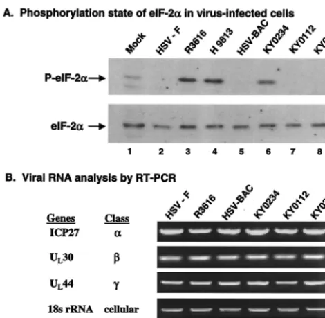
To explore whether in vitro eIF-2␣phosphatase activity cor-relates with the phosphorylation status of eIF-2␣ in virus-infected cells, we performed Western blot analysis. Specifically, monolayers of mouse 10T1/2 cells were mock infected or in-fected with HSV-1(F), R3616, HSV-BAC, KY0112, KY0234, KY0233, or H9813, which has Val193Glu and Phe195Leu
sub-stitutions in the PP1 binding motif, at 5 PFU per cell (8). At 20 h postinfection, cell lysates were prepared and processed for immunoblotting with antibodies against phosphorylated and total eIF-2␣. As shown in Fig. 5A, eIF-2␣ was present at comparable levels in mock-infected and virus-infected cells. A small amount of phosphorylated eIF-2␣was present in mock-infected cells. Interestingly, eIF-2␣ remained unphosphory-lated in cells infected with HSV-1(F), HSV-BAC, KY0112, or KY0233 (Fig. 5A, lanes 2, 5, 7, and 8, respectively). Essentially, viruses expressing the wild-type ␥134.5 protein prevented
eIF-2␣phosphorylation, yet the KY0112 mutant with a dele-tion of amino acids 1 to 52 of the␥134.5 protein did not induce
eIF-2␣phosphorylation. In contrast, eIF-2␣became phosphor-ylated in cells infected with R3616, KY0234, or H9813 (Fig. 5A, lanes 3, 4, and 6), which resulted from the failure to express the␥134.5 protein or from point mutations in the PP1
binding motif in the␥134.5 protein. These results correlate well
with eIF-2␣phosphatase activity analyzed in vitro.
An important finding emerging from these experiments is that in HSV-infected cells, eIF-2␣ dephosphorylation medi-ated by the␥134.5 protein is tightly coupled to viral resistance
to interferon, but not necessarily to efficient viral replication. Consistent with the data from the baculovirus system, the
␥134.5 mutant KY0112 with a deletion of amino acids 1 to 52
showed full activity in mediating eIF-2␣dephosphorylation. In correlation with these results, KY0112 exhibited an interferon-resistant phenotype like wild-type virus (Table 2). These data strongly support the notion that functional interaction of the
␥134.5 protein and PKR is involved in HSV resistance to
in-terferon. However, it is surprising to find that replication of the
␥134.5 mutant KY0112 was defective in infected cells. In this
respect, it resembled the␥134.5 null mutant. As restoration of
the wild-type␥134.5 gene completely reversed the phenotype,
the decrease in viral replication for KY0112 is attributable only to a defect in the␥134.5 gene. Although the molecular basis
remains unknown, it appears that the region containing amino acids 1 to 52 of the␥134.5 protein is required for efficient viral
replication. A simple explanation is that this region represents a distinct functional module. However, an alternative interpre-tation is that the region containing amino acids 1 to 52 has an indirect role. Deletion of this region may distort conformation of the ␥134.5 protein, which indirectly disrupts one or more
activities associated with the␥134.5 protein.
To test whether deletions in the␥134.5 gene altered gene
expression, we measured the levels of RNA transcript for ICP27 (␣ gene), UL30 (-gene), and UL44 (␥gene) in cells
infected with viruses. Mouse 10T1/2 cells were infected with HSV-1(F), R3616, HSV-BAC, KY0112, KY0234, or KY0233 at 5 PFU per cell. At 20 h postinfection, total RNA was ex-tracted and subjected to reverse transcription-PCR (RT-PCR) amplification. As shown in Fig. 5B, ICP27, UL30, and UL44
mRNAs were expressed at comparable levels in all virus-in-fected cells. These results indicated that deletions of the␥134.5
gene have no effect on the expression of mRNA in HSV-infected mouse 10T1/2 cells. Since RT-PCR analysis failed to detect major differences in mRNA expression between wild-type and mutant viruses in 10T1/2 cells, it is likely that the defect is at a step(s) after mRNA expression. Given that de-phosphorylation of eIF-2␣is essential for viral replication (8), these results suggest that efficient viral replication requires additional activities of the␥134.5 protein.
The␥134.5 protein of HSV is crucial for viral neurovirulence
in vivo (10, 27, 35, 37). Accumulating evidence suggests that the ␥134.5 protein is involved in different processes during
HSV infection (2–4, 6, 11, 21, 28, 34). A remarkable property of this viral factor is to recruit PP1, forming a high-molecular-weight-complex that dephosphorylates eIF-2␣ and thereby evades the host antiviral response. The fact that eIF-2␣ phos-phatase activity is not necessarily sufficient for efficient viral replication is consistent with the proposal that an additional function(s) associated with the␥134.5 protein contributes to
viral infection. FIG. 5. (A) Phosphorylation state of eIF-2␣in intact cells. Mouse
10T1/2 cells were mock infected or infected with the indicated viruses at 5 PFU per cell. At 20 h postinfection, cells were harvested. The lysates were prepared and processed for Western blotting with anti-bodies against phosphorylated eIF-2␣(P-eIF-2␣) and total eIF-2␣in the same membrane (Cell Signaling Tech, Inc.). (B) Viral gene tran-scription pattern in 10T1/2 cells. Cells were infected with the indicated viruses as described above, and total RNA was extracted using an RNeasy kit by Qiagen. Equal amounts of RNA from the samples were then subjected to RT-PCR amplification of specific viral cDNAs (ICP27, UL30, and UL44). As a control, the cellular 18s rRNA was included in the assay. The kinetic classes of selected genes are indi-cated to the left of the blots. The data are representative of three independent experiments.
on November 8, 2019 by guest
http://jvi.asm.org/
[image:7.603.45.280.68.297.2]We thank Bernard Roizman for providing HSV-1(F) and R3616 and Brian Horsburgh and Frank Tafaro for providing HSV-BAC plasmid. We are grateful to Melissa Cerveny for critical reading of the manu-script.
This work was supported in part by grant AI 46665 (B.H.) from the National Institute of Allergy and Infectious Diseases.
REFERENCES
1. Aitken, A., and P. Cohen.1982. Isolation and characterisation of active fragments of protein phosphatase inhibitor-1 from rabbit skeletal muscle. FEBS Lett.147:54–58.
2. Bower, J. R., H. Mao, C. Durishin, E. Rozenbom, M. Detwiler, D. Rempinski, T. L. Karban, and K. S. Rosenthal.1999. Intrastrain variants of herpes simplex virus type 1 isolated from a neonate with fatal disseminated infection differ in the ICP34.5 gene, glycoprotein processing, and neuroinvasiveness. J. Virol.73:3843–3853.
3. Brown, S. M., J. Harland, A. R. MacLean, J. Podlech, and J. B. Clements. 1994. Cell type and cell state determine differential in vitro growth of non-neurovirulent ICP34.5-negative herpes simplex virus types 1 and 2. J. Gen. Virol.75:2367–2377.
4. Brown, S. M., A. R. MacLean, J. D. Aitken, and J. Harland.1994. ICP34.5 influences herpes simplex virus type 1 maturation and egress from infected cells in vitro. J. Gen. Virol.75:3679–3686.
5. Brown, S. M., A. R. MacLean, E. A. McKie, and J. Harland.1997. The herpes simplex virus virulence factor ICP34.5 and the cellular protein MyD116 complex with proliferating cell nuclear antigen through the 63-amino-acid domain conserved in ICP34.5, MyD116, and GADD34. J. Virol. 71:9442–9449.
6. Cheng, G., M. E. Brett, and B. He.2002. Signals that dictate nuclear, nucle-olar, and cytoplasmic shuttling of the␥134.5 protein of herpes simplex virus
type 1. J. Virol.76:9434–9445.
7. Cheng, G., M. E. Brett, and B. He.2001. Val193and Phe195of the␥ 134.5
protein of herpes simplex virus 1 are required for viral resistance to inter-feron␣/. Virology290:115–120.
8. Cheng, G., M. Gross, M. E. Brett, and B. He.2001. AlaArg motif in the carboxyl terminus of the␥134.5 protein of herpes simplex virus type 1 is
required for the formation of a high-molecular-weight complex that dephos-phorylates eIF-2␣. J. Virol.75:3666–3674.
9. Chou, J., J. J. Chen, M. Gross, and B. Roizman.1995. Association of a Mr
90, 000 phosphoprotein with protein kinase PKR in cells exhibiting enhanced phosphorylation of translation initiation factor eIF-2␣and premature shutoff of protein synthesis after infection with␥134.5- mutants of herpes simplex
virus 1. Proc. Natl. Acad. Sci. USA92:10516–10520.
10. Chou, J., E. R. Kern, R. J. Whitley, and B. Roizman.1990. Mapping of herpes simplex virus-1 neurovirulence to␥134.5, a gene nonessential for
growth in culture. Science250:1262–1266.
11. Chou, J., and B. Roizman.1992. The␥134.5 gene of herpes simplex virus 1
precludes neuroblastoma cells from triggering total shutoff of protein syn-thesis characteristic of programmed cell death in neuronal cells. Proc. Natl. Acad. Sci. USA89:3266–3270.
12. Chou, J., and B. Roizman.1994. Herpes simplex virus 1␥134.5 gene function,
which blocks the host response to infection, maps in the homologous domain of the genes expressed during growth arrest and DNA damage. Proc. Natl. Acad. Sci. USA91:5247–5251.
13. Chou, J., and B. Roizman.1990. The herpes simplex virus 1 gene for ICP34.5, which maps in inverted repeats, is conserved in several limited-passage isolates but not in strain 17syn⫹. J. Virol.64:1014–1020. 14. Cohen, P. T.2002. Protein phosphatase 1—targeted in many directions.
J. Cell Sci.115:241–256.
15. Egloff, M. P., D. F. Johnson, G. Moorhead, P. T. Cohen, P. Cohen, and D. Barford.1997. Structural basis for the recognition of regulatory subunits by the catalytic subunit of protein phosphatase 1. EMBO J.16:1876–1887. 16. Ejercito, P. M., E. D. Kieff, and B. Roizman.1968. Characterization of
herpes simplex virus strains differing in their effects on social behaviour of infected cells. J. Gen. Virol.2:357–364.
17. Farassati, F., A. D. Yang, and P. W. Lee.2001. Oncogenes in Ras signalling pathway dictate host-cell permissiveness to herpes simplex virus 1. Nat. Cell Biol.3:745–750.
18. Grishin, A. V., O. Azhipa, I. Semenov, and S. J. Corey.2001. Interaction between growth arrest-DNA damage protein 34 and Src kinase Lyn nega-tively regulates genotoxic apoptosis. Proc. Natl. Acad. Sci. USA98:10172– 10177.
19. He, B., J. Chou, D. A. Liebermann, B. Hoffman, and B. Roizman.1996. The carboxyl terminus of the murine MyD116 gene substitutes for the
corre-sponding domain of the␥134.5 gene of herpes simplex virus to preclude the
premature shutoff of total protein synthesis in infected human cells. J. Virol. 70:84–90.
20. He, B., M. Gross, and B. Roizman.1998. The␥134.5 protein of herpes
simplex virus 1 has the structural and functional attributes of a protein phosphatase 1 regulatory subunit and is present in a high molecular weight complex with the enzyme in infected cells. J. Biol. Chem.273:20737–20743. 21. He, B., M. Gross, and B. Roizman.1997. The␥134.5 protein of herpes
simplex virus 1 complexes with protein phosphatase 1␣to dephosphorylate the alpha subunit of the eukaryotic translation initiation factor 2 and pre-clude the shutoff of protein synthesis by double-stranded RNA-activated protein kinase. Proc. Natl. Acad. Sci. USA94:843–848.
22. Hirano, K., F. Erdodi, J. G. Patton, and D. J. Hartshorne.1996. Interaction of protein phosphatase type 1 with a splicing factor. FEBS Lett.389:191–194. 23. Hollander, M. C., Q. Zhan, I. Bae, and A. J. Fornace, Jr.1997. Mammalian GADD34, an apoptosis- and DNA damage-inducible gene. J. Biol. Chem. 272:13731–13737.
24. Horsburgh, B. C., M. M. Hubinette, D. Qiang, M. L. MacDonald, and F. Tufaro.1999. Allele replacement: an application that permits rapid manip-ulation of herpes simplex virus type 1 genomes. Gene Ther.6:922–930. 25. Leib, D. A., M. A. Machalek, B. R. Williams, R. H. Silverman, and H. W.
Virgin.2000. Specific phenotypic restoration of an attenuated virus by knock-out of a host resistance gene. Proc. Natl. Acad. Sci. USA97:6097–6101. 26. Lord, K. A., B. Hoffman-Liebermann, and D. A. Liebermann.1990.
Se-quence of MyD116 cDNA: a novel myeloid differentiation primary response gene induced by IL6. Nucleic Acids Res.18:2823.
27. MacLean, A., L. Robertson, E. McKay, and S. M. Brown.1991. The RL neurovirulence locus in herpes simplex virus type 2 strain HG52 plays no role in latency. J. Gen. Virol.72:2305–2310.
28. Mao, H., and K. S. Rosenthal.2002. An N-terminal arginine rich cluster and a proline-alanine-threonine repeat region determines the cellular localiza-tion of the herpes simplex virus type-1 ICP34.5 protein and its ligand, protein phosphatase 1. J. Biol. Chem.277:11423–11431.
29. Mao, H., and K. S. Rosenthal.2003. Strain-dependent structural variants of herpes simplex virus type 1 ICP34.5 determine viral plaque size, efficiency of glycoprotein processing, and viral release and neuroinvasive disease poten-tial. J. Virol.77:3409–3417.
30. Mohr, I., D. Sternberg, S. Ward, D. Leib, M. Mulvey, and Y. Gluzman.2001. A herpes simplex virus type 1␥34.5 second-site suppressor mutant that exhibits enhanced growth in cultured glioblastoma cells is severely attenu-ated in animals. J. Virol.75:5189–5196.
31. Novoa, I., H. Zeng, H. P. Harding, and D. Ron.2001. Feedback inhibition of the unfolded protein response by GADD34-mediated dephosphorylation of eIF2␣. J. Cell Biol.153:1011–1022.
32. Novoa, I., Y. Zhang, H. Zeng, R. Jungreis, H. P. Harding, and D. Ron.2003. Stress-induced gene expression requires programmed recovery from trans-lational repression. EMBO J.22:1180–1187.
33. Tang, P. M., J. A. Bondor, K. M. Swiderek, and A. A. DePaoli-Roach.1991. Molecular cloning and expression of the regulatory (RG1) subunit of the glycogen-associated protein phosphatase. J. Biol. Chem.266:15782–15789. 34. Trgovcich, J., D. Johnson, and B. Roizman.2002. Cell surface major
histo-compatibility complex class II proteins are regulated by the products of the
␥134.5 and UL41 genes of herpes simplex virus 1. J. Virol.76:6974–6986.
35. Valyi-Nagy, T., M. U. Fareed, J. S. O’Keefe, R. M. Gesser, A. R. MacLean, S. M. Brown, J. G. Spivack, and N. W. Fraser.1994. The herpes simplex virus type 1 strain 17⫹ ␥34.5 deletion mutant 1716 is avirulent in SCID mice. J. Gen. Virol.75:2059–2063.
36. Van Eynde, A., S. Wera, M. Beullens, S. Torrekens, F. Van Leuven, W. Stalmans, and M. Bollen.1995. Molecular cloning of NIPP-1, a nuclear inhibitor of protein phosphatase-1, reveals homology with polypeptides in-volved in RNA processing. J. Biol. Chem.270:28068–28074.
37. Whitley, R. J., E. R. Kern, S. Chatterjee, J. Chou, and B. Roizman.1993. Replication, establishment of latency, and induced reactivation of herpes simplex virus␥134.5 deletion mutants in rodent models. J. Clin. Investig.
91:2837–2843.
38. Williams, K. R., H. C. Hemmings, Jr., M. B. LoPresti, W. H. Konigsberg, and P. Greengard.1986. DARPP-32, a dopamine- and cyclic AMP-regulated neuronal phosphoprotein. Primary structure and homology with protein phosphatase inhibitor-1. J. Biol. Chem.261:1890–1903.
39. Zhan, Q., K. A. Lord, I. Alamo, Jr., M. C. Hollander, F. Carrier, D. Ron, K. W. Kohn, B. Hoffman, D. A. Liebermann, and A. J. Fornace, Jr.1994. The
gaddandMyDgenes define a novel set of mammalian genes encoding acidic proteins that synergistically suppress cell growth. Mol. Cell. Biol.14:2361– 2371.
VOL. 77, 2003 NOTES 10161