EFFECTIVENESS OF NORMAL SALINE MOUTH
WASH VERSUS SODIUM BICARBONATE MOUTH
WASH ON ORAL MUCOSITIS AMONG PATIENTS
UNDERGOING RADIATION THERAPY IN ONCOLOGY
WARD AT GOVERNMENT RAJAJI HOSPITAL
MADURAI
M.Sc (NURSING) DEGREE EXAMINATION
BRANCH – I-MEDICAL SURGICAL NURSING
COLLEGE OF NURSING
MADURAI MEDICALCOLLEGE, MADURAI -20.
A dissertation submitted to
THE TAMILNADU DR.M.G.R. MEDICAL UNIVERSITY,
CHENNAI - 600 032.
In partial fulfillment of the requirement for the degree
MASTER OF SCIENCE IN NURSING
EFFECTIVENESS OF NORMAL SALINE MOUTH
WASH VERSUS SODIUM BICARBONATE MOUTH
WASH ON ORAL MUCOSITIS AMONG PATIENTS
UNDERGOING RADIATION THERAPY IN ONCOLOGY
WARD AT GOVERNMENT RAJAJI HOSPITAL
MADURAI
Approved by dissertation committee on………
Professor in Nursing Research ___________________________
Mrs.S.POONGUZHALI, M.Sc (N), M.A,M.BA, PhD.,
Principal ,College of Nursing, Madurai Medical College, Madurai-20.
Clinical Specialty Expert ________________
Mrs.J.ALAMELUMANGAI, M.Sc (N), MBA (HM).,
Faculty in Nursing,Department of Medical Surgical Nursing, College of Nursing,
Madurai Medical College, Madurai-20.
Medical Expert ___________________
Dr.S.VASANTHAMALAI, B.Sc, M.D., DMRT.,
Professor and Head of the Department,Department of Radiation Oncology, Madurai Medical College,
Madurai-20
A dissertation submitted to
THE TAMILNADU DR.M.G.R. MEDICAL UNIVERSITY,
CHENNAI- 600 032.
In partial fulfillment of the requirement for the degree of
MASTER OF SCIENCE IN NURSING
CERTIFICATE
This is to certify that this Dissertation titled, “EFFECTIVENESS OF NORMAL SALINE MOUTH WASH VERSUS SODIUM BICARBONATE MOUTH WASH ON ORAL MUCOSITIS AMONG PATIENTS UNDERGOING RADIATION THERAPY IN ONCOLOGY WARD AT GOVERNMENT RAJAJI HOSPITAL MADURAI ” is a bonafide work done by Mrs.SUNITHA.G, M.Sc (N) Student, College of Nursing, Madurai Medical College, Madurai - 20, submitted to THE TAMILNADU DR M.G.R. MEDICAL UNIVERSITY, CHENNAI-32, in partial fulfillment of the university rules and regulations towards the award of the degree of MASTER OF SCIENCE IN NURSING, Branch- I Medical Surgical Nursing, under our guidance and supervision during the academic period from 2013—2015.
Mrs.S.POONGUZHALI, M.Sc (N)., M.A., M.B.A., Ph.D.,
CAPTAIN Dr.B.SANTHAKUMAR,M.Sc(F.Sc), M.D(F.M)., PGDMLE, Dip.N.B(F.M).,
PRINCIPAL, DEAN,
COLLEGE OF NURSING, MADURAIMEDICAL COLLEGE,
MADURAI MEDICAL COLLEGE, MADURAI-20.
MADURAI-20.
CERTIFICATE
This is to certify that the dissertation entitled “EFFECTIVENESS OF NORMAL SALINE MOUTH WASH VERSUS SODIUM BICARBONATE MOUTH WASH ON ORAL MUCOSITIS AMONG PATIENTS
UNDERGOING RADIATION THERAPY IN ONCOLOGY WARD AT GOVERNMENT RAJAJI HOSPITAL MADURAI” is a bonafide work done
by Mrs.SUNITHA.G, M.Sc (N) College of Nursing, Madurai Medical College, Madurai - 20, in partial fulfillment of the university rules and regulations for award of MASTER OF SCIENCE IN NURSING, Branch- I- Medical Surgical Nursing, under my guidance and supervision during the academic year 2013-15.
Name and signature of the guide________________ Mrs.J.ALAMELUMANGAI, M.Sc (N), MBA (HM)., Faculty in Nursing,
Department of Medical Surgical Nursing, College of Nursing,
Madurai Medical College, Madurai - 20.
Name and signature of the Head of Department___________________________ Mrs.S.POONGUZHALI, M.Sc. (N), M.A, M.B.A., Ph.D.
Principal,
College of Nursing, Madurai Medical College, Madurai - 20.
Name and signature of the Dean
CAPTAIN Dr.B.SANTHAKUMAR, M.Sc, F.Sc, MD(FM), PGDMLE, Dip.N.B(FM) Dean,
ACKNOWLEDGEMENT
The satisfaction and pleasure that accompany the successful completion of any task would be incomplete without mentioning the people who made it possible, whose constant guidance and encouragement rewards, any effort with success. I consider it a privilege to express my gratitude and respect to all those who guided and inspired me in the completion of this study.
First of all I praise and thank God Almighty for heavenly richest blessings and abundant grace, which strengthened me in each and every step throughout this endeavor.
I wish to express my deep and sincere gratitude to
CAPTAIN Dr. B.SHANTHAKUMAR, MSC(FSC)., M.D(FM), PGDMLE., DIPNB(FM)., Dean, Madurai Medical College, Madurai, for giving this opportunity to conduct this study.
My deepest gratitude is to Mrs.S.POONGUZHALI, M.sc (N).M.A., M.B.A., Ph.D, Principal College of Nursing, Madurai Medical College, Madurai. I have been amazingly fortunate to have a teacher who guided me to recover when my steps faltered. Her patience and support helped me overcome many crisis situations and finish this dissertation.
My heartful and faithful thanks to MRS.J.ALAMELU MANGAI,M.Sc (N), MBA (HM), Clinical speciality guide, Medical Surgical Nursing Department, College of Nursing, Madurai Medical College, Madurai for her immense help and valuable suggestions.
I am indebted and privileged to express my deep sense of gratitude to my esteemed teachers Mrs.P.GOKILAMANI, M.sc.,(N), Lecturer in Nursing,
Mrs. S.MUNIAMMAL,M.Sc N),Mrs.S.SUROSEMANI,M.Sc (N),
I extend my immense thanks to Dr.S.VASANTHAMALAI, B.Sc, M.D., DMRT., Professor and Head of the Department, Department of Radiation Oncology, Madurai Medical College Madurai for rendering their greatest help and for discussions and lectures on related topics which provoked me to select the topic in Radiation Oncology department and helped me to improve my knowledge in the area.
I extend my special thanks to ALL THE FACULTY MEMBERS of College of Nursing, Madurai Medical College, Madurai-20 for the support and assistance given by them in all possible manners to complete this study.
It’s my pleasure and privilege to express my deep sense of gratitude to Dr.SARASRINISHA M.Sc. (N) (Ph.D), Reader In Nursing, Rani Meyyammai College of Nursing, Annamalai University, Mrs.G.JEYA THANGA SELVI.,M.sc(N), Professor, Head of the Department, Medical Surgical Nursing, CSI Jeyaraj Annapackiyam College of Nursing, Madurai, Mrs.G.SUMATHI.,M.sc(N), Associate Professor, Head of the Department, Medical Surgical Nursing, Dhanalakshmi Srinivasan College of Nursing-Perambalur. Mr.ANAND, M.Sc., (N), Lecturer, College of Nursing, NEIGRIHMS, Shillong for validating the tool for this study and commenting on my views and helping me understand and enrich my ideas.
I would like to acknowledge Mr.MANI VELUSAMY, M.sc, Lecturer in Statistics for his expert guidance and help in the statistical presentation of data involved in the study.
I thank, Mrs.A.KALAVATHI, M.A,M.Ed, M.Phil., Tamil Literature, for her help in editing the manuscript.
I also thank, Mrs.G.SAKUNTHALADEVI, M.A. B.Ed,PG Assistant in English for her help in editing the manuscript in English.
I am thankful to Mr.S.KALAISELVAN,M.A, B.LISc, librarian, college of Nursing, Madurai Medical College, Madurai for his abundant book and journal supply and enthusiastic helpful support throughout the study.
I wish to express my affectionate thanks to my Daughter R.VISMAYA for her patience and understanding to complete my study successfully.
It extend my immense pleasure to express my affectionate thanks to my beloved parents,brothers,friends and relatives for their care, assistance and support throughout this study which cannot be expressed in words.
ABSTRACT
TABLE OF CONTENTS
CHAPTER CONTENT PAGE
NO
I INTRODUCTION 1
1.1 Need for the study 10
1.2 Statement of the problem 15
1.3 Objectives of the study 16
1.4 Hypothesis 16
1.5 Operational definition 16
1.6 Assumption 18
1.7 Delimitation 18
1.8 Projected outcome 18
II REVIEW OF LITERATURE
PART I-Review of Literature 20
PART-II Conceptual frame work 49
III METHODOLOGY
3.1 Research approach 53
3.2 Research design 54
3.3 Research variables 54
3.4 Setting of the study 55
3.5 Study population 56
3.6 Sample 56
3.7 Sampling size 56
3.8 Sampling technique 56
CHAPTER CONTENTS PAGE NO 3.10 Development and Description of the
tool
57
3.11 Content validity 58
3.12 Reliability 59
3.13 Report of Pilot study 59
3.14 Data collection procedure 60
3.15 Statistical analysis 60
3.16 Ethical Consideration 61
IV DATA ANALYSIS AND
INTERPRETATION
63
V DISCUSSION 93
VI SUMMARY AND CONCLUSION
6.1 Summary 108
6.2 Major findings of the study 110
6.3 Conclusion 113
6.4 Implications 113
6.5 Recommendations 116
6.6.Limitations 116
REFERENCES 117
LIST OF TABLES
TABLE
NO TITLE
PAGE NO 1. Frequency and percentage distribution of subjects according
to their Demographic variables
65
2. Frequency and percentage distribution of subjects according to their Clinical variables
72
3. Assessment of Level of Oral mucositis among subjects undergoing Radiation therapy for Head and Neck Cancer
82
4. Effectiveness of intervention in Experimental group I 84 5. Effectiveness of intervention in Experimental group II 85 6. Comparison of interventions between Experimental group I
and II
86
7. Association between the level of Oral mucositis in
Experimental group I with their Demographic and clinical variables
87
8. Association between the level of Oral mucositis in
Experimental group II with their Demographic and clinical variables
[image:11.595.107.526.101.487.2]LIST OF FIGURES
TABLE NO
TITLE PAGE
NO
1 Conceptual framework 52
2 Schematic representation of the methodology 62
3 Distribution of subjects according to Age 68
4 Distribution of subjects according to Gender 69 5 Distribution of subjects according to Occupation 70 6 Distribution of subjects according to the Family monthly
income
71
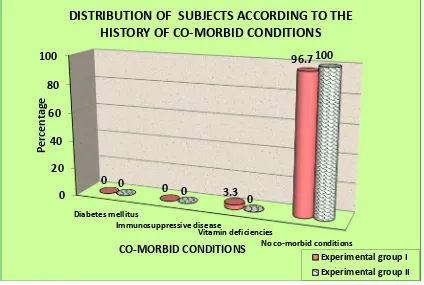
7 Distribution of subjects according to the stage of Cancer 76 8 Distribution of subjects according to the Nutritional status 77 9 Distribution of subjects according to the History of
co-morbid conditions
78
10 Distribution of subjects according to the frequency of taking oral hygiene
79
11 Distribution of subjects according to the Radiation dosage 80 12 Distribution of subjects according to the history of using any
dentures
81
13 Assessment of level of Oral mucositis among patients under going Radiation therapy for Head and Neck cancer
LIST OF APPENDICES
APPENDIX
NO
TITLE
I Letter seeking and granting permission to conduct the study in Radiation Oncology ward, Government Rajaji Hospital, Madurai II Ethical committee approval letter
III Content validity certificate
IV Informed consent form V Research Tool
VI English Editing Certificate VII Tamil Editing Certificate VIII Procedure
IX Photographs
LIST OF ABBREVIATIONS
RT : Radiation therapy
RT-AF : Altered Fractionation Radiotherapy cGy : Centigray
WHO : World Health Organization EBT : External Beam therapy
IMRT : Intensity Modulated Radiation therapy TNF : Tumor Necrosis Factor
NCI-CTC : National Cancer Institute-Common Toxicity Criteria RIM : Radiation induced Mucositis
HNC : Head and Neck Cancer OM : Oral Mucositis
TPN : Total Parenteral Nutrition DNA : Deoxyribo Nucleic acid FUO : Fever of unknown origin NaCl : Sodium Chloride
CHAPTER I
INTRODUCTION
“The block of granite which was an obstacle in the pathway of the weak, became a stepping-stone in the pathway of the strong.”
-Thomas Carlyle
Cancer refers to a large group of potentially lethal disorders characterized by abnormal cell growth and metastasis .Because of its diversity and complexity, cancer has no single treatment nor it can be attributed to a single etiologic agent.The word Cancer came from the Greek words, carcinos and carcinoma to describe tumors, thus calling cancer "karkinos." The Greek terms actually were words to describe a crab, which Hippocrates thought a tumor resembled. Although Hippocrates may have named "Cancer," he was certainly not the first to discover the disease. The history of cancer actually begins much earlier.
Cancer, also known as a malignant tumor, is a group of diseases involving abnormal cell growth with the potential to invade or spread to other parts of the body. Not all tumors are cancerous; benign tumors do not spread to other parts of the body.
about 2.3 lakhs (33%) cancers are tobacco related. In Tamilnadu, there would be about 1.5 lakhs cancer cases at any given time and about 35,000 new cancer cases are added to this pool each year. (Jaypee International scientific Journal-vol 2.mar 2013).
In India, around 555 000 people died of cancer in 2010, according to estimates published in The Lancet today (March 28, 2012). The study, led by Dr Prabhat Jha, the Director of the Centre for Global Health Research at St. Michael's Hospital, Toronto, in a collaboration with Indian national institutions and the International Agency for Research on Cancer (IARC), used a unique method of projecting cancer deaths for the whole of India based on the patterns of cancer mortality in 2000-2003 in a sample of households. Cancer mortality is a key measure of the cancer burden in a given country and provides an important basis for implementing public health preventive measures.
From the Kidwai Memorial Institute of Oncology: The estimated number of new cancers in India per year is about 7 lakhs and over 3.5 lakhs people die of cancer each year. Out of these 7 lakhs new cancers about 2.3 lakhs (33%) cancers are tobacco related.
Tobacco use is the cause of about 22% of cancer deaths. Another 10% is due to obesity, a poor diet, lack of physical activity, and drinking alcohol. Other factors include certain infections, exposure to ionizing radiation, and environmental pollutants. In the developing world nearly 20% of cancers are due to infections such as hepatitis B, hepatitis C, and human papillomavirus. Approximately 5–10% of cancers are due to genetic defects inherited from a person's parents.
Warning signs of Cancer includes the following: C change in bowel habits -sign of colorectal cancer
A sore that does not heal on the skin or in the mouth could be malignant
Unusual bleeding or discharge from rectum, bladder or vagina could be colorectal, prostate, bladder or cervical cancer
Thickening of breast tissue or a new lump in breast
Indigestion or trouble swallowing -cancer of the mouth thoart esophagus or stomach. Obvious changes to moles or warts could be skin cancer
Nagging cough or hoarseness that persists for four to six weeks could be cancer of lung or throat cancer.
Overall 57.5% of global Head and Neck cancer occurs in Asia, especially in India. Head and neck cancer includes cancer of the paranasal sinuses, nasal cavity, oral cavity, tongue, salivary glands, larynx, and pharynx (including the nasopharynx, oropharynx, and hypopharynx). Head and Neck cancer in India accounted for 30% in all cancers. In India, 60-80% patients present with advanced disease as compared to 40% in developed countries.(10.5005/JP-Journals-10001-1132,Manik Rao Kulkarni)
without chemotherapy, 34 to 43% develop severe mucositis. The severity of oral mucositis increases in (1) patients with primary tumors in the oral cavity, oropharynx or nasopharynx, (2) treated with concomitant chemotherapy, (3) receiving a total dose over 5000 Centigray, and (4) treated with altered fractionation radiation schedules. (International Scientific Journals from Jaypee).
There are four standard methods of treatment for cancer: surgery, chemotherapy, radiation therapy, and immunotherapy/biologic therapy. When initially diagnosed with cancer, a cancer specialist (called an oncologist) will provide the patient withcancer treatment options.
Radiation therapy, radiotherapy, or radiation oncology, is therapy using ionizing radiation, generally as part of cancer treatment to control or kill malignantcells. Radiation therapy is commonly applied to the cancerous tumor because of its ability to control cell growth. Ionizing radiation works by damaging the cancerous tissue leading to cellular death. To spare normal tissues (such as skin or organs which radiation must pass through to treat the tumor), shaped radiation beams are aimed from several angles of exposure to intersect at the tumor, providing a much larger absorbed dose there than in the surrounding, healthy tissue.
Typically, one of the following radiation therapy procedures may be used to treat Head and Neck Cancer:
External beam therapy (EBT): a method for delivering a beam of high-energy x-rays to the location of the tumor. The beam is generated outside the patient (usually by a linear accelerator) and is targeted at the tumor site.
precise radiation doses to a malignant tumor or specific areas within the tumor(2014-TexasOncology).
Aggressive treatment of malignant disease may produce unavoidable toxicities to normal cells. The mucosal lining of the gastrointestinal tract, including the oral mucosa, is a prime target for treatment-related toxicity by virtue of its rapid rate of cell turnover. The oral cavity is highly susceptible to direct and indirect toxic effects of cancer chemotherapy and ionizing radiation.
Oral mucositis is probably the most common, debilitating complication of cancer treatments, particularly chemotherapy and radiation.
Oral mucositis refers to erythematous and ulcerative lesions of the oral mucosa. ~Davidson (2003)'
Incidence as well as severity may vary from patient to patient. The probability of developing mucositis is dependent upon the treatment. It is estimated that about 40% of patients treated with standard chemotherapy develop mucositis . The risk of developing mucosal injury increases with the number of chemotherapy cycles and previous episodes of chemotherapy-induced mucositis. There is a qualitative difference between the severity of oral mucositis induced by radiation and that of induced by chemotherapy.
mucosa, smoking, alcohol consumption, and oral hygiene. Mucosal erythema occurs in the first week in patients treated with standard 200 Centigray of daily fractionated radiotherapy programs. With daily fractionated programs of <200 Centigray, the severity of mucositis is expected to be low.( Neoplasia. 2004 September; 6).
At Government Rajaji Hospital- Madurai, patients with Head and Neck cancer are receiving around 200-300 Centigray of daily fractionated dose of Radiotherapy.
A variety of patient-related factors appears to increase the potential for developing mucositis after chemoradiotherapy, including the age of the patient, nutritional status, type of malignancy, pretreatment oral condition, oral care during treatment, and pretreatment neutrophil counts.
Today, mucositis is recognized as an epithelial and sub epithelial injury and is thought to develop in a five-stage model: (1)initiation; (2) up-regulation with generation of messengers;(3) signaling and amplification; (4) ulceration with inflammation; and (5) healing (from Sonis ST. A Biological Approach to Mucositis. J Support Oncol 2004; 2:21–36).
1. Initiation of tissue injury: Radiation and/or chemotherapy induce cellular damage resulting in death of the basal epithelial cells. The generation of reactive oxygen species (free radicals) by radiation or chemotherapy is also believed to exert a role in the initiation of mucosal injury. These small highly reactive molecules are byproducts of oxygen metabolism and can cause significant cellular damage.
transmit signals from receptors on the cellular surface to the inside of the cell. This leads to upregulation of pro-inflammatory cytokines, tissue injury and cell death.
3. Signaling and amplification: Upregulation of proinflammatory cytokines such as tumor necrosis factor- alpha (TNF-α), produced mainly by macrophages, causes injury to mucosal cells, and also activates molecular pathways that amplify mucosal injury.
4. Ulceration and inflammation: There is a significant inflammatory cell infiltrate associated with the mucosal ulcerations, based in part on metabolic byproducts of the colonizing oral microflora. Production of pro-inflammatory cytokines is also further upregulated due to this secondary infection .
5. Healing: This phase is characterized by epithelial proliferation as well as cellular and tissue differentiation , restoring the integrity of the epithelium.
The degree and extent of oral mucositis that develops in any particular patient and site appears to depend on factors such as age, gender, underlying systemic disease and race as well as tissue specific factors (e.g. epithelial types, local microbial environment and function).
Signs and symptoms of mucositis include: -Red, shiny, or swollen mouth and gums -Blood in the mouth
-Sores in the mouth or on the gums or tongue -Soreness or pain in the mouth or throat -Difficulty swallowing or talking
-Soft, whitish patches or pus in the mouth or on the tongue -Increased mucus or thicker saliva in the mouth
Diagnosis of Mucositis is based on the symptoms the patient is experiencing and the appearance of the tissues of the mouth following chemotherapy, bone marrow transplants or radiotherapy. Red burn-like sores or ulcers throughout the mouth is enough to diagnose mucositis.
Prophylactic measures and treatment options should be employed by practitioners for patients in the appropriate clinical settings. Specific recommendations for minimizing oral mucositis include the following:
Good oral hygiene.
Avoidance of spicy, acidic, hard, and hot foods and beverages.
Use of mild-flavored toothpastes.
Use of saline-peroxide mouthwashes 3 or 4 times per day.
Prophylaxis, such as ice-chip cryotherapy, Palifermin (keratinocyte growth factor), and antiviral medications
Some mucosal pharmacologic alterations that have been tried include cryotherapy, Normal saline, Sodium bicarbonate, allopurinol, propantheline, and pilocarpine.
Focal topical application of anesthetic agents is preferred over widespread oral topical administration, unless the patient requires more extensive pain relief. Products such as the following may provide relief:
2% viscous lidocaine
One of the many extemporaneously prepared mixtures combining the following coating agents with topical anesthetics:
o Milk of magnesia.
o Kaolin with pectin suspension. o Mixtures of aluminum.
o Magnesium hydroxide suspensions (many antacids).
Systemic analgesics should be administered when topical anesthetic strategies are not sufficient for clinical relief. Opiates are typically used; the combination of chronic indwelling venous catheters and computerized drug administration pumps to provide Patient controlled analgesia has significantly increased the effectiveness of controlling severe mucositis pain while lowering the dose and side effects of narcotic analgesics.
Normal saline solution is also recommended to treat radiation induced mucositis.It can be prepared by adding approximately 1 teaspoon of table salt to 250ml of water. The solution can be administered at room or refrigerated temperatures, depending on patient preference. The patient should rinse and swish approximately 1 tablespoon, followed by expectoration; this can be repeated as often as necessary to maintain oral comfort. Sodium bicarbonate can be added, if viscous saliva is present. Saline solution can enhance oral lubrication directly as well as by stimulating salivary glands to increase salivary flow.
as bi-carb, for treating oral mucositis. The Latin name for sodium bicarbonate is Saleratus, which means, 'aerated salt'.
Toothpaste containing sodium bicarbonate has in several studies shown to have a better whitening and plaque removal effect than toothpastes without it. Sodium bicarbonate is also used as an ingredient in some mouthwashes. It works as a mechanical cleanser on the teeth and gums, neutralizes the production of acid in the mouth and also acts as an antiseptic to help prevent infections.(Oral complications of Chemotherapy and Head /Neck Radiation (PDQ/R-11-08-2013).
It is important that cancer patients be on the lookout for signs of mucositis, which should be treated as soon as possible once diagnosed. The consequences of mucositis can be mild, requiring little intervention, but they can also be severe--such as hypovolemia, electrolyte abnormalities, and malnutrition--and even result in fatality.
1.1 NEED FOR THE STUDY
“Every area of trouble gives out a ray of hope; and the one
underlying stromal connective tissue due to loss of epithelial cells is found in the most severe form of mucositis; this condition is usually seen 5 to 7 days following medication.
Oral mucositis is a distressing toxic effect of radiotherapy and systemic chemotherapy in cancer patients. Mucositis is characterized by atrophy of squamous epithelial tissue, vascular damage, and an inflammatory infiltrate concentrated at the basement membrane and is followed by ulceration. The erythematous atrophic and ulcerative lesions that develop are a consequence of epithelial damage and death mediated through a complex series of molecular and cellular events.It is associated with significant morbidity characterized by pain, odynodysphagia, dysgeusia, malnutrition, dehydration and it also increases the risk for systemic infections in immunocompromised patients.(International cancer of Head and Neck surgery, May-Aug 2010;(2):1-67).
Oral mucositis can occur with cumulative radiotherapy doses as low as 1000-2000 Centigray with therapy administered at a rate of 200 Centigray per day.In greater than half of patients with mucositis, the condition is of such severities so as to require parenteral analgesia, interruption of Radiotherapy, and hospitalization, all of which increase the cost of cancer therapy and have a negative impact on quality of life.
drug assumption and require a particular diet. About 40% of patients treated with chemotherapy at standard doses develop mucositis and of these, around 50% develop lesions that require modifications or a suspension of the treatment programme. Oral care protocols are based on two levels of intervention: non-medicated vs medicated strategies. The non-medicated oral care protocol focuses on topical therapy and
emphasizes frequent rinsing with 0.9% saline or sodium bicarbonate solutions. (Iranian Journal of Cancer prevention, Vol 5, No 4, Autumn 2012).
The severity of oral mucositis can be evaluated using several different assessment tools. Two of the most commonly used are the World Health Organization (WHO) Oral Toxicity score and the National Cancer Institute Common Toxicity Criteria (NCI-CTC) for Oral Mucositis.World Health Organization (WHO) grading of
mucositis: This scoring system is widely used in routine clinical practice and clinical trials for the evaluation of mucositis. It is graded from 0 to 4. If the patient has no signs and symptoms, it is graded as 0. If the patient has painless ulcers, edema, or mild soreness, it is graded as 1. If there is painful erythema, edema, or ulcers but able to eat, it is graded as 2. If there is painful erythema, edema, or ulcers but unable eat, it
is graded as 3. If there a requirement for parenteral or enteral support, it is graded as 4.
Necrosis or deep ulceration; may include bleeding not induced by minor trauma or abrasion, it is considered as grade 4.
The morbidity of all mucositis can be profound. It is estimated that approximately 15% hospitalization for treatment-related complications . In addition, severe oral mucositis may interfere with the ability to deliver the intended course of therapy, leading to significant interruptions in treatment, and possibly impacting on local tumor control and patient survival.
Parulekar et al. have estimated that chemotherapy-induced mucositis varies from 40 to 76% in patients treated respectively with standard and high-dose chemotherapy.Nearly all (90% to 97%9,24) patients receiving radiotherapy in the head and neck will develop some degree of mucositis.16 Of these patients treated with radiotherapy with or without chemotherapy, 34% to 43% will present severe mucositis. As a result, the patient’s quality of life is affected, hospital admittance rates are higher, the use of total parenteral nutrition is increased and interruption of treatment is more frequent, all of which compromise tumor control. Mucositis causes 9% to 19% of chemotherapy and radiotherapy interruption.
Mucositis may limit the patient's ability to tolerate chemotherapy or radiation therapy, and nutritional status is compromised. It may drastically affect cancer treatment as well as the patient's quality of life. Thus, the treatment aimed to reduce the symptoms of mucositis should also aim to improve the quality of life.
patients with oral mucositis are significantly more likely to have severe pain and a weight loss of ≥ 5%.Approximately 16% of patients receiving radiation therapy for head and neck cancer were hospitalized due to mucositis. Further, 11% of the patients receiving radiation therapy for head and neck cancer had unplanned breaks in radiation therapy due to severe mucositis. Thus, oral mucositis is a major dose-limiting toxicity of radiation therapy to the head and neck region.(Inernational journal on head and neck surgery-Manik Rao Kulkarni).
healing time from treatments. Physical problems may interfere with food intake and proper nutrition. Patients with head and neck tumors may have mouth or throat pain that can interfere with chewing and compound difficulties in swallowing. Tooth and gum disease can also exacerbate issues.
Mucositis can have a negative impact on the overall treatment experience, especially when severe pain or infections occur. In general, mucositis should be treated conservatively to avoid further tissue irritation and damaging the remaining cells from which the epithelium will regenerate.Plaque control and oral hygiene should be maintained.Hence,Nurses have a critical role in all aspects of managing mucositis, including assessing it, teaching oral care, administering pharmacologic interventions, and helping patients cope with symptom distress.
The researcher, during the clinical posting observed that the oral mucositis induced by cancer therapy can be reduced by the use of Normal saline or Sodium bicarbonate oral wash. Hence the researcher was intended to assess the extent of effectiveness of Normal saline and Sodium bicarbonate oral wash in reducing oral mucositis among cancer patient.
1.2 STATEMENT OF THE PROBLEM
A study to compare the effectiveness of Normal saline mouth wash versus Sodium bicarbonate mouth wash on Oral mucositis among patients undergoing Radiation therapy in oncology ward at Government Rajaji Hospital Madurai.
1.3 OBJECTIVES OF THE STUDY
• To evaluate the effectiveness of Normal saline mouth wash in Experimental group I and Sodium bicarbonate mouth wash in Experimental Group II • To compare the effectiveness between Normal saline mouth wash and
Sodium bicarbonate mouth wash in Experimental group I and II
• To associate the level of Oral mucositis among patients undergoing Radiation therapy with selected demographic and clinical variables.
1.4 HYPOTHESES
• H1: There is a significant difference between the pre and post test level of Oral mucositis among patients undergoing Radiation therapy for Head and neck cancer in Experimental group I and II
• H2:There is a significant difference between the post test level of Oral mucositis between Experimental group I and II.
• H3:There is a significant association between the level of Oral mucositis with selected demographic and clinical variables.
1.5 OPERATIONAL DEFINITIONS 1. EFFECTIVENESS:
2. NORMAL SALINE MOUTH WASH:
In this study, it refers to rinsing oral cavity of patients with Oral mucositis by using 40 ml of Normal saline solution, which is prepared by adding 1teaspoon of salt (6grams) in 250 ml of water which contains Sodium 150mmol/litre and chloride 150mmol/litre) for 1 minute , thrice a day (8am, 2 pm and 8 pm) for 2 weeks.
3. SODIUM BICARBONATE MOUTH WASH:
In this study, it refers to rinsing oral cavity of patients with Oral mucositis by using 40 ml of Sodium bicarbonate solution, which is prepared by adding 1teaspoon of Sodium bicarbonate (1.3 grams) in 250 ml of water for 1 minute , thrice a day (8am, 2 pm and 8 pm) for 2 weeks.
4. ORAL MUCOSITIS:
In this study it refers to redness, swelling, pain and ulceration that occurs in the oral mucosa as a side effect of Radiation therapy for Head and Neck cancer which can be measured by National Cancer Institute-Common Toxicity Criteria- Oral Mucositis grading scale.
5. PATIENTS UNDERGOING RADIATION THERAPY:
In this study, it refers to patients with Head and Neck Cancer receiving Radiation therapy in Radiation oncology ward at Government Rajaji Hospital Madurai.
6. ONCOLOGY WARD:
1.6 ASSUMPTION The study assumes that,
1. The patients receiving radiation therapy for Head and Neck Cancer develops varying level of Oral mucositis
2. Oral mucositis patients will cooperate for the Normal saline and Sodium bicarbonate mouth wash.
3. Normal saline and sodium bicarbonate mouth wash has no side effects and it helps to heal Oral mucositis.
1.7 DELIMITATIONS The study is limited to:
1. Patients receiving Radiation therapy for Head and Neck Cancer at Radiation oncology ward, Government Rajaji Hospital Madurai.
2. The sample size is limited to 60 patients with Radiation induced Oral mucositis
3. Data collection period is limited to 4-6weeks
1.8 PROJECTED OUTCOME
CHAPTER -II
REVIEW OF LITERATURE
“For the creation of a masterwork of literature two powers must concur, the power of the man and the power of the moment, and the man is not enough without the moment”.
-James Allen
A review of relevant literatures was collected to generate a picture of what is
known about a particular situation. Relevant literature to those sources that are
important in providing in depth knowledge related to make changes in practice or to
study a selected problem.
This chapter is divided into two parts:
PART I:
Review of related literature on the study
PART II:
PART I
REVIEW OF RELATED LITERATURE
A literature review is a text written by someone to consider the critical points of current knowledge including substantive findings, as well as theoretical and methodological contributions to a particular topic.
-BT Basavanthappa(2012)
A literature review is the process of reading, analyzing, evaluating, and summarizing scholarly materials about a specific topic.
-Polit (2010)
Literatures relevant for this study reviewed and have been organized under the
following headings.
1. Review related to the prevalence of Oral mucositis
2. Review related to the effectiveness of Normal saline mouth wash on other
conditions
3. Review related to the effectiveness of Sodium bicarbonate mouth wash on
other conditions
4. Review related to the effectiveness of Normal saline mouth wash on Oral
mucositis
5. Review related to the effectiveness of Sodium bicarbonate mouth wash on
1. REVIEW RELATED TO THE PREVALENCE OF ORAL MUCOSITIS:
Bjarnason.,(2012).A prospective observational study was conducted at
Boston University to examine the burden of mucositis and risk of complications in
head and neck cancer patients receiving radiation with or without chemotherapy at
Chicago. Oral mucositis was assessed two, four and six weeks by using questionnaire
for head and neck cancer. A 12 team instrument was used to measuring mouth and
throat soreness and pain and limitation in oral functions. Data was collected at every
weeks and results showed that oral mucositis was initially developed who is with
radiation therapy and severe mucositis and throat soreness occurred in 76 percent of
patients.
David I. Rosenthal, et al;(2013).conducted a Randomized control trial at
Mumbaito identify the toxicity associated with Radiation therapy.Radiation-induced
mucositis (RIM) is a common toxicity for head and neck cancer (HNC)patients. The
frequency has increased because of the use of more intensive altered radiation
fractionation and concurrent chemotherapy regimens. The extent of the injury is
directly related to the mucosal volume irradiated, anatomic subsite exposed, treatment
intensity, and individual patient predisposition.
Fayed,L;(2009).conducted a retrospective study on the various modalities of
cancer therapies at California and identified that Chemotherapy and radiation therapy
are the most effective treatments of cancer. Both will damage the cancerous and
normal cells, which leads to systemic adverse effect. It works by targeting rapidly
multiplying cancer cells. Unfortunately, other types of cells in bodies also multiply at
high rates. This is why both can cause side effects like hair loss and mucosal damage.
cells. It works by damaging a cancer cell's DNA, making it unable to multiply. Cancer
cells are highly sensitive to radiation and typically die when treated. Nearby healthy
cells can be damaged and leads to complications such as Mucositis.
Fayed, L.,(2010).A study was conducted to explore the relationship between
oral mucositis and selected clinical and economic outcomes of patients with radiation
and chemotherapy. Subjects who were participated in this study consisted of 92
patients from eight centers. Oral mucositis scoring system (Oral Mucositis
Assessment Scale) was used to assess oral mucositis and examined the relationship
between patients peak oral mucositis scores and days with fever, the occurrence of
infection, days of total parenteral nutrition (TPN), and days of injectable narcotic
therapy, days in hospital, total hospital charges for the index admission, and vital
status at 100 days. Results showed that Patients’ peak oral mucositis scores reached
the full range of possible values (0 to 5) and were significantly (P<0.05) correlated
with all of the outcomes and it revealed that oral mucositis is associated with
significantly worse clinical and economic outcomes in cancer treatment.
Floyd; (2011). conducted a Randomized clinical trial at Boston and found out
tissues with a larger blood supply or a higher cell turnover rate respond more
intensely to radiation. In the oral cavity, these areas are the lateral borders and ventral
surface of the tongue as well as the soft palate and floor of the mouth. Large amounts
of fine vasculature exist in these areas, and radiation leads to vascular congestion and
increased interstitial permeability. Within the irradiated fields, mucositis can occur
anywhere in the oral cavity. However, it may be found more frequently on the uvula
and soft palate because these sites have a higher cell turnover rate than other area.
the salivary glands or metallic dental restorations. Extensive irradiation of the salivary
glands leads to production of glycoproteins and an increased acidity of saliva all of
which render patients at higher risk for mucositis.
Jai prakash Agarwal;(2012). conducted a Randomized control trial at
Mumbai on the prevalence of Radiation induced oral mucositis among patients
undergoing radiation terapy for head and neck cancer.Oral mucositis is one of the
debilitating and dose-limiting acute toxicity during (chemo) radiation or for HNC
having a major impact on the patient daily functioning, well-being and quality of life.
The unplanned interruption of treatment secondary to mucositis may compromise the
treatment and the outcomes if not adequately addressed.
John Henry;(2010).A retrospective study was done in the department of
Clinical Oncology, Netherlands, to assess the incidence and severity of
Radiotherapy-associated oral mucositis on 150 subjects. Mucositis was scored using the World
Health Organization (WHO) criteria. Eighty-seven episodes of mucositis occurred in
47 (31%) patients. Twenty-six patients each experienced only one episode, whereas
21 patients had up to eight episodes of mucositis. The 1,281 Radiotherapy cycles that
have been analyzed included 87 cycles in which mucositis was observed. In 16
patients (11%) only slight oral mucosal changes were recorded (maximum WHO
score 1), while 25 patients (17%) experienced mild to moderate mucositis (maximum
WHO score 2), and in 6 patients (4%) mucositis was moderate to severe (maximum
WHO score 3). No grade 4 mucositis developed. It was concluded that almost
one-third of patients receiving chemotherapy for solid tumors experienced one or more
Kumar;(2009).conducted a study to identify the prevalence of Radiation
induced oral mucositis among patients receiving Receiving radiotherapy or
chemotherapy for Head and Neck cancer. Patients receiving radiotherapy or
chemotherapy for Head and Neck cancer will develop some degree of oral mucositis.
The incidence of oral mucositis was especially high in patients: (i) With primary
tumors in the oral cavity, oropharynx, or nasopharynx; (ii) who also received
concomitant chemotherapy; (iii) who received a total dose over 5,000cGy; and (iv)
who were treated with altered fractionation radiation schedules. Radiation-induced
oral mucositis affects the quality of life of the patients and the family concerned.
Loyd V. Allen;(2011). conducted a Bibliographical review on Oral
mucositis.Oral mucositis is a widespread and potentially serious consequence of
high-dose chemotherapy and radiotherapy. It seems to be particularly associated with
fluorouracil, doxorubicin, and methotrexate. Symptoms, which may include altered
taste perception, sores, and varying degrees of pain, usually appear 4 to 5 days after
treatment initiation. Treatment is mainly supportive, involving both
nonpharmacologic and pharmacologic methods. For compounded preparations such
as mouthwashes, there are various formulations that pharmacists can use based on the
experience and needs of the individual physician and patient, respectively.
Naidu.R;(2012).conducted a study and concluded that Oral mucositis remain
a major source of illness despite the use of a variety of agents to prevent them. Oral
mucositis is defined as inflammation and ulceration of the mouth mucosa with pseudo
membrane formation; it is a potential source of infection which may lead to death. It
continue to thin, ulceration eventually occurs. It is at this stage that the primary
symptom of severe debilitating oral pain is most severe.
Napenas J;(2007)conducted a study and identified that the incidence and
severity of cancer radiotherapy-associated mucositis is caused in part by changes in
the oral bacterial microflora. This systematic review examined the role of oral
bacterial microflora changes in the development of oral mucositis during
radiotherapy. Thirteen prospective clinical trials were identified, involving 300
patients with 13 different cancer diagnoses.The most frequent Gram-negative species
isolated during chemotherapy were from the Enterobacteriaceae family, Pseudomonas
sp. and E. coli.
Ramana.V;(2010). conducted a study on the prevalence of Oral mucositis.It
occurs secondary to radiotherapy for various solid tumors, the exact pathophysiology
of development is not known, but it is thought to be divided into direct and indirect
mucositis.Chemotherapy or radiation therapy will interfere with the normal turnover
of epithelial cells, leading to mucosal injury; subsequently, it can also occur due to
indirect invasion of gram-negative bacteria and fungal species because most of the
cancer therapy will cause changes in blood counts.
Ronald., (2011)conducted aprospective studyto assess the toxicity on patients
who receives high-dose therapy. Two recently published retrospective analyses of
patient complaints following radiotherapy have identified,oral mucositis as the worst
toxicity reported by patients, and what is more important is that patients indicated that
oncology healthcare team members do a poor job of managing and providing methods
of symptom relief. Twenty percent of patients surveyed indicated they received no
Steven;(2012). A prospective study was conducted in the Cancer Institute
Hospital of Japanese Foundation for Cancer Research, Tokyo, Japan to evaluate of
incidence and severity of oral mucositis induced by Radiotherapy in solid tumors and
malignant lymphomas.Two hundred twenty-seven patients who received
chemotherapy for head and neck cancer, esophageal cancer, colorectal cancer, breast
cancer, and malignant lymphomas at the Cancer Institute Hospital between January
2011 and December 2012 were recruited. It was found that OM frequently occurs in
patients with various tumors receiving Radiotherapy. Despite low-grade OM, they
might cause gastrointestinal adverse events.
Stokman M A, Spijkervet F K, et al;(2009).conducted a cross sectional
study which aim to evaluate the effectiveness of interventions for the prevention of
oral mucositis in cancer patients treated with head and neck radiotherapy and/or
chemotherapy, with a focus on randomized clinical trials,the aim of which was the
prevention of mucositis in cancer patients undergoing head and neck radiation,
chemotherapy, or chemoradiation. The control group consisted of a placebo, no
intervention, or another intervention group. Mucositis was scored by either the WHO,
the National Cancer Institute-Common Toxicity Criteria (NCI-CTC) score, or the
absence or presence of ulcerations, or the presence or absence of grades 3 and 4
mucositis. The meta-analyses included 45 studies fulfilling the inclusion criteria, in
which 8 different interventions were evaluated: i.e., local application of
chlorhexidine; iseganan; PTA (polymyxin E, tobramycine, and amphotericin B);
granulocyte macrophage-colony-stimulating factor/granulocyte colony-stimulating
factor (GM-CSF/G-CSF); oral cooling; sucralfate and glutamine; and systemic
administration of amifostine and GM-CSF/G-CSF. Four interventions showed a
with an odds ratio (OR) = 0.61 (95% confidence interval [CI], 0.39-0.96); GM-CSF,
OR = 0.53 (CI: 0.33-0.87); oral cooling, OR = 0.3 (CI: 0.16-0.56); and amifostine,
OR = 0.37 (CI: 0.15-0.89).
Suman. A;(2010).conducted a study on the incidence and severity of oral
mucositis. It will vary from patient to patient, and treatment to treatment.
Approximately 400,000 patients per year may develop acute or chronic oral
complications during chemotherapy and radiation therapy. It is estimated that there is
40 percent incidence of mucositis in patients treated with chemotherapy, patients
receiving radiation have 30 to 60 percent chance and patients receiving radiation
therapy in particular to head and neck have chance of 98%.Severe mucositis is
commonly seen in patients who receive radiation therapy for cancer of the oral cavity
and surrounding structures.
Trotti A, Bellm L A;(2013).conducted a Randomized clinical trial on patients
with head and neck cancer receiving RT with or without chemotherapy that reported
one or more outcomes of interest. Thirty-three studies (n=6181 patients) met inclusion
criteria. Mucositis was defined using a variety of scoring systems. The mean
incidence was 80%. Over one-half of patients (56%) who received altered
fractionation RT (RT-AF) experienced severe mucositis (grades 3-4) compared to
34% of patients who received conventional RT. Rates of hospitalization due to
mucositis, reported in three studies (n=700), were 16% overall and 32% for RT-AF
patients. Eleven percent of patients had RT regimens interrupted or modified because
of mucositis in five studies (n=1267) reporting this outcome. It gives a conclusion that
Mucositis is a frequent, severe toxicity in patients treated with RT for head and neck
Verdi.,(2011).A descriptive study was conducted to find out the incidence of
oral mucositis in cancer treatment. Patients receiving radiation therapy and
chemotherapy were included in the study. Patients oral cavity was assessed weekly
and identified that patients receiving chemotherapy, oral mucositis usually develops
from 10 to 12 days of administration and in radiation therapy mucositis occurred after
7 to 10 days of administration, the incidence and severity was high in patients
receiving both.
\
2. REVIEW RELATED TO THE EFFECTIVENESS OF NORMAL SALINE
MOUTH WASH ON OTHER CONDITIONS
Boston, Denman;(2011) conducted a comparative evaluation of 0.9% Normal
saline mouthwash with 0.2% chlorhexidine gluconate in prevention of plaque and
gingivitis at department of Periodontology, Pune, Maharashtra, to assess the efficacy
of 0.9% Normal saline mouthwash as an anti-plaque agent and its effect on gingival
inflammation and to compare it with 0.2% chlorhexidine gluconate by evaluating the
effect on plaque and gingival inflammation and on microbial load on 60 subjects.
Group A-30 subjects were advised chlorhexidine gluconate mouthwash. Group B-30
subjects were advised experimental Normal saline mouthwash. Parameters were
recorded for plaque and gingival index at day 0, on 14th day, and 21st day. On
comparison between chlorhexidine and Normal saline mouthwash, percentage
reduction of the Plaque Index between 0 and 21 st day were 64.207 and 69.072,
respectively (P=0.112), percentage reduction of Gingival Index between 0 and
21st day were 61.150 and 62.545 respectively (P=0.595) and percentage reduction of
BAPNA values between 0 and 21st day were 42.256 and 48.901 respectively
Einberg. Stephen;(2012) conducted a systematic review to assess the
effectiveness of mouthwashes in preventing and ameliorating chemotherapy-induced
oral mucositis at Boston University. Based on study quality, three out of five
randomized controlled trials were included in a meta-analysis. The results failed to
detect any beneficial effects of chlorhexidine as compared with sterile water, or NaCl
0.9%. Patients complained about negative side-effects of chlorhexidine, including
teeth discoloration and alteration of taste in two of the five studies on chlorhexidine.
The severity of oral mucositis was shown to be reduced by 30% using 0.9% normal
saline mouthwash as compared with sterile water in a single randomized controlled
trial.
Felix. Fernandes (2012). A study was conducted to evaluate the oral care of
patients with cancer at Pune.The effects of povidone-iodine and normal saline
mouthwashes on oralmucositis after high dose chemotherapy on 132 patients who
were randomized to use normal saline (n=65) or povidone-iodine diluted 1:100 (n=67)
mouthwashes for oral mucositis prophylaxis and treatment after high-dose
chemotherapy followed by autologous peripheral stem cell transplantation. No
significant difference was found between the groups in respect of oral mucositis
characteristics, fever of unknown origin and other infections.
Hadi Darvishi Khezri. Mohammad Ali Haidari Gorji.et
al;(2013)conducted a double blinded clinical trial atMazandaran University of
Medical Sciences, Sari, Mazandaran, Iran.This study is aimed to determine and
compare anti-bacterial effects of the chlorhexidine gluconate 0.2%, herbal mouthwash
of matrica (chamomile extracts) 10%, PersicaTM 10% and normal saline in intensive
into four groups of 20 patients each one. Researchers applied PersicaTM to group one,
chlorhexidine gluconate mouthwash 0. 2% to group two and third group received
matrica, finally in the control group, normal saline were used. In order to culturing of
Staphylococcus aureus and Streptococcus pneumoniae, salivary samples were obtained without any stimulation after six minimums oral rinsing. The result showed
that decreased rate of bacterial colonies after intervention in the whole four groups
was significant (p < 0.001). The mouthwash of chlorhexidine (p < 0.001), PersicaTM
(p = 0.008) and Normal saline(p = 0.01) had a significant antibacterial effect on S. aureus and S. pneumoniae (p < 0.001). Hence it is concluded that Herbal oral mouthwash of PersicaTM and Normal saline has the effect on S. pneumoniae and S. aureus of oropharynx area in mechanical ventilation patients.
Muskan. Ronald et al;(2013) conducted a study at Lansdowne, Uttrakhand,
India to compare the efficacy of 3 mouth washes such as Aloe vera, Chlorhexidine
and Normal saline on Dental plaque. A total of 300 systemically healthy subjects were
randomly allocated into 3 groups: Aloe vera mouthwash group (n=100), control group
(=100)-chlorhexidene group and saline water-Placebo (n=100). To begin with,
Gingival index (GI) and plaque index (PI) were recorded. Then, baseline plaque
scores were brought to zero by professionally cleaning the teeth with scaling and
polishing. After randomization of the participants into three groups, Subjects were
asked to swish with respective mouthwash (Aloe vera mouthwash, 0.2%chlorhexidine
gluconate mouthwash, or normal saline) as per therapeutic dose for 4 days. There was
a significant reduction on plaque in Normal saline and chlorhexidine groups and no
statistically significant difference was observed among them (p>0.05). Normal saline
Normal saline may prove an effective mouthwash due to its ability in reducing dental
plaque.
Parwani SR.Parwani RN. et al; (2013) conducted a Comparative evaluation
of anti-plaque efficacy of herbal and 0.2% chlorhexidine gluconate mouthwash in a
4-day plaque re-growth study at Modern Dental College campus, Bijasan road, Madhya
Pradesh.In this clinical trial, 90 pre-clinical dental students with gingival index (GI)
≤1 were enrolled.The baseline plaque scores were brought to zero by professionally
cleaning the teeth with scaling and polishing. After that, randomized 3 groups were
made (of 30 subjects each - after excluding the drop-outs) who were refrained from
regular mechanical oral hygiene measures. Subjects were asked to swish with
respective mouthwash (0.2% chlorhexidine gluconate mouthwash, herbal mouthwash,
or normal saline) as per therapeutic dose for 4 days. Then, GI and PI scores were
re-evaluated on 5 th day by the same investigator, and the differences were compared
statistically by ANOVA and Student's 't'- test. It was concluded that 0.2%
chlorhexidine gluconate and Norml saline mouthwash remains the best anti-plaque
agent. However, when socio-economic factor and/or side-effects of chlorhexidine
need consideration, presently tested normal saline mouthwash may be considered as a
good alternative.
Rahn,Adamietz et al; (2011)conducted a comparative study at University of
Caulifornia on 60 subjects. The present study demonstrated that rinsing with salt and
soda reduced the incidence and severity of Dental plaque, when compared to
Chlorhexidine and other control mouthwashes. It has given the conclusion that rinsing
with salt and soda, in addition to a standard prophylaxis regimen, reduced the
Samuel Vokurka.Eva Bystrická et al; (2012) conducted a randomized
multicentre study on chemotherapy induced oral mucositis at Department of
Haemato-oncology, University Hospital, Alej Svobody. In this study, 132 patients
were randomized to use normal saline (n=65) or povidone-iodine diluted 1:100 (n=67)
mouthwashes for OM prophylaxis and treatment after high-dose chemotherapy
comprising BEAM or HD-L-PAM. The study groups were well balanced in respect of
age, sex, chemotherapy and the number of CD34+ cells in the graft. No significant
difference was found between the groups in respect of OM characteristics, fever of
unknown origin (FUO) and other infections. The antimicrobial solution was less
tolerable for patients. OM occurred significantly more often in females than in males
(86% vs 60%, P=0.0016).The mechanical effect of mouthwashes might have a certain
importance in FUO prevention. When indicating oral rinses, the patient's individual
preference and tolerance of solutions offered should be considered.
Shabanloei. Ahmadi et al; (2011) conducted a randomized, double-blind
clinical trial on 83 patients receiving chemotherapy to determine and compare the
efficacy of Alloporinol, Chamomile and normal saline mouthwashes in the prevention
of chemotherapy-induced Stomatitis, Tarbiat Modares University of Tehran-(Iran).
Significant differences were found between Alloporinol, Chamomile and normal
saline groups in the scores of the severity of Stomatitis (P=0.017), Stomatitis pain
(P=0.027) and in the persistence of Stomatitis. No significant differences were noted
among the mean Stomatitis (P=0.59), Stomatitis pain (0.071) and the severity scores
of the Alloporinol and Normal saline groups. These findings indicate the equal
efficacy of Alloporinol and Normal saline in the prevention of
chemotherapy-induced Stomatitis as compared to the Chamomile group. Considering the cost and
reduction of the severity of chemotherapy-induced Stomatitis, it has been implied for
the prevention of the same.
Zohreh Taraghi. Hadi Darvishi Khezri. et al;(2011)conducted a
randomized clinical trial at Imam Khomeini Hospital, Sari, Iran to determine and
compare the antibacterial effects of persica® mouthwash 10% (miswak extract) and
chlorhexidine gluconate 0.2% and 0.9% normal saline in mechanically ventilated
patients in intensive care unit (ICU). In this trial, 60 patients who were admitted in a
surgical ICU and met the inclusion criteria were randomly divided in two equal
intervention and one control groups. In the first intervention group, chlorhexidine
gluconate mouthwash 0.2% was used, in the second one, the researchers used
persica® herbal mouthwash 10% and finally in the control group, normal saline was
used. Data were analyzed using Chi-square and ANOVA tests in SPSS 17 software.
Decrease of bacterial counts was significant in all three groups after intervention
(p<0.001). The findings of this study indicated that herbal persica® mouthwash and
normal saline can be considered as an effective mouth wash in ICU patients due to
high resistance of the bacteria to synthetic mouthwashes and side effects of these
drugs.
3. REVIEW RELATED TO THE EFFECTIVENESS OF SODIUM
BICARBONATE MOUTH WASH ON OTHER CONDITIONS
Berry.Davidson et al; (2011) conducted a single blind randomised
comparative study in a 20-bed adult intensive care unit in a university hospital.
Patients with an expected duration of mechanical ventilation more than 48 h were
eligible. Patients were randomised to one of three study regimens (Group A control,
second hourly, and Group C twice daily irrigations with chlorhexidine 0.2% aqueous
oral rinse and second hourly irrigations with sterile water).Data from a total of 109
patients were analyzed. Group A 43, Group B 33 and Group C 33 (mean age: 58 ± 17
years, simplified acute physiology score II: 44 ± 14 points). On admission no
significant differences were found between groups for all clinical data. While Group
B showed a greater trend to reduction in bacterial colonization,(p=0.302). The
incidence of ventilator associated pneumonia was evenly spread between Groups A
and C (5%) while Group B was only 1%.
Dixon.Berlin et al; (2013) A Study was conducted to see the effect of three
test mouthwashes and a control were studied. 0.12% chlorhexidine, 1%
povidone-iodine, Salt/sodium bicarbonate, Plain water (control) Coloring agents, sweeteners,
and flavoring agents were added to the mouthwashes so that all had identical color
and taste. All were alcohol free, 76 completed Compliance was assessed weekly by
WHO Stomatis scale. Significant difference in mean Stomatitis scores were observed
among all four groups. Post hoc analysis for repeated measure showed a statistically
significant difference between the povidone group and control group (p = 0.013) at
the end of week 1.At the end week 4, significant difference also were observed
between the povidone and salt/soda groups (p =0.16). Thus the study concluded that
all the 3 mouthwashes were effective in reduction of Stomtitis.
Eun Choi ;(2011) conducted a randomized controlled trial study at
Department of Nursing, Nambu University, Gwangju, South Korea was to compare
the effectiveness of sodium bicarbonate (SB) solution with chlorhexidine (CHX)
mouthwash in oral care of acute leukemia patients under induction chemotherapy.
CHX-based product group according to acute myelogenous leukemia or acute
lymphoblastic leukemia. Patients were asked to rinse their mouth four times a day
from the day before chemotherapy started until discharge. The oral microbial count
was assessed on a weekly basis from the 1st day of chemotherapy started to the 28th
day or to the day of discharge from the hospital. Of all the patients in the SB group,
25.0% developed ulcerative oral mucositis, whereas 62.5% in the CHX group did. As
a result of this study, it was found that oral care by SB solution for acute leukemia
patients undergoing chemotherapy was an effective intervention to improve oral
health.
Irwin;(2010) conducted a randomized clinical trial to compare the
effectiveness of two different durations of Soda bicarb mouth wash for prevention of
5- Fluorouracil related stomatitis at New york. The trial involved patients who were
receiving their first course of a treatment regimen– Fluorouracil plus leucoverin
chemotherapy. These patients were randomized to receive Soda bicarb mouth wash
twice a day. Evaluation was done using physician judgement of Stomatitis and patient
interview. Out of the total 178 patients evaluated it was found that both Soda bicarb
groups had less degrees of Stomatitis.
Janjan,N.A et al., (2010) conducted a study to compare the effectiveness of
povidone iodine mouthwash and Soda bicarb mouthwash on Stomatitis. The result of
the study which reveal that both povidone iodine mouthwash and Soda bicarb mouth
wash have effect in reducing the grade of stomatis, but need more evidence for to
identify the more effective mouthwash on Stomtitis and need to integrate into health
Kumar,M., (2012) conducted a randomized clinical trial on the effectiveness
of povidone iodine mouthwash and Soda bicarb mouth wash on stomatitis at
University of Lucknow. Eighty patients with Stomatitis were randomly assigned to
receive one of the two alcohol-free test mouthwashes (1% povidone-iodine and Soda
bicarb). The patients were instructed to rinse with 10 ml of the mouthwash, twice a
day, for a period of 6 weeks. Mucositis was assessed at baseline and at weekly
intervals during radiation therapy, using the World Health Organization criteria for
grading of mucositis. Among the 76 patients who completed the study, patients in the
Soda bicarb group had significantly lower scores when compared to the povidone
iodine group. This study shows that use of Soda bicarb mouthwash can reduce the
severity and delay the onset of Stomatitis.
Lewin;(2012) A study was conducted on client to see the effect of baking
soda oral rinse in reducing the severity of stomatitis. The patient presented with a
change in his voice, weight loss, and pain in his throat for two months. Nurse planed
(a) rinsing his mouth with baking soda several times a day, (b) using abioadherent
oral gel mixed with water every eight hours, and (c) applying the patient already had
been prescribed nystatin to rinse with and expectorate. As the treatment sessions
continued, performance status remained at 90 and his stomatitis scale wavered from
2.0–3.0 (on a scale from0 = no stomatitis to 4 = tissue necrosis, significant bleeding,
and life-threatening consequences).Through the use of multiple interventions during
his therapy, the client was able to reduce his pain, maintain a good performance
status, and maintain his lifestyle without severe changes. The study concluded that
Michael;(2011) conducted a systematic review to assess the effectiveness of
mouthwashes on chemotherapy-induced Stomatitis at British University. Based on
study quality, three out of five randomized controlled trials were included. The resul