Variations in subclinical left ventricular dysfunction,
functional capacity, and clinical outcomes in different
heart failure aetiologies
Ying Wang
1, Hong Yang
1, Mark Nolan
1, Faraz Pathan
1, Kazuaki Negishi
1and Thomas H. Marwick
1,2*
1Menzies Institute for Medical Research, Hobart, Australia;2Baker Heart and Diabetes Institute, Melbourne, Australia
Abstract
Aims Patients with heart failure (HF) risk factors are described as being in Stage A of this condition (SAHF). Management is directed towards prevention of HF progression, but to date, no evidence has been described to align the intensity of this in-tervention to HF risk. We sought to what extent SAHF of Type 2 diabetes mellitus (T2DM) and other HF risks showed differ-ences in subclinical left ventricular function, exercise capacity, and prognosis.
Methods and results We recruited 551 elder asymptomatic SAHF patients (age 71 ± 5 years, 49% men, 290 T2DM) with at least one risk factor from a community-based population with preserved ejection fraction. All underwent a comprehensive echocardiogram including global longitudinal strain (GLS) and a 6 min walk test and were followed for 2 years. The primary endpoints were new-onset HF and all-cause mortality. The T2DM group was associated with reduced 6 min walk test distance (451 ± 111 vs. 493 ± 87 m,P<0.001), worse diastolic function (E/e0 9.2 ± 2.7 vs. 8.7 ± 2.4,P= 0.028), and impaired GLS (17.7 ± 2.6% vs.19.0 ± 2.6%,P<0.001). Over a median follow-up of 1.6 years, 49 T2DM-SAHF and 27 other-SAHF met the primary endpoint. T2DM-SAHF had significantly worse outcome than other-SAHF (P= 0.021). In Cox models, obesity [haz-ard ratio (HR) = 2.46;P= 0.007], atrialfibrillation (HR = 2.39;P= 0.028), 6 min walk distance (HR = 0.99;P= 0.034), and GLS (HR = 1.14; P= 0.033) were independently associated with the primary endpoint in T2DM-SAHF, independent of age and glycaemic control.
Conclusions The T2DM-SAHF has worse subclinical left ventricular function, exercise capacity, and prognosis than other-SAHF. Impaired GLS, atrial fibrillation, exercise capacity, and obesity are associated with a worse prognosis in T2DM-SAHF but not in other-SAHF.
Keywords Heart failure; Diabetes mellitus; 6 min walk; Global longitudinal strain
Received: 12 June 2017; Revised: 8 December 2017; Accepted: 17 December 2017
*Correspondence to: Prof Thomas H. Marwick, Baker Heart and Diabetes Institute, 75 Commercial Road, Melbourne, VIC 3004, Australia. Tel: +61 3-8532 1550. Email: [email protected]
Introduction
The current American College of Cardiology/American Heart Association heart failure (HF) guidelines define Stage A HF (SAHF) as existing in subjects with risk factors for HF, in the ab-sence of structural heart disease or symptoms.1These risk fac-tors include obesity, hypertension, atherosclerosis, diabetes mellitus (DM), exposure to cardiotoxins, and family history of HF, and they may be present in up to a third of subjects aged ≥45 years. The natural history of SAHF is that patients may progress to functional or structural abnormalities without symptoms [Stage B HF (SBHF)], clinical manifestations of
HF (Stage C), and eventually, refractory or end-stage HF (Stage D).2,3The identification of HF risk should stimulate ef-forts to identify SBHF, as therapeutic interventions in this set-ting may prevent the development of clinical HF and improve prognosis.2,3However, an effective screening strategy necessi-tates an understanding of underlying risk.4
A recent systematic review of the relative risk of a large range of HF risk factors showed that Type 2 DM (T2DM) had nearly double the risk of incident HF, while the risk with hypertension was lower.5 The frequency of HF in patients with DM is even higher among elderly subjects.6 The pres-ence of T2DM adversely affects the prognosis of patients with
O R I G I N A L R E S E A R C H A R T I C L E
ESC Heart Failure2018;5: 343–354
HF, with contributions from coronary heart disease, diabetic cardiomyopathy, and hypertension.7,8 Even in the absence of ischaemic heart disease, T2DM is over-represented in HF.9 We hypothesized that non-ischaemic SAHF due to T2DM would have more subclinical left ventricular (LV) dys-function and worse dys-functional capacity and clinical outcomes, compared with other SAHF. Thisfinding might justify a more aggressive stance towards screening for SBHF in T2DM.
Methods
Patient selection
We prospectively recruited 551 asymptomatic patients aged ≥65 years, with preserved ejection fraction (EF) but at least one Stage A risk factor for HF (DM, obesity, hypertension, or known cardiac disease), from a community-based population in Tasmania. Patients with existing HF or known ischaemic heart disease were excluded, as were patients with more than moderate valve disease, history of HF, LV EF<40%, inability to acquire adequate echocardiographic images for speckle track-ing imagtrack-ing analysis at baseline. All participants provided writ-ten informed consent, and the study protocol was approved by the Tasmanian Human Research Ethics Committee.
Clinical features
Type 2 DM was based on self-report of diagnosis including medication. Obesity was defined as body mass index (BMI) ≥30 kg/m2.
Demographics, disease and family history, and medication use were obtained using a standardized questionnaire. BMI was calculated as weight in kilograms divided by height in metres squared. In addition to standardized weight and height measurements, waist circumference was measured with a tape measure to the nearest millimetre at the mid-point between the lower costal margin and the iliac crest by a trained examiner. Supine resting blood pressure (BP) was measured twice and averaged in each patient after at least 10 min of rest in a quiet room. Uncontrolled hyperten-sion was defined by averaged systolic BP≥140 mmHg or a di-astolic BP ≥ 90 mmHg.10 The International Federation of Clinical Chemistry standardized haemoglobin A1c (HbA1c) (cut-off 64 mmol/mol) level, estimated glomerularfiltration rate (eGFR), and creatinine were obtained from local pathol-ogy records. Missing values for HbA1c, eGFR, and creatinine were estimated by imputation using linear regression.
Six-minute walk test
The 6 min walk test (6MWT) was performed by traversing back and forth along a marked pathway on a hard, flat
surface, to test the distance an individual was able to walk over a total of 6 min. Patients were allowed to self-pace, and the test was performed using a standardized protocol.11
Echocardiography
A comprehensive echocardiogram including standard trans-thoracic 2D, Doppler echocardiographic studies, and speckle tracking echocardiogram using sensitive systolic and diastolic
function parameters was performed using the same
ultrasound machine (Siemens ACUSON SC2000, 4V1c and 4Z1c probes, Siemens Healthcare, Mountain View, CA) in accordance with the American Society of Echocardiography guidelines.12,13 Images were saved in raw data format and analysed offline. LV internal dimensions and wall thickness, chamber volumes, and valvular morphology were assessed. LV mass index was obtained from LV mass measurement using standard criteria and normalized for body size (body surface area or height to the power of 1.7). LV EF measurement used the modified Simpson biplane method. LV inflow was obtained using pulsed wave Doppler in the apical four-chamber view; peak early (E) and late (A) diastolic velocities, deceleration time, and E/A ratio were assessed. Peak early diastolic medial and lateral mitral annular velocities (e0) and the ratio of mitral inflow early diastolic velocity to average e0 velocity were obtained from pulsed tissue Doppler; E/e0>13 was used as an indicator of diastolic dysfunction. For deformation analysis, standard greyscale two-dimensional images were acquired in conventional four-chamber, two-chamber, three-chamber, parasternal short-axis views at the mid-level, basal level, and apical level. Global longitudinal strain (GLS) was calculated by averaging three apical views using standard software and greater than 18% was considered consistent with LV dysfunction.14,15
Outcomes
Potential HF symptoms were assessed through regular follow-up phone calls, followed by symptom surveillance questionnaires and clinical visits. Records of all-cause hospi-talization were obtained and collected. Suspicious HF symp-toms and signs were reviewed by three independent cardiologists. The diagnosis of HF was established according to the Framingham HF criteria.16The primary endpoint for study was new onset of HF and all-cause mortality.
Statistical analysis
for comparison of non-normally distributed variables. Univariable Cox regression was used in order to identify the predictors with the primary endpoint among clinical, demo-graphic, and echocardiographic variables. Multivariable Cox proportional hazards model was performed to determine the independent predictors and reported as hazard ratio (HR) with 95% confidence interval, guided by univariable analyses. Analyses were performed with standard statistical computer software (SPSS 22, IBM, Chicago, IL); P < 0.05 was deemed to be statistically significant.
Results
Patient characteristics
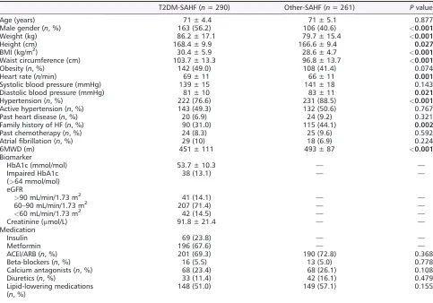
[image:3.594.56.539.349.687.2]All 551 SAHF patients (age 71 ± 5 years, 49% male) were eligi-ble for inclusion in thefinal analysis. T2DM was present in 290 subjects (53%). The clinical and demographic characteristics of the SAHF patients according to T2DM status are listed in
Table 1. T2DM-SAHF was characterized by more obesity as
well as central obesity, higher heart rate, and lower diastolic BP. The 6 min walk distance (6MWD) was significantly lower in T2DM-SAHF patients compared with other-SAHF patients (451 ± 111 vs. 493 ± 87 m,P<0.001). Among T2DM-SAHF pa-tients, 24% patients were treated with insulin and 68% with metformin. The mean HbA1c level is 53.7 ± 10.3 mmol/mol with 13% having impaired HbA1c level (>64 mmol/mol). For eGFR, 14% patients had eGFR > 90 mL/min/1.73 m2, 71%
had eGFR 60–90 mL/min/1.73 m2, and 15% had
eGFR<60 mL/min/1.73 m2. The mean creatinine value was 92 ± 21μmol/L.
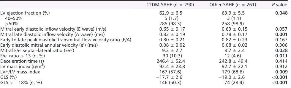
Echocardiographic characteristics
Table 2shows the echocardiographic characteristics between T2DM-SAHF and the remaining SAHF group. Overall, the mean EF was 63 ± 6% (range 40–80%), and only 2% and 1% had EF within 40–50% in T2DM-SAHF and other-SAHF patients, re-spectively. Average GLS was18.4 ± 2.7% (range10.4% to 26.0%). T2DM-SAHF had higher E/e0 ratio, higher LV mass
Table 1 Demographic and clinical characteristics of the SAHF population according to T2DM status
T2DM-SAHF (n= 290) Other-SAHF (n= 261) Pvalue
Age (years) 71 ± 4.4 71 ± 5.1 0.877
Male gender (n, %) 163 (56.2) 106 (40.6) <0.001
Weight (kg) 86.2 ± 17.1 79.7 ± 15.4 <0.001
Height (cm) 168.4 ± 9.9 166.6 ± 9.4 0.027
BMI (kg/m2) 30.4 ± 5.9 28.6 ± 4.7 <0.001
Waist circumference (cm) 103.7 ± 13.3 96.8 ± 13.7 <0.001
Obesity (n, %) 142 (49.0) 108 (41.4) 0.074
Heart rate (n/min) 69 ± 11 66 ± 11 0.001
Systolic blood pressure (mmHg) 139 ± 15 141 ± 18 0.143
Diastolic blood pressure (mmHg) 81 ± 10 83 ± 11 0.021
Hypertension (n, %) 222 (76.6) 231 (88.5) <0.001
Active hypertension (n, %) 143 (49.3) 132 (50.6) 0.767
Past heart disease (n, %) 20 (6.9) 24 (9.2) 0.321
Family history of HF (n, %) 90 (31.0) 115 (44.1) 0.002
Past chemotherapy (n, %) 24 (8.3) 25 (9.6) 0.592
Atrialfibrillation (n, %) 29 (10) 18 (6.9) 0.224
6MWD (m) 451 ± 111 493 ± 87 <0.001
Biomarker
HbA1c (mmol/mol) 53.7 ± 10.3 — —
Impaired HbA1c (>64 mmol/mol)
38 (13.1) — —
eGFR
>90 mL/min/1.73 m2 41 (14.1) — —
60–90 mL/min/1.73 m2
207 (71.4) — —
<60 mL/min/1.73 m2 42 (14.5) — —
Creatinine (μmol/L) 91.8 ± 21.4 — —
Medication
Insulin 69 (23.8) — —
Metformin 196 (67.6) — —
ACEI/ARB (n, %) 201 (69.3) 190 (72.8) 0.368
Beta-blockers (n, %) 16 (5.5) 13 (5.0) 0.778
Calcium antagonists (n, %) 68 (23.4) 68 (26.1) 0.108
Diuretics (n, %) 33 (11.4) 42 (16.1) 0.479
Lipid-lowering medications (n, %)
148 (51.0) 149 (57.1) 0.155
index, and worse GLS than the other-SAHF group. The T2DM group was associated with worse diastolic function (E/e0 9.2 ± 2.7 vs. 8.7 ± 2.4, P = 0.028) as well as impaired GLS (17.7 ± 2.6 vs.19.0 ± 2.6%,P<0.001). T2DM-SAHF also had a much higher prevalence of abnormal GLS (50% vs. 28%,P<0.001) and abnormal E/e0(10% vs. 5%,P= 0.011).
[image:4.594.55.545.82.230.2]In order to determine the incremental impact of T2DM on subclinical systolic and diastolic functions over hypertension,
Table 3 lists echocardiographic parameter comparisons between patients with isolated active hypertension (HT-SAHF) and patients with both active hypertension and T2DM (HT + T2DM-SAHF). HT + T2DM-SAHF had worse GLS than the HT-SAHF group. HT + T2DM-SAHF also had a much higher prevalence of abnormal GLS (52% vs. 30%,P<0.001).
Follow-up
Over a median follow-up period of 1.6 ± 0.6 years (range 0.6–3.2 years), 49 (16.9%) T2DM-SAHF and 27 (10.3%) other-SAHF met the primary endpoint of new onset of
HF and all-cause mortality. On examination of individual com-ponents of the primary endpoint, 46 T2DM-SAHF and 24 other-SAHF developed HF, and three T2DM-SAHF and three other-SAHF died. In the entire cohort, the annualized event rate of HF and all-cause mortality was 8.8%—varying from 11.2% in T2DM-SAHF to 6.4% in other-SAHF.
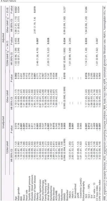
Stepwise nested Cox models were constructed to deter-mine the predictors of primary outcome in SAHF patients with and without T2DM. HRs of the primary outcome from univariable and multivariable analyses are listed in Table 4. Significant variables from univariable analyses were entered into the final age-adjusted and sex-adjusted model. Among T2DM-SAHF patients, obesity [HR = 2.46 (1.28 to 4.70);
P = 0.007], atrial fibrillation (AF) [HR = 2.39 (1.10 to 5.22);
[image:4.594.56.542.547.678.2]P= 0.028], 6MWD [HR = 0.99 (0.99 to 1.00);P= 0.034], and GLS [HR = 1.14 (1.01 to 1.30);P= 0.033] were independently associated with the primary endpoint after adjustment for age, gender, and glycaemic control. In the other-SAHF cohort, history of heart disease [HR = 2.97 (1.19 to 7.4);P= 0.019] was predictive after adjustment for age [HR = 1.08 (1.01 to 1.15);P= 0.022] and gender.
Table 3 Echocardiographic characteristics of the SAHF population according to T2DM and hypertension status
HT-SAHFa(n= 132) HT + T2DM-SAHFb(n= 143) Pvalue
LV ejection fraction (%) 63.6 ± 5.6 62.7 ± 6.9 0.247
Mitral early-diastolic inflow velocity (E wave) (m/s) 0.61 ± 0.15 0.67 ± 0.17 0.008 Mitral late diastolic inflow velocity (A wave) (m/s) 0.80 ± 0.16 0.85 ± 0.20 0.016
Early-to-late peak diastolic transmitralflow velocity ratio (E/A) 0.78 ± 0.19 0.79 ± 0.23 0.513 Early diastolic mitral annular velocity (e0) (m/s) 0.07 ± 0.02 0.08 ± 0.02 0.220
Mitral E/e0septal–lateral ratio (E/e0) 9.0 ± 2.5 9.4 ± 2.8 0.185
E/e0ratio>13 (n, %) 8 (6.1) 18 (12.6) 0.065
Deceleration time (s) 247.5 ± 49.7 246.5 ± 52.1 0.882
LV mass index (g/m2) 93.4 ± 21.5 95.1 ± 25.1 0.547
LVH/LV mass index 41 (31.1) 44 (30.8) 0.989
GLS (%) 19.1 ± 2.4 17.7 ± 2.5 <0.001
GLS>18% (n, %) 39 (29.5) 74 (51.7) <0.001
GLS, global longitudinal strain; HT, hypertension; LV, left ventricular; LVH, left ventricular hypertrophy; SAHF, Stage A heart failure; T2DM, Type 2 diabetes mellitus. BoldPvalues are significant.
a
HT-SAHF: Patients had active hypertension without diabetes. bPatients had both T2DM and active hypertension.
Table 2 Echocardiographic characteristics of the SAHF population according to T2DM status
T2DM-SAHF (n= 290) Other-SAHF (n= 261) Pvalue
LV ejection fraction (%) 62.9 ± 6.5 63.9 ± 5.5 0.048
40–50% 5 (1.7) 3 (1.1)
>50% 285 (98.3) 258 (98.9)
Mitral early diastolic inflow velocity (E wave) (m/s) 0.65 ± 0.17 0.63 ± 0.15 0.057 Mitral late diastolic inflow velocity (A wave) (m/s) 0.83 ± 0.19 0.78 ± 0.17 0.001
Early-to-late peak diastolic transmitralflow velocity ratio (E/A) 0.80 ± 0.21 0.82 ± 0.23 0.167 Early diastolic mitral annular velocity (e0) (m/s) 0.08 ± 0.02 0.08 ± 0.02 0.306
Mitral E/e0septal–lateral ratio (E/e0) 9.2 ± 2.7 8.7 ± 2.4 0.028
E/e0ratio>13 (n, %) 30 (10.3) 12 (4.6) 0.011
Deceleration time (s) 246.4 ± 52.4 242.8 ± 49.4 0.414
LV mass index (g/m2) 92.4 ± 23.8 92.7 ± 22.1 0.912
LVH/LV mass index 167 (57.6) 179 (68.6) 0.009
GLS (%) 17.7 ± 2.6 19.0 ± 2.6 <0.001
GLS>18% (n, %) 146 (50.3) 74 (28.4) <0.001
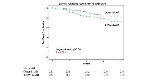
Kaplan–Meier survival curves were constructed for the pri-mary endpoint, with log-rank testing for significance between strata. Among the whole cohort, T2DM-SAHF had significantly worse outcome than other-SAHF (χ2 = 5.36; P = 0.021;
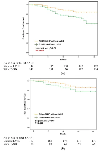
Figure 1). Subclinical LV systolic dysfunction (LVSD) (defined as GLS > 18%) was predictive of primary outcome (HF and all-cause mortality) in T2DM-SAHF in Kaplan–Meier anal-ysis (χ2= 6.75;P= 0.009;Figure 2A). However, there was no statistically significant difference in survival when comparing other-SAHF with or without LVSD (χ2 = 2.68; P = 0.101;
Figure 2B). There were significantly more events in
T2DM-SAHF with obesity compared with those without
(χ2= 11.86;P= 0.001;Figure 3A), but there were no differ-ences when other-SAHF was divided by obesity (χ2 = 0.09;
P= 0.770;Figure 3B). In T2DM-SAHF patients, the most seri-ous prognosis pertained to those with impaired GLS or obe-sity. However, as shown in Figure 4, in other-SAHF individuals, the most serious prognosis was seen in those with a history of heart disease (χ2= 12.49;P<0.001).
Discussion
In these data from a prospectively enrolled cohort, we exam-ined the prevalence of subclinical LV dysfunction, impaired exercise capacity, and prognosis in a community-based popu-lation with HF risk factors. The results suggest that SAHF due to T2DM has a worse echocardiographic manifestation,
functional capacity, and clinical outcome than other-SAHF. This work builds on previous reports of an association be-tween increasing HF stage and worse functional status and prognosis,17by adding evidence about clinical features and functional capacity within the early asymptomatic phases of HF.
Left ventricular mechanics in Stage A heart failure
Previous studies have shown that changes of subclinical LV longitudinal systolic and diastolic function begin before struc-tural LV changes in asymptomatic patients with HF risk fac-tors.18–21 However, comparisons of the degree of LV myocardial dysfunction in early diabetic cardiomyopathy and in other-SAHF have not been documented previously. Our results show that abnormalities of diastolic function are more common in T2DM than in other-SAHF groups, as are disturbances of LV longitudinal systolic function (GLS:
17.7 ± 2.6% vs. 19.0 ± 2.6%,P <0.001). In the current study, we report that the prevalence of systolic longitudinal dysfunction is 50% among community population-based T2DM-SAHF and 28% among other-SAHF. Alterations of sys-tolic strain may exist despite normal diassys-tolic function.22 Im-portantly, impaired GLS as a marker of SBHF is associated with an increased risk of further transition to symptomatic Stage C HF with preserved EF in diabetes.2However, in real life, few asymptomatic SAHF patients undergo tests of car-diac morphology and function, and perhaps because of this,
many of them develop into Stage C/D directly without experiencing SBHF. Effectively, many SBHF ‘hide’ in SAHF, and our results highlight the importance of echocardiography for identifying SBHF and initiating cardioprotection.
These findings highlight the fact that the severity of the early LV function impairment is not necessarily analogous within different aetiologies of SAHF. Despite rigorous
[image:7.594.123.506.93.614.2]adjustment for covariables, each unit decrement of GLS in subjects with T2DM showed a 1.14-fold higher risk of HF and mortality, whereas in subjects with other risks, the pre-dictive value of GLS was not found. The worse LV function in T2DM-SAHF may be attributable to diabetic complications and associated co-morbidities as well as the presence of a distinct diabetic cardiomyopathy.4,18 The pathogenesis of Figure 2 Prognostic implications of T2MD-SAHF and other-SAHF and the role of left ventricular systolic dysfunction. SAHF, Stage A heart failure; T2DM, Type 2 diabetes mellitus.
(A)
the latter is complex and likely multifactorial, involving car-diac autonomic neuropathy, altered myocardial metabolism, small-vessel and large-vessel damage, insulin resistance, and myocardialfibrosis.18Additionally, in our study, patients with uncontrolled hypertension showed relatively preserved GLS, but a combined group with active hypertension and T2DM had more abnormal GLS (19.1 ± 2.4% vs. 17.7 ± 2.5%,
P<0.001;Table 3), as well as a higher prevalence of abnor-mal GLS than in T2DM without hypertension (52% vs. 30%).
Although hypertension and T2DM are the two leading aetiol-ogies of early HF, impaired GLS is more likely due to diabetic cardiomyopathy than hypertensive heart disease.23
Functional capacity in Stage A heart failure
[image:8.594.115.531.85.584.2]In this study, functional capacity at submaximal stress was assessed using 6MWT. This is a simple, safe test of functional Figure 3 Prognostic implications of T2MD-SAHF and other-SAHF and the role of obesity. SAHF, Stage A heart failure; T2DM, Type 2 diabetes mellitus.
(A)
capacity that is a well-established diagnostic procedure in cardiovascular and pulmonary disease, which has prognostic value.24 A systematic review25to investigate the reliability between 6MWT and other methods to assess functional ca-pacity showed that 6MWT has moderate to good ability to predict VO2 in chronic HF, especially in those patients who
are unable to walk>490 m. Compared with healthy controls, patients with T2DM have lower 6MWD.26The results of this study show that, compared with that in other-SAHF, func-tional capacity in T2DM-SAHF was more impaired.
[image:9.594.124.503.95.620.2]The determinants of functional capacity are complex and depend on both psychological and physical factors. Although Figure 4 Prognostic implications of T2MD-SAHF and other-SAHF and the role of heart disease history. SAHF, Stage A heart failure; T2DM, Type 2 di-abetes mellitus.
(A)
it might be considered that the association of T2DM with functional capacity could be driven by obesity—and indeed, advanced age and obesity had strong associations with func-tional capacity—our results suggest that the association of T2DM with functional capacity was independent of these fea-tures. Subclinical cardiac dysfunction, which is often present in both T2DM-SAHF and other-SAHF, could influence func-tional capacity but cannot fully explain the difference of exer-cise capacity between the two SAHF subgroups. Previous work has shown that exercise intolerance in T2DM is inde-pendent of obesity and even presents with good glycaemic control and without clinically apparent cardiovascular dis-ease.27The presence of endothelial dysfunction, decreased myocardial perfusion, decreased muscle mitochondrial func-tion, abnormal tissue haemoglobin oxygen saturafunc-tion, and in-sulin resistance may be mediators in the relationship between diabetic oxidative dysfunction and defects in func-tional capacity.27 In addition, there is evidence of
involve-ment of both cardiac sympathetic and cardiac
parasympathetic nervous system dysfunctions.28
Parasympa-thetic control may be weakened by persistent
hyperglycaemia, with relative enhancement of sympathetic activity.29The association of heart rate recovery with vagal tone leads to T2DM being associated with poor heart rate re-covery and chronotropic incompetence.30Cardiac autonomic dysfunction is associated with reduced cardiac output re-sponse to functional capacity in diabetes, which is probably due to haemodynamic instability during exercise.31Left atrial (LA) dysfunction is also common in patients with HF with pre-served EF and asymptomatic T2DM and contributes to exer-cise intolerance. However, while LA enlargement in diabetes is an independent predictor of LA dysfunction, probably ow-ing to a combination of diastolic dysfunction and diabetic atrial myopathy,32 the specific influence of diabetes on LA dysfunction is more controversial.
Determinants of prognosis in Stage A heart failure
Despite current epidemiological evidence of the greater risk of development of HF in patients with diabetes, the natural his-tory of asymptomatic subjects at risk of HF remains poorly identified.33,34This study extends present knowledge about the prognosis of the entire SAHF cohort in 2 years, based on a community (as opposed to a clinic-based) population. In the entire SAHF cohort, the annualized event rate of incident HF and all-cause mortality was 8.8%. The rate in T2DM-SAHF (11.2%) was almost twice that of other-SAHF (6.4%).
Type 2 DM was associated with more serious outcome, irrespective of whether or not other risk factors were pres-ent. A recent systematic review of 15 observational studies indicated that diabetes (HR = 2.0) showed the strongest predictive value for incident HF among the non-ischaemic SAHF risks, followed by hypertension (HR = 1.61) and BMI
(per 5 kg/m2) (HR = 1.15).5In the present study, the prognosis was associated with obesity, exercise capacity, and abnormal GLS independent of glycaemic control and renal function. However, while hyperglycaemia may be associated with the development of HF, the association between intensive glycaemic control and the reduction of cardiovascular compli-cations remains controversial.35 Although a large cohort study reported an HbA1c≥10% was associated with 1.6-fold risk of incident HF relative to an HbA1c <7%, several large clinical trials have reported no significant benefit for primary cardiovascular events with intensive glycaemic control.35Our
findings do not show that poor glycaemic control
(HbA1c > 64 mmol/mol) is a marker of the composite endpoint (HF and all-cause death) in this elderly asymptom-atic cohort.
Previous research showed that reduced eGFR is a risk factor of cardiovascular events and death in diabetes patients without advanced renal disease, but the risks are small when considering other risk factors.36Although we found no asso-ciation between renal impairment and outcomes in T2DM-SAHF, such an association might become apparent with longer follow-up.
A recent systematic review and meta-analysis of 15 cohort studies confirmed the association of AF with myocardial in-farction, HF, and all-cause mortality.37 In our study, AF showed an independent association with increased risk of new-onset HF and all-cause mortality in T2DM-SAHF but not in other-SAHF. This inconsistent result in other-SHAF may be due to the relatively low prevalence of AF (7%) and the exclusion of ischaemic heart disease at baseline.
The staging system serves as a reminder to clinicians of the importance of early detection and prevention of patients at risks of transitioning to higher stages of HF. In the absence of screening, many SAHF patients (i.e. patients with HF risk factors) have structural or functional abnormalities that that are unrecognized. However, echo screening in SAHF is currently not supported by guidelines or payers, and if these patients had not been recruited to this trial, they would have been indistinguishable from those without any echo abnor-malities (i.e.‘pure’SAHF). Nonetheless, these results highlight the notion that SAHF associated with diabetes is clinically different from other causes of SAHF and perhaps should have different targets for prevention. Thesefindings have impor-tant implications for awareness and identification of high-risk individuals and optimal management to prevent or delay pro-gression to adverse outcome in SAHF.
Limitation
asymptomatic LVSD. In addition, the prevalence of T2DM is relatively high in this SAHF population, as we preferentially recruited patients with diabetes by specific advertising to this population. Second, patients with Type 1 DM were excluded from our study, as the frequency and mechanism of cardiac involvement may not be the same as in T2DM. Third, we chose longitudinal strain as the most robust of the myocardial deformation parameters and did not record circumferential strain, which could have provided additional details about myocardial mechanics. Finally, we do not have data on bio-markers, which may also be used as a potential predictor of HF and adverse outcome.
Conclusion
In this community cohort of patients with HF risks, T2DM-SAHF had worse subclinical LV function, functional capacity, and adverse outcome than other causes of SAHF. Impaired GLS, worse exercise capacity, and the presence of obesity or AF were independently associated with prognosis in T2MD-SAHF, whereas a history of heart disease was the driver of subsequent HF and mortality in other causes of SAHF. The clinical application of this study provides an important caveat that not all types of SAHF are the same. Better targeting of interventions at the most vulnerable SAHF group—those with T2DM—seems appropriate.
Acknowledgements
The authors gratefully acknowledge the contribution of our tireless volunteer coordinators, Diane Binns, Jasmine Prichard, and Jane Mitchell.
Con
fl
ict of interest
None declared.
Funding
This study was partially supported by Tasmanian Community Fund and Diabetes Australia Research Trust. Neither of these agencies had any role in the design, analysis, or interpreta-tion of this study.
Ethics
The study was approved by the Tasmanian Human Research Ethics Committee.
References
1. Hunt SA, Abraham WT, Chin MH, Feldman AM, Francis GS, Ganiats TG, Jessup M, Konstam MA, Mancini DM, Michl K, Oates JA, Rahko PS, Silver MA, Stevenson LW, Yancy CW. 2009 focused update incorporated into the ACC/AHA 2005 Guidelines for the Diagnosis and Management of Heart Failure in Adults: a report of the American College of Car-diology Foundation/American Heart As-sociation Task Force on Practice Guidelines: developed in collaboration with the International Society for Heart and Lung Transplantation. Circulation 2009;119: e391–e479.
2. Lam CS. Diabetic cardiomyopathy: an expression of Stage B heart failure with preserved ejection fraction. Diab Vasc Dis Res2015;12: 234–238.
3. Ammar KA, Jacobsen SJ, Mahoney DW, Kors JA, Redfield MM, Burnett JC Jr, Rodeheffer RJ. Prevalence and prognos-tic significance of heart failure stages: application of the American College of Cardiology/American Heart Association heart failure staging criteria in the com-munity. Circulation 2007; 115: 1563–1570.
4. Murtagh G, O’Connell JO, O’Connell EO, Tallon E, Watson C, Gallagher J, Baugh J, Patle A, O’Connell L, Griffin J, O’Hanlon R, Voon V, Ledwidge M, O’Shea D, McDonald K. Importance of risk factor management in diabetic patients and re-duction in Stage B heart failure. QJM 2015;108: 307–314.
5. Yang H, Negishi K, Otahal P, Marwick TH. Clinical prediction of incident heart failure risk: a systematic review and meta-analysis. Open Heart 2015; 2: e000222.
6. Bertoni AG, Hundley WG, Massing MW, Bonds DE, Burke GL, Goff DC Jr. Heart failure prevalence, incidence, and mor-tality in the elderly with diabetes. Diabe-tes Care2004;27: 699–703.
7. Krumholz HM, Chen YT, Wang Y, Vaccarino V, Radford MJ, Horwitz RI. Predictors of readmission among elderly survivors of admission with heart failure. Am Heart J2000;139: 72–77.
8. Zarich SW, Nesto RW. Diabetic cardio-myopathy. Am Heart J 1989; 118: 1000–1012.
9. Johansson I, Dahlstrom U, Edner M, Nasman P, Ryden L, Norhammar A.
Prognostic implications of Type 2 diabe-tes mellitus in ischemic and nonischemic heart failure.J Am Coll Cardiol2016;68: 1404–1416.
10. Mancia G, Fagard R, Narkiewicz K, Redon J, Zanchetti A, Bohm M, Christiaens T, Cifkova R, De Backer G, Dominiczak A, Galderisi M, Grobbee DE, Jaarsma T, Kirchhof P, Kjeldsen SE, Laurent S, Manolis AJ, Nilsson PM, Ruilope LM, Schmieder RE, Sirnes PA, Sleight P, Viigimaa M, Waeber B, Zannad F. 2013 ESH/ESC guidelines for the management of arterial hyper-tension: the Task Force for the manage-ment of arterial hypertension of the European Society of Hypertension (ESH) and of the European Society of Cardiology (ESC). J Hypertens 2013; 31: 1281–1357.
11. ATS Committee on Proficiency Standards for Clinical Pulmonary Func-tion LaboratoriesATS statement: guide-lines for the six-minute walk test.Am J Respir Crit Care Med 2002; 166: 111–117.
FA, Foster E, Goldstein SA, Kuznetsova T, Lancellotti P, Muraru D, Picard MH, Rietzschel ER, Rudski L, Spencer KT, Tsang W, Voigt J-U. Recommendations for cardiac chamber quantification by echocardiography in adults: an update from the American Society of Echocardiography and the European As-sociation of Cardiovascular Imaging.Eur Heart J Cardiovasc Imaging 2015; 16: 233–271.
13. Nagueh SF, Appleton CP, Gillebert TC, Marino PN, Oh JK, Smiseth OA, Waggoner AD, Flachskampf FA, Pellikka PA, Evangelisa A. Recommendations for the evaluation of left ventricular dia-stolic function by echocardiography. Eur Heart J Cardiovasc Imaging 2009; 10: 165–193.
14. Yingchoncharoen T, Agarwal S, Popovic ZB, Marwick TH. Normal ranges of left ventricular strain: a meta-analysis. Jour-nal of the American Society of Echocardi-ography: Official Publication of the American Society of Echocardiography 2013;26: 185–191.
15. Marwick TH, Leano RL, Brown J, Sun JP, Hoffmann R, Lysyansky P, Becker M, Thomas JD. Myocardial strain measure-ment with 2-dimensional speckle-tracking echocardiography: definition of normal range.JACC Cardiovasc Imaging 2009;2: 80–84.
16. Ho KK, Pinsky JL, Kannel WB, Levy D. The epidemiology of heart failure: the Framingham Study. J Am Coll Cardiol 1993;22: 6A–13A.
17. Azevedo A, Bettencourt P, Alvelos M, Martins E, Abreu-Lima C, Hense H-W, Barros H. Health-related quality of life and stages of heart failure.Int J Cardiol; 129: 238–244.
18. Ng AC, Delgado V, Bertini M, van der Meer RW, Rijzewijk LJ, Shanks M, Nucifora G, Smit JW, Diamant M, Romijn JA, de Roos A, Leung DY, Lamb HJ, Bax JJ. Findings from left ventricular strain and strain rate imaging in asymptomatic patients with Type 2 diabetes mellitus. Am J Cardiol2009;104: 1398–1401. 19. Imbalzano E, Zito C, Carerj S, Oreto G,
Mandraffino G, Cusma-Piccione M, Di Bella G, Saitta C, Saitta A. Left ven-tricular function in hypertension: new insight by speckle tracking echocardiog-raphy. Echocardiography (Mount Kisco, NY)2011;28: 649–657.
20. Ho E, Brown A, Barrett P, Morgan RB, King G, Kennedy MJ, Murphy RT. Sub-clinical anthracycline- and trastuzumab-induced cardiotoxicity in the long-term follow-up of asymptomatic breast cancer survivors: a speckle tracking
echocardiographic study.Heart (British Cardiac Society)2010;96: 701–707. 21. Kusunose K, Yamada H, Nishio S,
Mizuguchi Y, Choraku M, Maeda Y, Hosokawa S, Yamazaki N, Tomita N, Niki T, Yamaguchi K, Koshiba K, Soeki T, Wakatsuki T, Akaike M, Sata M. Vali-dation of longitudinal peak systolic strain by speckle tracking echocardiog-raphy with visual assessment and myo-cardial perfusion SPECT in patients with regional asynergy.Circulation Jour-nal: Official Journal of the Japanese Cir-culation Society2011;75: 141–147. 22. Ernande L, Bergerot C, Rietzschel ER,
De Buyzere ML, Thibault H, Pignonblanc PG, Croisille P, Ovize M, Groisne L, Moulin P, Gillebert TC, Derumeaux G. Diastolic dysfunction in patients with Type 2 diabetes mellitus: is it really the
first marker of diabetic cardiomyopathy? J Am Soc Echocardiogr2011;24: 1268, e1261–1275.
23. Yang H, Wang Y, Negishi K, Nolan M, Marwick TH. Pathophysiological effects of different risk factors for heart failure. Open Heart2016;3: e000339. 24. Lucas C, Stevenson LW, Johnson W,
Hartley H, Hamilton MA, Walden J, Lem V, Eagen-Bengsten E. The 6-min walk and peak oxygen consumption in advanced heart failure: aerobic capacity and survival. Am Heart J 1999; 138: 618–624.
25. Pollentier B, Irons SL, Benedetto CM, Dibenedetto AM, Loton D, Seyler RD, Tych M, Newton RA. Examination of the six minute walk test to determine functional capacity in people with chronic heart failure: a systematic re-view.Cardiopulmonary Physical Therapy Journal2010;21: 13–21.
26. Stewart T, Caffrey DG, Gilman RH, Mathai SC, Lerner A, Hernandez A, Pinto ME, Huaylinos Y, Cabrera L, Wise RA, Miranda JJ, Checkley W. Can a sim-ple test of functional capacity add to the clinical assessment of diabetes?Diabetic Medicine: A Journal of the British Diabetic Association 2016; 33: 1133–1139.
27. Reusch JE, Bridenstine M, Regensteiner JG. Type 2 diabetes mellitus and exercise impairment. Rev Endocr Metab Disord 2013;14: 77–86.
28. Sucharita S, Bantwal G, Idiculla J, Ayyar V, Vaz M. Autonomic nervous system function in Type 2 diabetes using conventional clinical autonomic tests, heart rate and blood pressure variability measures. Indian Journal of Endocrinology and Metabolism 2011; 15: 198–203.
29. Vanninen E, Uusitupa M, Lansimies E, Siitonen O, Laitinen J. Effect of meta-bolic control on autonomic function in obese patients with newly diagnosed Type 2 diabetes. Diabetic Medicine: A Journal of the British Diabetic Association 1993;10: 66–73.
30. Loimaala A, Huikuri HV, Koobi T, Rinne M, Nenonen A, Vuori I. Exercise training improves baroreflex sensitivity in Type 2 diabetes. Diabetes2003; 52: 1837–1842.
31. Roy TM, Peterson HR, Snider HL, Cyrus J, Broadstone VL, Fell RD, Rothchild AH, Samols E, Pfeifer MA. Au-tonomic influence on cardiovascular per-formance in diabetic subjects.Am J Med 1989;87: 382–388.
32. Kadappu KK, Boyd A, Eshoo S, Haluska B, Yeo AE, Marwick TH, Thomas L. Changes in left atrial volume in diabetes mellitus: more than diastolic dysfunction? Eur Heart J Cardiovasc Imaging 2012; 13: 1016–1023.
33. Dandamudi S, Slusser J, Mahoney DW, Redfield MM, Rodeheffer RJ, Chen HH. The prevalence of diabetic cardiomyopa-thy: a population-based study in Olmsted County, Minnesota.J Card Fail 2014;20: 304–309.
34. Xanthakis V, Enserro DM, Larson MG, Wollert KC, Januzzi JL, Levy D, Aragam J, Benjamin EJ, Cheng S, Wang TJ, Mitchell GF, Vasan RS. Prevalence, neurohormonal correlates, and progno-sis of heart failure stages in the commu-nity. JACC Heart failure 2016; 4: 808–815.
35. Giorgino F, Leonardini A, Laviola L. Cardiovascular disease and glycemic control in Type 2 diabetes: now that the dust is settling from large clinical trials. Ann N Y Acad Sci 2013; 1281: 36–50.
36. Drury PL, Ting R, Zannino D, Ehnholm C, Flack J, Whiting M, Fassett R, Ansquer J-C, Dixon P, Davis TME, Pardy C, Colman P, Keech A. Estimated glomerularfiltration rate and albumin-uria are independent predictors of car-diovascular events and death in Type 2 diabetes mellitus: the Fenofibrate In-tervention and Event Lowering in Dia-betes (FIELD) study. Diabetologia 2011;54: 32–43.