Nigel Esteven Gapper
A thesis submitted for the degree of
Master of Science,
in Plant Biology,
Massey University,
Palmerston North
,
New Zealand
Abstract
The asparagus spear is a rapidly growing shoot, dependent on the crown and storage
roots for substrate. Once harvested, spears have a very short shelf-life. Investigations to date point to a physiological cause of this deterioration rather than a pathogenic one. Since loss of membrane integrity is a notable feature of the postharvest deterioration, spears were treated immediately following harvest with cytokinin
(which promotes membrane integrity), and jasmonic acid (produced by deteriorating membranes). Treated plant material was collected and monitored for
physiological and compositional changes. Results show a reduction in postharvest elongation of spears treated with cytokinin, and a reduction of shelf-life of spears treated with jasmonic acid, when compared with control spears treated with water. Also an extension of shelf-life was observed for spears treated with cytokinin. We quantified jasmonates using ELISA in spears after harvest, and also in naturally senescing cladophylls. Jasmonate concentration increased in spears rapidly after harvest, which is most likely to be in response to wounding. Results also showed that
jasmonates may be involved in desiccation stress and cessation of elongation in asparagus spears. Jasmonate production and metabolism appears to be more ordered during natural foliar senescence than during harvest induced senescence of the spear.
Jasmonic acid and dihydrojasmonic acid are metabolised to cucurbic acid during the later stages of natural foliar senescence. The presence of jasmonates in asparagus spears was confirmed by electrospray ionisation mass spectrometry. This analysis also enabled to identify a novel jasmonate, tryptophan-dihydrojasmonic acid amino acid
Acknowledgements
I would like to express my thanks to the many people who have helped me throughout the duration of my MSc program.
Firstly, I would like to express thanks to my two supervisors, Professor Paula Jameson (Institute of Molecular Biosciences, Massey University), and Dr Ross Lill (Crop & Food Research, Palmerston North), whose knowledge, patience, advice and encouragement was much appreciated.
Thank you Dr Gill Norris for the electrospray mass spectrometry work you did for me. Without your knowledge this section of the thesis would not have been achieved.
Thank you Dr John Koolaard, whose statistical knowledge helped me out no end.
To the people in the Plant Physiology Laboratory, Lyn Watson, Don Hunter, Trish Murray, Simone Downs, Celia Zhang, Deming Gong, San Dong Yoo, Rainer Hoffman thank you for the advice and for making the lab a happy environment to work.
A very special thanks to Huaibi Zhang, Marian McKenzie and Geraldine Wood of the cytokinin group. You all helped me very much in different ways and I probably would not have been able to get my project underway without your help and advice.
I would like to acknowledge the assistance I received from Dr Sean Clarke when setting up the ELISA for jasmonate quantification. I learnt a lot from you in the short time you were in Palmerston North.
To my friends at Creative Sounds (The Stomach), and the wider live music scene here in Palmerston North ,thank you for giving me the opportunity to meet new people and forget about science for a while. In particular I would Like to thank Jarrod Love, Malcolm McKinnon, Rob Williams, Craig Black, Dave Bloxham, James Lissette, Mitchell and Ryan Watson and Kelvin Shearman.
Thank you to all the people of the Institute of Molecular Biosciences in particular the students and staff of the old Department of Plant Biology and Biotechnology.
I would like to express my gratitude to Crop & Food Research for financial assistance throughout the project. Without this funding I would not have been able to start this project.
Abstract
Ack.now ledgements
Table of Contents
List of Figures and Tables
Table of Contents
Chapter 1. Introduction
11 ill v
Vlll
1
1.1 Overview 1
1.2 Postharvest deterioration of asparagus 2
1.3 Cytokinins and plant development 5
1.3 .1 Cytokinins and senescence 5
1.4 Jasmonates 6
1.4.1 Jasmonate structure and biosynthesis 6
1.4.2 Metabolism 9
1.4.3 Jasmonates and plant development 12
1.4.4 Interactions between jasmonates and other plant hormones 15
1.4.5 Jasmonate identification and measurement 17 1.5 Objectives
Chapter 2. Materials and Methods 2.1 Plant material
2.2 Plant growth regulator treatments 2.3 Spear quality
2.4 Tiprot
2.5 Spear extension
2.6 Extraction and quantification of soluble sugars 2.7 Chlorophyll extraction and quantification 2.8 Asparagine
2.8.1 Extraction of amino acids
2.8.2 Pre-column derivatisation of amino acids 2.8.3 High performance liquid chromatography
2.9 Jasmonic acid purification and detection 2.9 .1 Plant material
2.9.2 Extraction of jasmonates 2.9.3 Chromatography systems
2.9.4 High performance liquid chromatography 2.9.5 Purification of antiserum
2.9.6 Preparation of JA:OVA conjugate 2.9.7 ELISA for jasmonic acid
2.9.8 Cross-reactivity of jasmonates
2.9.9 Immuno-affinity of jasmonates for mass spectrometry 2. 9 .10 Electro-spray mass spectrometry
2. 9 .11 Statistical analysis Chapter 3. Results
3.1 Physiological changes postharvest 3.1.1 Spear quality
3.1.2 Tiprot
3.1.3 Spear extension
3.2 Biochemical changes postharvest 3.2.1 Chlorophyll
3.2.2 Soluble sugars 3.2.3 Asparagine
3.3Changes in endogenous jasmonates postharvest 3.3.1 High performance liquid chromatography 3.3.2 Optimisation of ELISA
3.3.3 Effect of harvest on endogenous jasmonates
26 26 26 27 27 28 28 29 29 29 30 30 31 31 31 33 33 36 36 36 38 40 40 41 43 3.3.4 Effect of natural foliar senescence on endogenous jasmonates 48 3.3.5 Electrospray mass spectrometry
Chapter Four. Discussion
4.1 Cytokinin and jasmonate-induced physiological and biochemical changes
4.2 Postharvest changes in endogenous jasmonates 4.3 Summary and conclusion
53 57
Bibliography
68
Appendix I
77
Appendix II
78
Appendix III
79
Appendix IV
80
Appendix V
81
List of Figures and Tables
Figures
Figure 1.1 A. Photograph showing the deterioration spear tips after harvest. B. Photograph showing interrupted development of asparagus following harvest 3
Figure 1.2 Structure of jasmonic acid and some of its derivatives 7
Figure 1.3 Biosynthetic pathway of jasmonic acid 8
Figure 1.4 Metabolism of jasmonic acid and dihydrojasmonic acid 11
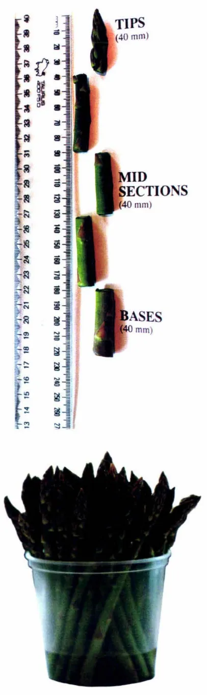
Figure 2.1 A. Photograph showing tips, mid sections and bases of spears harvested in Year 2 for ja monate analysis. B. Photograph showing water-vase technique 21
Figure 3.1 Influence of exogenous cytokinin and jasmonic acid treatment on shelf-life
of asparagus spears postharvest 32
Figure 3.2 Influence of exogenous cytokinin and jasmonic acid treatment on tiprot of
asparagus spears postharvest 34
Figure 3.3 Influence of exogenous cytokinin and jasmonic acid treatment on extension
of asparagus pears postharvest 35
Figure 3.4 Influence of exogenous cytokinin and jasmonic acid treatment on
chlorophyll content in asparagus spears postharvest 37
Figure 3.5 Influence of exogenous cytokinin and jasmonic acid treatment on soluble
Figure 3.1 Influence of exogenous cytokinin and jasmonic acid treatment on
asparagine content in asparagus spears postharvest 40
Figure 3.7 HPLC separation profiles of jasmonate standards 41
Figure 3.8 Anti-jasmonic acid IgG titer 42
Figure 3.9 ELISA standard curve for jasmonic acid (JA)cross reacted with anti-JA
antibodies 42
Figure 3.10 Jasmonate profiles of asparagus spear tips 44
Figure 3.11 Changes in endogenous jasmonic acid in spears of asparagus 47
Figure 3.12 Changes in endogenous dihydrojasmonic acid in spears of asparagus 49
Figure 3 .13 Changes in endogenous cucurbic acid in spears of asparagus 50
Figure 3.14 Changes in endogenous methyl jasmonate in spears of asparagus 51
Figure 3.15 Changes in endogenous jasmonates in naturally senescing foliar tissue of
asparagus 52
Figure 3.16 Electrospray mass spectra of jasmonate samples 55
Figure 3.17 Proposed fragmentation pattern of dihydrojasmonic acid tryptophan
conjugate obtained from the [M + H]+ ion using E/S mass spectrometry 56
Appendix Figure 1 HPLC separation profile of amino acids 81
Appendix Figure 3 Standard curves for soluble sugars 83
Tables
Table 2.1 Visual assessment scale of spear quality
Table 3.1 Effect of harvest and exogenous cytokinin and jasmonic acid treatment postharvest on endogenous jasmonate levels within asparagus spear tips
22
45
Table 3.2 Key ions in the E/S mass spectra of the [M +Ht-ion of methyl jasmonate
(MeJA) and cucurbic acid (CA) obtained by E/S mass spectrometry using positive
ionisation mode (mlz)
53
Table 3.3 Key ions in the E/S mass spectra of the [M - Hf-ion of jasmonic acid (JA)
and dihydrojasmonic acid (DJA) obtained by E/S mass spectrometry using negative
ionisation mode (mlz) 54
Table 3.4 Key ions in the E/S mass spectra of the [M +Ht-ion of an unknown
jasmonate obtained by E/S mass spectrometry using positive ionisation mode (mlz) 54
Appendix Table 1 Cross-reactivity of anti-JA antibody with various jasmonates and
Chapter One
INTRODUCTION
1.1 Overview
Fresh asparagus (Asparagus officinalis L.) is a sought after vegetable crop and only available during spring. As it has a high value to weight ratio it is a suitable crop for export to supply off season markets in the northern hemisphere but asparagus spears are metabolically very active and highly perishable during handling and storage (Lill
1980; King et al. 1988). The New Zealand industry favours a shelf-life of four to five days after three to four weeks of storage to enable sea freight of asparagus but this is not yet attainable due to tissue deterioration during shelf-life (Lill 1980; King et al. 1986, 1988).
The asparagu spear is an immature, fast growing shoot. After cutting at harvest the spear undergoes many physiological, biochemical and gene expression changes, many of which occur very rapidly (King et al. 1993). Altogether these changes lead to loss of visual appeal and freshness which are important attributes for the sale of fresh asparagus.
While the physiology, biochemistry and gene expression of senescing asparagus spears has been investigated (Lipton 1990; Hurst et al. l 994a; King et al. 1992, 1995), there has been little work on plant hormones in this proce s. The cytokinin and jasmonates were selected for this project because the harvested asparagus spear is a system where substantial membrane degradation occurs (Hurst et al. 1994b). Cytokinins have been shown to delay senescence in a number of crop plants, such as broccoli (Clarke et al.
during lipid/membrane degradation. Jasmonates have not been reported in asparagus
and although implicated in senescence there appear to be no studies of jasmonate
levels over time in a senescing system. Furthermore, there are suggestions in the
literature which indicate that cytokinins and jasmonates may have opposing effects on
leaf senescence (Ueda and Kato 1982; Weidhase et al. 1987a; Parthier et al. 1992),
that their modes of action are different and that they do not compete for the same
target (Weidhase et al. 1987a). For example, the action of jasmonates and cytokinins
were negatively correlated in studies comparing the effects of exogenously applied
jasmonates and cytokinins in isolated barley segments (Weidhase et al. 1987a). It was thought that methyl jasmonate might act indirectly by accelerating chloroplast
senescence. Furthermore, it has been suggested that jasmonates promote senescence
processes and that the cytokinins partially counteract their effects (Hermann et al.
1992).
1.2 Postharvest deterioration of asparagus
The asparagus spear is a rapidly growing immature shoot capable of up to 70 mm
growth per day at 20°C. While outwardly asparagus spears appear green, they are
relatively non photosynthetic and thus dependant on the crown and storage roots for
substrate. The spear also has a very high rate of respiratory metabolism, producing
9-40 µmol C02 .g-1
FW.h-1• Deterioration is rapid and spears can become unsaleable within five to six days after harvest if left under shelf-like conditions at 20°C (see
Figure 1.1).
Deterioration coincides with a trait known as tip-rot or melting tip (King et al. 1993).
Investigations to date point to a physiological cause of tiprot rather than a pathogenic
one (Carpenter et al. 1988). Tiprot starts with a softening of the spear tip, the tip
bracts then become grey and feathered, and the tip tissue becomes waterlogged. Soft
rots, mainly of bacterial origin invade the affected tissue resulting in a slimy
B
temperature (King et al. 1993), genotype and concentration of soluble sugars (Lill et al. 1994) and physiological status of the spear at the time of harvest and preharvest temperature (Lill et al. 1996). Lill et al. ( 1998) noted that controlled atmosphere conditions (5 % 02, IO% C02) extended shelf-life significantly and prevented development of tiprot.
Biochemical changes in spears after harvest include degradation of chlorophyll (King et al. 1995), reduction in ascorbic acid (Scott and Kramer 1949; Lill 1980),
accumulation of ammonia and free amino acids (in particular asparagine) (Lill et al.
1990), loss of proteins (King et al. 1990), loss of soluble sugars (Hurst et al. l 993a), and loss of lipids (Hurst et al. 1994b). Changes in gene expression in spears
postharvest (consistent with the compositional changes seen) include up-regulation of
the genes encoding asparagine synthetase (King and Davies 1992) and
~-galactosidase (King and Davies 1994 ), and down-regulation of the gene encoding
glutamine synthetase (Hurst et al. 1993c ).
Consequently, because some of the same proteins expressed during natural foliar
senescence are also present during the harvest-induced changes in spears, the
deterioration in the spear has been considered to be due to a programmed senescence
process which was induced by harvest. King et al. (1995) showed that the artificial postharvest-induced "senescence" of the spear had similarities to the natural foliar
senescence in asparagus fronds. Transcripts for three harvest-induced cDNA clones were found to accumulate during natural foliar senescence, suggesting that the regulatory mechanisms may be similar in both situations. Two of the transcripts
showed homology with B-galactosidase and asparagine synthetase. Both of these enzymes are known to be involved with the remobilisation of carbon and nitrogen
respectively. However, Hurst et al. (1993a) demonstrated that there were no differential effects on nitrogen metabolism in asparagus spear tips during early postharvest senescence, irrespective of whether spears were stored in the dark or the light. This differs markedly from other naturally senescing plant systems. They
concluded that the observed shifts in nitrogen metabolism were storage-related rather
1.3 Cytokinins and plant development
The cytokinins are a group of plant hormones which are involved with a number of
plant growth and developmental processes (Mok 1994) including de novo bud
formation during tissue culture (Stabel et al. 1990; Zhang 1998), germination and
seed dormancy (Shultz and Small 1991 ), release of apical dominance (Wickson and
Thimann 1958; Li and Bangerth 1992), leaf expansion (Brock and Cleland 1989), cell
division (Miller et al. 1956; Lewis et al. 1996), reproductive development (van der
Krieken et al. 1991) and delay of senescence (Clarke et al. 1994; Gan and Amasino
1995). Reviews by Horgan (1984) and Mok (1994) cover these topics in more detail.
1.3.1 Cytokinins and senescence
The cytokinins appear to be the major group of senescence-retarding hormones in
plants. Many studies have reported that leaf senescence is usually correlated with a decrease in cytokinin activity in the leaves, and roots have been implicated as the
major source of cytokinins in mature leaves (Nooden et al. 1990 and papers within).
These root-produced cytokinins are carried through the xylem into the leaves with the
transpiration stream. It is thought that cytokinins act by preventing oxidising reactions which produce free radicals (Grossman and Lesham 1978). Lipoxygenase mediated
lipid oxidation produces free radicals, which are hyperactive and can cause damage to
biological materials within plants, which is an essential facet of senescence. Grossman
and Lesham (1978) further showed that exogenous cytokinin application to pea plants
lowered lipoxgenase activity considerably. They suggested that lipoxygenase
repression induced by cytokinin is a contributing factor to the overall anti-senescence
action of the hormone.
Recently developed molecular biology techniques have enabled the possibility of
genetically modifying cytokinin biosynthesis and metabolism to influence plant
development. Genes controlling cytokinin biosynthesis have been isolated from
Agrobacterium tumefaciens, the bacterium responsible for crown gall disease
(McKenzie et al. 1994). The genes responsible for the regulation of cytokinin
codes for the enzyme isopentenyltransferase and has been used to raise cytokinin
levels in a number of transformed plants including tobacco, arabidopsis, petunia and
kalanchoe (Medford et al. 1989; Smart et al. 1991; McKenzie et al. 1998). Increased
cytokinin production in these plants caused a variety of growth responses including
axillary bud release, reduced root growth and delayed senescence. Undesirable
responses include inhibition of root growth and increased bushiness, a result of
constitutive over-production of cytokinins. The development of artificially or
developmentally regulated or tissue-specific gene promoters is being studied in the
hope of better regulating the elements necessary to control the location and timing of
gene expression (McKenzie et al. 1994; Gan and Amasino 1995).
1.4 Jasmonates
The jasmonates, namely, (-)-jasmonic acid and its methyl ester, methyl jasmonate,
have been detected in a wide variety of plants (Vick and Zimmerman 1984) and in a
wide variety of plant tissues (Meyer et al. 1984). Jasmonates are considered by some
to be a new group of plant hormone for a number of reasons, including their
ubiquitous occurrence in the plant kingdom (Meyer et al. 1984), specificity of
structure in physiological responses, and their interaction with other phytohormones
(Koda et al. 1992; Sembdner and Parthier 1993; Staswick et al. 1992). Other
members of the group include cucurbic acid (6-hydroxy-jasmonic acid), 9,
10-dihydrojasmonic acid and ( + )-7-iso-jasmonic acid.
1.4.1 Jasmonate structure and biosynthesis
The structures of different jasmonates are shown in Fig. 1.2. Jasmonic acid comprises
a cyclopentanone ring with two attached side chains. An acetic acid side chain is
attached at C3, and a pentenyl side chain is attached at C7• The biosynthesis of
jasmonic acid and its derivatives is shown in Fig. 1.3. Jasmonic acid is synthesised
2
Jasmonic acid
Cucwbic: acid
Dibydro-dematins
Dihydrojasmonic acid
OH
11-Hydroxyjasmonic acid
Glucose I
0
l 1-0-glucosyljasmonic acid
AllliftO acid derinitives
COO-isoleucine
N-jasmonyl-isolcucinc
Methyl jumonatc
Dihydroaicurbic acid
Glucose I
0
[image:18.558.81.480.48.783.2]6-0-Clucosyk:ucurbic acid
COOH
O,
~ic
acid Lipuygen=OH
h COOH
Lh1olenic acid hydroperoxide
1
Hydroperoxide cyclase COOHReductasc
COOH
~
o·
~,
.
,,
·
~~·,·
~
~r·:::: ~d
P-<>•idation
[image:19.562.139.421.50.737.2]3 ·OXO· 2 ·( 2· ·pcnrcnyl )·cyclopcn cane acetic :acid (Jnsmon;c acid)
acids which contain a cis,cis-1,4-pentadiene system. The product of this enzymatic
reaction is a highly reactive polyunsaturated fatty acid hydroperoxide.
13-hydroperoxylinolenic acid is then converted to an 18-carbon fatty
acid containing a cyclopentanone ring by a hydroperoxide cyclase (Vick and
Zimmerman 1984). The product 3-oxo-2-(2-pentenyl)-4-cyclopentene-octanoic acid
was given the common name 12-oxo-phytodienoic acid (12-oxo-PDA).
12-oxo-PDA is saturated to 3-oxo-2-(2'-pentenyl)-cyclopentaneoctanoic acid
(OPC-8:0). This is followed by three cycles of oxidation, each being a chain shortening
reaction. Vick and Zimmerman ( 1984) observed that oxidation produced fatty acids
with only even numbers of carbons meaning ~-oxidation was occurring and not
a.-oxidation. OPC-8:0 undergoes ~-oxidation to produce
3-oxo-2-(2'-pentenyl)-cyclopentanehexanoic acid (OPC-6:0), which in tum undergoes ~-oxidation to form
3-oxo-2-(2'-pentenyl)-cyclopentanebutanoic acid (OPC-4:0), which is finally
converted to 3-oxo-2-(2'-pentenyl)-cyclopentaneacetic acid (OPC-2:0 or jasmonic
acid).
The site of jasmonate biosynthesis is still unknown, although lipoxygenase has been
found to have a plasma membrane binding motif and its activity increases following
disruption of the plasma membrane (Parthier 1990). Further, it has been suggested
that jasmonic acid is spontaneously produced in the cytosol and the conversion of
linolenic acid to its fatty acid hydroperoxidase is the rate limiting step of the
biosynthesis. However, Vick and Zimmerman (1987) reported that some steps were
catalysed by membrane bound enzymes in chloroplasts. Isotope labelling experiments
in vivo have shown jasmonate biosynthesis to occur in fruits, cotyledons and leaves
(Vick and Zimmerman 1984).
1.4.2 Metabolism
The major metabolites which form during jasmonic acid metabolism are described in
and Parthier ( 1993). Metabolic routes of jasmonates detected by Sembdener and
Parthier ( 1993) are illustrated in Fig. 1.4. Biotransformation of dihydrojasmonic acid
and jasmonic acid is characterised by four major reactions:
• Hydroxylation, normally at C-11, sometimes at C-12, giving either 11-0H or 12-0H
derivatives
• 0-glucosylation of the hydroxy lated metabolites gives either 0( 11 )-or 0(
12)-glucosides, or 0(6)-glucosides of the cucurbic acid related compounds.
• Reduction of the C-6 keto group resulting in formation of cucurbic acid related
compounds.
• Conjugation at C-1 with amino acids leucine, isoleucine and valine of (the
non-metabolised) dihydrojasmonic acid and jasmonic acid, their 11-0H and 12-0H
metabolites and the cucurbic acid related compounds (Bruckner et al. 1986; Bruckner
et al. 1988; Sembdner et al. 1988).
Conjugation of jasmonates with amino acids was found to be widespread in plants but
was not found in cell suspension cultures by Sembdner and Parthier (1993). Instead,
conjugation at C 1 with sugars took place in cell suspension cultures of tomato and
potato, giving jasmonic acid glucosyl and jasmonic acid gentiobiosyl as the major
metabolites. Neither of these metabolites has been found in plant tissues, but all other
jasmonates detected after exogenous application of jasmonic acid are known to occur
as endogenous metabolites in plants (Sembdner and Parthier 1993).
None of the enzymes involved with jasmonate metabolism has been isolated (Helder
et al. 1993; Parthier et al. 1992). Furthermore, the biological functions of all these
jasmonates is yet to be determined (Staswick 1995). Of all of these compounds only
the 0-12-glucoside of jasmonic acid (tuberonic acid) has been given a role (in
tuberisation) (Helder et al. 1993). Furthermore, Helder et al. (1993) concluded that
the enzyme responsible for the hydroxylation of jasmonic acid to tuberonic acid is
probably important for tuberisation in potatoes. Further work is needed to elucidate
the biological functions of the many jasmonates present in plant tissues, and the
CA-1 IOG
CA-I !OH CA-120H
CA-60G
JA-1 IOG JA-120G DJA-1 IOG
JA-1 IOH JA-120H DJA-1 IOH DJA-120H
JA•··············· ~ DJA
Amino acid conjugates: -Valine -Isoluecine -Tyrosine -Tryptophan
[image:22.559.78.485.147.509.2]DCA Amino acid conjugates.
1.4.3 Jasmonates and plant development
Since their discovery over 30 years ago jasmonates have been found in a number of
plant tissues including tubers (Helder et al. 1993), hypocotyls (Creelman et al. 1992),
flowers (Knofel et al. 1990), and seeds (Ranjan et al. 1994). Jasmonates have been
implicated in a number of developmental processes in plants including tendril coiling
(Weiler et al. 1993), tuberisation (Helder et al. 1993), seed dormancy (Ranjan et al.
1994), plant defence (Farmer and Ryan 1992; Nojiri et al. 1996), osmotic stress (Parthier et al. 1992), desiccation stress (Xin et al. 1997), abscission and cell
elongation (Ueda et al. 1996; Miyamoto et al. 1997), wounding (Creelman et al 1992;
Pena-Cortes et al. 1993; Hildmann et al. 1992) and senescence (Ueda and Kato 1980;
Parthier et al. 1992).
Jasmonates and senescence
The ability of jasmonates to promote senescence has been reported previously (Ueda
and Kato 1980, 1981). Application of jasmonate caused a marked loss of chlorophyll
(Ueda and Kato 1980, 1981; Weidhase et al. 1987b). In ripening tomato fruits methyl
jasmonate prevented lycopene accumulation and stimulated ~-carotene synthesis
(Saniewski and Czapski 1983). Other typical senescence symptoms including cellular
respiration, and proteolytic as well as peroxidase activities, increased in leaf segments
treated with methyljasmonate (Weidhase et al. 1987b). Structural damage to
chloroplasts (U. zur Nieden, unpublished results [from Parthier 1990]) and the
reduction of photosynthetic activity (markers of normal leaf senescence) were also
observed following treatment of jasmonate (Popova et al. 1988). Also a rapid decline
in the activity of and an increase in the protein degradation of
ribulose-1,5-bisphosphate carboxylase has been observed (Weidhase et al. 1987b) as well as the
cessation of ribulose-1,5-bisphosphate carboxylase synthesis (Popova and V aklinova 1988; Weidhase et al. 1987b).
These phenomena resemble symptoms of natural senescence (Thimann 1985; Thomas
and Stoddart 1980; Parthier 1988; Woolhouse 1984), although functional damage and
Further, Parthier (1990) suggested that there may be differences between
jasmonate-induced senescence and naturally occurring senescence even in the early stages of the
process.
Lesham (1987) suggested that sufficient endogenous jasmonates could be produced to
induce senescence, as the production of jasmonic acid may be a consequence of a
cascade process triggered by membrane peroxidation, the production of linolenic acid
and subsequently jasmonic acid.
Another idea for the role of jasmonates refers to the jasmonate-induced changes in
gene expression of specific genes resulting in the synthesis of novel polypeptides
(Parthier 1990). Parthier (1990) found thatjasmonate-induced proteins (JIPs)
accumulated in the leaf tissues of a large number of monocotyledonous and
dicotyledonous plants in response to jasmonate treatment. This indicates a dramatic
alteration in gene expression in these tissues. These proteins were not induced by salt
stress, ethylene-treatment, or anaerobic conditions. JIPs are synthesised de nova, as
demonstrated by both labelling and inhibitor experiments (Weidhase et al. 1987a). The
number and relative molecular masses of JIPs can differ in various plant species.
Furthermore, Hermann et al. ( 1989) reported that even barley cul ti vars differed in
their JIP patterns.
In recent reviews Creel man and Mullet ( 1997 a and b) suggested that there is little
evidence supporting a causal role for jasmonates in senescence. Kinetics of 35
S-methionine-labelling experiments, as well as in vitro translation of JIP mRNAs,
indicate a molecular response within 3-5 h after methyl jasmonate treatment of barley
leaf segments (Mueller-Uri et al. 1988). Although JIPs are detectable much earlier
than many of the senescence symptoms, a causative role for JIPs in the induction of
senescence has yet to be established (Parthier 1990).
J asmonates and wounding
Early jasmonate studies focused on their potential role in plant growth and
development. However, there has been a resurgence in interest in recent years due to
(Farmer and Ryan 1992; Nojiri et al. 1996). Increasing amounts of evidence support
the hypothesis that jasmonates are involved with stress responses in plants. One stress
worthy of mention is that of wounding. During harvest asparagus is wounded and this
must trigger a stress response within the spear.
Jasmonates have been reported to be involved in the wound stress signal by many
authors (Creelman et al. 1992; Conconi et al. 1996; Ohnmeiss et al. 1997). Almost
immediately following wounding, jasmonate concentration increases in the wounded
tissue (Creelman et al. 1992; Conconi et al. 1996; Clarke 1996). Changes in gene
expression takes place including induction of proteinase inhibitor II (pin2) in tomato
and potato leaves (Pena-Cortes et al. 1993) and the accumulation of vegetable storage
proteins (VSPs) in soybean hypocotyls (Creelman et al. 1992). Jasmonates have been
observed to induce the expression of both pin2 (Farmer and Ryan 1990; Farmer et al.
1992; Pena-Cortes et al. 1992) and vsp genes in vivo (Staswick 1994 and papers
therein), so it is postulated that elevated levels of jasmonates due to wounding
induced this gene expression. In some plants jasmonates also stimulated the
production of lipoxygenase enzyme, a key enzyme in the jasmonic acid biosynthesis
pathway. This may suggest ajasmonate signal amplification mechanism. Small
increases in jasmonate levels could stimulate the jasmonic acid biosynthesis pathway,
causing further induction of jasmonate-inducible genes (Stas wick 1994 and papers
therein). Although wounding induces VSP gene expression, there is no indication that
VSPs are involved in plant defence as are the wound-inducible proteinase inhibitors
(Staswick 1994).
As already mentioned jasmonic acid is synthesised in plants by an oxidative pathway
that starts with linolenic acid produced by deteriorating membranes. This pathway has
similarities to the pathways which produce prostaglandins in animals, both having
lipoxygenase-dependant pathways (Anderson 1989). Furthermore, jasmonates are
similar in structure to mammalian eicosanoids (prostaglandins), which are potent
modulators of smooth muscle contraction and inflammatory responses (Creelman et
al. 1992). These similarities in structure, biosynthesis and function or response have
led to the idea that both situations share a common ancestral mechanism for defence
which prevents inflammatory responses in mammalian systems, also prevented
wound-induced gene expression in tomato leaves by blocking jasmonic acid
biosynthesis.
The asparagus spear is an ideal system in which to study endogenous jasmonates. As
senescence progresses a significant amount of lipid breakdown occurs in asparagus
spears (Hurst et al. 1994). Further, the process of harvesting should cause a
substantial wound response. Monitoring endogenous jasmonate levels in asparagus
spears after harvest may help elucidate whether jasmonates are involved in senescence
and/or wounding.
1.4.4 Interactions betweenjasmonates and other plant hormones
Although the biological role of jasmonates is not yet completely elucidated, their
interactions with other plant hormones should not be neglected. Only a few reports
evaluate the interactions between jasmonates and cytokinins. Ueda and Kato (1982)
observed the effects of jasmonate treatment on cytokinin-induced callus growth of
soybean. Both jasmonic acid and methyl jasmonate treatment caused inhibition of
callus growth induced by cytokinin whilst abscisic acid was only inhibitory at high
concentrations. They suggested that jasmonates possibly strongly inhibit cellular
metabolism in relation to the actions of cytokinin. Weidhase et al. ( 1987 a)
demonstrated a reversal of methyl jasmonate-induced degradation of
ribulose-1,5-bisphosphate carboxylase in senescing barley leaf segments, by counteraction with
cytokinin. Cytokinin treatment before MeJA treatment could not protect tissues
against senescence promoting actions of methyl jasmonate. Also cytokinin treatment
can stop or even restore the chlorophyll loss caused by methyl jasmonate only when
cytokinin is added after methyl jasmonate has disappeared as an active compound.
These observations suggest different modes of action for the two substances rather
than competition for the same target (Weidhase et al. 1987a).
Dermatsia et al. ( 1994) suggested that jasmonates may have a role in the regulation of
cytokinin metabolism. Although the total amount of cytokinins in jasmonate treated
change in cytokinin metabolism was seen. Jasmonate treatment caused the
accumulation of the cytokinin ribosides, a reduction in the cytokinin free bases, and a
reduction in the cytokinin 9-glucosides. In contrast to the metabolically active
ribosides and free bases, 9-glucosides are regarded as the non-active forms of
cytokinins. The ratio between active and non-active forms of the cytokinins increased
from 1.2 in control plantlets, to 2.1 in jasmonic acid- treated plantlets. Although the
molecular basis of the jasmonate/cytokinin interaction is not known there is evidence
that jasmonic acid-induced growth responses are associated with increased levels of
cytokinin ribosides. Jasmonates have been reported to interact with ethylene as well as
cytokinin during senescence. Tsai et al. ( 1996) demonstrated that an increase in
ethylene sensitivity was associated with jasmonate-promoted senescence of detached
rice leaves.
Jasmonates also interact with other plant hormones during wounding. Sano et al.
( 1996) demonstrated that levels of jasmonic acid increased in transgenic tobacco
plants expressing a gene for a small OTP-binding protein, eighteen hours earlier upon
wounding than in wild-type tobacco plants. These transgenic plants constitutively
produced four- to six-fold higher amounts of endogenous cytokinins than wild-type
plants. Cytokinin treatment of wounded tobacco plants also caused early
accumulation of jasmonic acid, and addition of a cytokinin antagonist (
2-chloro-4-cyclo-hexylamino-6-ethylamino-s-triazine) erased these effects (Sano et al. 1996).
They concluded that cytokinins were indispensable for the control of endogenous
jasmonates. Jasmonates have also been postulated to interact with abscisic acid upon
mechanical wounding. Hildmann et al. (1992) demonstrated that treatment of both
wild-type and AB A-deficient plants with jasmonates caused expression of transcripts
of ABA-responsive genes. Wounding, however, did not cause transcription of
ABA-responsive genes in ABA-deficient plants. These results support the hypothesis that
jasmonic acid is an intermediate in the signalling pathway that leads from ABA
1.4.5 Jasmonate identification and measurement
Jasmonates are present at trace levels in plant tissues so extensive separation and
purification is essential to ensure against interference of substances during
immunoassay. Since losses of jasmonates during purification can be relatively high, it
is preferable to use an internal standard so that losses can be accounted for (Knofel
1984). In this study, radiolabelled cucurbic acid c3H-cucurbic acid) was used as an
internal standard to monitor recovery.
Extraction:
Jasmonates have been extracted into a number of different solvents including ethanol
(Gundlach et al. 1992), hexane (Creelman et al. 1992) and methanol (Clarke 1996). In
this thesis, jasmonates along with an internal standard were extracted into 80%
methanol (Meyer et al. 1984 ).
Purification:
Jasmonates are most commonly purified by partitioning steps, using carbonate and
chloroform phases (Miersch et al. 1991; Clarke 1996). During partitioning jasmonic
acid and methyl jasmonate move into different phases. Frequently, the extract is
methylated and combined as a total. In this thesis, we wished to monitor methyl
jasmonate and jasmonic acid separately. Jasmonates were separated from phenolic
compounds and pigments by combined polyvinylpolypyrrolidone (PVPP) and DEAE
cellulose (DE52) anion exchange column chromatography (Glenn et al. 1972; Clarke
1996). Low pressure C18 column chromatography was then used to separate
jasmonates from more polar compounds (Gundlach et al. 1992; Nojiri et al. 1996).
Separation:
Separation of jasmonic acid from other analogues has routinely been achieved by thin
layer chromatography (TLC) (Miersch et al. 1991; Clarke 1996). In this thesis
jasmonates were separated by high performance liquid chromatography (HPLC).
Separation of individual jasmonate derivatives is crucial as each has differing
biological activity and differing cross reactivity in the immunoassay, due to differences
jasmonates (Bruckner et al. 1986; Weiler et al. 1993; Nojiri et al. 1996).
Detection:
J asmonates have been commonly detected by enzyme linked immunosorbent assay
(ELISA) (Clarke 1996) and radioimmunoassay (RIA) (Knofel 1984). In this thesis,
jasmonates were quantified by ELISA. Antibodies raised against jasmonic acid
(Clarke 1996) were used in a competitive binding reaction between jasmonic acid
bound to the well walls of the ELISA plate and an unknown amount of jasmonate in
the sample. ELISAs are fast, simple and very specific, do not involve the use of
radioactivity, and for jasmonic acid have detection limits as low as 10 pmol.
Identification/confirmation:
Occasionally jasmonates have been identified by use of combined gas
chromatography/mass spectrometry (GC/MS) (Miersch et al. 1991). GC/MS uses a
gas phase so jasmonates must be methylated, to become volatile, but only jasmonic
acid and methyljasmonate are usually identified by this method (Miersch et al. 1991).
However, during this project electrospray mass spectrometry has been used to identify
jasmonates. Electrospray MS/MS uses a liquid phase so no methylation is required.
J asmonic acid and jasmonic acid amino acid conjugates have been identified using
electrospray mass spectrometry (Schmidt et al. 1995). Mass spectrometry provides
structural identification of the derivatives cross reactive in the ELISA.
1.5 Objectives
This project was a component a FRST-funded programme, Biochemistry and
Genetics of Vegetable Quality (Crop & Food Research). The overall objective of the
programme was to determine the biochemical and genetic factors affecting quality
attributes of vegetable crops so that improved cultivars can be developed. This project
is part of a study of how shelf-life of very perishable vegetables is controlled.
Consequently the aims of this project were:
harvest on shelf-life, tiprot, spear extension postharvest, chlorophyll loss, soluble
sugar loss and asparagine accumulation.
• To determine the effects of exogenous treatment of cytokinin and jasmonic acid on endogenous jasmonate concentration in spear tips.
• To determine the effects of harvest and dry or wet storage of spears on the endogenous jasmonate concentration in tips, mid sections and bases of spears.
• To determine the jasmonate concentration during natural foliar senescence of the asparagus frond and compare the pattern of harvest-induced changes in jasmonates
Chapter Two
MATERIALS AND METHODS
2.1 Plant material
Spears of Asparagus officinalis L. var. Jersey Giant from a commercial crop located at Crop & Food Research in Levin were harvested at 180 to 240 mm in length. The
spears were washed with water and cut to 180 mm in length. They were then
subjected to a number of different postharvest treatments for physiological analyses,
and 30 mm spear tips were quartered longitudinally and stored at -80°C for later
analysis. Spears analysed in Year 1 were harvested from four year old plants
between November 25 and December 3, 1996. Spears analysed in Year 2 were
harvested from a mixture of four year old and five year old plants between
November 10 and December 13, 1997. Spears harvested in Year 2 for endogenous
jasmonate analyses were harvested at 180 to 240 mm in length and stored either
untreated or using the water-vase technique in water (see section 2.2, Fig. 2. lB).
Tips, mid sections and bases of spears were then excised as shown in Fig. 2.1 A.
Naturally senescing foliar tissue was harvested from the plants in the autumn of
1996, at six different stages of senescence (King et al. 1995).
2.2 Plant growth regulator treatments
Spears were treated with exogenous cytokinin and jasmonate, using the "water vase"
technique (Fig 2. lB). This technique involved the treatment of spears standing in
plastic 1 litre buckets, with 250 ml of solution (giving a basal depth of 20-25 mm),
which was replenished on a daily basis. Two control treatments were also used, a
water control, and a non treated dry control. In year 1 all spears were treated in a
temperature controlled room at 20°C in the light with plastic film draped over the
top. In year 2 all spears were treated in a temperature controlled room at 20°C in the
dark. Jasmonic acid (JA; 50 nM and 50 µM) and dihydrozeatin riboside (DZR; 2 nM
B
[image:32.558.177.380.66.745.2]2.3 Spear quality
Spear quality is a visual assessment for determining shelf-life. Spear quality was
visually assessed and ranked to a scheme developed by R. Lill. (Table 2.1) Spears
were ranked, from 1 through to 9, where 1 indicates freshly harvested, and anything
[image:33.558.71.521.251.488.2]scoring over 6 is unsaleable.
Table 2.1 Visual assessment scale of spear quality. Scale adapted from Lill [1980].
Score Description
1 fresh
3 slight wilt, very slight wrinkle on stem
5 browning of stem bracts, more pronounced
wilting
7 soft rots starting to develop, some
browning of spears, wilting and feathering
of bud bracts, occasional spears with stem
collapse
9 more extensive rotting and stem collapse,
bud wilting, browning of spears
2.4 Tiprot
The proportion of spears which had tiprot was determined after 9 d of treatment.
Spears with tiprot collapse under a slight downward pressure exerted by the finger
on the spear tip. Spears were scored either with or without tiprot.
2.5 Spear extension
Spear extension was measured during treatment to provide an estimate of growth
2.6 Extraction and quantification of soluble sugars
Soluble sugars were extracted as described by Irving and Hurst (1993). Samples (10
mg) of freeze dried tissue were extracted with 1 ml 62.5% (v/v) methanol at 55°C for
15 min. This extraction was followed by precipitation of interfering substances from
the supernatant with 10 ml saturated lead acetate (Haslemore & Roughan 1976). Sugars (mg/g dry weight) were determined enzymatically with
sucrose/D-fructose/D-glucose kits (Boehringer Mannheim Biochemicals). The methodology was altered to use micro amounts using a plate reader instead of a spectrophotometer
(Dr Jocelyn Eason, unpublished).
Determination of D-glucose and D-fructose
Sample (10 µl) was mixed with 154 µl milli-Q water and 100 µl of solution 2
(triethanolamine buffer, pH 7.6, NADP 2.4 mg/ml, ATP 5.8 mg/ml), and left to
incubate for 3 min at room temperature. Absorbance was read at 340 nm (AJ
Suspension 3 (hexokinase and glucose-6-phosphate dehydrogenase), diluted 10-fold,
(20 µ1) was then added and mixed. When the reaction was complete (10-15 min), the
absorbance was again read at 340 nm (A). Suspension 4 (phosphoglucose
isomerase), diluted 10-fold (20 µl) was added and mixed. After completion of the
reaction absorbances were again read at 340 nm (AJA standard curve was made
and sample amounts were calculated from the standard.
A2 - A
1
=
D-glucose ~AA3 - A
1
=
D-fructose ~ADetermination of sucrose
Sample (10 µl) was mixed with 20 µl solution 1 (~-fructosidase). This reaction
hydrolysed sucrose to form D-glucose and D-fructose. The above protocol for
D-glucose determination was then followed to determine sucrose concentration and a
sucrose standard was used to calculate sucrose content.
A1 - A
2.7 Chlorophyll extraction and quantification
Chlorophyll was extracted from the asparagus spear tips by the method described by
Moran and Porath (1980), who used dimethyl formamide (DMF) rather than other
methods which are more time consuming and require more in the way of chemicals
and materials.
Year 1
Spear tips were stored at -80°C for approximately two months. Spear tips (upper 30
mm; 75 mg FW total) was extracted in 1.5 ml DMF for 24 hat 4°C. Samples were
then centrifuged briefly to separate out debris.
Year 2
Spear tips were stored for a maximum of 5 d at -80°C. Spear tips (upper 20 mm; 4 g
FW total) was extracted in 5 m1 DMF for 48 h at 4°C. Samples were centrifuged
briefly to separate out debris.
Absorbances were taken at wavelengths 625, 647, and 664.5 nm, corresponding to
protochlorophyll (Pehl), chlorophyll b (Chi b), and chlorophyll a (Chi a),
respectively. The formulae used by Inskeep et al. (1985) to estimate total
chlorophyll, can also be used to estimate the components of Pehl, Chi b and Chl a.
Chlorophyll content was measured in mg/g FW.
Chi a.
=
12.65 A664 - 2.99 A 641 - 0.04 A625 Chi b
=
-5.48 A 66-1+23.44A6-11 - 0.97 A 625 Pehl=
-3.49 A664 - 5.25 A647 + 28.3 A6252.8 Asparagine
2.8.1 Extraction of amino acids
Amino acids were extracted by a procedure developed by Mr Jason Johnson
(personal communication). Amino acids were extracted from l 0 mg freeze dried
material in 4.5 ml methanol/chloroform/water (MCW), (12:5:3, v/v/v), at 4°C. The
extract was centrifuged at 4000 g for 10 minutes, and the supernatant collected. Chloroform ( 1.5 ml) was then added to the supernatant. Milli-Q H20 (3 ml) was
added to the pellet, which was vortexed then heated at 55°C for 10 min, cooled and
centrifuged at 4000 g for 10 min. The supernatents were combined and two phases
were created. These were vortexed and again centrifuged at 4000g for 5 min. The upper aqueous phase (amino acids and sugars) was aspirated away and stored at
-20°C. The lower chloroform phase (lipids and phospholipids)was also retained and
stored at -20°C. The aqueous phase was sub equently dried by Speed Vac (Savant
SC200 Speed Vac & Savant RT4104 Refrigerated Condensation Trap) and
resuspended in 0.5 ml milli-Q H20. The resuspension was then centrifuged in a bench top microfuge for 3 min, and the supernatant filtered through a 22 µm syringe
tip filter. The filtrate was then stored at -20°C until required for HPLC analysis.
2.8.2 Pre-column derivatisation of amino acids
Amino acids were determined in subpicomole amounts by the pre-column
fluorescence derivatisation with o-phthaldahyde (OPA) as described by Lindroth and
Mopper (1979). The OPA (270 mg) was dissolved in 5 ml ethanol.
2-Mercaptoethanol (200 µ1) was added. The volume was then made up to 50 ml with
0.4 M borate buffer pH 9.5, (pH to 9.5 with lM NaOH). The OPA derivatising
mixture was then aged for 48 h before use. Reagent strength was maintained by
addition of 20 µl 2-mercaptoethanol every 3-4 d, and was stable for up to two months at 4°C. The OPA derivatisation agent (100 µ1) was mixed with 20 µI of
amino acid solution, (standard or sample), and incubated at room temperature for 2
2.8.3 High performance liquid chromatography (HPLC)
The HPLC used was a Waters 600 multisolvent controller with a Waters U6K
injection loop and a Waters 490E programmable multiwave UV detector. UV absorbance was measured at 335 nm and all solvents used were of HPLC grade. All solvents were filtered through a 0.45 µm filter and degassed prior to use. Separation of amino acids was achjeved on an octadecyl silica C,8 column, (Phenomenex Prodigy 5µm ODS (3) lOOA 150 x 4.6 mm), using a phosphate buffer/methanol gradient. (Refer to Appendix II for gradient table). The separation of asparagine from other amino acids was verified using four amino acid standards, asparagine, serine, glutamine and l-aminocyclopropane-1-carboxylic acid (ACC). The HPLC gradient table is shown in Appendix II.
2.9 Jasmonic acid purification and detection
2.9.1 Plant material
Plant material came from the same crop as described in section 2.1. Year 1 material came from spear tips (upper 30 mm). Year 2 spear material was harvested slightly
differently. Spears were harvested between 180 and 240 mm in length, but were not trimmed to 180 mm nor were they washed in water before treatment and sampling.
Spear material came from spear tips (upper 40 mm), mid sectjon (mid section 90-130 mm from top of tip), and base section (lower 40 mm, thus from 140 - 180 mm thru to200 - 240 mm). Spear material was harvested differently in Year 2, because we
were interested in the initial wound response at harvest in the field, and also needed to reduce the amount of plant material harvested.
2.9.2 Extraction of jasmonates
added. The extract was then centrifuged at 3000 g for 10 min. The supernatant was
decanted off and stored at 4°C. The pellet was resuspended in a further 100 ml of
80% methanol for 48 h at 4°C. The extract was again centrifuged at 3000 g for 10
min, and the supematants were combined. The volume of the extract was then
reduced in the Speed Vac or by rotary evaporation, to ca. 10 ml aqueous.
2.9.3 Chromatography systems
Combined PVPP+DE52 column chromatography: The PVPP and DE52 columns
were prepared as described in Appendix III. The column system was equilibrated
with 0.1 M phosphate buffer pH 6.5. The jasmonate extract was loaded onto the
PVPP+DE52 combined column system and washed through with 15 ml phosphate
buffer. The PVPP column was then removed and a further 20 ml phosphate buffer
was washed through the DE52 column. Jasmonates eluted off in the wash, and were
collected. The volume was then reduced to 5 ml using a Speed Vac. The concentrate
was then acidified to pH 2.5 using dropwise addition of cone HCI.
Low pressure Cu chromatography: The low pressure C,8 column was
pre-equilibrated in 100% methanol, then 1 % acetic acid, (10 column volumes of each).
The sample was then loaded and washed through with 15 ml l % acetic acid, 15 ml
1 % acetic acid in 30% methanol, and 30 ml 1 % acetic acid in 60% methanol.
Jasmonates eluted in the 60% methanol wash and were collected. The eluent was
then dried completely, and then resuspended in 70 µl 0.05% Trifluoroacetic acid in
40% acetonitrile (methyl cyanide, MeCN), prior to HPLC injection.
2.9.4 High performance liquid chromatography ofjasmonic acid and derivatives
The HPLC used was a Waters 600 multisolvent controller with a Waters U6K
injection loop and a Waters 490E programmable multiwave UV detector. UV
absorbance was measured at 218 nm and all solvents used were of HPLC grade. All
solvents were filtered through a 0.45 µm filter and degassed prior to use. Separation
of jasmonates was achieved on an octadecyl silica C,8 column, (Beckman ultrasphere
were placed in a water bath at 20°C to stabilise retention times. The separation of
jasmonates was verified using four jasmonate standards; jasmonic acid, methyl
jasmonate, dihydrojasmonic acid and cucurbic acid.
2.9.5 Purification of antiserum
The antiserum was a gift from Dr Sean Clarke. It had been produced at the
University of Otago by the inoculation of a New Zealand white rabbit with jasmonic
acid conjugated to the immunogen keyhole limpet haemocyanin (KLH-JA).
Ovalbumin - jasmonic acid conjugate was used in the competitive immunoassays.
The blood serum was stored in 50% glycerol at 4°C, and purified as described by
Clarke ( 1996).
Blood serum ( 1 ml) was diluted with 9 ml H20. The antiserum was then precipitated
by dropwise addition of 10 ml saturated ammonium sulphate. The precipitate was
then centrifuged at 3000 g for 15 min, and the pellet resuspended in half strength
PBS buffer. (PBS buffer: 1.4 mM KH2P04, 8.0 mM Na2HP04, 0.14 M NaCl, 2.6
mM KCl, 0.2% NaN). The resuspension was then dialysed against half strength PBS buffer for 48 hat 4°C. DE23 (1 g) was equilibrated overnight in half-strength PBS
buffer. The dialysed antibody solution was loaded then washed through the column
with half strength PBS buffer. Five to seven fractions (2 ml) were collected and the
concentration of antibody determined and altered to 1 mg/ml using a
spectrophotometer at 280 nm with crystal cuvettes. Sodium azide NaN3 (0.2%) and 5
mg bovine serum albumin (BSA) were then added.
2.9.6 Preparation of JA:OVA conjugate
OVA ( 10 mg) was dissolved in 1 ml water. Jasmonic acid (10 mg) was then
dissolved in 2.5 ml of conjugation buffer (0.1MMES,0.9 M NaCl, 0.02% NaN3,
pH 4.7). The two solutions were combined and 10 mg of EDC was added and left at
room temperature for 4 h. The solution was then dialysed against MES buffer (0.1
MMES, 0.9 M NaCl, 0.02% NaN3, pH 4.7) for 12 h followed by three 12 h changes
2.9.7 ELISA for jasmonic acid
The ELISA for jasmonic acid was developed by Clarke (1996). OVA-JA (100µ1),
diluted 1: 1000 using coating buffer (Appendix I) was added to the ELISA well for
48 hat 4°C. Plates were washed five times with PBST buffer (Appendix I), and 50 µl
jasmonic acid (diluted in ELISA buffer (Appendix I) to give a standard curve
ranging from 0.001 to 100 nmol JA) was added to each well. Sample (50 µl) was
added to remaining wells. Anti-JA IgG (50 µl) diluted 1:500 was then added to each
well. The plate was then incubated at room temperature for 18 h. Plates were washed
five times with PBST buffer. Anti-rabbit IgG conjugated to alkaline phosphatase
(100 µl) (Sigma Immunochemical), diluted 1:2000 with ELISA buffer, was added to
each well and incubated at 37°C for 1 h. The plate was then washed five times with
PBST buffer and 100 µl of substrate was added to each well (0.5 mg/ml p
-nitrophenol phosphate (Sigma Chemicals) in substrate buffer (Appendix I)). The
plates were then incubated at room temperature for 1 h and absorbance measured
using the ELISA microplate reader at 405 nm.
2.9.8 Cross-reactivity of jasmonates
Jasmonates were tested for cross-reactivity with the anti-JA IgG previously (Clarke
1996). Cross-reactivity of jasmonates is shown in Appendix IV.
2.9.9 Immuno-affinity of jasmonates for mass spectrometry
Fractions were collected every 30 sec during HPLC separation of jasmonates
extracted from sample. The fractions containing individual jasmonates were reduced
to dryness in a speed vac, and resuspended in 1 ml anti-J A IgG (diluted 1: 50) in
ELISA buffer. The resuspended jasmonates (0.5 ml) were transferred to micro
concentrators with a protein cut off of 10 K (Nanosep™, PALL FILTRON), and
spun at 12000 rpm for 15 min. The remaining 0.5 ml was then placed in the
concentrators and again spun at 12000 rpm for 15 min. The micro concentrators
were washed and spun with 0.5 ml ELISA buffer (twice), and then washed again
concentrators and spun at 12000 rpm for 20 min, and the elutates were collected. The
elutates were reduced to near dryness and resuspended in 0.2 ml methanol (HPLC
grade), ready for electrospray mass spectrometry analysis.
2.9.10 Electro-spray mass spectrometry
Electrospray (ES) mass spectrometry measurements were carried out on a PE Sciex
API 300 triple quadruple mass spectrometer fitted with articulated ionspray plenums
and atmospheric pressure ionisation sources. The abundance of low-mass jasmonate
marker ions in electrospray mass spectra was enhanced by collision-induced
decomposition of the parent ions. This was accomplished by stepping the potential,
which controls the extent of the collision-induced decomposition of source-produced
ions from a high voltage, to maximise fragment-ion production during acquisition of
the low m/z ions, to a lower voltage to yield intact ionised molecules during the
remainder of the scan (Carr et al. 1993; Huddleston et al. 1993). All samples were
infus"'rl into the mas pectrnm teLat5 µLnlln-1 by a syringe pump. Meth)'ljasmona~
cucurbic acid and the unknown jasmonate (dwell time 1 ms each) were monitored in
positive ion mode, with an orifice potential of 15 V, ring potential of 90 V and
ionisation potential of 4800 V, and scans were acquired at m/z 100 -600 (0.1 amu
steps). Jasmonic acid and dihydrojasmonic acid (dwell time 1 ms each) were
monitored in negative ion mode, with an orifice potential of 60 V, ring potential of
-200 V and ionisation potential of-5000 V, and scans were acquired at m/z 100 - 600
(0.1 amu steps).
2.9.11 Statistical analysis
The data were analysed using standard analysis of variance (ANOV A) procedures on
Chapter Three
RESULTS
3.1 Physiological changes postharvest
3.1.1 Spear quality
Year 1
The influence on spear quality of postharvest treatment with cytokinin and jasmonic
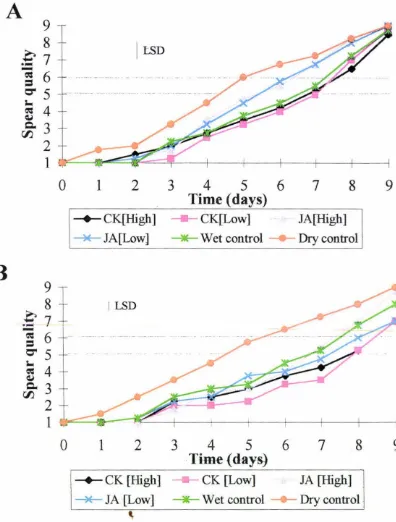
acid for spears harvested in Year 1 is shown in Fig. 3. lA. Spears with a spear quality
of 6 and higher become unsaleable (Lill 1980). Taking a horizontal line across the
graph at a spear quality of 5-6 it can be seen that spears subjected to dry storage
deteriorated faster than all other wet treatments having a shelf-life of around 4.5-5 d.
Spears treated with jasmonic acid deteriorated faster than the water control and both
of the cytokinin treatments, having a shelf-life of 5.5-6.5 d. Spears subjected to
water treatment and both cytokinin treatments showed an extended shelf-life to
around 7-7.5 days.
Year 2
The influence of cytokinin and jasmonic acid treatment on postharvest spear quality
for spears harvested in Year 2 is shown in Fig. 3. lB. Again focussing on a spear
quality of 5-6 it can be seen that spears subjected to dry storage deteriorated at a
similar rate to those in Year 1, but the spears subjected to wet treatments appeared to
have an extended shelf-life compared to wet treated spears in Year 1. Shelf-life of
dry control spears was only around 4.5-5 days. Spears treated with the high
concentration of jasmonic acid or with water share a shelf-life of around 7-7.5 d.
Spears treated with both cytokinin treatments share had an extended shelf-life of
around 8-8.5 d, compared to spears treated with the low concentration of jasmonic
A
9
8
I
I::SD
.€
7
-
eo:6
=
O"
5
... .---; 4
QJ
Q.
3
00
2
1
0
1
2
3
4
5
6
7
8
9
Time (days)
--+-CK[High]
- - -
CK[Low]
JA[High]
~JA[LowJ----
Wet control
Drycontrol
B
9
8
I LSD£1
-
eo:6
=
O"
5
;
4
QJ
Q.
3
00
2
1
0
1
2
3
4
5
6
7
8
9
·
Time (days)
-+-
CK
[High]
CK
[Low]
JA [High]
[image:43.558.90.486.67.589.2]----*-
JA [Low]
----
Wet control
Drycontrol
'
Figure 3.1 ,Influence of exogenous cytokinin and jasmonic acid treatment on
shelf-life of asparagus spears postharvest. A = spears harvested in Year I. B =
spears harvested in Year 2. (CK [High]= 2 µM DZR, CK [Low]= 2 nM DZR, JA
[High]= 50 µM JA, JA [Low]= 50 nM JA, wet control= H20, dry control=
untreated). Least significant differences (LSD {p = 0. 05}) for the whole analysis are
given as vertical bars. Each point in the above graphs represents the mean value of
3.1.2 Tiprot
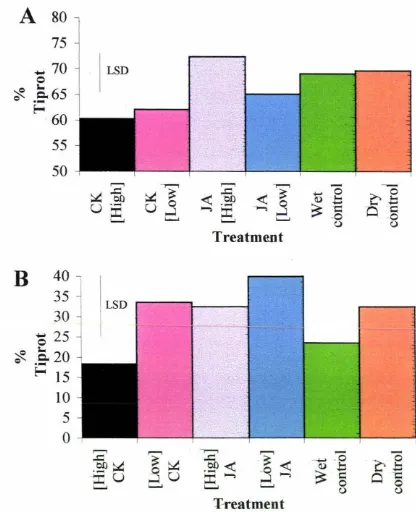
Year 1
The effect of postharvest treatment of spears with cytokinin and jasmonic acid in
relation to tiprot for Year 1 is shown in Fig 3.2A. Approximately 60 % of spears
treated with cytokinin contracted tiprot. 72 % of spears treated with the high
concentration of jasmonic acid contracted tiprot. Tiprot was contracted in
approximately 70 % of spears in both control groups, and 65 % of spears contracted
tiprot that were subjected to the low concentration jasmonic acid postharvest.
Year 2
The effect of postharvest treatment of spears with cytokinin and jasmonic acid in
relation to tiprot for Year 2 is shown in Fig 3.2B. The proportion of spears having
ti prot in year two was lower than that of year one. 18 % of spears treated with high
concentration cytokinin contracted tiprot, compared to spears treated with low
concentration jasmonic acid with 40 % contracting tiprot. The low cytokinin, high
jasmonic acid, and dry control treatments all led to between 32-34 % of spears
acquiring the disorder, whilst 24 % of the wet control treated spears had the disorder.
3.1.3 Spear extension
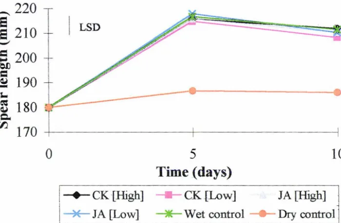
The effect of postharvest cytokinin and jasmonic acid treatment on spear extension is
shown in Fig 3.3. All spears subjected to wet treatments tended to grow or extend
after harvest at least 25 mm more than the dry untreated spears. The spears treated
with a low concentration of cytokinin were slightly, but not significantly, shorter after
A
so
75
..,. 70
LSD0
~
a65
e ·-~
60
55
50
B
40
-35
~
I LSD30
~
0
25
~
a2o
e ·
-~
15
10
5
0
Treatment
T-reatment
c1
0
-0=
[image:45.557.69.485.69.581.2]u
Figure 3.2 Influence of exogenous cytokinin and jasmonic acid treatment on
tiprot of a~paragus spears postharvest. A = spears harvested in year one. B
=
spears harvested in year two. Plant hormone concentrations are shown in Fig. 3 .1.
LSDs (p = Q.05) for full analysis are given as vertical bars. Each bar in the above
_220
e
e
210
,__,
; 200
~
=
~
190
J,.. a'3
180
c.
00.
170
0
I
LSD5
Time (days)
-+-CK-[High] - -CK.[Low]
10
JA [High]
[image:46.566.85.435.109.338.2]- * -IA [Low] - * -Wet control - Dry control
Figure 3.3 ;Influence oT exogenous cytokinin and jasmonic acid treatment on
extension of asparagus spears postharvest. Spears harvested in year one. Plant
hormone cqncentrations are shown in Fig. 3.1. LSD (p
=
0.05) for full analysis isgiven as vertical bar. Each point in the above graph represents the mean value of four
3.2 Biochemical changes postharvest
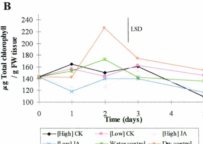
3.2.1 Chlorophyll
The influence of exogenous cytokinin and jasmonic acid treatment after harvest on
chlorophyll content is shown in Fig. 3.4.
Year 1
The graph and statistics clearly show that there is a high degree of variability within
samples. However, the overal trend is a decline in chlorophyll content over time, in all
treatments except the dry (control) spears. The downward trend in chlorophyll
content seen was due to the loss of chlorophyll a (data not shown).
Year2
The chlorophyll content in spears harvested in year two was also variable. Chlorophyll
content rose in some treatments and then declined two or three days after harvest. All
samples were held at -80°C for no longer than five days before extraction of
chlorophyll to stop degradation occurring. Contents of chlorophyll a and
protochlorophyll declined whilst chlorophyll b content increased slightly (data not
shown).
3.2.2 Soluble sugars
Sucrose
The effect of exogenous cytokinin and jasmonic acid treatment on sucrose content in
spear tips is shown in Fig. 3.5A. Sucrose content declined rapidly in all spear tips
within the first 24 h after harvest. Sucrose levels declined faster in all spear tips
subjected to wet treatment compared to dry storage. By two days after harvest
sucrose levels in spear tips had declined to around the same level in all treatments.
130 125
-
120~
Q. ~ 115e
=
fl.l110 0 .~
:a -
105i~
1000 Oil E-c -... 95
Oil
::s. 90
85 80
-0 1 2. 3 4 5
Time (days)
-+-[High] CK - [Low] CK [High] JA
~[Low] JA --*--Wet control - Dry control
B
240->.
220.c ~
e- :
200 0 .~::c -
180i
~
1600 Oil
E-c -Oil 140
~=---::s. 120
100 +-~~~'---~~-+~~~--+-~~~-+-~~---1
0 1
I
-+-[High] CKl
~
[Low]JA
2 3 4 5
Time (days)
- [Low]CK [High] JA
[image:48.567.76.400.335.565.2]---water control - Dry control
Figure 3.4 Influence of exogenous cytokinin and jasmonic acid treatment on
chlorophyll content in asparagus spears postharvest. A= spears harvested in year
one. B = spears harvested in year two. Plant bormone concentrations are shown in
Fig 3 .1. LSDs (p = 0. 05) for full analysis are given as vertical bars. Each point in the
above graphs represents the mean value of four individual observations performed J.n
![Table 2.1 Visual assessment scale of spear quality. Scale adapted from Lill [1980].](https://thumb-us.123doks.com/thumbv2/123dok_us/8377794.319887/33.558.71.521.251.488/table-visual-assessment-scale-spear-quality-scale-adapted.webp)