This is a repository copy of Photocatalytic effects of wool fibers modified with solely TiO2 nanoparticles and N-doped TiO2 nanoparticles by using hydrothermal method.
White Rose Research Online URL for this paper: http://eprints.whiterose.ac.uk/82900/
Version: Accepted Version
Article:
Zhang, H, Yang, Z, Zhang, X et al. (1 more author) (2014) Photocatalytic effects of wool fibers modified with solely TiO2 nanoparticles and N-doped TiO2 nanoparticles by using hydrothermal method. Chemical Engineering Journal, 254 (10). pp. 106-114. ISSN 1385-8947
https://doi.org/10.1016/j.cej.2014.05.097
© 2014 Elsevier B.V. This is an author produced version of a paper published in Chemical Engineering Journal. Uploaded in accordance with the publisher's self-archiving policy. This version licensed under the Creative Commons
Attribution-NonCommercial-NoDerivatives 4.0 International (http://creativecommons.org/licenses/by-nc-nd/4.0/).
eprints@whiterose.ac.uk
Reuse
Unless indicated otherwise, fulltext items are protected by copyright with all rights reserved. The copyright exception in section 29 of the Copyright, Designs and Patents Act 1988 allows the making of a single copy solely for the purpose of non-commercial research or private study within the limits of fair dealing. The publisher or other rights-holder may allow further reproduction and re-use of this version - refer to the White Rose Research Online record for this item. Where records identify the publisher as the copyright holder, users can verify any specific terms of use on the publisher’s website.
Takedown
If you consider content in White Rose Research Online to be in breach of UK law, please notify us by
This is a post-refereeing final draft. When citing, please refer to the published version: H Zhang, Z Yang, X Zhang and N Mao (2014). Photocatalytic effects of wool fibers modified with solely TiO2 nanoparticles and N-doped TiO2 nanoparticles by using hydrothermal method, Chemical Engineering Journal, Volume 254, 15 October 2014, Pages 106–114
DOI: 10.1016/j.cej.2014.05.097
Photocatalytic Effects of Wool Fibers Modified with Solely TiO2 Nanoparticles and
N-doped TiO2 Nanoparticles by Using Hydrothermal Method
Hui Zhang*1, Zhenwei Yang1, Xingtao Zhang1, and Ningtao Mao1,2
1School of Textile & Materials, Xi an Polytechnic University, Xi an 710048, China
2School of Design, University of Leeds, Leeds, LS2 9JT, United Kingdom
*Corresponding author: Hui Zhang, School of Textile & Materials, Xi an Polytechnic
University, Xi’an City, Shannxi Province, China
E-mail: hzhangw532@xpu.edu.cn; hzhangw532@163.com
Phone: 0086 029 13002929736
Abstract: The surfaces of wool fibers are modified with N-doped TiO2 nanoparticles by
treating the fibers with tetrabutyl titanate and ammonium chloride under low temperature
hydrothermal conditions to obtain wool fibers with photocatalytic functions in the visible light
spectrum. The effects of nitrogen and sulfur in amino acids in keratin on the photocatalytic
activity of TiO2 particle coated wool fibers are investigated. Changes of various fiber
properties such as tensile strength, surface friction, photocatalytic activity, and self-cleaning
performance of untreated, TiO2-coated and N-doped TiO2-coated wool fibers are studied. It is
found that N-doped anatase TiO2 nanoparticles with an average grain size of 11.2 nm are
synthesized and simultaneously grafted onto the wool fibers. After treatments, the
crystallization index of the wool fibers is slightly reduced. The capability to protect against
ultraviolet radiation is much enhanced. The performances of photocatalytic degradation of
methylene blue dye and self-cleaning of red wine under both UV and visible light irradiation
are endowed. It is also found that wool fibers coated with TiO2 particles without being doped
by nitrogen still have apparent photocatalytic reactions and self-cleaning effects under visible
light irradiation due to the formation of C-Ti3+, O-Ti3+, and N-Ti3+ bonds between TiO2 and
wool keratin on the wool fiber surfaces. Thus wool fabrics might not need to be coated with
N-doped TiO2 nanoparticles to realize its self-cleaning effect under visible light. Such
technical products such as wastewater treatment.
Keywords: Wool fibers; N-doped TiO2, Hydrothermal; Modification
1. Introduction
Titanium dioxide (TiO2) loaded textile structures are very attractive not only in functional
clothing and home textiles, but also in technical textile applications. One of the typical
potential applications of TiO2 loaded textile structure in technical textiles might be its use in
the treatment of pollution effluents. While photocatalytic mineralization of organic pollutants
by using photocatalyst TiO2 particles is effective to reduce water and air pollutions, the
post-treatment removal of TiO2 in either wastewater slurries or air effluent through filtration
and re-suspension processes can be a time consuming and costly process [1, 2]. The solution of
this problem is to immobilize photocatalyst TiO2 particles on a porous substrate such as fibrous
structures, to facilitate the practical application of the photocatalytic wastewater treatment
process [3]. Wool fibers are of particular interests in both functional clothing and wastewater
treatment substrates. On one hand, wool clothing is notorious for its dimensional stability after
scouring and many efforts are made in achieving easy-care wool fabrics, and thus a TiO2
treated and scouring-free wool fabric is long desired. On the other hand, wool fibers are proved
an effective keratin substrate to absorb and adsorb organic pollutants and heavy metal ions
from waste effluents. Therefore, a visible-light-induced self-decontamination wool fabric has
become our research interests.
However, the photocatalytic activities of the TiO2 nanoparticles treated fabrics mentioned
expand the photocatalytic activities of the TiO2 nanoparticles into visible light spectrum. One is
to lower the unoccupied molecular orbital level of the conduction band by ion implantation of
metallic elements into TiO2 nanoparticles and the other is to raise the occupied molecular
orbital level of the valence band (usually by using organic photosynthesizing dyes or pigments)
[4]. The photosynthesizing dyes have the capability of absorbing photons from visible light in a
conventional TiO2 thin film, and then transfer them to TiO2, thus creating electricity [5].
Pelaez et al. [6] and Daghrir et al. [7] have systematically reviewed the development of the
visible light active TiO2 synthesized by different strategies, including non metal doping (boron,
nitrogen, carbon, fluorine, sulfur, etc), noble metal and transition metal doping, dye
sensitization, and coupling semiconductors. However, only modest progress in increasing the
photocatalytic activity have been made from those efforts because the increased absorption of
visible light cannot be straightforwardly related to the reaction rate and the foreign species
often work as recombination centers for the photogenerated electron/hole pairs [8].
When doping of TiO2 with nitrogen, the origin of the photoresponse at higher wavelengths is
the mixing of the 2p nitrogen level with the oxygen 2p orbital to form the valence band, which
results in a lower band gap resulting in visible light absorption [9]. The element nitrogen can
be easily incorporated into the TiO2 structure either in the bulk or as a surface dopant because
of its comparable atomic size with oxygen, small ionization energy, and high stability [10]. PL
analysis confirms that nitrogen atoms in substitutional sites enhance the photocatalysis of TiO2
under visible light more effectively than nitrogen atoms in interstitial sites [11]. Substitution of
N in place of oxygen in the TiO2 lattice causes a decrease in oxygen vacancies which inhibits
formation of surface oxygen vacancies [13]. The orbit of doped nitrogen is not responsible for
modifying the electron structure of TiO2 but some local unusual structures forming when the
N-doping process generates the visible-light activity, such as crystal defects or distortion [14].
Nitrogen and other elements co-doped TiO2 have also been widely investigated. In nitrogen
co-doped TiO2, the interstitial nitrogen doping sites rather than substitutional sites seem to
improve the photocatalytic activity of TiO2 because of the better charge separation [15].
Moreover, the synergic effect of substitutional and interstitial nitrogen sites are more efficient
to improve the electrochemical photoactivity of TiO2 due to the good light absorption, charge
transfer in substitutional doping, and the good charge separation induced by the interstitial
nitrogen doping [16]. Results indicate that the increased photocatalytic activity of the V-N
co-doped TiO2 can be attributed to the synergistic effects caused by V and N that increase the
visible light absorption and simultaneously act as electron and hole trapping sites thus
decreasing the rate of charge recombination [17]. The visible light photocatalytic activity of
N-S co-doped TiO2 is not only influenced by the value of energy gap, the distribution of
impurity state, but also depends on the property of impurity state, the location of Fermi level
and the energy in the edge of band gap [18]. Examples to achieve visible light photocatalytic
functions in TiO2 coated textile fabrics are also reported. Wool fabrics are treated with citric
acid as a cross-linking agent and with TiO2/Ag nanocomposites, in which the lowest
unoccupied molecular orbital level of Ti3+ is lowered by ion implantation of Ag elements into
TiO2 nanoparticles, to achieve superior self-cleaning properties [19]. A significantly improved
visible-light-induced self-cleaning effect can be achieved in cotton fabrics by using N-doped
Unlike cotton fiber, wool fibers are composed of keratin protein and are often not stable in
both UV and visible light irradiation [20]. While surface gratification of TiO2 clusters on wool
fibers has been realized mainly by the sol-gel process [21, 22] and nanocrystalline TiO2 is
found to act primarily as a UV absorber in dry conditions [20] and as a UV photocatalyst in
wet conditions [23], there is hardly any research done to achieve visible-light-induced
self-cleaning effect in wool fabrics and little is known if nitrogen or sulfur element in amino
acids of keratin wool fibers would affect such visible-light-induced photochemical reactions in
TiO2 coated wool fibers. In addition, TiO2 nanoparticles are required to be securely bound into
the surface of targeted wool fibers in order to increase the durability of the desired
photochemical properties and not to release nanoparticles into surrounding environment to
meet ecological requirements, also the mechanical properties of the wool fibers need to be
maintained.
In this study, wool fibers coated with nitrogen doped TiO2 nanoparticles are achieved by
using tetrabutyl titanate as the precursor and ammonium chloride as the doping agent under
low temperature hydrothermal conditions, in order to increase its photocatalytic activity of
TiO2 nanoparticles under visible light irradiation. The functions of N-doped TiO2 particles and
pure TiO2 particles coated wool fibers are compared with each other to find out if nitrogen or
sulfur element in amino acids in wool fibers would play a role in self-cleaning effect in TiO2
coated wool fibers. The differences between the tensile properties, friction coefficient,
photocatalytic activity, and self-cleaning capability of both pure TiO2 particles and N-doped
TiO2 particles coated wool fibers are investigated. The changes of the surface morphology,
the wool fibers before and after treatments are investigated to establish an understanding of the
photochemistry mechanism of TiO2 treated wool fibers.
2. Experimental section
2.1. Materials
The merino wool fibers with an average diameter of 20 m and nonionic surfactant W900 were
obtained from the local textile mill. The reagent-grade chemicals used include tetrabutyl
titanate (Ti(OC4H9)4), ammonium chloride (NH4Cl), methylene blue (MB), acetone, and
anhydrous ethanol. Deionized water was used throughout this study.
2.2. Modification of wool fibers with TiO2 and N-doped TiO2 nanoparticles
About 3.0 g of wool fibers was immersed in 200 ml of mixed solution containing 2.0 g/l of
sodium carbonate and 0.5% of nonionic surfactant W900 at 50°C for 15 min, and subsequently
treated with a 100 ml of acetone and anhydrous ethanol solution at 50°C for 10 min,
respectively, and then washed thrice in deionized water at room temperature for 10 min and
dried at 60°C for 8 h. About 0.5 ml of tetrabutyl titanate was added dropwise into 10 ml of
ethanol solution under vigorous stirring at room temperature. The solution was then diluted
with 70 ml of deionized water and 8.0 g of ammonium chloride was added into. About 0.5 g of
pretreated wool fibers were dipped in the above suspension for 10 min, and then transferred to
a 100 ml PTFE-lined stainless steel autoclave. Six same autoclaves were simultaneously
prepared according to the above method, which were placed in a furnace and run at a speed of
10 r/min. The temperature was raised to 110°C at a heating rate of 2.0°C/min. After 2 h, the
ethanol, and deionized water at 50°C for 10 min, respectively, and finally dried at 60°C. The
resultant precipitate was centrifuged after discarding the upper solution, and successively
washed with acetone, anhydrous ethanol, and deionized water, and finally dried in an oven at
120°C for 10 h. For comparative analysis, the wool fibers were also treated with Ti(OC4H9)4
without NH4Cl based on the above described procedure.
2.3. Characterization and measurement
The morphology, structure, composition, thermal behavior, and optical properties of the wool
fiber samples before and after treatments were characterized by using field emission scanning
electron microscopy (FESEM), X-ray diffraction (XRD), Fourier transform infrared
spectroscopy (FT-IR), X-ray photoelectron spectroscopy (XPS), thermal gravimetric (TG)
analysis, differential scanning calorimetry (DSC), diffuse reflectance spectroscopy (DRS), and
photoluminescence (PL) spectroscopy techniques, respectively. The fiber tensile properties
were tested by using a standard electromechanical apparatus. The fiber surface friction
properties were measured by using a standard single fiber friction measurement device. The
photocatalytic activity was performed by monitoring the discoloration of MB dye. The details
of the above characterization methods are provided in the Supporting Information S1 of this
paper.
The self-cleaning performance of the wool fiber samples was assessed by the discoloration
of red wine. A bunch of wool fibers was tied onto a slide glass and about 2 ml of red wine was
dropped onto one end of wool padding. After 3 min of absorption of red wine, the wool
padding was irradiated with a 1500W Xenon arc lamp which produces UV and visible light
1.55×10⁵ lux for visible light irradiation, respectively. The color changes of the red wine
absorbed into the wool padding were monitored by comparing the images taken before and
after 84 h of irradiation by using a Samsung ST700 digital camera.
3. Results and discussion
TiO2 particles have been used as a delustrant and UV blocker in synthetic fibers for years due
to its good reflective properties and UV absorption ability. But in wool fibers, there is
possibilities for reactive oxygen species (superoxide radical anions O2·-, hydroxyl radicals ·OH,
and hydrogen peroxide) forming by the interaction between UV radiation and TiO2 in the
surface of wool fibers to ultimately degrade disulfide bond and produce unwanted yellow
photoproducts. In this section, the morphology, chemistry composition, and surface properties
of wool fibers coated with TiO2 and N-doped TiO2 nanoparticles in hydrothermal process are
examined and discussed.
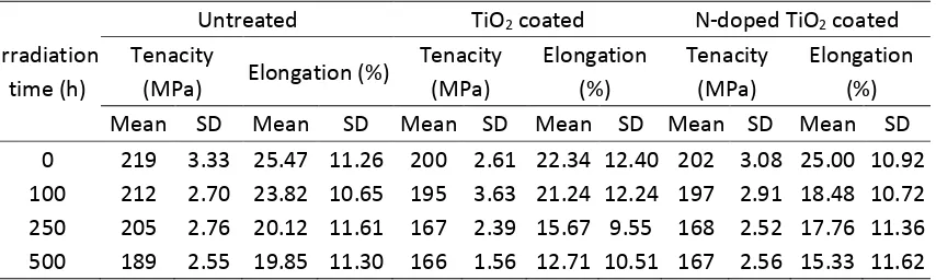
3.1. Tensile properties
The results of the tensile testing of the untreated, TiO2 coated and N-doped TiO2 coated wool
fibers before and after irradiation are listed in the Supporting Information S2 (see Table S1). It
is found that after treatment with Ti(OC4H9)4, the average tenacity and elongation of wool
fibers decrease slightly from 219 MPa to 200 MPa and from 25.5% to 22.3%, respectively.
After being added with NH4Cl, the average tenacity has almost no further change (202 MPa)
and the average elongation increases slightly (25.0%). This seems that the detrimental effect on
the tensile properties of wool fibers is due to the hot and high pressure water together with
It is also noticed that, with the increases of the irradiation time, the average tenacity and
elongation of the untreated wool fibers gradually decrease. After 500 h of continuous
irradiation, the average tenacity and elongation decrease by 13.7% and 25.6%, respectively.
However, for both TiO2 coated and N-doped TiO2 coated wool fibers, the average tenacity
decreases with the increase of the irradiation time at first, and then levels off beyond 250 h.
The average elongation gradually decreases with increasing irradiation time. The average
tenacity decreases about 17% for the TiO2 coated and N-doped TiO2 coated wool fibers after
being irradiated for 500 h. The decrease of the average elongation of the TiO2 coated wool
fibers (43%) is greater than that of the N-doped TiO2 coated wool fibers (39%). Therefore, it is
evident that TiO2 coating on wool regardless of N-doped will accelerate the photo-degradation
process of wool fibers to some degree. Besides the photodegradation of wool fibers, it is
guessed that the increased light absorption of the TiO2 coated and N-doped TiO2 coated wool
fibers generates the electron-hole pairs and further forms the active groups [8], which can
destroy the structure of wool fibers under UV and visible light irradiation. This is a very
interesting phenomenon that TiO2 coated wool fibers have the similar photocatalytic reaction to
that of N-doped TiO2 under UV and visible light irradiation. However, because there is not
much decrease of wool tensile strength, the photocatalytic reaction site is probably limited to
the surface layer of wool fibers. The mechanism of action will be studied in the future work.
3.2. Friction properties
The results of the friction testing of the wool fibers before and after treatments are listed in the
Supporting Information S3 (see Table S2). After treatment with Ti(OC4H9)4 and NH4Cl, as the
morphological changes of wool fibers (became coarse), the static and dynamic friction
coefficients of the wool fibers (against-scale and with-scale) increase to some extent. However,
the differential frictional coefficients are reduced from 28.0% to 22.7% for static friction
testing and reduced from 12.5% to 11.8% for dynamic friction testing, respectively. Because
felting effect of wool fibers are determined by directional frictional effect, the smaller the
differential frictional coefficient is, the less felting effect the wool fiber has [41]. It is thus
anticipated that the felting phenomenon of the N-doped TiO2 coated wool fibers will be
alleviated during wet processing including laundering and scouring owing to a reduction of
these frictional difference ( a − w). Compared with the N-doped TiO2 coated wool fibers, the
static and dynamic friction coefficients of the TiO2 coated wool fibers change little. Thus the
nitrogen doping has no effects on the friction properties of the TiO2 coated wool fibers.
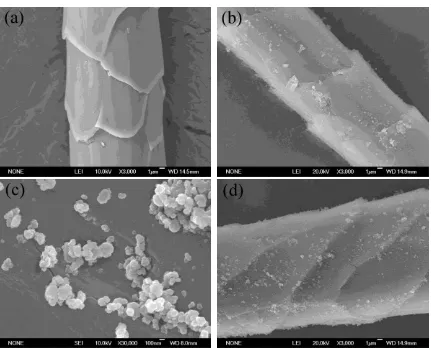
3.3. Surface morphology
The FESEM images of the wool fibers before and after treatments are shown in the Supporting
Information S4 (see Figure S1). It is seen that the surface of the untreated wool fibers is very
clean without any substances on. After being modified with Ti(OC4H9)4 and NH4Cl, a thin
layer of materials is uniformly coated on the surface of the N-doped TiO2 coated wool fibers.
Meanwhile, some large granules in micrometer scale adhere onto the fiber surfaces due to the
agglomeration of nanoparticles. As illustrated in the high resolution FESEM image, such
nanoscale particles with a diameter around 100 nm are tightly anchored to the substrate of the
wool fibers. In addition, there is no distinct difference in the surfaces of the TiO2 coated and
N-doped TiO2 coated wool fibers. The high pressure and hot water are believed to have
3.4. Thermal properties
The TG and DSC curves of wool fibers before and after treatments are shown in Figure 1, and
the results of TG analysis of wool fibers are listed in the Supporting Information S5 (see Table
S3). It is evident from the TG curves that the onset and endset decomposition temperatures of
wool fibers decrease from 263.3°C and 381.0°C to 259.7°C and 370.7°C after being treated
with Ti(OC4H9)4, respectively. The lost mass increases from 63.3% to 67.0% at a temperature
of 550°C. After treatment with Ti(OC4H9)4 and NH4Cl, the onset and endset decomposition
temperatures of the N-doped TiO2 coated wool fibers decrease to 261.9°C and 372.8°C,
respectively. The corresponding lost mass is 70.7%.
It is clear from the DSC curves that the initial endothermic peak increases from 81.2°C in
untreated wool fibers to 85.1°C in the TiO2 coated wool fibers, which is ascribed to the
removal of surface absorbed water or the residual water molecules caused by TiO2 coating. The
major endothermic peak at 277.4°C decreases to 273.9°C. That is attributed to the
decomposition of both polypeptide and amino acid as well as the incorporation of TiO2
nanoparticles. The initial endothermic peak of the N-doped TiO2 coated wool fibers further
decreases to 80.7°C, whereas its major endothermal peak increases to 296.0°C. Obviously,
such change in thermal stability of the N-doped TiO2 coated wool fibers is mainly due to the
doping of N into TiO2. Therefore, the hydrothermal treatment has a little effect on the thermal
properties of wool fibers.
3.5. XRD analysis
The XRD patterns of the wool fibers before and after treatments as well as the remaining
N-doped TiO2 coated fibers are found at around 2 =9° and 20°, which belong to the
characteristic positions of wool fibers. However, the characteristic peaks of TiO2 are not
observed in the XRD patterns of both modified wool fibers. This might be because the amount
of TiO2 nanoparticles loaded onto the fiber surfaces is relatively small so that its diffraction to
X-ray cannot be detected by the instrument. The crystallization indexes of wool fibers are
reduced from 59.8% to 51.7% for the TiO2 coated wool fibers and 57.3% for the N-doped TiO2
coated wool fibers under hydrothermal conditions, respectively. This might be attributed to the
structural change of microfibrils in wool surface. For both the remaining particles left in the
hydrothermal processing medium, a series of characteristic peaks at 25°(101), 38°(004),
48°(200), 54°(105), 56°(211), 63°(204), 68°(116), 70°(220) and 75°(215) are in agreement
with the data list in JCPDS card No.21-1272 [24]. At the same time, three diffraction peaks are
identified at 2 of 23°, 32°, and 58° in the XRD pattern of the N-doped TiO2 particles, which
are attributed to the (100), (110), and (211) planes of salammoniac (JCPDS card No.07-0007).
Therefore, the crystalline structure of the as-synthesized particles by using Ti(OC4H9)4 and
NH4Cl can be indexed to the anatase TiO2 doped with nitrogen. The average crystallite sizes of
the remaining particles in the processing liquid are determined to be 9.6 nm for the TiO2
particles and 11.2 nm for the N-doped TiO2 particles by measuring the FWHM of (101), (004),
and (200) reflections and using Scherrer’s equation, respectively. Combined with the FESEM
observation, the cluster of 100 nm particles is mainly caused by growth of the smaller particles
near the surface of wool fibers due to the continuous deposition of small particles.
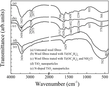
3.6. FT-IR analysis
nanoparticles are shown in Figure 3. Compared with the spectrum of the untreated wool fibers,
the O-H band of the TiO2 coated wool fibers shifts from 3425 to 3443 cm-1. This is ascribed to
the surface absorbed water induced by the TiO2 coating [25], which might make the TiO2
coated fiber generate much stronger oxidative free radicals than those of untreated one [26].
The peak at 2932 cm-1 (CH2 asymmetric stretching) decreases to 2929 cm-1, while the band at
1450 (CH3 asymmetric bending) increases to 1452 cm-1. The amide I and III bands shift from
1636 (C=O stretching) and 1234 cm-1 (C-N stretching) to 1638 and 1236 cm-1, respectively.
Furthermore, influenced by the Ti-O band at 462 cm-1 of TiO2, the N-H band at 568 increases
to 588 cm-1 [27].
For the N-doped TiO2 coated wool fibers, the peak at 3430 cm-1 is intensified because of the
introduction of N-H band. The amide I and III bands decrease to 1628 and 1229 cm-1. The
peaks at 2932, 2877 (CH3 symmetric stretching), 1708 (carbonyl group), 1450, 933 (C-O
stretching), and 568 cm-1 shift to 2926, 2875, 1717, 1445, 937, and 578 (influenced by the Ti-O
band at 582 cm-1 of N-doped TiO2) cm-1, respectively. Therefore, it can be concluded that the
TiO2 and N-dope TiO2 nanoparticles are immobilized onto the wool fibers by chemical
grafting.
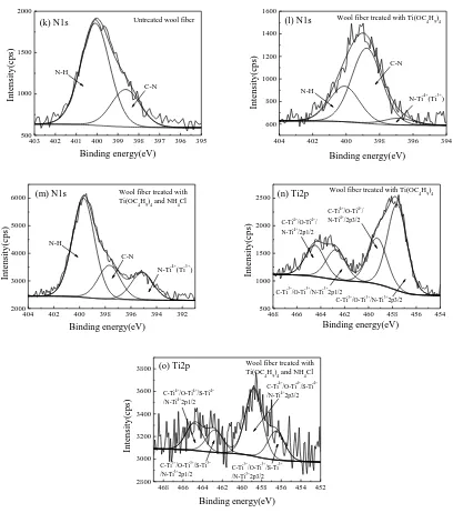
3.7. XPS analysis
To study the bonding mechanism between TiO2 nanoparticles and wool fibers, the survey
spectra and core level single spectra of the bonding partners (C1s, O1s, S2p, N1s, and Ti2p) of the
wool fibers before and after treatments are shown in the Supporting Information S6 (see Figure
S2). The quantitative XPS data are listed in the Supporting Information S7 (see Table S4).
coated wool fibers, while it is a much smaller intensity peak in Ti2p in N-doped TiO2 coated
wool fibers than in TiO2 coated wool fibers. The percentage atomic concentration of Ti2p in
N-doped TiO2 coated wool fibers is about 16% of that in TiO2 coated wool fibers, which
indicates a small amount of TiO2 is coated in the N-doped TiO2 coated wool fibers. Also, In
comparison with TiO2 coated wool fibers, there are much greater intensity peaks appearing in
both N1s and C1s binding energy band in N-doped TiO2 coated wool fibers, and the percentage
atomic concentrations of both of these two elements are very close to those of untreated wool
fibers as shown in Table S4.
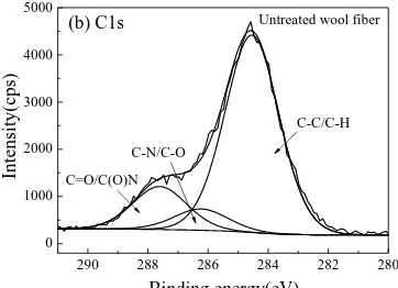
Compared with the C1s XPS spectra of the untreated wool fibers, the sub-peaks of C-C/C-H,
C-N/C-O, and C=O/C(O)N of the TiO2 coated fibers shift from 284.54, 286.23, and 287.61 eV
to 284.60, 285.89, and 287.77 eV, respectively. A new sub-peak at the binding energy of
283.47 (C-Ti4+/C-Ti3+) eV is observed [28]. For the N-doped TiO
2 coated fibers, the sub-peaks
of C-C/C-H, C-N/C-O, and C=O/C(O)N shift to 284.39, 285.90, and 287.78 eV, respectively
[29, 30]. Meanwhile, two new sub-peaks at the binding energies of 280.08 (C-Ti3+) and 282.37
(C-Ti4+) eV are identified [31]. These are attributed to C atoms of wool fibers bound to Ti of
N-doped TiO2.
The O1s peak of the untreated wool fibers is deconvoluted into two subpeaks. After treatment
with Ti(OC4H9)4, the subpeaks at 531.42 (O=C) and 532.39 (O-C) eV shift to 531.80 and
533.50 eV, respectively. Moreover, two new subpeaks are found. The subpeak at 530.44 eV is
assigned to O atoms bound to Ti of TiO2 (O-Ti4+). The subpeak at 528.73 eV is ascribed to O
atoms of wool fibers bound to Ti of TiO2 (O-Ti3+). After being modified with Ti(OC4H9)4 and
Also, two new subpeaks are identified. The subpeak at 529.16 eV is attributed to O atoms
bound to Ti of N-doped TiO2 (O-Ti4+) [32]. The subpeak at 526.88 eV is assigned to O atoms
of wool fibers bound to Ti of N-doped TiO2 (O-Ti3+) [33].
With respect to the S2p XPS spectra of the untreated wool fibers, the subpeaks at 163.42 (S-S)
and 164.67 (S-H) eV change little (163.21 and 164.75 eV) when wool fibers are modified with
Ti(OC4H9)4. After treatment with Ti(OC4H9)4 and NH4Cl, the subpeaks at 163.42 and 164.67
eV are reduced to 161.90 and 163.87 eV, respectively. Furthermore, a shoulder at lower
binding energy of 159.18 eV is noticed. This has been assigned to S atoms of wool fibers
bound to Ti of N-doped TiO2 (S-Ti3+) [34]. However, it has been reported that the peak of a
binding energy between 163–164 eV is assigned to element sulfur or TiS [35], and that TiS2
nanoparticles is fabricated by sol-gel process [36, 37], therefore, there might be the new
bindings of S-Ti4+, S-Ti3+, and S-Ti2+ forming between wool fibers and TiO
2 nanoparticles in
the hydrothermal process.
For the N1s XPS spectra of three wool fiber samples, the subpeaks at 398.62 (C-N) and
400.08 (N-H) eV in untreated wool fibers slightly shift to 398.77 and 400.10 eV after treatment
with Ti(OC4H9)4, respectively. A small subpeak at 396.86 eV also forms in the wool fibers
treated with Ti(OC4H9)4, and this new subpeak is ascribed to the bonding of nitrogen atoms of
wool fibers with Ti of TiO2 [38]. This means that the N atoms of wool fibers are bound to Ti of
N-doped TiO2 (N-Ti4+/N-Ti3+). When wool fibers are treated with Ti(OC4H9)4 and NH4Cl, the
subpeaks at 398.62 and 400.08 eV are reduced to 397.68 and 399.69 eV, respectively.
Meanwhile, a new subpeak at 395.12 eV is found, which can be attributed to the nitrogen
of C1s, S2p, and N1s in N-doped TiO2 coated wool fibers are not the same as those of TiO2
coated wool fibers.
The Ti2p XPS spectra for both modified wool fibers consist of four distinct subpeaks. For the
TiO2 coated wool fibers, the subpeaks at 464.48 and 459.28 eV represent
C-Ti4+/O-Ti4+/N-Ti4+2p1/2 and C-Ti4+/O-Ti4+/N-Ti4+2p3/2, respectively [14]. The subpeaks at
462.73 and 457.60 eV correspond to C-Ti3+/O-Ti3+/N-Ti3+
2p1/2 and C-Ti3+/O-Ti3+/N-Ti3+2p3/2
respectively. But for the N-doped TiO2 coated wool fibers, the subpeaks at 464.85 and 458.80
eV are in accord with C-Ti4+/O-Ti4+/S-Ti4+/N-Ti4+2p1/2 and C-Ti4+/O-Ti4+/S-Ti4+/N-Ti4+2p3/2,
respectively. The subpeaks at 462.77 and 456.51 eV agree with C-Ti3+/O-Ti3+/S-Ti3+/N-Ti3+2p1/2
and C-Ti3+/O-Ti3+/S-Ti3+/N-Ti3+2p3/2, respectively. Therefore, the results testify that the
N-doped TiO2 nanoparticles are grafted onto the wool fibers via the C-Ti4+(Ti3+), O-Ti4+(Ti3+),
S-Ti4+(Ti3+, Ti2+), and N-Ti4+(Ti3+) bonds.
While it is known that titanium trivalent (Ti3+) on the surface of TiO2 particles is very
reactive [41] and plays an essential role in photocatalytic process over TiO2 photocatalyst [42],
and that it can be generated by using UV irradiation and thermal annealing on the surface of
anatase TiO2 particles, we thus propose that the stable bonding between TiO2 particles and
wool fibers are C-Ti4+, S-Ti4+(Ti2+), O-Ti4+, and N-Ti4+, and the reaction mechanism between
TiO2 nanoparticles and wool fibers is summarized in Figure 4.
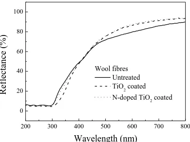
3.8. DRS analysis
The diffuse reflectance spectra of the untreated, TiO2 coated and N-doped TiO2 coated wool
fiber samples in the wavelength range of 200-800 nm are shown in Figure 5.
coated fiber samples decreases in the UV region (200nm~400nm) and increases significantly in
the visible light region (450nm~800nm); and that such decrease of the reflectance in the UV
region in N-doped TiO2 coated wool fiber samples is relatively small but the increase of the
reflectance in the visible light region (450nm~800nm) is still significant. This increase of the
reflectance in the visible light region is a band gap narrowing effect which might be due to N2p
and S2p states mixing with O2p states in N-doped TiO2 coated fibers, as it is known that the
substitutional doping of nitrogen into the TiO2 lattice causes a shift of the absorption edge
towards the visible spectral region [9]. With the increase of the substitutional nitrogen sites in
the TiO2 lattice, the UV activity decreases but not the visible light photoactivity [16]. The
photocatalytic activity of the N-doped TiO2 coated wool fibers is thus enhanced in visible
region but weakened in UV region because of the N doping.
However, there is hardly any existing theory that can explain why TiO2 coated wool samples
(without N-doping) has such a significant decrease of reflectance in UV band and increase in
visible light band. We suspect that such decrease of reflectance in UV band is due to the role of
existing C, O, and N elements existing in keratin protein, which forms a certain amount of
C-Ti3+, O-Ti3+ and N-Ti3+ bands with TiO2 as indicated in XPS analysis and thus promote
photocatalytic activities in the TiO2 coated wool fibers [28, 38, 43]. The increase of the
reflectance in visible light region is mainly due to the increase of the whiteness of TiO2 coated
wool fibers.
It is also shown that, in comparison with the average reflectance of wool fibers coated with
pure TiO2, the average reflectance of the wool fibers coated with N-doped TiO2 is about 1.3%
the fact that the amount of TiO2 applied onto wool fibers in N-doped TiO2 coated wool is much
smaller than that in pure TiO2 coated wool, as indicated in XPS results.
The effect of such small differences in the average diffuse reflectance on the photocatalysis
and self-cleaning performance between TiO2 coated and N-doped TiO2 coated wool fibers will
be investigated further in next few sections.
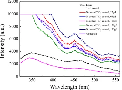
3.9. PL analysis
The PL spectra of the wool fibers before and after treatments are shown in the Supporting
Information S8 (see Figure S3). It is known that the PL spectrum of TiO2 is related to its
transfer behavior of photo-induced electrons and holes (TiO6 octahedra and oxygen vacancies),
reflecting the separation and recombination of charge carriers [44]. For the TiO2 coated wool
fibers, a broad emission band can be observed in the range of 320–560 nm (direct electron-hole
radiative recombination for 320-400 nm, indirect band gap and surface recombination for
400-500 nm, and charge transfer transition of trapped electron in an oxygen vacancy and Ti3+
defects for 500-560 nm), which is consistent with the previous studies [45–47]. Although the
shape and position of the PL emission peaks are almost identical, the PL intensity of the TiO2
coated wool fibers is lower than those of the N-doped TiO2 coated wool fibers expect for the
sample doped with 100g/l of NH4Cl. This is because doping of N into the TiO2 lattice results in
the effective quenching of photoluminescence [48].
It is also observed that the PL intensity of the N-doped TiO2 coated wool fibers gradually
decreases with the increase of the amount of NH4Cl at first, and then reaches the lowest value
when the amount of NH4Cl is 100 g/l, indicating the low recombination rate of photogenerated
photocatalytic activity [49]. After that, the PL intensity increases with increasing the amount of
NH4Cl. This is due to an increase in the formation of vacancy sites, which can increase the
probability of charge recombination [50]. As a result, the N-doping level causes the change in
the valence band level of N-doped TiO2, which results from the photocatalytic formation of
OH· radicals induced by irradiation with visible light.
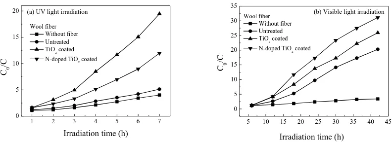
3.10. Photocatalysis
The photocatalytic activity of the untreated, TiO2 coated and N-doped TiO2 coated wool fibers
are characterized by measuring the absorbance of MB solution under both UV and visible light
irradiation. The change of normalized C0/C of MB concentration with irradiation time is shown
in the Supporting Information S9 (see Figure S4). It is evident that the normalized C0/C
gradually increases with the increase of irradiation time for all samples under both UV and
visible light irradiation.
While the UV rays can decolorize the MB dye to some degree due to UV photo-bleaching
[51], it is found in Figure S4(a) that, the TiO2 coated wool fibers behave much better than the
N-doped TiO2 coated ones under UV irradiation condition. The apparent rate constant of the
normalized C0/C for the TiO2 coated wool fibers (0.41 h-1, squared correlation coefficient,
R2=0.96) is as about 1.2 times as that for the N-doped TiO
2 coated ones (0.34 h-1, R2=0.99).
This might be because there is a greater amount of TiO2 particles coated on the surface of wool
fibers. After being irradiated by visible light irradiation for a specific time, it appears in Figure
S4(b) that the apparent rate constant of the normalized C0/C of the N-doped TiO2 coated wool
fibers (0.15 h-1, R2=0.93) is as about 1.15 times as that of the TiO2 coated fibers (0.13 h-1,
R2=0.94), although there are much less amount of N-doped TiO
samples than the amount of pure TiO2 particles on wool fibers as indicated in XPS results. The
visible light activity of the N-doped TiO2 coated wool fibers during the MB degradation
process might be attributed to the N- and/or S-induced mid-gap level forming above the
valence band of TiO2 [51, 52]. Based on the analyses of XPS, DRS and PL, it is concluded that
the nitrogen and sulfur anions act as the hole traps, reducing the recombination rate of the
hole–electron couples [53]. Moreover, the oxygen deficient sites forming in the grain
boundaries are responsible for the visible light response [54], whilst the presences of nitrogen
and sulfur improve the stabilization of these oxygen vacancies [55, 56].
3.11. Self-cleaning performance under the mixture of UV and visible lights
The images of the untreated, TiO2 coated and N-doped TiO2 coated wool fibers stained by red
wine before and after irradiation are shown in Figure 6. It is interesting to note that the
self-cleaning performance of the wool fibers after treatment with Ti(OC4H9)4 can be achieved
regardless of NH4Cl being added. After 84 h of UV and visible light irradiation, the intensity of
the red color on the untreated wool padding stained by red wine still remains intact. However,
the red color on the surface of the TiO2 coated wool padding is almost completely discolored
while the red color of N-doped TiO2 coated wool padding can still be seen. When exposed to
UV and visible light irradiation, a photon with energy greater than the band gap of TiO2
generates an electron hole pair. The positive hole in the valence band can react with the
absorbed water to produce H+ and ·OH radicals, and the electron in the conduction band can
reduce oxygen to produce O2·- anions. Both hydroxyl radicals and peroxide anions are
extremely reactive species, and they can oxidize the organic compounds of red wine until
oxygen vacancies (VO), O2 adsorbed as superoxo (O2–) at fivefold-coordinated Ti sites can be
transformed to peroxo (O22-) and placed into an anion surface lattice site as an interstitial (O2)O
species, which is also an important intermediate in the photooxidation of water [58]. The
photocatalytic activity of the N-doped TiO2 coated wool fibers can be enhanced in visible
region because of the N doping, but weakened in UV region. In addition, it is noticed that the
introduction of NH4Cl into hydrothermal process reduces the amount of TiO2 particles forming
on the surface of wool fibers and thus reduces the total photocatalytic activity of the N-doped
TiO2 coated wool fibers under UV and visible light irradiation. In contrast, the wool fibers after
treatment solely with Ti(OC4H9)4 have much greater amount of TiO2 particles coated on the
surface of wool and also have C-Ti3+ and N-Ti3+ bonds forming on the surface of the wool fiber
and could thus have photocatalytic activities under both UV and visible light irradiation.
Therefore, the self-cleaning ability of the TiO2 coated wool fibers is much better than that of
the N-doped TiO2 coated ones under a combination of UV and visible light irradiation.
4. Conclusions
The N-doped TiO2 nanoparticles have been prepared by using tetrabutyl titanate as the
precursor and ammonium chloride as the doping agent in the presence of wool fibers under
hydrothermal conditions. The photocatalytic effects have been compared with the wool fibers
treated solely using tetrabutyl titanate as the precursor under similar hydrothermal conditions.
The SEM and XRD results confirm that the surfaces of the wool fibers are coated with a thin
film of anatase-type N-doped TiO2 nanoparticles with an average nanocrystal size of 11.2 nm.
wool fibers through the chemical bonds of C-Ti4+, S-Ti4+(Ti2+), and N-Ti4+. There are also N-
and S-Ti3+ bonds forming between TiO
2 particles and the surface of wool fibers, which enable
the photocatalytic activities of TiO2 nanoparticles under visible light irradiation. The TG and
DSC results confirm that the thermal properties of wool fibers have only small changes. The
coefficients of friction increase, while the differential frictional coefficients decrease. The DRS,
PL and tensile results confirm that there are photocatalytic activities and photodegradation
reactions happening in the TiO2 coated and N-doped TiO2 coated wool fibers. A certain amount
of N-dopant incorporated into TiO2 accelerates the evolution of O2. The self-cleaning
performances confirm that wool keratin polymers could form C-, N-, and O-Ti3+ bonding with
TiO2 particles to have photocatalysis effect and self-cleaning performance under visible light
irradiation.
Acknowledgement
The authors are grateful for the supporting fund from the Education Department of Shaanxi
Province of China (Grant No.12JK0564). The corresponding author also acknowledges the
Youth Leading Scholar Supporting Plan of Xi’an Polytechnic University.
References
TiO2 Powder for the Treatment of Polluted Water. Appl. Catal. B–Environ, 17 (1998) 25–36.
[2] N. M. Mahmoodia, M. Arami, N. M. Mahmoodi, M. Arami, Bulk Phase Degradation of
Acid Red 14 by Nanophotocatalysis Using Immobilized Titanium(IV) Oxide Nanoparticles. J.
Photochem. Photobiol. A. 182 (2006) 60–66.
[3] D. Wu, M. Long, Realizing Visible-light-induced Self-cleaning Property of Cotton through
Coating N-TiO2 Film and Loading AgI Particles. ACS Appl. Mater. Inter. 3 (2011) 4770–4774.
[4] M. A. Henderson, A Surface Science Perspective on TiO2 Photocatalysis. Surf. Sci. Rep. 66
(2011) 185−297.
[5] Y. Li, Z. Y. Fu, B. L. Su, Hierarchically Structured Porous Materials for Energy Conversion
and Storage. Adv. Funct. Mater. 22 (2012) 4634–4667.
[6] M. Pelaez, N. T. Nolan, S. C. Pillai, M. K. Seery, P. Falaras, A. G. Kontos, P. S. M. Dunlop,
J. W. J. Hamilton, J. A. Byrne, K. O’Shea, M. H. Entezari, D. D. Dionysiou, A Review on the
Visible Light Active Titanium Dioxide Photocatalysts for Environmental Applications. Appl.
Catal. B–Environ. 125 (2012) 331–349.
[7] R. Daghrir, P. Drogui, D. Robert, Modified TiO2 for Environmental Photocatalytic
Applications: A Review. Ind. Eng. Chem. Res. 52 (2013) 3581–3599.
[8] A. Di Paola, E. Garcia-Lopez, G. Marci, L. Palmisano, A Survey of Photocatalytic
Materials for Environmental Remediation. J. Hazard. Mater. 211–212 (2012) 3–29.
[9] T. L. Thompson, J. T. Yates, Surface Science Studies of the Photoactivation of TiO2– New
Photochemical Processes. Chem. Rev. 106 (2006) 4428–4453.
[10] D. Wu, M. Long, W. Cai, C. Chen, Y. Wu, Low Temperature Hydrothermal Synthesis of
289–294.
[11] S. Lee, I. S. Cho, D. K. Lee, D. W. Kim, T. H. Noh, C. H. Kwak, S. Park, K. S. Hong, J. K.
Lee, H. S. Jung, Influence of Nitrogen Chemical States on Photocatalytic Activities of
Nitrogen-doped TiO2 Nanoparticles under Visible Light. J. Photochem. Photobiol. A. 213
(2010) 129–135.
[12] K. Selvam, M. Swaminathan, Nano N-TiO2 Mediated Selective Photocatalytic Synthesis
of Quinaldines from Nitrobenzenes. RSC Adv. 2 (2012) 2848–2855.
[13] H. Y. Li, S. L. Zhang, Q. Zhong, Effect of Nitrogen Doping on Oxygen Vacancies of
Titanium Dioxide Supported Vanadium Pentoxide for Ammonia-SCR Reaction at Low
Temperature. J. Colloid Interf. Sci. 402 (2013) 190–195.
[14] T. Sano, N. Mera, Y. Kanai, C. Nishimoto, S. Tsutsui, T Hirakawa, N. Negishi, Origin of
Visible-light Activity of N-doped TiO2 Photocatalyst: Behaviors of N and S Atoms in a Wet
N-doping Process. Appl. Catal. B–Environ. 128 (2012) 77–83.
[15] Y. L. Pang, A. Z. Abdullah, Effect of Carbon and Nitrogen Co-doping on Characteristics
and Sonocatalytic Activity of TiO2 Nanotubes Catalyst for Degradation of Rhodamine B in
Water. Eng. J. 214 (2013) 129–138.
[16] H. Fakhouri, J. Pulpytel, W. Smith, A. Zolfaghari, H. R. Mortaheb, F. Meshkini, R. Jafari,
E. Sutter, F. Arefi-Khonsari, Control of the Visible and UV Light Water Splitting and
Photocatalysis of Nitrogen Doped TiO2 Thin Films Deposited by Reactive Magnetron
Sputtering. Appl. Catal. B–Environ. 144 (2014) 12–21.
[17] R. Jaiswal, N. Patel, D. C. Kothari, A. Miotello, Improved Visible Light Photocatalytic
47–54.
[18] P. Zhou, J. Yu, Y. Wang, The New Understanding on Photocatalytic Mechanism of
Visible-light Response N–S Codoped Anatase TiO2 by First-principles. Appl. Catal. B–Environ.
142–143 (2013) 45–53.
[19] M. Montazer, A. Behzadnia, M. B. Moghadam, Superior Self-cleaning Features on Wool
Fabric Using TiO2/Ag Nanocomposite Optimized by Response Surface Methodology. J. Appl.
Polym. Sci. 125 (2012) E356–E363.
[20] H. Zhang, K. R. Millington, X. G. Wang, The Photostability of Wool Doped with
Photocatalytic Titanium Dioxide Nanoparticles. Polym. Degrad. Stab. 94 (2009) 278–283.
[21] W. A. Daoud, S. K. Leung, W. S. Tung, J. H. Xin, K. Cheuk, K. Qi, Self-cleaning Keratins.
Chem. Mater. 20 (2008) 1242–1244.
[22] W. S. Tung, W. A. Daoud, Photocatalytic Formulations for Protein Fibers: Experimental
Analysis of the Effect of Preparation on Compatibility and Photocatalytic Activities. J. Colloid
Interf. Sci. 32 (2008) 283–288.
[23] M. Montazer, E. Pakdel, Reducing Photo-yellowing of Wool Using Nano TiO2.
Photochem. Photobiol. 86 (2010) 255–260.
[24] H. Liu, M. Wang, Y. Wang, Y. Liang, W. Cao, Y. Su, Ionic Liquid-templated Synthesis of
Mesoporous CeO2–TiO2 Nanoparticles and Their Enhanced Photocatalytic Activities under UV
or Visible Light. J. Photochem. Photobiol. A. 223 (2011) 157–164.
[25] S. H. Hsieh, F. R. Zhang, H. S. Li, Anti-ultraviolet and Physical Properties of Woolen
Fabrics Cured with Citric Acid and TiO2/chitosan. J. Appl. Polym. Sci. 100 (2006) 4311–4319.
Core-shell Nano Particle with Magnetic Performance and High Visible Light Photocatalytic
Activity. Opt. Mater. 31 (2008) 380–384.
[27] M. Ye, Q. Zhang, Y. Hu, J. Ge, Z. Lu, L. He, Z. Chen, Y. Yin, Magnetically Recoverable
Core-shell Nanocomposites with Enhanced Photocatalytic Activity. CHEM-EUR J. 16 (2010)
6243–6250.
[28] Y. Huang, W. Ho, S. Lee, L. Zhang, G. Li, J. C. Yu, Effect of Carbon Doping on the
Mesoporous Structure of Nanocrystalline Titanium Dioxide and Its Solar-light-driven
Photocatalytic Degradation of NOx. Langmuir, 24 (2008) 3510–3516.
[29] R. J. Ward, H. A. Willis, G. A. George, G. B. Guise, R. J. Denning, D. J. Evans, R. D.
Short, Surface Analysis of Wool by X-ray Photoelectron Spectroscopy and Static Secondary
Ion Mass Spectrometry. Text. Res. J. 63 (1993) 362–368.
[30] B. Kidd, C. M. Carr, K. J. Dodd, J. Vickermanand, K. Byrne, X-ray Photoelectron
Spectroscopic Study of Wool Modified by Gaseous Fluorine. Text. Res. J. 65 (1995) 504–506.
[31] S. Atul, The Pearson Guide to Physical Chemistry for the Aipmt, first ed., Pearson
Education, India, 2011.
[32] Y. C. Zhang, M. Yang, G. Zhang, D. D. Dionysiou, HNO3-involved One-step Low
Temperature Solvothermal Synthesis of N-doped TiO2 Nanocrystals for Efficient
Photocatalytic Reduction of Cr(VI) in Water. Appl. Catal. B–Environ. 142–143 (2013) 249–
258.
[33] V. V. Atuchin, V. G. Kesler, N. V. Pervukhina, Z. Zhang, Ti 2p and O 1s Core Levels and
Chemical Bonding in Titanium-bearing Oxides. J. Electron. Spectrosco. Relat. Phenom., 152
[34] S. Sune, Atomic and Molecular Spectroscopy: Basic Aspects and Practical Applications,
first ed., Springer, Berlin, 2004.
[35] S. N. Dutta, D. Dowerah, D. C. Frost, Study of Sulphur in Assam Coals by X-ray
Photoelectron Spectroscopy. Fuel. 62 (1983) 840–841.
[36] S. Sakka, Handbook of Sol-gel Science and Technology: Processing, Characterization and
Applications, V. I - Sol-Gel Processing, first ed., Springer, Berlin, 2005.
[37] M. A. Sriram, P. N. Kumta, The Thio-sol-gel Synthesis of Titanium Disulfide and
Niobium Disulfide. Part 1.–Materials Chemistry. J. Mater. Chem. 8 (1998) 2441–2451.
[38] M. N. Uddin, S. U. A. Shibly, R. Ovali, S. Islam, M. M. R. Mazumder, M. S. Islam, M. J.
Uddin, O. Gulseren, E. Bengu, An Experimental and First-principles Study of the Effect of B/N
Doping in TiO2 Thin Films for Visible Light Photo-catalysis. J. Photochem. Photobiol. A. 254
(2013) 25–34.
[39] C. W. H. Dunnill, Z. A. Aiken, J. Pratten, M. Wilson, D. J. Morgan, I. P. Parkin, Enhanced
Photocatalytic Activity under Visible Light in N-doped TiO2 Thin Films Produced by APCVD
Preparations Using T-butylamine as a Nitrogen Source and Their Potential for Antibacterial
Films. J. Photochem. Photobiol. A. 207 (2009) 244–253.
[40] F. Napoli, M. Chiesa, S. Livraghi, E. Giamello, S. Agnoli, G. Granozzi, G. Pacchioni, C.
Di Valentin, The Nitrogen Photoactive Centre in N-doped Titanium Dioxide Formed via
Interaction of N Atoms with the Solid. Nature and Energy Level of the Species. Chem. Phys.
Lett. 477 (2009) 135–138.
[41] A. Sirisuk, E. Klansorn, P. Praserthdam, Effects of Reaction Medium and Crystallite Size
Catal. Commun. 9 (2008) 1810–1814.
[42] H. Liu, H. T. Ma, X. Z. Li, W. Z. Li, M. Wu, X. H. Bao, The Enhancement of TiO2
Photocatalytic Activity by Hydrogen Thermal Treatment. Chemosphere. 50 (2003) 39–46.
[43] B. Li, Z. Zhao, F. Gao, X. Wang, J. Qiu, Mesoporous Microspheres Composed of
Carbon-coated TiO2 Nanocrystals with Exposed {001} Facets for Improved Visible Light
Photocatalytic Activity. Appl. Catal. B–Environ. 147 (2014) 958–964.
[44] Z. Q. Liu, Y. C. Wang, W. Chu, Z. H. Li, C. C. Ge, Characteristics of Doped TiO2
Photocatalysts for the Degradation of Methylene Blue Waste Water under Visible Light. J.
Alloy. Compd. 501 (2010) 54–59.
[45] X. Liu, Z. Q. Liu, J. Zheng, X. Yan, D. D. Li, S. Chen, W. Chu, Characteristics of
N-doped TiO2 Nanotube Arrays by N2-plasma for Visible Light-driven Photocatalysis. J. Alloy.
Compd. 509 (2011) 9970– 9976.
[46] X. Xiang, X. Y. Shi, X. L. Gao, F. Ji, Y. J. Wang, C. M. Liu, X. T. Zu, Effect of N-doping
on Absorption and Luminescence of Anatase TiO2 Films. Chin. Phys. Lett. 29 (2012) 027801.
[47] Z. Z. Zhang, J. L. Long, X. Q. Xie, H. Lin, Y. G. Zhou, R. S. Yuan, W. X. Dai, Z. X. Ding,
X. X. Wang, X. Z. Fu, Probing the Electronic Structure and Photoactivation Process of
Nitrogen-doped TiO2 Using DRS, PL, and EPR. ChemPhysChem. 13 (2012) 1542–1550.
[48] L. Gomathi Devi, B. Nagaraj, K. Eraiah Rajashekhar, Synergistic Effect of Ag Deposition
and Nitrogen Doping in TiO2 for the Degradation of Phenol under Solar Irradiation in Presence
of Electron Acceptor. Chem. Eng. J. 181–182 (2012) 259–266.
[49] Y. L. Chen, X. X. Cao, B. Z. Lin, B. F. Gao, Origin of the Visible-light Photoactivity of
845–852.
[50] H. J. Yun, David M. Lee, S. Yu, J. Yoon, H. J. Park, J. Yi, Effect of Valence Band Energy
on the Photocatalytic Performance of N-doped TiO2 for the Production of O2 via the Oxidation
of Water by Visible Light. J. Mol. Catal. A–Chem. 377 (2013) 221–226.
[51] M. B. Fisher, D. A. Keane, P. Fernandez-Ibanez, J. Colreavy, S. J. Hinder, K. G.
McGuigan, S. C. Pillai, Nitrogen and Copper Doped Solar Light Active TiO2 Photocatalysts for
Water Decontamination. Appl. Catal. B–Environ. 130–131 (2013) 8–13.
[52] S. S. Umare, A. Charanpahari, R. Sasikala, Enhanced Visible Light Photocatalytic Activity
of Ga, N and S Codoped TiO2 for Degradation of Azo Dye. Mater. Chem. Phys. 140 (2013)
529–534.
[53] M. D. Arienzo, R. Scotti, L. Wahba, C. Battocchio, E. Bemporad, A. Nale, F. Moraz-zoni,
Hydrothermal N-doped TiO2: Explaining Photocatalytic Properties by Electronic and Magnetic
Identification of N Active Sites. Appl. Catal. B–Environ. 93 (2009) 149–155.
[54] V. Etacheri, M. K. Seery, S. J. Hinder, S. C. Pillai, Oxygen Rich Titania: A Dopant Free,
High Temperature Stable, and Visible-light Active Anatase Photocatalyst. Adv. Funct. Mater.
21 (2011) 3744–3752.
[55] T. Ihara, M. Miyoshi, Y. Iriyama, O. Matsumoto, S. Sugihara, Visible-light-active
Titanium Oxide Photocatalyst Realized by an Oxygen-deficient Structure and by Nitrogen
Doping. Appl. Catal. B-Environ. 42 (2003) 403–409.
[56] K. M. Parida, Nrupara Jsahu, A. K. Tripathi, V. S. Kamble, Gold Promoted S,N-doped
TiO2: An Efficient Catalyst for CO Adsorption and Oxidation. Environ. Sci. Technol. 44 (2010)
[57] Y. L. Kuo, T. L. Su, F. C. Kung, T. J. Wu, A Study of Parameter Setting and
Characterization of Visible-light Driven Nitrogen-modified Commercial TiO2 Photocatalysts. J.
Hazard. Mater. 190 (2011) 938–944.
[58] M. Setvín, U. Aschauer, P. Scheiber, Y. F. Li, W. Hou, M. Schmid, A. Selloni, U. Diebold,
Reaction of O2 with Subsurface Oxygen Vacancies on TiO2 Anatase (101). Science. 341 (2013)
988–991.
Figure captions:
Figure 1. TG (a) and DSC (b) curves of wool fibers before and after treatments
Figure 2. X-ray patterns of wool fibers before and after treatments and the remaining particles
Figure 3. FT-IR spectra of wool fibers before and after treatments and the remaining particles
Figure 4. Proposed mechanism of wool fiber surface-grafted with N-doped TiO2 nanoparticles
Figure 5. DRS of the untreated, TiO2 coated and N-doped TiO2 coated wool fibers
Figure 6. The self-cleaning effect of wool fibers before and after 84 h of UV and visible light
100 200 300 400 500 20 40 60 80 100 Re la ti v e mass (%)
Temperature (oC)
(a) TG
Untreated wool fibres
Wool fibres treated with Ti(OC4H9)4
Wool fibres treated with Ti(OC4H9)4 and NH4Cl
100 200 300 400 500
0.0 0.3 0.6 0.9 1.2 Heat flo w ( mW/mg)
Temperature (oC)
Untreated wool fibres Wool fibres treated
with Ti(OC4H9)4
Wool fibres treated with Ti(OC4H9)4 and NH4Cl
[image:33.595.102.490.115.249.2](b) DSC
Figure 1 TG (a) and DSC (b) curves of wool fibres before and after treatments
10 20 30 40 50 60 70 80
(e) (d) Int en sit y (arb. u n it s)
2θ( o )
(a) Untreated wool fibres (b) Wool fibres treated with Ti(OC4H9)4 (c) Wool fibres treated with Ti(OC4H9)4 and NH4Cl (d) TiO2
(e) N-doped TiO2
Anatase JCPDF 21-1272 Salammoniac JCPDF 07-0007
(a)
(b)
(c)
(100)
(101) (110)
(004) (200) (105) (211) (211) (204) (116) (220) (215)
Figure 2 X-ray patterns of wool fibres before and after treatments and the remaining particles
4000 3000 2000 1500 1000 500
588 1236 1452 1638 2929 3443 937 1717 462 3424 582 578 1229 (e) (d)
(a) Untreated wool fibres (b) Wool fibres trated with Ti(OC4H9)4
(c) Wool fibres trated with Ti(OC
4H9)4 and NH4Cl
(d) TiO2 nanoparticles
(e) N-doped TiO2 nanoparticles
Tran smit ta n ce (arb. u n it s)
Wavenumber (cm-1)
(a) (b) (c) 3425 2932 2877 1708 1636 1450 1234 933 568 3430 2926 2875 1628 1445 3436 1629
[image:33.595.200.387.304.453.2] [image:33.595.202.387.508.654.2]Figure 4 Proposed mechanism of wool fibre surface-grafted with N-doped TiO2 nanoparticles
200 300 400 500 600 700 800
0 20 40 60 80 100
Re
flec
ta
n
ce
(%)
Wavelength (nm)
[image:34.595.194.389.289.435.2]Wool fibres Untreated TiO2 coated N-doped TiO2 coated
Figure 5 DRS of the untreated, TiO2 coated and N-doped TiO2 coated wool fibres
(a) before irradiation (b) after irradiation
[image:34.595.92.508.497.644.2]Supporting Information
Photocatalytic Effects of Wool Fibers Modified with Solely TiO2 Nanoparticles and
N-doped TiO2 Nanoparticles by Using Hydrothermal Method
Hui Zhang*1, Zhenwei Yang1, Xingtao Zhang1, and Ningtao Mao1,2
1S T M X P U X C
2School of Design, University of Leeds, Leeds, LS2 9JT, United Kingdom
S1. Characterization and measurement
The surface morphologies of the wool fibers before and after treatments were examined by
using a field emission scanning electron microscope (FESEM, JEOL JSM-6700).
The X-ray diffraction (XRD) patterns of the fiber samples and as-obtained particles were
C K 1 radiation ( =0.154056 nm), with a 7000S diffractometer at 40 kV and
40 mA with an angle of 2 from 5° to 80° at a scan speed of 8 deg/min. The crystallite size of
the remaining particles was determined by the Scherrer formula (D=K D is the
X- ull width at half maximum (FWHM)
K is a constant 0.89) [S1]. The
crystallization index CI of the fiber samples was calculated by equation 1 [S2].
% 100
9 14 9
o o o
I I I
CI (1)
Where I9° and I14° = =14°, respectively.
Functional groups of untreated and treated wool fibers were analyzed by using Fourier
transform Infrared spectroscopy (FT-IR) in an FTIR 7600 spectrophotometer (Lambda Scientific
Systems, Inc.). The spectra were recorded in the range of 400 4000 cm-1 with a resolution of
4 cm-1 as the KBr pellets.
The composition and chemical states of key elements in the fiber surfaces were obtained by
using a Thermo Scientific K-Alpha X-ray photoelectron spectrometer (XPS) system. The fiber
A K X-ray source (1486.68 eV, 12 kV, 6 mA) and
the vacuum of the analysis chamber was less than 8×10⁻⁶ Pa. All binding energies were
calibrated relative to the C1s peak (284.6 eV) from hydrocarbons absorbed on the surface of
from XPS peak areas and peak decomposition which were determined using the Thermo
Scientific Avantage Data System. The spectrum was first smoothed by using the Savitsky-Golay
algorithm (autoapply changes), and the peak background was then processed by using the
smart algorithm, and finally the peak was fitted by using the Gauss-Lorentz mixed algorithm.
The changes of the thermal properties of the wool fibers were investigated by using
thermogravimetric (TG) and differential scanning calorimetry (DSC) analyses in a NETZSCH STA
449F3 instrument. The percentage weight change and heat flow with respect to temperature
were evaluated from 40°C to 550°C with a 10oC/min heating rate under a nitrogen
atmosphere, respectively. The onset and endset decomposition temperatures and peak
decomposition temperature were determined using the attached NETZSCH Proteus Thermal
Analysis Software.
The optical properties of the fiber samples were measured by using diffuse reflectance
spectroscopy (DRS) in U-3010 spectrophotometer, which was equipped with an integrating
sphere (ø150 mm) and barium sulfate (BaSO4). The spectra were recorded at ambient
temperature in the 200-800 nm range at a scanning speed of 600 nm/min.
The photoluminescence (PL) spectra of the fiber samples at room temperature were
recorded using a Hitachi F-4500 fluorescence spectrophotometer with a 150 W xenon lamp
as the light source under photoexcitation at 300 nm.
The photocatalytic degradation of the MB dye in an aqueous solution was performed by
using the untreated and treated wool fibers. About 0.4 g of the fiber sample was immersed
into 50 ml of a 5 mg/l MB solution, which was placed in a dark box. Every 8 h, the reaction
kept at 0.467 at max 662 nm. The MB solution was then exposed to a Philips 40W UV lamp
with a main wavelength of 254 nm at a distance of 10 cm. The intensity of UV irradiation was
W -2 by a TM-213 UV illumination meter. To simulate the visible light
irradiation, a 125W metal halide lamp was hung over the MB solution at a distance of 15 cm.
The solution was covered with a UV filter to block out the UV rays. The intensity of visible light
was measured to be 1.25×10⁴ lux by a TES 1332A digital light intensity meter. The absorbance
of the MB solution at max 662 nm was measured by using a VIS-7220N spectrophotometer
(Beijing Rayleigh Analytical Instrument Corp.) at set intervals. The standard curve between the
concentration and the absorbance for MB solution was calibrated based on the
Beer-L V C0/C of MB concentration was plotted against
irradiation time t and the apparent photo-degradation rate constant k of MB solution was
calculated by equation 2 [S3].
kt t f C
C
) (
ln 0 (2)
Where C0 is the initial concentration of MB aqueous solution, C is the concentration of MB
solution at time t.
The wool fibers were irradiated with the aforementioned metal halide lamp at a distance of
15 cm. The room temperature is 26°C and the relatively humidity is 72%. The intensity in
visible light waveband is 1.75×10⁴ lux and the intensity of UV irradiation is W -2. The
tensile strength of the fiber samples before and after irradiation was conducted in accordance
with GB/T 13835.5-2009 on a YG001N electromechanical measurement device. The gage
length was 20 mm and the constant extension rate was 20 mm/min with a pretension of 0.2
The changes of the static and dynamic friction coefficients of the wool fibers against a
smooth stainless steel rod surface were evaluated based on the capstan method [S4] by using
a Y151 single fiber friction measurement device. A defined weight (W) was fixed at one end of
a single fiber sample and the free end of the single fiber was connected to the hook of a
torsion balance, which was hung onto the rod perpendicular to the rotation axis of the rod.
The static and dynamic friction coefficients ( s and d) were determined by evaluating the
loads imposed on the fiber sample when the cylinder was not rotating and when it was
rotating, respectively. The value of either s or d was calculated by equation 3 [S5].
) /( log 733 . 0 )
( d W W m
s
(3)
Where W is the initial weight loaded onto one of the ends of fiber sample and m is the weight
recorded on the torsion balance. The average of 50 tests was calculated. The differential
frictional coefficient (static s or dynamic d) was calculated by equation 4.
% 100 ) ( w a w a d
s
(4)
Where a and w -
-respectively.
S2.The results of the tensile testing of the untreated, TiO2 coated and N-doped TiO2 coated
[image:39.595.87.514.629.757.2]wool fibers before and after irradiation
Table S1. The results of tensile properties of wool fibers
Irradiation time (h)
Untreated TiO2 coated N-doped TiO2 coated
Tenacity
(MPa) Elongation (%)
Tenacity (MPa) Elongation (%) Tenacity (MPa) Elongation (%)
Mean SD Mean SD Mean SD Mean SD Mean SD Mean SD
Note: Mean is denoted as the average value; SD is denoted as the standard deviation
[image:40.595.83.514.286.635.2]S3. The results of the friction testing of the wool fibers before and after treatments
Table S2. The results of friction properties of wool fibers
Wool fibers
Friction coefficient Differential frictional coefficient (%) Against-scale With-scale
Static Dynamic Static Dynamic Static Dynamic
Untreated 0.48 0.27 0.27 0.21 28.0 12.5
TiO2 coated 0.53 0.39 0.35 0.31 20.5 11.4
N-doped TiO2 coated 0.54 0.38 0.34 0.30 22.7 11.8