This is a repository copy of
Anterior knee pain and evidence of osteoarthritis of the
patellofemoral joint should not be considered contraindications to mobile-bearing
unicompartmental knee arthroplasty; a 15-year follow-up
.
White Rose Research Online URL for this paper:
http://eprints.whiterose.ac.uk/109179/
Version: Accepted Version
Article:
Hamilton, TW, Pandit, HG orcid.org/0000-0001-7392-8561, Maurer, DG et al. (5 more
authors) (2017) Anterior knee pain and evidence of osteoarthritis of the patellofemoral joint
should not be considered contraindications to mobile-bearing unicompartmental knee
arthroplasty; a 15-year follow-up. The Bone & Joint Journal, 99-B (5). pp. 632-639. ISSN
2049-4394
https://doi.org/10.1302/0301-620X.99B5.BJJ-2016-0695.R2
(c) 2017, The British Editorial Society of Bone and Joint Surgery. This is an author
produced version of a paper published in the Bone & Joint Journal. Uploaded in
accordance with the publisher's self-archiving policy. The version of record can be found
online; https://doi.org/10.1302/0301-620X.99B5.BJJ-2016-0695.R2
eprints@whiterose.ac.uk https://eprints.whiterose.ac.uk/ Reuse
Items deposited in White Rose Research Online are protected by copyright, with all rights reserved unless indicated otherwise. They may be downloaded and/or printed for private study, or other acts as permitted by national copyright laws. The publisher or other rights holders may allow further reproduction and re-use of the full text version. This is indicated by the licence information on the White Rose Research Online record for the item.
Takedown
If you consider content in White Rose Research Online to be in breach of UK law, please notify us by
Title: Pre-operative anterior knee pain and evidence of patellofemoral degeneration should not be considered contraindications to mobile-bearing UKR: A fifteen-year follow up.
Abstract
Aims: It is unclear whether anterior knee pain and patellofemoral joint (PFJ) disease are contraindications to medial unicompartmental knee replacement (UKR). This study investigates the long-term outcomes of a consecutive series of patients with anterior knee pain and PFJ disease managed with UKR.
Patients and methods: The ten-year functional outcomes and fifteen-year implant survival of 805 knees (677 patients) that underwent medial mobile-bearing UKR were assessed. The intra-operative status of the PFJ was documented and, with the exception of bone loss with grooving to the lateral facet, it and the presence of anterior knee pain was not considered a contraindication. To examine the impact of radiographic findings and anterior knee pain a sub-study of 100 knees (91 patients) was performed.
Results: At a mean of 10 years (range 5 to 17) no correlation was seen between the intra-operative PFJ disease and outcomes assessed using OKS, AKSS-O, AKSS-F in the medial facet (p=0.27; p=0.66; p=0.67), lateral facet (p=0.99; p=0.92; p=0.49) or trochlea (p=0.32; p=0.14; p=0.95). Knees with radiographic lateral PFJ disease (Altman score≥2) and intra -operative exposed bone at the lateral facet saw smaller improvements from baseline OKS. However, overall no difference in absolute long-term functional outcomes or implant survival was seen between knees with intra-operative full thickness cartilage loss, evidence of radiographic PFJ degenerative disease (Altman score≥2) or knees with anterior pain compared to those knees without these features.
Conclusions: These findings provide evidence that in the majority of cases the status of the PFJ and presence of pre-operative anterior knee pain can be safely ignored and as such these factors should not be considered contraindications to mobile-bearing UKR. Knees with lateral PFJ disease may not improve as much from baseline but will achieve similar functional outcomes with no difference in implant survival.
Level of Evidence: Level III
Introduction
Anterior knee pain and patellofemoral joint (PFJ) disease have previously been reported as contraindications for unicompartmental knee replacement (UKR)1,2. Whilst these contraindications were initially specified for fixed-bearing UKR there is uncertainty about whether anterior knee pain and PFJ disease represent contraindications for mobile-bearing UKR. Long term follow up of series of mobile-bearing UKR have demonstrated that the Oxford UKR appears to be PFJ friendly with revision secondary to PFJ pain or progression of disease rarely reported3-8. This has, in part, been attributed to the spherical design of the femoral component, which means that the anterior part of the component does not impinge on the patella, which contrasts with the fixed bearing designs where this can happen, and revision for PFJ problems is common, particularly in the second decade 3,9. Additionally, the design of the Oxford UKR maintains normal knee kinematics and as such avoids overloading of the PFJ which is seen in other implant designs10.
PFJ disease and anterior knee pain are common in the population of patients undergoing joint replacement and there is uncertainty as to whether these factors represent a contraindication to UKR. Short term data has demonstrated that the whilst the presence of medial facet PFJ disease and location of pre-operative knee pain do not influence functional outcome following mobile-bearing UKR in knees with lateral facet PFJ disease, lower improvements from baseline function and absolute functional outcome at two years post-operatively have been reported11-15. However, the influence of PFJ disease and anterior knee pain on long-term functional outcomes and implant survival following mobile-bearing UKR have not been reported.
Patients and materials
Intra-operative Assessment of the PFJ
The status of PFJ and trochlea was assessed intra-operatively in the first 1000 consecutive cemented Phase 3 Oxford medial UKRs performed via a minimally invasive approach by two designer surgeons (DWM & CAFD, June 1998 to March 2009)16. The outcomes at a mean two year follow-up in this cohort have been published previously11. In this series UKR was performed independent of the status of the PFJ, with the exception of cases of bone loss with grooving to the lateral patella facet which were considered contraindicated for UKR. All patients met the recommended indications as described by Goodfellow et al. with the location of pre-operative knee pain and/or presence of anterior knee pain or symptoms not considered contraindications17.
Scoring of the PFJ was performed intra-operatively with the medial and lateral patella facets as well as trochlea scored according to the size and depth of damage: No damage, superficial, focal (≤2cm2) full thickness cartilage loss (FTCL), extensive (>2cm2) FTCL2.
Patients were assessed and followed up independently. Assessments were performed pre-operatively and at one, five, seven, ten, twelve and fifteen years post pre-operatively by a senior physiotherapist who was blinded to the state of the PFJ. Functional outcomes were assessed using the: Oxford Knee Score (OKS), American Knee Society Score Objective (AKSS-O), and Functional (AKSS-F), and the Tegner Activity Score18-21. Clinical examination was performed in all patients with the exception of those who were unable to attend when the OKS, AKSS-F and Tegner Activity Scores were administered via postal questionnaire.
A correlation analysis was performed to assess whether there was an association between degree of cartilage loss at operation and functional outcomes at ten-years. To assess the impact of full thickness cartilage loss at different sites within the PFJ knees were grouped into those with full thickness cartilage loss and those without full thickness cartilage loss based on the following groupings: any site within the PFJ, medial facet, lateral facet and trochlea. Additionally outcomes were assessed based on whether the full thickness cartilage loss affected the medial facet only, lateral facet only or both medial and lateral facets and also whether differences were seen between knees with full thickness cartilage loss at either the medial or lateral facet with reciprocal full thickness cartilage loss at the trochlea and those without this finding. Absolute functional outcomes at ten-years using OKS, AKSS-O, AKSS-F, Tegner Activity Scale and Question 12 (Q12) of the OKS as well improvement from baseline to ten-years were assessed. Independent analysis of Q12 of the OKS, ‘In the last four weeks could you walk down a flight of stairs’, was performed as it provides further information on the
function of the PFJ. Implant survival at fifteen-years was assessed using a broad definition of failure, which included any re-operations in which components were changed, in which the meniscal bearings were replaced for dislocation, and any re-operations in which new components were inserted as an endpoint.
Radiographic and Clinical Assessment of the PFJ
The presence and location of pre-operative pain was assessed by a physiotherapist, independent of the clinical team, who was blinded to radiographic findings. Pain was classified as medial, anterior, lateral or generalised with patients were grouped based on the presence or absence of anterior knee pain.
A correlation analysis was performed to assess whether there was an association between Altman score and functional outcomes at last follow-up. To assess the impact of radiographic changes on outcome knees were sub-divided into groups divided based on their Altman scores. Using a broad definition of radiographic degenerative change within the PFJ knees were divided into those with an Altman score ≥2, considered to have evidence of degenerative change, and compared with those with an Altman Score of 0 or 1, considered to have no evidence of degenerative change. To assess the impact of radiographic structural changes within the PFJ, knees were divided into knees with evidence of cartilage and bone loss (joint space narrowing including joint space obliteration; Altman score ≥2) and compared to knees without these features. The medial and lateral PFJ were considered separately with outcomes assessed using absolute functional outcomes at last follow-up using OKS, AKSS-O, AKSS-F, Tegner Activity Scale and Question 12 (Q12) of the OKS as well improvement from baseline to last follow-up. Implant survival at ten-years was assessed.
This study was approved by the local ethics committee chair person (Oxfordshire Research Ethics Committee C) who confirmed that the clinical and radiological follow up of these patients formed part of routine assessment and therefore does not need formal ethical approval. Consent was taken from all patients for involvement in this study including consent to use data from medical records and radiographs.
Statistical methods
using a standard deviation of 8, a sample size of 80 patients is required to detect a clinically important difference between groups. Due to differences in the number of knees in each group, with groups with PFJ disease having fewer knees than those without evidence of disease, it was established that a minimum of 20 knees in the smaller cohort was required to for the study to have adequate power11.
Differences in baseline demographics and functional scores differences were assessed using non-parametric tests (Mann Whitney U test, Kruskal Wallis test) with correlation analysis assessed using a Spearman Rank Test. Survival analysis was performed using life-table analysis with confidence intervals (CI) were calculated using the method described by Peto et al.25
Analysis was performed using SPSS Version 22 (IBM Corp., Chicago, Illinois) with a p value of less than 0.05 considered significant.
Results
Intra-operative assessment of the PFJ
Detailed intra-operative data on the status of the PFJ was available for 805 knees (677 patients). The mean age at operation was 66 years (32 to 89), 38% of patients were female and the mean body mass index (BMI) was 28 kg/m2 (15 to 52). A flow chart outlining the study is provided in Figure 1.
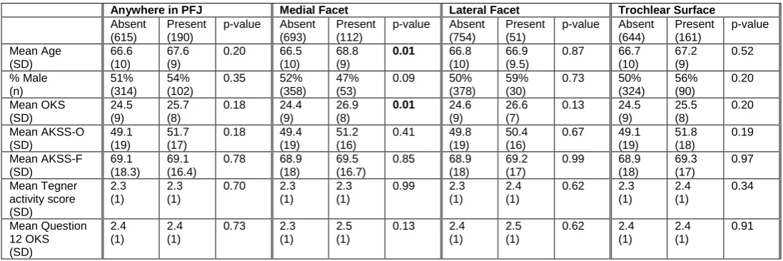
Knees with full thickness cartilage loss at the medial facet were significantly older and had better pre-operative OKS compared with knees without full thickness cartilage loss at the medial facet. No difference in baseline characteristics or function were detected between knees with or without full thickness cartilage loss at the lateral facet, trochlea or any site within the PFJ. Table 1.
or were lost to follow up (4). In the patients who died, withdrew from the study at any time point, all due to medical co-morbidities not associated with their knee, or were lost to follow up we are not aware of any revisions. The mean follow up was 10 years (range 5 to 17) with 347 knees having a minimum 10 year follow.
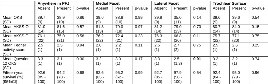
Of the 805 knees 74 had full thickness cartilage loss affecting the medial facet only, 13 had full thickness cartilage loss affecting the lateral facet only and 38 had full thickness cartilage loss affecting both the medial and lateral facets. Overall 96 knees had full thickness cartilage loss at either the medial or lateral facet with reciprocal full thickness cartilage loss at the trochlea. The functional outcomes at ten-years are outlined in Table 2. No difference in absolute functional scores at ten years or improvement from baseline to ten-years assessed by OKS, AKSS-O, AKSS-F or Tegner Activity Score was detected between groups. Analysis of Q12 of the OKS revealed that, compared to knees without exposed bone, knees with full thickness cartilage loss at the lateral patella facet had a lower ten-year Q12 score (p=0.01) and lower improvement from baseline to ten-year score (p=0.01). Additionally, knees with full thickness cartilage loss at the trochlea had a higher improvement from baseline to ten-year score (p=0.01). In all cases the difference was under one point and as such this is regarded to be unlikely to be clinically relevant.
No difference was observed between knees with medial facet exposed bone only, lateral facet exposed bone only or both medial and lateral facet exposed bone in absolute functional scores at ten years (OKS p=0.36, Q12 p=0.09, OKS AKSS-O p=0.81, AKSS-F p=0.39 or Tegner Activity Score p=0.26) or improvement from baseline to ten-years (OKS p=0.46, Q12 p=0.09, AKSS-O p=0.90, AKSS-F p=0.43 or Tegner Activity Score p=0.99).
baseline to ten-years (OKS p=0.23, Q12 p=0.98, AKSS-O p=0.16, AKSS-F p=0.29 or Tegner Activity Score p=0.07).
There was no correlation between functional outcome at ten years and the degree of intraoperative cartilage damage at the medial facet (OKS p=0.27, AKSS-O p=0.66, AKSS-F p=0.67), lateral facet (OKS p=0.99, AKSS-O p=0.92, AKSS-F p=0.49) or trochlea (OKS p=0.32, AKSS-O p=0.14, AKSS-F p=0.95).
Overall there were 32 implant related reoperations, with none performed due to progression of arthritis within the PFJ or due to PFJ symptoms. In patient who underwent revision to primary TKR for lateral progression at 6.9 years progression of PFJ degeneration was noted, however this was not considered to be symptomatic and the patella was not resurfaced with the patient subsequently progressing to a full recovery with no further surgery at three years post-revision. At fifteen-years no difference in implant survival was seen based on the presence, or location of full thickness cartilage loss in the PFJ. Table 2. Figure 2. No difference in survival was seen based on whether the full thickness cartilage loss affected the medial facet only, lateral facet only or medial and lateral facets (p=0.62) or whether the full thickness cartilage loss on the patella facet had a reciprocal area of full thickness cartilage loss on the trochlea or not (p=0.83).
Radiographic assessment of the PFJ (Altman score).
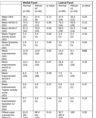
At last follow-up no difference in absolute functional outcome score or implant survival was seen between knees with radiographic degenerative disease of the PFJ (Altman Score ≥2) at either the medial or lateral facet. Table 3. Aside from a lower improvement from baseline OKS to OKS at last follow up no difference in improvement was seen between groups.
Clinical Assessment
Discussion
This study has demonstrated that neither the presence of anterior knee pain, radiographic medial PFJ disease or intra-operative exposed bone at the medial patella facet influence the long-term functional outcome or implant survival following medial-mobile bearing UKR and as such these factors should not be regarded as contraindications for this procedure. In the presence of radiographic lateral PFJ and intra-operative exposed bone at the lateral patella facet this study found that whilst the improvement from baseline function were less, for OKS and Q12 OKS respectively, compared to those knees with no lateral PFJ disease, no difference in absolute functional outcomes scores was seen. As such these findings, coupled with evidence of no difference in implant survival suggests that lateral PFJ disease may not represent an absolute contraindication to mobile-bearing UKR.
This study builds on short term functional outcome data which has previously provided evidence that, unlike fixed-bearing UKR, for mobile-bearing UKR anterior knee pain, radiographic medial facet PFJ disease and intra-operative exposed bone at the medial patella facet are not contraindications 11. The data presented here conflicts with the early results from this case-series which found that knees with lateral radiographic PFJ disease had significantly worse improvements from baseline function as well as absolute functional outcome at two-years post-operatively, as this study, at a mean follow up of ten-two-years, found no difference in absolute scores based on clinical, radiographic or intra-operative assessment.
patellofemoral joint, a decision was not made to resurface the PFJ and this patient has made a good post-operative recovery highlighting the lack of correlation between PFJ degenerative change and knee function.
The proposed reasons that anterior knee pain and the presence of PFJ disease do not affect functional outcomes or survival have been discussed previously11. Whilst cross-sectional studies of patients with knee pain have demonstrated an incidence of radiographic PFJ disease in 30% of those aged 34 to 55 post mortem studies have demonstrated that significant PFJ disease can occur in individuals who had not previously reported knee pain26,27. As such it is likely that many cases of PFJ disease are likely asymptomatic. This argument is supported by findings that that that the location of pre-operative pain does not correlate with the pattern and severity of intra-articular PFJ disease and that this study has found that PFJ disease does not influence post-operative outcomes following mobile-bearing UKR14,15.
In addition to implant design factors and avoiding overload of the PFJ by preserving knee kinematics, other factors in assuring good outcomes in the setting of PFJ disease may include operative factors, such as the removal of patella, trochlear or tibial anvil osteophytes which are undertaken as part of the UKR procedure may be responsible for the resolution in symptoms. Additionally, restoration of pre-disease limb alignment, as is achieved with mobile-bearing UKR would be expected to restore pre-disease patella tracking which may serve to mitigate any future complications and permit normal function of the PFJ 15.
up and, whilst the presence, or absence of radiographic PFJ progression is of interest it is the clinical outcomes that are the most clinically relevant.
Conclusion
Anywhere in PFJ Medial Facet Lateral Facet Trochlear Surface Absent
(615)
Present (190)
p-value Absent (693)
Present (112)
p-value Absent (754)
Present (51)
p-value Absent (644) Present (161) p-value Mean Age (SD) 66.6 (10) 67.6 (9)
0.20 66.5 (10)
68.8 (9)
0.01 66.8 (10)
66.9 (9.5)
0.87 66.7 (10) 67.2 (9) 0.52 % Male (n) 51% (314) 54% (102)
0.35 52% (358)
47% (53)
0.09 50% (378)
59% (30)
0.73 50% (324) 56% (90) 0.20 Mean OKS (SD) 24.5 (9) 25.7 (8)
0.18 24.4 (9)
26.9 (8)
0.01 24.6 (9)
26.6 (7)
0.13 24.5 (9) 25.5 (8) 0.20 Mean AKSS-O (SD) 49.1 (19) 51.7 (17)
0.18 49.4 (19)
51.2 (16)
0.41 49.8 (19)
50.4 (16)
0.67 49.1 (19) 51.8 (18) 0.19 Mean AKSS-F (SD) 69.1 (18.3) 69.1 (16.4)
0.78 68.9 (18)
69.5 (16.7)
0.85 68.9 (18)
69.2 (17)
0.99 68.9 (18) 69.3 (17) 0.97 Mean Tegner activity score (SD) 2.3 (1) 2.3 (1)
0.70 2.3 (1)
2.3 (1)
0.99 2.3 (1)
2.4 (1)
0.62 2.3 (1) 2.4 (1) 0.34 Mean Question 12 OKS (SD) 2.4 (1) 2.4 (1)
0.73 2.3 (1)
2.5 (1)
0.13 2.4 (1)
2.5 (1)
0.62 2.4 (1)
2.4 (1)
0.91
Table 1: Preoperative demographics and functional performance of knees with and without
[image:14.595.34.583.70.253.2]Anywhere in PFJ Medial Facet Lateral Facet Trochlear Surface
Absent Present p-value Absent Present p-value Absent Present p-value Absent Present p-value
Mean OKS (SD) 39.7 (9) 38.9 (10)
0.86 39.6 (9)
38.8 (10)
0.99 39.8 (9)
35.0 (11)
0.14 39.6 (9) 39.6 (9) 0.54 Mean AKSS-O (SD) 81.0 (14) 81.6 (15)
0.57 81.3 (13)
79.0 (18)
0.97 81.2 (14)
79.0 (23)
0.70 80.7 (14) 84.0 (14) 0.15 Mean AKSS-F (SD) 76.1 (22) 75.0 (21)
0.58 76.2 (22)
72.4 (21)
0.23 76.3 (22)
66.8 (26)
0.11 75.7 (22) 77.1 (20) 0.75 Mean Tegner activity score (SD) 2.5 (1) 2.5 (1)
0.94 2.6 (1)
2.2 (1)
0.11 2.5 (1)
2.7 (2)
0.75 2.5 (1) 2.6 (1) 0.25 Mean Question 12 OKS (SD) 3.3 (1) 3.1 (1)
0.30 3.2 (1)
3.0 (1)
0.17 3.3 (1)
2.5 (1.3)
0.01 3.2 (1) 3.2 (1) 0.74 Fifteen-year survival (%) (95%CI) 92.6 (85 - 100) 94.2 (78 - 100)
0.68 92.6 (85 - 100)
95.2 (62 - 100)
0.99 92.7 (85 - 100)
97.9 (58 - 100)
[image:15.595.35.580.70.242.2]0.54 92.4 (84 - 100) 95.0 (79 - 100) 0.96
Table 2: Ten-year functional outcomes and fifteen-year implant survival of knees with and without
Medial Facet Lateral Facet Normal (n=55) Altman ≥2 (n=45)
p-value Normal
(n=80) Altman ≥2 (n=20) p-value Mean OKS (SD) 36.1 (12) 37.5 (9)
0.72 37.5 (10) 33.5 (12) 0.25 Mean AKSS-O (SD) 74.5 (22) 78.2 (12)
0.95 78.2 (16) 59.8 (28) 0.15 Mean AKSS-F (SD) 67.4 (32) 70.0 (23)
0.90 69.0 (29) 61.5 (13) 0.66 Mean Tegner activity score (SD) 2.4 (1) 2.4 (1)
0.65 2.4 (1) 2.4 (2) 0.43 Mean Question 12 OKS (SD) 2.9 (1) 3.1 (1)
0.80 3.0 (1) 2.9 (1) 0.60 Mean improvement OKS (SD) 12.8 (10) 13.8 (10)
0.82 14.3 (10) 9.1 (9) 0.02 Mean improvement AKSS-O (SD) 24.2 (23) 25.4 (23)
0.67 26.4 (23) 11 (16) 0.20 Mean improvement AKSS-F (SD) 6.3 (26) 7.8 (28)
0.59 7.5 (27) 5 (25) 0.67 Mean improvement Tegner activity score (SD) 0.6 (1) 0.8 (1)
0.37 0.6 (1) 0.9 (1) 0.47 Mean improvement Question 12 OKS (SD) 0.7 (1) 0.9 (1)
0.44 0.9 (1) 0.5 (1) 0.19 Fifteen-year survival (%) (95%CI) 91.9 (83 - 100) 96.9 (91 - 100)
0.41 92.7 (85.6 - 100)
[image:16.595.35.355.68.461.2]100 0.92
Table 3: Functional outcomes at last follow-up, improvement from baseline function to function at last
follow-up and ten-year implant survival of knees with and without radiographic disease of the PFJ as
Anterior knee pain Absent (n=46) Present (n=54) p-value Mean OKS (SD) 37.8 (10.2) 35.7 (11) 0.28 Mean AKSS-O (SD) 80.3 (16) 73.1 (19) 0.37 Mean AKSS-F (SD) 74.2 (25) 64.0 (29) 0.11 Mean Tegner activity score (SD) 2.6 (1) 2.3 (1) 0.18 Mean Question 12 OKS (SD) 3.2 (1) 2.8 (1) 0.10 Mean improvement OKS (SD) 13.3 (10) 13.2 (10) 0.79 Mean improvement AKSS-O (SD) 20.3 (25) 28.1 (21) 0.19 Mean improvement AKSS-F (SD) 9.2 (24) 5.0 (30) 0.82 Mean improvement Tegner activity score (SD) 0.7 (1) 0.6 (1) 0.56 Mean improvement Question 12 OKS (SD) 0.9 (1) 0.7 (1) 0.63 Ten-year survival (%) (95%CI) 90 (80 - 100) 98 (93 - 100) 0.84
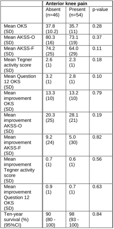
Table 4: Functional outcomes at last follow-up, improvement from baseline function to function at last
Figure Legend
Figure 1: Study flow chart
References
1. Kozinn SC, Scott R. Unicondylar knee arthroplasty. J Bone Joint Surg Am 1989;71-1:145-50.
2. Stern SH, Becker MW, Insall JN. Unicondylar knee arthroplasty. An evaluation of selection criteria.
Clin Orthop Relat Res 1993-286:143-8.
3. Hamilton TW, Pandit H, Jenkins C, Mellon SJ, Pegg E, Marks B, Dodd C, Murray DW. Fifteen year survival and functional outcome of 1000 minimally invasive unicompartmental knee arthroplasties.
American Academy of Orthopaedic surgeons. Las Vegas, 2015.
4. Lim HC, Bae JH, Song SH, Kim SJ. Oxford phase 3 unicompartmental knee replacement in Korean patients. J Bone Joint Surg Br 2012;94-8:1071-6.
5. Price AJ, Svard U. A second decade lifetable survival analysis of the Oxford unicompartmental knee arthroplasty. Clin Orthop Relat Res 2011;469-1:174-9.
6. Yoshida K, Tada M, Yoshida H, Takei S, Fukuoka S, Nakamura H. Oxford phase 3 unicompartmental knee arthroplasty in Japan - clinical results in greater than one thousand cases over ten years. J Arthroplasty 2013;28-9 Suppl:168-71.
7. Bergeson AG, Berend KR, Lombardi AV, Jr., Hurst JM, Morris MJ, Sneller MA. Medial mobile bearing unicompartmental knee arthroplasty: early survivorship and analysis of failures in 1000 consecutive cases. J Arthroplasty 2013;28-9 Suppl:172-5.
8. Faour-Martin O, Valverde-Garcia JA, Martin-Ferrero MA, Vega-Castrillo A, de la Red Gallego MA, Suarez de Puga CC, Amigo-Linares L. Oxford phase 3 unicondylar knee arthroplasty through a minimally invasive approach: long-term results. Int Orthop 2013;37-5:833-8.
9. Argenson JN, Blanc G, Aubaniac JM, Parratte S. Modern unicompartmental knee arthroplasty with cement: a concise follow-up, at a mean of twenty years, of a previous report. J Bone Joint Surg Am 2013;95-10:905-9.
10. Price AJ, Rees JL, Beard DJ, Gill RH, Dodd CA, Murray DM. Sagittal plane kinematics of a mobile-bearing unicompartmental knee arthroplasty at 10 years: a comparative in vivo fluoroscopic analysis.
J Arthroplasty 2004;19-5:590-7.
11. Beard DJ, Pandit H, Gill HS, Hollinghurst D, Dodd CA, Murray DW. The influence of the presence and severity of pre-existing patellofemoral degenerative changes on the outcome of the Oxford medial unicompartmental knee replacement. J Bone Joint Surg Br 2007;89-12:1597-601.
12. Pandit H, Jenkins C, Gill HS, Smith G, Price AJ, Dodd CA, Murray DW. Unnecessary contraindications for mobile-bearing unicompartmental knee replacement. J Bone Joint Surg Br 2011;93-5:622-8.
13. Goodfellow JW, O'Connor J. Clinical results of the Oxford knee. Surface arthroplasty of the tibiofemoral joint with a meniscal bearing prosthesis. Clin Orthop Relat Res 1986-205:21-42.
14. Liddle AD, Pandit H, Jenkins C, Price AJ, Dodd CA, Gill HS, Murray DW. Preoperative pain location is a poor predictor of outcome after Oxford unicompartmental knee arthroplasty at 1 and 5 years.
Knee Surg Sports Traumatol Arthrosc 2013;21-11:2421-6.
15. Beard DJ, Pandit H, Ostlere S, Jenkins C, Dodd CA, Murray DW. Pre-operative clinical and radiological assessment of the patellofemoral joint in unicompartmental knee replacement and its influence on outcome. J Bone Joint Surg Br 2007;89-12:1602-7.
16. Pandit H, Hamilton TW, Jenkins C, Mellon SJ, Dodd CA, Murray DW. The clinical outcome of minimally invasive Phase 3 Oxford unicompartmental knee arthroplasty: a 15-year follow-up of 1000 UKAs. Bone Joint J 2015;97-B-11:1493-500.
17. Goodfellow JW, Kershaw CJ, Benson MK, O'Connor JJ. The Oxford Knee for unicompartmental osteoarthritis. The first 103 cases. J Bone Joint Surg Br 1988;70-5:692-701.
18. Hamilton TW, Pistritto C, Jenkins C, Mellon SJ, Dodd CA, Pandit HG, Murray DW. Unicompartmental knee replacement: Does the macroscopic status of the anterior cruciate ligament affect outcome? Knee 2016;23-3:506-10.
20. Insall JN, Dorr LD, Scott RD, Scott WN. Rationale of the Knee Society clinical rating system. Clinical Orthopaedics and Related Research 1989-248:13-4.
21. Tegner Y, Lysholm J. Rating systems in the evaluation of knee ligament injuries. Clin Orthop Relat Res 1985-198:43-9.
22. Altman RD, Fries JF, Bloch DA, Carstens J, Cooke TD, Genant H, Gofton P, Groth H, McShane DJ, Murphy WA, et al. Radiographic assessment of progression in osteoarthritis. Arthritis Rheum 1987;30-11:1214-25.
23. Ahlback S. Osteonecrosis of the knee--radiographic observations. Calcif Tissue Res 1968:Suppl:36-b.
24. Clement ND, MacDonald D, Simpson AH. The minimal clinically important difference in the Oxford knee score and Short Form 12 score after total knee arthroplasty. Knee Surg Sports Traumatol Arthrosc 2014;22-8:1933-9.
25. Peto R, Pike MC, Armitage P, Breslow NE, Cox DR, Howard SV, Mantel N, McPherson K, Peto J, Smith PG. Design and analysis of randomized clinical trials requiring prolonged observation of each patient. II. analysis and examples. Br J Cancer 1977;35-1:1-39.
26. Kumm J, Tamm A, Lintrop M, Tamm A. The prevalence and progression of radiographic knee osteoarthritis over 6 years in a population-based cohort of middle-aged subjects. Rheumatol Int 2012;32-11:3545-50.