RESEARCH NOTE
Clinical and prognostic profile of Her2neu
positive (non-luminal) intrinsic breast cancer
subtype: comparison with Her2neu positive
luminal breast cancers
Atif Ali Hashmi
1, Raeesa Mahboob
1, Saadia Mehmood Khan
1, Muhammad Irfan
1, Mariam Nisar
1, Narisa Iftikhar
1,
Maham Siddiqui
1, Naveen Faridi
1, Amir Khan
2*and Muhammad Muzzammil Edhi
3Abstract
Objective: Her2neu receptor is proto-oncogene which can be over-expressed in both luminal and non-luminal breast cancers. In the present study, we aimed to compare the prognostic and predictive factors like tumor grade, T-stage, N-stage and ki67 index in Her2neu intrinsic breast cancer subtype with Her2neu over-expressed luminal breast cancers.
Results: 801 (41%) cases were Her2neu positive; out of which, 418 cases (52.2%) showed ER positivity and thus were classified as Her2neu positive luminal cancers whereas 383 cases (47.8%) were ER and PR negative and therefore were labeled as intrinsic Her2neu subtype (non-luminal). Her2neu positive (non-luminal) cancers were significantly associated with higher grades and Ki67 proliferative index compared to Her2neu positive luminal cancers. On the other no significant association was noted in T-stage and N-stage. We found a high frequency of her2neu positivity in our studied population of breast cancer. Moreover, association of her2neu positive (non-luminal) breast cancers with higher grade and ki67 index indicates a predictive value of ER/PR positivity in her2neu positive breast cancers. On the other hand, lack of association with respect to T and N stage, signifies no prognostic benefit of ER/PR in her2neu posi-tive breast cancers.
Keywords: Her2neu, Intrinsic breast cancer, ER, PR, Her2neu
© The Author(s) 2018. This article is distributed under the terms of the Creative Commons Attribution 4.0 International License (http://creat iveco mmons .org/licen ses/by/4.0/), which permits unrestricted use, distribution, and reproduction in any medium, provided you give appropriate credit to the original author(s) and the source, provide a link to the Creative Commons license, and indicate if changes were made. The Creative Commons Public Domain Dedication waiver (http://creat iveco mmons .org/ publi cdoma in/zero/1.0/) applies to the data made available in this article, unless otherwise stated.
Introduction
Her2neu receptor is a proto-oncogene located on long arm of chromosome 17 with intracellular tyrosine kinase activity [1, 2]. It belongs to epidermal growth factor receptor family and is expressed at low levels in many normal tissues of the body where it drives epithelial cell growth [3, 4]. Over-expression of Her2neu occurs in 18–20% of breast cancers [5–7]. Gene expression profil-ing defines breast cancer subtypes on the basis of expres-sion patterns of different genes. High expresexpres-sion of Her2neu gene, in the absence of estrogen receptor (ER)
and progesterone receptor (PR) related genes are cat-egorized as Her2neu intrinsic breast cancer subtype. On the other hand co-expression of Her2neu and ER related gene (although low) may be seen in luminal B subtype of breast carcinoma. Luminal B cancers with Her2neu positivity were found to be significantly associated with high tumor grade and higher frequency of nodal metasta-sis [8]. St. Gallen International Expert Consensus on the Primary Therapy of Early Breast Cancer 2013 proposed that intrinsic molecular subtypes of breast cancer can be defined without molecular diagnostics with the help of immunohistochemistry (IHC) for ER, PR, HER2neu and ki67 [9]. High expression of Her2neu is known to associ-ated with poor prognosis in untreassoci-ated patients [10, 11]. Introduction of herceptin therapy has made a remarkable
Open Access
*Correspondence: dramirkhan04@gmail.com
improvement in the survival of these patients. On the other hand, use of other chemotherapeutic and hormo-nal agents has been a matter of debate. Some authors suggested a decreased sensitivity to anthracycline based chemotherapy in Her2neu amplified breast cancer [12]. Similarly Her2neu over expression also leads to resist-ance to hormone therapy [13]. Ki67 index and other pathologic parameters may be important factors in these cases in predicting response to therapy. Pathologic parameters and proliferative index has not been widely studied in Her2neu positive breast cancers in our popula-tion. Therefore we aimed to compare the prognostic and predictive factors like tumor grade, T-stage, N-stage and ki67 index in Her2neu intrinsic breast cancer subtype with Her2neu over-expressed luminal breast cancers.
Main text Methods
The study included 1951 cases of breast cancers that underwent surgical resection or biopsy at Liaquat National hospital, Karachi Pakistan. The duration of study was 6 years from January 2011 till December 2016. Ethical approval was taken from institutional research and ethical committee. Medical and surgical records of all cases were reviewed. The surgical specimens were grossed according to standard protocols and histopatho-logical examination was done to reveal information regarding tumor size, grade, type and stage. Representa-tive tissue sections were than selected for IHC studies including ER alpha, PR, her2neu and ki67 by DAKO envi-sion method. Following antibodies are used.
1. FLEX Monoclonal Rabbit Anti-human Estrogen receptor alpha, clone EP1, ready to use, purchased from DAKO (code IR084).
2. FLEX Monoclonal Mouse Anti-human Progester-one receptor, clProgester-one PgR 636, ready to use, purchased from DAKO (code IR068).
3. Polyclonal Rabbit Anti-human c-erbB-2 oncoprotein, purchased from DAKO (code A0485).
4. FLEX Monoclonal Mouse human, Ki67 Anti-gen, Clone MIB-1, Ready to use, purchased from DAKO (code IR626).
One representative tumor block was selected for IHC staining for ER, PR, HER2neu and Ki67. IHC staining was done by DAKO envision method according to manu-facturers protocols. Thick sections of 4 mm were depar-affinized in xylene and then dehydrated. Antigen retrieval was done by boiling target DAKO Envision retrieval solu-tion at high ph for 40 min at 96–99 °C. DAKO Envision flex peroxidase blocking reagent was used for blocking endogenous peroxidase activity. The slides were then
incubated for 20–30 min in humidity chamber at room temperature followed by incubation with secondary anti-body. The substrate (Diamino benzidine + Chromogen) was used producing brown color at the site of target anti-gen. The hematoxylin was used as a counter stain. Appro-priate positive and negative controls were used (normal breast tissue for ER and PR as positive control, known case of her2neu amplified tumor as positive control for her2neu and lymph node as positive control for ki67).
For ER and PR, more than 1% nuclear staining was con-sidered positive expression.
Her2neu IHC scoring was done according to ASCO/ CAP guidelines (on a scale of 0 to 3 +). Both intensity of expression and percentage of positively stained cells were taken into account. Only membranous Her2neu expression was evaluated and scored from 0 (negative) to 3+ (positive) according to ASCO and CAP recom-mendations [14]. Cases with equivocal (2+) IHC expres-sion were further subjected to molecular testing by FISH technique. FISH testing was done utilizing FDA approved Path Vysion Her2 DNA Probe kit purchased from Abbot. Results were given as negative (not amplified) or positive (amplified) according to ASCO/CAP recommendations [14].
Ki67 IHC was scored according to number of positivity stained cells from 0 to 100. Number of positive cells was counted in at least five different areas of the tumor and then average score was calculated.
Her2neu positive luminal and non-luminal breast can-cers were defined as follows:
1. Her2neu positive intrinsic (non-luminal) breast can-cers: ER and ER negative, Her2neu positive (3+ by IHC or amplified on FISH).
2. Her2neu positive luminal breast cancers: ER positive, Her2neu positive (3+ by IHC or amplified on FISH).
Statistical package for social sciences (SPSS 21) was used for data analysis. Mean and standard deviation were calculated for quantitative variables. Frequency and per-centage were determined for qualitative variables. Chi square was applied to see association. P value ≤ 0.05 was considered significant.
Results
of Her2neu intrinsic subtype (Her2neu+, ER and PR−) with Her2neu positive luminal cancers (Her2neu+, ER+). Significant association was noted in tumor grade and Ki67 index categories where Her2neu positive (non-luminal) cancers showed higher grades and Ki67 pro-liferative index compared to Her2neu positive luminal cancers. On the other no significant association was noted in T-stage and N-stage. Although mean age of Her2neu intrinsic subtype cancers was low as compared to luminal cancers with Her2neu positivity, no significant association was noted in respect to age groups. Similarly, no significant association was noted when these two cat-egories of Her2neu positive cancers were compared in respect to lymphocytic infiltration of tumor, presence of insitu carcinoma, lymphovascular invasion, dermal lym-phatic invasion and pagetoid tumor spread in epidermis (Table 1). Table 2 depicts distribution of histologic sub-types in Her2neu and luminal subsub-types of breast cancers.
Frequency of lobular and mucinous carcinoma was found to be slightly higher in luminal cancers compared to Her2neu positive non-luminal cancers.
Discussion
In the current study we evaluated Her2neu positivity in a large group of patients with breast cancers and impact of ER/PR positivity in these cancers. Frequency of Her2neu positivity was 41% (801/1951 cases) and ER/PR positivity was associated with lower tumor grade and Ki67 index, however there was no significant difference in tumor stage and nodal metastasis.
Four intrinsic molecular subtypes of breast cancer are defined by gene expression analysis studies i.e. luminal A, luminal B, Her2neu enriched and triple negative. St. Gallen international expert consensus on primary ther-apy of early breast cancer 2013, defined clinicopatho-logical surrogate definitions of these intrinsic breast
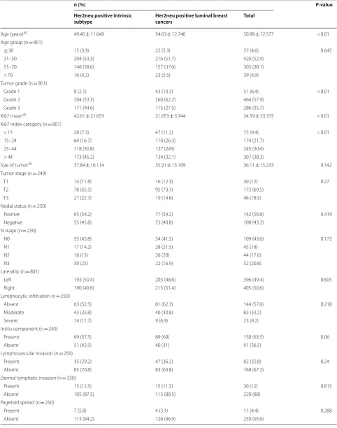
[image:3.595.57.541.88.442.2]Table 1 Association of clinico-pathologic parameters of Her2neu positive intrinsic breast cancer (non-luminal) with Her2neu positive luminal breast cancers (luminal B like)
n (%) P-value
Her2neu positive Intrinsic
subtype Her2neu positive luminal breast cancers Total
Age (years)ab 49.40 ± 11.649 54.63 ± 12.740 50.98 ± 12.577 < 0.01
Age group (n = 801)
≤ 30 15 (3.9) 22 (5.3) 37 (4.6) 0.645
31–50 204 (53.3) 216 (51.7) 420 (52.4)
51–70 148 (38.6) 157 (37.6) 305 (38.1)
> 70 16 (4.2) 23 (5.5) 39 (4.9)
Tumor grade (n = 801)
Grade 1 8 (2.1) 43 (10.3) 51 (6.4) < 0.01
Grade 2 204 (53.3) 260 (62.2) 464 (57.9)
Grade 3 171 (44.6) 115 (27.5) 286 (35.7)
Ki67 meanab 42.61 ± 21.653 21.653 ± 3.344 34.59 ± 23.375 < 0.01
Ki67 index category (n = 801)
< 15 28 (7.3) 47 (11.2) 75 (9.4) < 0.01
15–24 64 (16.7) 110 (26.3) 174 (21.7)
25–44 118 (30.8) 127 (245) 245 (30.6)
> 44 173 (45.2) 134 (32.1) 307 (38.3)
Size of tumorab 37.84 ± 16.114 35.21 ± 15.189 36.11 ± 15.233 0.142
Tumor stage (n = 249)
T1 14 (11.8) 16 (12.3) 30 (12) 0.27
T2 78 (65.5) 95 (73.1) 173 (69.5)
T3 27 (22.7) 19 (14.6) 46 (18.5)
Nodal status (n = 250)
Positive 65 (54.2) 77 (59.2) 142 (56.8) 0.419
Negative 55 (45.8) 53 (40.8) 108 (43.2)
N stage (n = 250)
N0 55 (45.8) 54 (41.5) 109 (43.6) 0.172
N1 17 (14.2) 28 (21.5) 45 (18)
N2 18 (15) 26 (20) 44 (17.6)
N3 30 (25) 22 (16.9) 52 (20.8)
Laterality (n = 801)
Left 193 (50.4) 203 (48.6) 396 (49.4) 0.605
Right 190 (49.6) 215 (51.4) 405 (50.6)
Lymphocytic infiltration (n = 250)
Absent 63 (52.5) 81 (62.3) 144 (57.6) 0.218
Moderate 43 (35.8) 40 (30.8) 83 (33.2)
Severe 14 (11.7) 9 (6.9) 23 (9.2)
Insitu component (n = 249)
Present 69 (57.5) 89 (69) 158 (63.5) 0.06
Absent 51 (42.5) 40 (31) 91 (36.5)
Lymphovascular invasion (n = 250)
Present 35 (29.2) 47 (36.2) 82 (32.8) 0.24
Absent 85 (70.8) 83 (63.8) 168 (67.2)
Dermal lymphatic invasion (n = 250)
Present 15 (12.5) 15 (11.5) 30 (12) 0.815
Absent 105 (87.5) 115 (88.5) 220 (88)
Pagetoid spread (n = 250)
Present 7 (5.8) 4 (3.1) 11 (4.4) 0.288
cancer subtypes including luminal A like, luminal B like, Her2neu positive (non-luminal) and triple nega-tive (ductal) [8]. It is important to recognize two distinct types of luminal B like breast cancers i.e. luminal B like (her2neu negative) and luminal B like (her2neu positive) as her2neu positivity can influence the sensitivity of can-cer cells to conventional hormonal and chemotherapy in breast cancer [11, 12].
We found a significantly higher grade and ki67 index in her2neu positive (non-luminal) breast cancers com-pared to Her2neu positive luminal cancers. This find-ing is of clinical importance as grade and ki67 index are among predictive factors which can influence sensitivity to chemotherapy. Higher grade and ki67 index have been reported in her2neu positive breast cancers in previous studies [15–19]. On the other hand, we didn’t find any significant difference in T and N stage between Her2neu positive luminal and non-luminal breast cancers, which are considered more important factors determining prognosis in breast compared to grade and ki67 index, which are more of predictive value in breast cancer.
Naeem et al. in a recent study found 12% of breast cancers to be her2neu positive, while all of those were found to have ki67 index more than 14% [20]. Schmidt et al. found that a her2neu score 0 can be considered as a poor prognostic factor in triple negative breast cancer [21]. In another study by Thangarajah et al. it was noted that high ki67 can be used as a poor prognostic factor in
terms of disease free survival in hormone receptor posi-tive tumors [22].
Another significant finding in our study is high fre-quency of her2neu positivity in our cases of breast cancer i.e. 41%; this contrasts to most of the previous studies in other parts of the world where her2neu posi-tivity ranges from 15 to 30% in breast cancer [23, 24]. One of the limitations of our study was that we didn’t evaluate survival and recurrence status in these two subtypes of her2neu positive breast cancers and there-fore we suggest that more prospective studies should be done to evaluate if there is any survival benefit of ER/ PR positivity in her2neu positive breast cancers.
Limitations
The main limitation of the study was lack of clinical fol-low up of patients; however, we found a high frequency of her2neu positivity in our studied population of breast cancer. Moreover, association of her2neu posi-tive (non-luminal) breast cancers with higher grade and ki67 index indicates a predictive value of ER/PR posi-tivity in her2neu positive breast cancers. On the other hand, lack of association with respect to T and N stage, signifies no prognostic benefit of ER/PR in her2neu positive breast cancers.
Abbreviations
IHC: immunohistochemistry; ER: estrogen receptor; PR: progesterone receptor. Authors’ contributions
AAH, RM and SMK: main author of manuscript, have made substantial contri-butions to conception and design of study. MI, MN, NI, MS, NF, AK and MME: have been involved in requisition of data, analysis of the data and revision of the manuscript. All authors read and approved the final manuscript. Author details
1 Department of Histopathology, Liaquat National Hospital and Medical College, Karachi, Pakistan. 2 Department of Medicine, Kandahar University, Kandahar, Afghanistan. 3 Department of Surgery, Brown University, Providence, RI, USA.
Acknowledgements
We gratefully acknowledge all staff members of Pathology, Liaquat National Hospital, Karachi, Pakistan for their help and cooperation.
Competing interests
The authors declare that they have no competing interests. Availability of data and materials
Please contact author, Atif Ali Hashmi (doc_atif2005@yahoo.com) for data requests.
Table 1 (continued)
Chi Square applied
a Mean ± SD b Independent t-Test
Table 2 Distribution of histological types of breast carcinoma among Her2neu intrinsic breast cancers (non-luminal) and Her2neu positive luminal breast cancers
Her2neu intrinsic subtype
Her2neu +ve luminal breast cancers
Total
Ductal (NOS) 363 (94.8) 379 (90.7) 742 (92.6) Lobular 5 (1.3) 17 (4.1) 22 (2.7) Cribriform 3 (0.8) 4 (1) 7 (0.9) Papillary 2 (0.5) 1 (0.2) 3 (0.4) Mucinous 1 (0.3) 8 (1.9) 9 (1.1) Micropapillary 0 (0) 3 (0.7) 3 (0.4)
Tubular 4 (1) 0 (0) 4 (0.5)
Metaplastic 5 (1.3) 4 (1) 9 (1.1) Mixed ductal and
lobular 0 (0) 2 (0.5) 2 (0.2)
[image:5.595.56.291.187.355.2]•fast, convenient online submission •
thorough peer review by experienced researchers in your field • rapid publication on acceptance
• support for research data, including large and complex data types •
gold Open Access which fosters wider collaboration and increased citations maximum visibility for your research: over 100M website views per year •
At BMC, research is always in progress.
Learn more biomedcentral.com/submissions
Ready to submit your research? Choose BMC and benefit from:
Consent to publish Not applicable.
Ethical approval and consent to participate
Ethics committee of Liaquat National Hospital, Karachi, Pakistan approved the study. Written informed consent was obtained from the patients for the participation.
Funding
There was no funding available for this manuscript.
Publisher’s Note
Springer Nature remains neutral with regard to jurisdictional claims in pub-lished maps and institutional affiliations.
Received: 9 May 2018 Accepted: 3 August 2018
References
1. King CR, Kraus MH, Aaronson SA. Amplification of a novel v-erbB-related gene in a human mammary carcinoma. Science. 1985;229(4717):974. 2. Schecter AL, Hung MC, Vaidyanathan L, et al. The neu gene: an
erbB-homologous gene distinct from and unlinked to the gene encoding the EGF receptor. Science. 1985;229:976–8.
3. Klapper LN, Glathe S, Vaisman N, Hynes NE, Andrews GC, Sela M, Yarden Y. The ErbB-2/HER2 oncoprotein of human carcinomas may function solely as a shared coreceptor for multiple stroma-derived growth factors. Proc Natl Acad Sci USA. 1999;96(9):4995.
4. Karunagaran D, Tzahar E, Beerli RR, Chen X, Graus-Porta D, Ratzkin BJ, Seger R, Hynes NE, Yarden Y. ErbB-2 is a common auxiliary subunit of NDF and EGF receptors: implications for breast cancer. EMBO J. 1996;15(2):254. 5. Slamon DJ, Clark GM, Wong SG, Levin WJ, Ullrich A, McGuire WL. Human
breast cancer: correlation of relapse and survival with amplification of the HER-2/neu oncogene. Science. 1987;235(4785):177.
6. Yaziji H, Goldstein LC, Barry TS, Werling R, Hwang H, Ellis GK, Gralow JR, Livingston RB, Gown AM. HER-2 testing in breast cancer using parallel tissue-based methods. JAMA. 2004;291(16):1972.
7. Hashmi AA, Edhi MM, Naqvi H, Khurshid A, Faridi N. Molecular subtypes of breast cancer in South Asian population by immunohistochemical profile and Her2neu gene amplification by FISH technique: association with other clinicopathologic parameters. Breast J. 2014;20(6):578–85. 8. Hashmi AA, Aijaz S, Khan SM, Mahboob R, Irfan M, Zafar NI, Nisar M,
Sid-diqui M, Edhi MM, Faridi N, Khan A. Prognostic parameters of luminal A and luminal B intrinsic breast cancer subtypes of Pakistani patients. World J Surg Oncol. 2018;16(1):1.
9. Goldhirsch A, Winer EP, Coates AS, Gelber RD, Piccart-Gebhart M, Thürli-mann B, Senn HJ. Panel members: personalizing the treatment of women with early breast cancer: highlights of the St Gallen International Expert Consensus on the Primary Therapy of Early Breast Cancer 2013. Ann Oncol. 2013;24:2206–23.
10. Gilcrease MZ, Woodward WA, Nicolas MM, et al. Even low-level HER2 expression may be associated with worse outcome in node-positive breast cancer. Am J Surg Pathol. 2009;33:759.
11. Chia S, Norris B, Speers C, et al. Human epidermal growth factor receptor 2 overexpression as a prognostic factor in a large tissue microarray series of node-negative breast cancers. J Clin Oncol. 2008;26:5697.
12. Di Leo A, Desmedt C, Bartlett JM, et al. HER2 and TOP2A as predic-tive markers for anthracycline-containing chemotherapy regimens as adjuvant treatment of breast cancer: a meta-analysis of individual patient data. Lancet Oncol. 2011;12:1134.
13. De Laurentiis M, Arpino G, Massarelli E, et al. A meta-analysis on the inter-action between HER-2 expression and response to endocrine treatment in advanced breast cancer. Clin Cancer Res. 2005;11:4741.
14. Wolff AC, Hammond ME, Hicks DG, et al. Recommendations for human epidermal growth factor receptor 2 testing in breast cancer: American Society of Clinical Oncology—College of American Pathologists (ASCO/ CAP) Clinical Practice Guideline Update (2013). Arch Pathol Lab Med. 2014;138(2):241–56.
15. Hoff ER, Tubbs RR, Myles JL, Procops GW. HER2/neu amplification in breast cancer. Am J Clin Pathol. 2002;117:916–21.
16. Shapochka DO, Zaletok SP, Gnidyuk MI. Relationship between NF-KB, ER, PR, HER2/neu, ki67, p53 expression in human breast cancer. Exp Oncol. 2012;34(4):358–63.
17. Haroon S, Hashmi AA, Khurshid A, Kanpurwala MA, Mujtuba S, Malik B, Faridi N. Ki67 index in breast cancer: correlation with other prognostic markers and potential in pakistani patients. Asian Pac J Cancer Prev. 2013;14(7):4353–8.
18. Hashmi AA, Naz S, Hashmi SK, Hussain ZF, Irfan M, Bakar SMA, Faridi N, Khan A, Edhi MM. Cytokeratin 5/6 and cytokeratin 8/18 expression in tri-ple negative breast cancers: clinicopathologic significance in South-Asian population. BMC Res Notes. 2018;11(1):372.
19. Hashmi AA, Edhi MM, Naqvi H, Faridi N, Khurshid A, Khan M. Clinico-pathologic features of triple negative breast cancers: an experience from Pakistan. Diagn Pathol. 2014;9:43.
20. Naeem S, Naz S, Riyaz A, Jehangir F, Afzal N. Immunohistochemical Analysis of breast cancer subtypes and their correlation with Ki 67 index. J Ayub Med Coll Abbottabad. 2018;30(1):94–6.
21. Schmidt G, Gerlinger C, Juhasz-Böss I, Stickeler E, Rody A, Liedtke C, Wimberger P, Link T, Müller E, Fehm T, Abel M, Stein S, Bohle R, Endrikat J, Solomayer EF. Her2-neu score as a prognostic factor for outcome in patients with triple-negative breast cancer. J Cancer Res Clin Oncol. 2016;142(6):1369–76.
22. Thangarajah F, Enninga I, Malter W, Hamacher S, Markiefka B, Richters L, Krämer S, Mallmann P, Kirn V. A retrospective analysis of Ki-67 index and its prognostic significance in over 800 primary breast cancer cases. Anticancer Res. 2017;37(4):1957–64.
23. Soloman JP, DellAquila M, Fadare O, Hasteh F. Her2/neu status determina-tion in breast cancer: a single institudetermina-tional experience using a dual-testing approach with immunohistochemistry and fuorescence in situ hybridization. Am J Clin Pathol. 2017;147(4):432–7.