Glucose-targeted niosomes deliver vasoactive
intestinal peptide (VIP) to the brain
Christine Dufes
a,b, Frederic Gaillard
c, Ijeoma F. Uchegbu
d, Andreas G. Sch¨atzlein
b,
Jean-Christophe Olivier
e, Jean-Marc Muller
a,∗aEquipe de Biologie des Interactions Cellulaires, LBSC CNRS UMR 6558, Facult´e des Sciences, Universite de Poitiers,
40 Avenue du Recteur Pineau, 86022 Poitiers Cedex, France
bCancer Research UK Department of Medical Oncology, University of Glasgow, Garscube Estate, Switchback Road,
Glasgow G61 1BD, United Kingdom
cEquipe du D´eveloppement Cortical, LBSC CNRS UMR 6558, Facult´e des Sciences, Universite de Poitiers,
40 Avenue du Recteur Pineau, 86022 Poitiers Cedex, France
dDepartment of Pharmaceutical Sciences, University of Strathclyde, Strathclyde Institute for Biomedical Sciences,
27 Taylor Street, Glasgow G4 0NR, United Kingdom
eEquipe ´emergente M´edicaments Anti-infectieux et Barri`ere H´ematoenc´ephalique, Laboratoire de Pharmacie Gal´enique et Biopharmacie,
Facult´e de M´edecine et Pharmacie, 34 rue du Jardin des Plantes, 86005 Poitiers Cedex, France
Received 13 July 2004; received in revised form 18 July 2004; accepted 19 July 2004 Available online 11 September 2004
Abstract
The aim of this study was to evaluate glucose-bearing niosomes as a brain targeted delivery system for the vasoactive intestinal peptide (VIP).
To this end, VIP/125I-VIP-loaded glucose-bearing niosomes were intravenously injected to mice. Brain uptake was determined
by measuring the radioactivity of125I-labeled VIP using␥-counting, after intravenous administration of VIP in solution or
encapsulated in glucose-bearing niosomes or in control niosomes. VIP integrity was assessed by reversed-phase HPLC analysis of brain extracts. Distribution of125I-VIP derived radioactivity was examined from serial brain slices.
HPLC analysis confirmed the presence of intact VIP in brain after administration of VIP-loaded niosomes, but not after administration of VIP solution. Encapsulation within glucose-bearing niosomes mainly allowed a significantly higher VIP brain uptake compared to control niosomes (up to 86%, 5 min after treatment). Brain distribution of intact VIP after injection of glucose-bearing niosomes, indicated that radioactivity was preferentially located in the posterior and the anterior parts of the brain, whereas it was homogeneously distributed in the whole brain after the administration of control vesicles.
∗Corresponding author. Tel.: +33 5 49 45 37 25; fax: +33 5 49 45 39 76.
E-mail address: [email protected] (J.-M. Muller).
In conclusion, this novel vesicular formulation of VIP delivers intact VIP to particular brain regions in mice. Glucose-bearing vesicles might be therefore a novel tool to deliver drugs across the blood–brain barrier (BBB).
© 2004 Elsevier B.V. All rights reserved.
Keywords: Niosomes; Glucose; Vasoactive intestinal peptide (VIP); Brain delivery; Blood–brain barrier
1. Introduction
The vasoactive intestinal peptide (VIP) is a 28-amino acid neuropeptide of the glucagon-secretin family (Mutt and Said, 1970) with a widespread distri-bution in both the central and peripheral nervous system (Fahrenkrug, 1979). It exerts diverse peripheral bio-logical functions, such as anti-inflammatory and im-munomodulatory effects, regulation of cell growth and differentiation and participation in the development of neural tissue (Muller et al., 1995). It also possesses neuroprotective properties that influence the survival of activity-dependent neurons in the central nervous sys-tem, and plays an important role in cerebral metabolism (Magistretti et al., 1981) and in cerebral carcinogene-sis (Leli`evre et al., 1998). VIP may have interesting applications in the treatment of various neurological disorders: a lipophilic VIP analogue was proposed as a therapy against Alzheimer’s disease (Gozes et al., 1996). However, like most endogenous peptides, its po-tential therapeutic applications are limited by its failure to cross the blood–brain barrier (BBB) which strictly controls the access of solutes to the brain (Begley, 1996) and by its rapid elimination after intravenous administration. Its blood half-life is less than 1 min in rat (Hassan et al., 1994) and approximately 1 min in man (Domschke et al., 1978). However, it has recently been shown that a weak amount of VIP appeared to reach the brain in conditions where a large amount of intravenous radiolabelled peptide was administrated (Dogrukol-Ak et al., 2003), which allowed to increase the sensitivity of the brain delivery assays utilized in these studies but also possible risks of side effects of this high concentration of free circulating radio-peptide.
In order to overcome this limitation, liposomal for-mulations for intravenous use have been proposed to protect VIP from degradation (Gao et al., 1994; Suzuki et al., 1996; S´ejourn´e et al., 1997; Gololobov et al., 1998; ¨Ony¨uksel et al., 1999). However, as liposomes were unable to cross the BBB, the sole
pharmacolog-ical effect obtained was a peripheral vasodilation. We recently delivered with success intact VIP to the brain in rats, by using an intranasal route of administration (Dufes et al., 2003). Through this pathway, VIP not only circumvents the BBB, but the polypeptide also avoids the hepatic first-pass effect and/or degradation in the blood compartment, a particularly important is-sue in the case of peptide drugs. Also for brain delivery, a VIP analogue was conjugated to the OX26 mono-clonal murine antibody directed against the rat trans-ferrin receptor (Bickel et al., 1993). This conjugation permitted a significant brain uptake of the analogue administered intravenously in rats, associated with a 65% increase in cerebral blood flow, compared to basal level (Bickel et al., 1993). Despite these attempts, no procedure promoting the passage of inherently non-penetrating drugs through the intact brain blood ves-sel endothelium in their unmodified form is currently available.
2. Materials and methods
2.1. Chemicals
N-Palmitoylglucosamine (NPG) (Dufes et al., 2000) and125I-VIP (Martin et al., 1986; Pineau et al., 2001) were synthesized and characterized as previously de-scribed. Human VIP was obtained from Neosystem (France). Bovine serum albumin (BSA), trifluoroacetic acid (TFA), sorbitan monostearate (Span 60) and cholesterol were all purchased from Sigma (France). Cholesteryl poly-24-oxyethylene ether (Solulan C24) was obtained from Amerchol (USA).
2.2. Animals
Male Swiss mice (30–40 g) obtained from D´epr´es Breeding Laboratories (St. Doulchard, France) were housed in the animal breeding facilities of the labora-tory (Authorization no. 0028) for 4–6 days before ex-periments. They were maintained in a light-controlled (12/12 h light–dark cycle) and temperature-controlled environment with water and food ad libitum. Experi-mental procedures adhered to the Principles of Labo-ratory Animal Care (NIH Publication # 86-23, revised 1985).
2.3. Preparation of VIP loaded niosomes
N-Palmitoylglucosamine (NPG) niosomes entrap-ping VIP were prepared by shaking a mixture of NPG (16 mg), Span 60 (65 mg), cholesterol (58 mg), Solulan C24 (54 mg) in PBS (0.1 M, pH = 7.4, 1.5 mL) at 90◦C for 30 min, followed by probe sonication (Soniprep 150, Fisher Scientific) for 5 min with the instrument set at 75% of its maximal capacity. The whole proce-dure of heating and sonication was repeated once. VIP was entrapped into niosomes by probe sonicating them in125I-VIP (5×10⁶cpm/mL final concentration) and unlabelled VIP (10−9mol/mL final concentration, pre-pared in PBS or in PBS supplemented with 1% (w/v) BSA), in order to reduce the adsorption of VIP onto containers.
Control niosomes (without glucose) entrapping VIP were prepared in the same manner from a mixture of Span 60 (73 mg), cholesterol (65 mg), Solulan C24 (54 mg) in PBS (1.5 mL) or in PBS supplemented with 1% (w/v) BSA).
To separate unencapsulated VIP from encapsulated VIP, the mixture was then centrifuged (150,000 × g, first for 1 h, then for 30 min, Optima LE-80K ul-tracentrifuge, Beckman Instruments, France) and the supernatant containing the unencapsulated VIP was discarded. After each ultracentrifugation step, the pel-leted vesicles were resuspended in PBS (2 mL).
2.4. Vesicle analysis
2.4.1. Assay for the amount of VIP entrapped by the vesicles
Radioactivity in control or glucose-bearing vesicles was counted using a Cobra II Auto-Gamma Counting System (Packard, USA).
2.4.2. Vesicle sizing
Vesicle sizing was performed by photon correlation spectroscopy on a Malvern Zetasizer (Malvern Instru-ments, UK).
2.5. Uptake of VIP entrapped in glucose-bearing vesicles in brain
Mice (n = 4 animals/group) received three intra-venous injections, at 5 min intervals, of VIP/125I-VIP, free or encapsulated in glucose-bearing vesicles or in control vesicles. These formulations were admin-istered at a dose equivalent to 6.6×10⁻¹⁰mol/kg VIP (100,000 cpm125I-VIP per 0.1 mL injection).
Five, 15 or 30 min after the last injection, the mice were sacrificed by cervical dislocation and a blood sam-ple was collected by intracardiac puncture. A 200 mL volume of saline (0.9% w/v sodium chloride) was then infused at room temperature into the clamped aorta to remove blood from brain vessels carefully. The brain was removed and weighed, and its radioactivity, as well as the radioactivity of the blood sample, was deter-mined by␥-counting.
2.6. HPLC analysis of intact VIP in the brain
In an additional experiment the VIP integrity was examined by HPLC, after administration of VIP, free or entrapped in control or glucose-bearing niosomes.
volumes of 10 mM Tris buffer (pH 8) containing protease inhibitors (1 mg/mL bacitracin, 1 mM phenyl-methylsulfonylfluoride, 0.2 mM antipa¨ıne and 2 mM orthophenanthroline) using a Teflon-glass Potter tis-sue homogenizer and centrifuged at 4◦C (1,000×g for 10 min). Supernatants were collected and stored on ice. The homogenization and centrifugation steps were applied two more times to the pellets. Finally, the three supernatants collected for each sample were pooled and ultracentrifuged at 100,000×g for 30 min at 4◦C to spin down any remaining tissue debris. The clear super-natant layers were collected and dried under vacuum using a Speed-Vac (Heto Maxi Dry Lyo FD1.0). Ex-tracts were dissolved in 200L mobile phase prior to HPLC analysis.
VIP reversed-phase HPLC was carried out using a 5m VYDAC C18column (Interchrom, France) with ␥-counting of125I-VIP. A two-step linear gradient was used with 0.1% (v/v) TFA in water as solvent A and 85:15 acetonitrile: 0.1% (v/v) TFA in water as solvent B. The column was equilibrated with solvent A at a 1 mL/min flow-rate. Injection volume was 100L. At each run, solvent B increased linearly from 0 to 35% in 2 min, then up to 100% in further 15 min, and was main-tained at 100% for another 5 min. Fractions of 1 mL were collected and␥-counted. VIP eluted in fractions 13–15.
2.7. Intracerebral distribution of VIP after administration in vesicles
VIP formulations were administered as described above. Mice were anesthetized by intraperitoneal in-jection of 0.6% (w/v) chloral hydrate (300 mg/kg body weight) and perfused through the heart with 100 mL physiological saline followed by 200 mL fixative (4% w/v paraformaldehyde in saline). Brains were removed and cut serially (2.5 mm-thick slices). Radioactivity was directly determined by␥-counting in each brain slices.
2.8. Statistical analysis
Results were expressed as means±S.E.M. Statis-tical significance was determined by one-way analy-sis of variance (ANOVA) followed by the Bonferroni’s multiple comparison test. Differences were considered significant for p < 0.05.
3. Results
3.1. VIP is efficiently encapsulated into vesicles
VIP was entrapped in the glucose-bearing vesicles (3.03×10⁻¹² ±1.05×10⁻¹³mol (1.01×10⁻⁸± 3.51×10⁻¹⁰g) per gram niosome, corresponding to 24.07±0.83% of the initial VIP) and in the control vesicles (3.28 × 10⁻¹² ± 7.27 × 10⁻¹⁴ mol (1.09
×10−8±2.42×10⁻¹⁰g) per gram niosome, corre-sponding to 25.32±0.56% of the initial VIP). Control vesicles loaded with VIP had a z-average mean diame-ter of 214 nm, while glucose vesicles loaded with VIP had a z-average mean diameter of 220 nm.
3.2. VIP entrapped in vesicles reaches the brain, and targeting with glucose improves the uptake
No unexpected mortality of any animals occurred after administration of the VIP formulations used in the present study. As a result, vesicles were considered to be safe at the dosing schedule used. Radioactivity could be detected in the brain following the adminis-tration of glucose-bearing vesicles, indicating a pas-sage of their content from the blood compartment into the brain parenchyma (Fig. 1). Brain radioactivity up-take was independent of the presence or not of BSA in the formulation: brain radioactivity level was 1.892
±329 cpm/g after administration of VIP vesicles pre-pared without BSA, not significantly different to 1.544
[image:4.842.288.501.498.613.2]±325 cpm/g observed after administration of the vesi-cles prepared with this additive. The blood radioactivity
Fig. 2. Time-related radioactivity in brain (A) and blood (B) after systemic administrations of VIP encapsulated in glucose-bearing vesicles (a, d, g); encapsulated in control vesicles (b, e, h) and in solution (c, f, i) (four animals per formulation). Significant differ-ence: a vs. (b, g), c vs. (b, f, i), d vs. (e, f). Blood radioactivity levels are not significantly different.
levels obtained after administration of these two for-mulations were also very similar (respectively 3.879± 883 cpm/g and 3.966 ±876 cpm/g with and without BSA) (Fig. 1).
Brain and blood radioactivity was then measured at various time points after administration of VIP in different formulations (in solution, entrapped in glucose-bearing vesicles or in control vesicles, both of them prepared with BSA), as shown inFig. 2. For vesi-cles prepared with and without glucose, the maximum brain uptake was reached 15 min post-administration (Fig. 2A). The glucose modified vesicles significantly increased brain radioactivity levels by comparison with control vesicles (by about 86% and 58% re-spectively 5 min and 15 min after treatment). With glucose-bearing vesicles, the brain radioactivity lev-els measured 15 min after the last intravenous injection represented 0.26%±0.04% of the total radioactivity administered. After 30 min of administration, a reduced
and similar residual level of radioactivity was found in the brain, as well in control (without glucose) or glucose-bearing vesicles. This might be due to a clear-ance (cerebrospinal fluid draining) or a degradation process limiting the amount of peptide inside the brain. Regardless of the formulations, the blood radioac-tivity remained constant at each time point examined (Fig. 2B).
3.3. Intact VIP is found in brain after administration in vesicles with BSA
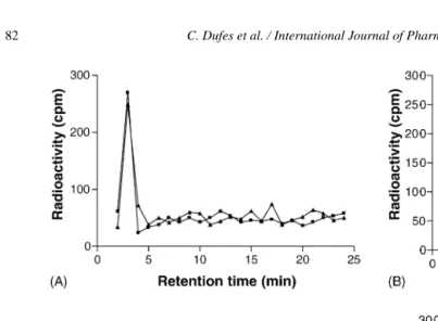
HPLC analysis performed on the brain extracts showed no peak corresponding to intact VIP after administration of glucose-bearing vesicles entrapping VIP without BSA (Fig. 3A). In contrast, a peak of radioactivity corresponding to intact VIP could be observed after administration of glucose-bearing vesi-cles entrapping VIP with BSA (Fig. 3B). Analysis also showed one peak corresponding to intact VIP as well after administration of vesicles without glucose but with BSA (Fig. 3C). The presence of BSA could de-crease the interaction of VIP to lipids in the niosome membrane, thus allowing VIP to be released out of the vesicle.
In our HPLC experimental conditions, free radioio-dine should elute very quickly, after few minutes of elution. Such early radioactive peak is not observed in experiments corresponding to glucose-bearing nio-somes, indicating that radiolysis of the125I-VIP does not occur in these experiments, at least for the fraction of radioligand that reached the brain.
No peak could be detected after administration of free VIP (Fig. 3D).
3.4. Glucose-bearing vesicles target specific cerebral regions
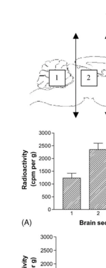
After administration of glucose-bearing vesicles, the radioactivity in the brain accumulated specifically in the posterior and the anterior parts of the brain (Fig. 4A). Counts for the posterior and the anterior sec-tors were respectively about two times and 1.5 times those read for the median sector or the cerebellum.
Fig. 3. Reversed-phase HPLC elution profiles of brain extracts obtained after intravenous administrations of VIP encapsulated in glucose-vesicles without BSA (A) or with BSA (B), VIP encapsulated in vesicles without glucose but with BSA (C) or as a solution (D), using125I-VIP as a tracer
(three animals per formulation).125I-VIP was evaluated by␥-counting. Intact VIP eluted in fractions 13–15. For more details see materials and
methods.
4. Discussion
In a previous work (Dufes et al., 2000), we have re-ported the design and the characterization of glucose-bearing niosomes, as well as their ability to bind to concanavalin A. The present study is an in vivo evalu-ation of these vesicles as a potential system to deliver the neuropeptide VIP into the brain.
There are three key findings of this study. First, we demonstrated that niosomes seem to be able to rapidly deliver intact VIP into brain provided that this neu-ropeptide is encapsulated in presence of BSA. Sec-ondly, the presence of glucose ligands on the vesicles seems to improve this uptake, at least for the two first time points studied. Finally, only the administration of glucose-bearing vesicles induced a characteristic brain distribution of radioactivity, among which is intact VIP.
Fig. 4. Brain radioactivity distribution profiles after intravenous ad-ministration of VIP encapsulated in glucose-vesicles (A) or in vesi-cles without glucose (B), or free VIP (C), using125I-VIP as a tracer (three animals per formulation). Radioactivity was evaluated by ␥-counting of the 2.5-mm thick brain slices. Arrows on the mouse brain schema denote the approximate spacing of the brain slices. For more details, seeSection 2. In A, significant differences in radioactivity levels were found betweenSections 1–4. Differences in B and C are not significant.
of the vesicular bilayer to trypsin. In view of the abil-ity of liposome constituents to interact with VIP, it is therefore hypothesized that VIP is “locked in” the vesi-cles by binding to lipids in the niosome membranes (Gololobov et al., 1998). The presence of BSA could decrease this interaction, thus allowing VIP to be re-leased out of the vesicle.
When considering the uptake of radioactivity into the brain, the glucose modified vesicles led to signifi-cantly increased brain radioactivity levels compared to control vesicles (Fig. 2A). VIP degradation products, like other low molecular weight molecules, may enter cells by passive non-specific diffusion. High molecular weight carriers could eventually:
• be taken up via endocytosis, a comparatively slower but potentially more specific process, if their size allows this passage;
• or bind the BBB and release their content through the luminal membrane of the endothelial cells.
The glucose-targeted vesicles increased the brain uptake when compared to non-targeted vesicles.
The importance of carbohydrate-mediated recogni-tion by specific cells has already been demonstrated. Umezawa and Eto (1988)reported that liposomes bear-ing aminophenyl mannoside were efficiently taken up into the mouse brain, suggesting that mannose could be recognized by the cells of the BBB. The BBB cells are known to express high levels of the glucose trans-porter GLUT1 (Dick et al., 1984). Therefore, an active targeting based on GLUT1 ligands could be a useful strategy for drug delivery in the brain. To our knowl-edge, our study is the first demonstration that VIP ad-ministered intravenously in targeted systems such as glucose-bearing niosomes is efficiently delivered to the brain.
transporter concentration in some brain regions. This idea is supported by previous studies showing the pres-ence of GLUT1 in considerable amounts throughout the brain, but with some regional variations (Brant et al., 1993; Rayner et al., 1994; Dobrogowska and Vorbrodt, 1999). Western blot studies revealed a rela-tively higher density in the posterior region of the brain such as the cerebellum (Brant et al., 1993; Rayner et al., 1994), whereas immunohistochemistry approaches in-dicated that GLUT1 was present in equal high amounts in the anterior cortex, the hippocampus and the cere-bellum (Dobrogowska and Vorbrodt, 1999; Choeiri et al., 2002).
Moderate to low GLUT1 levels have been reported in the same studies (Dobrogowska and Vorbrodt, 1999; Choeiri et al., 2002) for the olfactory bulb, the stria-tum, the thalamus, the hypothalamus, the brainstem and the medulla. Interestingly, we observed high levels of radioactivity inSection 4(including frontal, orbital and motor cortical areas) andSection 2(including ret-rosplenial, visual and auditory cortices, hippocampus and midbrain). Sections with lower levels of radioactiv-ity include brain regions containing lower densities of GLUT1 (striatum, thalamus, hypothalamus forSection 3; pons and medulla forSection 1). It should be noted in addition that orbital, retrosplenial, visual and auditory cortices exhibit significantly higher levels of glucose utilization (LCGU indices) than any other cortical area in the normal rat (Zilles and Wree, 1995). Together, high density of GLUT1 receptors and important rate of metabolic activity in these cortical areas could be responsible of the high VIP uptake observed in the an-terior and posan-terior parts of the brain (Dobrogowska and Vorbrodt, 1999).
In conclusion, the present study demonstrated that the administration of systemic glucose-bearing vesi-cles encapsulating VIP could deliver intact VIP to specific brain areas. Therefore, glucose-bearing vesi-cles represent a novel tool to deliver drugs across the BBB.
References
Begley, D.J., 1996. The blood-brain barrier: principles for target-ing peptides and drugs to the central nervous system. J. Pharm. Pharmacol. 48, 136–146.
Bickel, U., Yoshikawa, T., Landaw, E.M., Faull, K.F., Pardridge, W.M., 1993. Pharmacologic effects in vivo in brain by
vector-mediated peptide drug delivery. Proc. Natl. Acad. Sci. U.S.A. 90, 2618–2622.
Bodanszky, M., Bodanszky, A., Klausner, Y.S., Said, S.I., 1974. A preferred conformation in the vasoactive intestinal peptide (VIP). Molecular architecture of gastrointestinal hormones. Bioorg. Chem. 3, 133–140.
Brant, A.M., Jess, T.J., Miligan, G., Brown, C.M., Gould, G.W., 1993. Immunological analysis of glucose transporters expressed in different regions of the rat brain and central nervous system. Biochem. Biophys. Res. Commun. 192, 1297–1302.
Choeiri, C., Staines, W., Messier, C., 2002. Immunohistochemical lo-calization and quantification of glucose transporters in the mouse brain. Neuroscience 111, 19–34.
Dick, A.P.K., Harik, S.I., Walker, B.M., 1984. Identification and char-acterization of the glucose transporter of the blood-brain barrier by cytochalasin B binding and immunological reactivity. Proc. Natl. Acad. Sci. U.S.A. 81, 7233–7237.
Dobrogowska, D.H., Vorbrodt, A.W., 1999. Quantitative immuno-cytochemical study of blood-brain barrier glucose transporter (GLUT-1) in four regions of mouse brain. J. Histochem. Cy-tochem. 47, 1021–1029.
Dogrukol-Ak, D., Banks, A.W., Tuncel, N., Tuncel, M., 2003. Pas-sage of vasoactive intestinal peptide across the blood-brain bar-rier. Peptides 24, 437–444.
Domschke, S., Domschke, W., Bloom, S.R., Mitznegg, P., Mitchell, S.J., Lux, G., Strunz, U., 1978. Vasoactive intestinal peptide in man: pharmacokinetics, metabolic and circulatory effects. Gut 19, 1049–1053.
Dufes, C., Sch¨atzlein, A.G., Tetley, L., Gray, A.I., Watson, D.G., Olivier, J.C., Couet, W., Uchegbu, I.F., 2000. Niosomes and poly-meric chitosan based vesicles bearing transferrin and glucose ligands for drug targeting. Pharm. Res. 17, 1250–1258. Dufes, C., Olivier, J.C., Gaillard, F., Gaillard, A., Couet, W., Muller,
J.M., 2003. Brain delivery of vasoactive intestinal peptide (VIP) following nasal administration to rats. Int. J. Pharm. 255, 87–97. Fahrenkrug, J., 1979. Vasoactive intestinal polypeptide: measure-ment, distribution and putative neurotransmitter function. Diges-tion 19, 149–169.
Gao, X.P., Noda, Y., Rubinstein, I., Paul, S., 1994. Vasoactive intesti-nal peptide encapsulated in liposomes: effect on systemic arterial blood pressure. Life Sci. 54, PL247–PL252.
Gololobov, G., Noda, Y., Sherman, S., Rubinstein, I., Baranowska-Kortylewicz, J., Paul, S., 1998. Stabilization of vasoactive intesti-nal peptide by lipids. J. Pharmacol. Exp. Ther. 285, 753–758. Gozes, I., Bardea, A., Reshef, A., Zamostiano, R., Zhukovsky, S.,
Rubinraut, S., Fridkin, M., Brenneman, D.E., 1996. Neuroprotec-tive strategy for Alzheimer disease: intranasal administration of a fatty neuropeptide. Proc. Natl. Acad. Sci. U.S.A. 93, 427–432. Halmos, T., Santarromana, M., Antonakis, K., Scherman, D., 1997. Synthesis of glucose – chlorambucil derivatives and their recog-nition by the human Glut-1 glucose transporter. Eur. J. Pharma-col. 318, 477–484.
Hassan, M., Refai, E., Andersson, M., Schnell, P.O., Jacobsson, H., 1994. In vivo dynamical distribution of131I-VIP in the rat studied
by gamma-camera. Nucl. Med. Biol. 21, 865–872.
Leli`evre, V., Pineau, N., Du, J., Wen, C.H., Nguyen, T., Janet, T., Muller, J.M., Waschek, J.A., 1998. Differential effects of peptide histidine isoleucine (PHI) and related peptides on stimulation and suppression of neuroblastoma cell proliferation. J. Biol. Chem. 273, 19685–19690.
Magistretti, P.J., Morrison, J.H., Shoemaker, W.J., Sapin, V., Bloom, F.E., 1981. Vasoactive intestinal polypeptide induces glycogenol-ysis in mouse cortical slices: a possible regulatory mechanism for the local control of energy metabolism. Proc. Natl. Acad. Sci. U.S.A. 78, 6535–6539.
Martin, J.L., Rose, K., Hughes, G.J., Magistretti, P.J., 1986. [Mono[125I]iodo-Tyr10-MetO17]-vasoactive intestinal polypep-tide. Preparation, characterization, and use for radioimmunoas-say and receptor binding. J. Biol. Chem. 25, 5320–5327. Muller, J.M., Lelievre, V., Becq-Giraudon, L., Meunier, A.C., 1995.
VIP as a cell-growth and differentiation neuromodulator role in neurodevelopment. Mol. Neurobiol. 10, 115–134.
Mutt, V., Said, S.I., 1970. Polypeptide with broad biological activity: isolation from small intestine. Science 169, 1217–1218. ¨
Ony¨uksel, H., Ikezaki, H., Patel, M., Gao, X.P., Rubinstein, I., 1999. A novel formulation of VIP in sterically stabilized micelles am-plifies vasodilation in vivo. Pharm. Res. 16, 155–160. Pineau, N., Lelievre, V., Goursaud, S., Hilairet, S., Waschek, J.A.,
Janet, T., Muller, J.M., 2001. The polypeptide PHI discriminates a GTP-insensitive form of VIP receptor in liver membranes. Neu-ropeptides 35, 1–10.
Polt, R., Porreca, F., Szabo, L.Z., Bilsky, A.J., Davis, P., Albruscato, T.J., Davis, T.P., Horvath, R., Yamamura, H.I., Hruby, V.J., 1994. Glycopeptide enkephalin analogues produce analgesia in mice: evidence for penetration of the blood-brain barrier. Proc. Natl. Acad. Sci. U.S.A. 91, 7114–7118.
Rayner, D.V., Thomas, M.E., Trayhurn, P., 1994. Glucose trans-porters (GLUTs 1-4) and their mRNAs in regions of the rat brain: insulin-sensitive transporter expression in the cerebellum. Can. Pharmacol. J. 72, 476–479.
Robinson, R.M., Blakeney Jr., E.W., Mattice, W.L., 1982. Lipid-induced conformational changes in glucagon, secretin and va-soactive intestinal peptide. Biopolymers 21, 1217–1228. S´ejourn´e, F., Rubinstein, I., Suzuki, H., Alkan- ¨Ony¨uksel, H., 1997.
Development of a novel bioactive formulation of vasoactive in-testinal peptide in sterically stabilized liposomes. Pharm. Res. 14, 362–365.
Suzuki, H., Noda, Y., Gao, X.P., S´ejourn´e, F., Alkan- ¨Ony¨uksen, H., Paul, S., Rubinstein, I., 1996. Encapsulation of VIP into lipo-somes restores vasorelaxation in hypertension in situ. Am. J. Physiol. 271, H282–H287.
Umezawa, F., Eto, Y., 1988. Liposome targeting to mouse brain: man-nose as a recognition marker. Biochem. Biophys. Res. Commun. 153, 1038–1044.