Copyright i 1975 AmericanSociety for Microbiology PrintedinU.S.A.
RNA
Synthesis
in Cells Infected
with Herpes Simplex
Virus
X. Properties of Viral
Symmetric Transcripts and
of
Double-Stranded
RNA
Prepared from Them
BERNARD JACQUEMONT AND BERNARD ROIZMAN*
Departments of Microbiology and Biophysics, The University of Chicago, Chicago, Illinois 60637 Received forpublication17 October1974
HEp-2 cells infected with herpes simplex 1 virus contain RNA transcripts
capable of forming double-stranded (DS) RNAon annealing. The properties of
purified DS RNAwere asfollows: (i) DS RNA is resistanttodepolymerization by RNase AorT1 in2x 0.15 M NaCl, plus 0.015 M sodium citrate (SSC) butnot
0.1x SSCorfollowing thermal denaturation. (ii) The Tm of the viral DS RNAwas
100C in 0.1x SSC. (iii)Undenatured DS RNA doesnothybridize withviralDNA;
upondenaturation, excessunlabeled RNA drove 50to55%oflabeled DNAinto
DNA-RNA hybrid. The kinetics of hybridization indicate that the DS RNA
con-sists ofat least two populations of transcripts arising from 29and 26%ofviral DNA and differing40-fold in molar concentration.
This paper details a series of experiments designed to detect and analyze viral RNA
sequences
capable
of formingdouble-stranded
structures by base pairing in HEp-2 cells
in-fected with human herpesvirus 1 (herpes sim-plex 1,HSV-1). Thecircumstanceswhichled to
this studyare asfollows.
(i) In principle, analyses of the processes
which regulate transcriptionrequire some
infor-mation concerning the process of transcription
itself. We could envision several possibilities, i.e., asymmetric transcription of one or both strands ofDNA, partially symmetric transcrip-tion
resulting
fromoverlapping transcription ofboth
strands, and totally symmetric transcrip-tion. The first and lastimply entirely
different regulatory mechanisms for making transcriptsavailable
forfunction asmRNA.(ii) Transcripts arising from symmetric
tran-scription and therefore
capable
ofbase pairing havebeen
detected in cells infected with anumber of DNA viruses but especially with poxviruses, adenoviruses, and papovaviruses (1, 2, 4, 12). Suggestive evidence that
comple-mentary, self-annealing viral RNA sequences
exist in
HSV-1-infected
cellsemerged
fromthe observations that nuclear RNA from 8-h in-fected cells preannealed under conditions, which promoted self-annealing, droveonly 35% ofviral DNA into DNA-RNAhybrid,
i.e., 15%less than the nonpreannealed RNA. However,
the capacity to drive 50% of viral DNA into
hybrid was restored by heat denaturing the preannealedRNApriortohybridization.
More-over, theself-annealing ofRNAwasdependent
on RNA concentration andwas notaffected
by
mild alkaline hydrolysis which decreases the size of the RNA. These data suggested the
presence ofabundant symmetric transcriptsin
the nuclei of infected cells (11) but did not
reveal the extent to which viral DNA was
symmetricallytranscribedorthecharacteristics
of the double-stranded
(DS)
RNA formedby
self-annealing the RNA.
The presentstudieswere
designed
toanalyze
the properties of the DS RNA
prepared
by
self-annealing RNA from infected cells and to
estimate the extent ofthe DNA giving rise to
symmetrictranscripts.
MATERIALS AND METHODS
Solutions and chemicals. Standard saline citrate consisted of 0.15 M sodium chloride plus 0.015 M sodium citrate(1 xSSC);reticulocyte standard buffer consisted of 0.01 Msodium chloride-0.0025 M magne-sium chloride-0.01 M Tris-hydrochloride (pH 7.5).
Escherichia coli DNAwasthe kindgiftof A. Marko-vitz of the University of Chicago. Sodium dodecyl
sulfate and formamidewereobtained fromMatheson,
Coleman and Bell; sarkosyl (NL97) from Geigy
Chemical Company, sodium deoxycholate from Schwarz MannBioResearch,Orangeburg, N.Y.; crys-tallized DNase freeof RNase, RNase
T1,
andRNase A, from Worthington Biochemicals, Freehold, N.J.;and
[5-'HJuridine
(specific activity 28Ci/mmol)from NewEngland Nuclear, Boston, Mass.Cells and virus. The procedures for propagation
andmaintenance of humanepidermoidcarcinomano. 2 (HEp-2) cells andproduction, assay, andpertinent
7(07
on November 10, 2019 by guest
http://jvi.asm.org/
properties of the F strain ofherpes simplex 1 virus [HSV-1(F)]weredescribed elsewhere(2, 8, 11, 15,16).
Preparation and labeling of viral DNA. The procedure for preparation and purification of viral DNAwasthesameaspreviouslydescribed(8)except
that the final product wasdigested with RNase and extracted withphenol and chloroform. All batchesof DNA used in thisstudywererepurified by isopycnic banding inCsClsolution. HEp-2DNAwasextracted
bythemethodofMarmur (13).
The procedure for in vitro labeling of HSV-1(F) DNAby repairsynthesis using E. coli polymer-aseI wasdescribed elsewhere (7). The E. coli
polym-erase I was the kind gift of A. Kornberg, Stanford
University.
RNA purification. Total RNA from infected and uninfected cells was extracted as follows: cells were washed in phosphate buffered saline, suspended in reticulocyte standardbuffer, lysed bythe addition of sodium deoxycholate (0.5%final concentration), and digested briefly with RNase-free DNase (50 jg/ml, room temperature) todiminish viscosity. The
mate-rial wasthen extracted withsodium dodecyl sulfate,
phenol and chloroform, precipitated with ethanol, dissolved in0.01M Tris(pH 7.5)plus0.025M MgCl2, dialyzed againstthesame buffer, anddigested again with DNase. The RNAwasthenagainextractedwith
sodium dodecyl sulfate and phenol. This cycle was
repeatedonceagain. In the laststepof the extraction
procedure,the RNAwasdialyzed againstthe hybridi-zation bufferfor 3days.
PurificationofDSRNA. The RNAwasextracted asdescribed above, then allowedtoreannealat50C for 24 h in 0.01 M Tris (pH 7.5)-0.75 M sodium chloride-1 mM EDTA-50% (vol/vol)formamide. After reannealing the RNA was diluted and precipitated with 2 volumes of ethanol. The RNA pellet was
solubilized in 0.01 M Tris (pH 7.5) plus 2.5 mM magnesiumchloride, dialyzed against this buffer, and digestedwith DNase1(50 gg/ml)for1hat37C. Then
0.1 volumesof 10mM EDTA in 0.01 M Tris(pH 7.5) wasadded, the salt concentrationwasadjustedto0.35 M NaCl, and the RNA wasdigested for 1 h at37C
with 50,gofRNase A and 10U of RNase T1 perml.
To remove degradation products, RNA was passed
through SephadexG50 (1by60 cm)columns, and the excluded material wasprecipitated with ethanol.
Hybridizationof labeled RNAtoDNAonfilters. In several experiments, the RNA was denatured in
0.1x SSCat115C for 5minandhybridizedtoE.coli, HEp-2, and HSV-1 DNAs fixed to nitrocellulose filters. The DNAswerefixedto25-mmSchleicher and Schuell B6 filtersas previously described (9). Small
disks (6mm in diameter) were then punchedout. A
set of disks containing E. coli, HEp-2, and HSV-1 DNAs, respectively, plus a blank were placed in a
Beem capsule containing the denatured RNAin200,u1
of hybridization buffer (0.75 M NaCl-5 nM
EDTA-0.25% sodium dodecyl sulf'ate-0.01 M Tris, pH
7.5).After incubation for20hat66C, the diskswere
removed, washed two times for 30 min each in 2x
SSC, digested with RNase A (50 4g/ml) for 1 h at
roomtemperature, washed asbefore, anddried.
Hybridizationofunlabeled RNAtolabeled DNA in solution. Excess unlabeled RNA and sheared in
vitro labeled DNA were denatured by heatingfor 7
minat 115 C, then hybridizedin 0.3 M Na+-0.04M phosphatebufferat75C.Theamountof DNAdriven into DNA-RNA hybrid was monitored by digestion
with aNeurosporacrassanuclease (14) which
depo-lymerizes single-stranded DNA but does not attack DNA-RNA hybrid. The proceduresforpreparationof
the enzyme and digestion of the hybridization
mix-ture were as previously described (6, 11). In these experiments,theamountof denatured DNA resistant
toenzymedigestion didnotexceed 5% ofinput DNA
counts.
RESULTS
Preparationof DS RNA fromproductively infected cells. In this series of experiments HEp-2 cellswereinfectedwithamultiplicity of 100PFU/cell, labeled from 4.8to5 h
postinfec-tion with
[3Hjuridine
(50 ,Ci/ml), thenhar-vested. The extractionofRNAand purification
ofDS RNA were as described above. DS RNA
purification was monitored by digesting in 2x SSC samplestaken throughoutthepurification procedure with RNase Aorwith both RNaseA
and RNase T1. The salt concentration was chosen on the basis of the report (3) that DS RNA isdepolymerized withRNase inthe pres-enceof <0.02 M Na2+ butnot atconcentrations of Na+ > 0.2 M. The results of a typical
experiment are as shown in Table 1. In this
experiment, the fraction of labeled RNA resist-antin 2x SSC tonucleases rosefrom 2%before
self-annealing to 5 to 10% after self-annealing and 60 to 70% after nuclease treatment. The RNA included in the G50 Sephadex was 28%
resistant toRNase A. The excluded fractionwas
90to 95% resistant in the same salt concentra-tion toRNaseA,93% resistanttodigestionwith RNase A + T1, and 87%7c resistant to digestion with DNase followed by RNase A and
T,.
The sameRNA wasonly 1.7%'/ resistant toRNase A in0.1x SSC and only1.8%7cresistanttothesame enzyme in 2x SSC after denaturation in O.lxSsC.
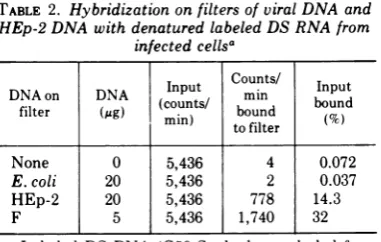
Demonstration of virus-specific DS RNA. DS RNA prepared as described above was tested for the presence of virus-specif'ic RNA sequences. In the experiment summarized in Table 2, the DS RNAwasdenatured by heating in 0.1x SSCat 115Cfor 5min,thenhybridized
toHSV-1(F), E. coli, and HEp-2 DNA fixedto
nitrocellulose f'iltersasdescribedinthe footnote
to Table 2. The data show that 32% of' the labeled RNA hybridized to HSV-1(F) DNA
bound to filters. This is probably a minimal
estimate since a test of' the fluid phase after
hybridization showed that 16%, of' the labeled
RNA in solutionwasresistant todigestionin 2x SSC with 50,gofRNaseApermlfor 30minat 37 C. The DS RNA formed during the
incuba-708
on November 10, 2019 by guest
http://jvi.asm.org/
TABLE 1. Purification of DS RNA from HSV-1-infected cells
3H-labeled RNA resistant todigestion
Preparation tested Nuclease Salt with nucleases (%)
Native
Dens-tureda
Initial RNA RNase Al 2x SSC 2
Afterself-annealing (lst digestion) RNaseAb 2x SSC 5-10
AfterRNaseA +T,+ DNase (2nddigestion) RNase A' 2x SSC 60-70 AfterG50 Sephadex chromatography
Included material RNase Al 2x SSC 27.5 3.7
Excluded material RNase A" 2x SSC 90-94 1.8
RNaseA" 0ix SSC 1.7
RNase A + Tc 2x SSC 93
DNased RSBe
Followedby RNases 2xSSC 87 3
aDenatured by heating at 115 C for5min,thencooled quickly in dry ice.
b50ug ofRNase A per ml, 30 minat 37C.
c50ggof RNase Aand10U of RNase T l perml,30minat37C.
d50
sg
ofribonuclease-free DNase per ml, 1 hat 37C.eRSB, Reticulocyte standard buffer.
TABLE 2. Hybridization on filters of viral DNA and HEp-2 DNA with denatured labeled DS RNA from
infectedcellsa
Input
Counts/
InputDNAon DNA (cut/ min bon
filter (Mg)
(coun
ts boundIto
filter (%None 0 5,436 4 0.072
E. coli 20 5,436 2 0.037
HEp-2 20 5,436 778 14.3
F 5 5,436 1,740 32
aLabeled DS RNA (G50Sephadex-excluded frac-tion) prepared from infected cells as described in Materials and Methodswasdenatured byheatingat 115C in 0.lx SSC for 5 min and chilled in dry ice. HSV-1(F), HEp-2, and E. coli DNAs bound tofilters wereprepared asdescribed and incubated together for 20hat66C in200Mlofhybridizationbuffer contain-ing, inaddition to the denatured RNA, 0.75 MNaCl, 5mMEDTA, 0.2% sodium dodecyl sulfate and0.01M Tris (pH 7.5). The filters werewashed, digested with RNase A (50 ug/ml, 2h at roomtemperature in 2x
SSC), washed again, and dried. Radioactivity was measured in aPackard scintillationspectrometer.
tionwasthereforeunavailablefor
hybridization
toeither host orviral DNA onfilters.Thermaldenaturationprofilesof DS RNA. Thermal denaturation profilesofDS RNA
puri-fied from infected and uninfected cells were
based on susceptibilityto RNaseA in 2x SSC.
Specifically, the G50-excluded fraction of DS
RNA prepared from infected cells as described
above and inthe footnotes toTable1, aswell as fromuninfected cells pulse labeled for the same
lengthoftime, were heated at various
tempera-tures for 5-min intervals. Samples taken after
heating at various temperatures were quickly chilled indryice, made2x SSC bytheaddition
of concentrated solution, and digested with RNase A (50
Ag/ml,
30 min at 37 C). Corre-sponding DS RNA heated, but untreated with RNase, served as a control. Figure 1 showsthe fraction of total labeled DS RNAs depolymer-ized by RNase A as a function of thetempera-tureofdenaturation. The thermal denaturation
of virus-specific sequence in the infected cell DNA were analyzed in a similar fashion, but in
this instance the denaturation was monitored
by measuringthe ability of the heated RNA to
hybridizeto viral DNA fixed to filters. Specifi-cally, samples heated to various temperatures
as above were quickly chilled in dry ice and
hybridized to HSV-1(F) and E. coli DNAs bound to filters. The hybridization conditions
were as thosedescribed in the footnotetoTable
2. Figure 1 shows the fraction of the
virus-specificsequences madeavailablefor
hybridiza-tionto viral DNA as a function oftemperature
of denaturation. The data were corrected for
nonspecificbindingtofiltersbysubtracting the
counts bound to E. coli DNA and normalized with respect to the maximum amount ofRNA
hybridizing to DNA on filters observed after
on November 10, 2019 by guest
http://jvi.asm.org/
[image:3.507.54.454.62.289.2] [image:3.507.59.250.345.466.2]1W 1
,20
70tlo
0 G0
30 70
30
60
0 N infectedcell0in 00.
tvial DN=ieofles() otosicue
30A heatd toidcatlluNAtitepeaurs
digestedwith RNAse
20 2
A o
o 10
10 ~~~~~~~~~~~~without
The DRmInfectedcell DS-RNA digestions digested withINAse
30 40 so 60 10 80 90 190 P0
Temperature
~TC
FIG. 1. Thermal transition of self-annealed
3H-labeledRNAfrom HSV- -infected cellsin o.nxSSC.
Thermal transition was measured by sensitivity to
RNaseA (Aand0) in 2x SSCand by hybridization
to viral DNA fixed tofilters(R). Controls included RNA heated to indicated temperatures but not
di-gested bynucleases.
denaturation of the DS RNAat115 C for 5mi. The DS RNA from infected cells shows three thermal
transitions, i.e.,
one between 20 and 40C,
probably resulting
frompoorly
matched basepairing,
oneatapproximately
78C,
corre-sponding
tothat ofuninfectedcells,
andone atapproximately
100 C. Thevirus-specific
se-quences show one thermal transition with a
Tm,
ofapproximately
100C(Fig.
1).Estimation of the fraction of viral DNA
giving
rise to DS RNA. In thepreceding
sections, we demonstrated that infected cells contain
virus-specifilc
DS RNA. The purposeof' theseexperimentswastodetermine the fractionof the DNA from which the DS RNA arose.
Unlike the
preceding
section, in which we usedlabeled DS RNA and
hybridized
it to DNA onf'ilters,
in theexperiments
described below wehybridized
insolutionexcess unlabeled RNAto trace amountsoflabeled DNA. Thistechnique,
described in detail
by
Frenkel and Roizman (6),allows direct estimate of' the amount of' DNA which served as a
template
to the RNA with which ithybridizes.
DS RNA was
prepared
from RNA extracted f'rom 8-h infected cells as describedabove,
but since the RNA wasnot labeled thepurification
was doneasfollows. The RNAwasdivided into
twounequal portions.Tothe smallportionwere
added smallvolumes of labeled viral DNA and RNA from 8-h infected cells. The twoportions
were then processed asdescribed above except that RNAwas diluted forRNase digestion and
chromatographed on G50 Sephadex. The small portion served as acheck for thecompleteness
ofdigestion and purity of the DS RNA in the larger, unlabeled RNA portion. The
hybridiza-tion tests werethen done with the unlabeled DS RNA excluded from the G50Sephadex
column)
asdescribed above. The results, summarized in Fig. 2A, show the following.(i) Undenatured DS RNA hybridizes with at
most 5% viral DNA. This could represent
con-taminating single-stranded viral RNA, or DS RNA withpoorly matched base pairs, such that could arise from intrastrand base pairing of short segments.
(ii) Denatured DS RNA drove 55% of viral DNAintoDNA-RNA hybrid. The labeled DNA hybridized in the presence of the same concen-tration ofuninfected cell RNA reassociated to
only 1%during the 8 h of hybridizationrequired
for the maximum Rot values reached in this test.
(iii) To test thepossibilitythatthe DNA was driven into DNA-DNAhybrid by traceamounts
of viral DNA contaminating the preparation, DS RNA at a concentration of 500,ug/ml was denatured at 115 C for 5 min, chilled, mixed with DNase-free RNase (50
4ig/ml),
and incu-bated for 2 h at 37C. After digestion labeled viralDNA was added, the concentration of salt wasincreased to 0.3 M final concentration, and the mixture wasallowed tohybridize totheRot value shown in Fig. 2. The amount of DNA in hybrid was found to be approximately 1%, i.e., the same asthatreassociatinginthe absence of' viral RNA.(iv) The hybridization kinetics ofdenatured DS RNA with labeled viral DNAwereanalyzed
by twodifferent methods todetermine whether all viral sequences inDS RNA were present at the same molar concentration. In the first, we applied the analytical treatmentofFrenkel and Roizman (6). Specifically, if a, ...
a,n
is the f'raction of DNA serving as template for n classesofRNA, i.e.,R,
... Rndiffering in abun-dance expressed in molesofRNA perliter, K is the hybridization rate constant, andDJDo
isthe fraction of'DNA remaining singlestranded, then the hybridization of excess nuclear RNA with trace amounts of labeled DNA, under conditions in which the reassociationofDNAis
insignificant, is described by the following equation (6).
on November 10, 2019 by guest
http://jvi.asm.org/
[image:4.507.63.252.57.310.2]10
20
30
Rot
(
molesnucleotides-sec
Iiter-I
)
FIG. 2. Hybridization ofexcessDSRNAtoinvitrolabeled, sheared, and denatured viral DNA. Symbols:0,
Reassociationof viral DNA in thepresenceof undenaturedDS RNA; *, denatured DS RNA;V,reassociation
of viral DNA in thepresenceof uninfected cell RNAduring thesametimeintervalasthe experimentalpoints;
0,reassociationof viral DNA in thepresenceofdenaturedDS RNA depolymerized by RNase.
D,/D.
Fraction of DNAremaining single stranded. The basis for the plots in A and Bisstated in thetext.-= aiIe
Do
+ ane-KRnt+
1-(al
...a)The values a...an and the ratios of R1 to
R2...Rn were estimated by determining the
best fit of a nonlinear regression of
DJDo
against tforn= 1,2, 3,etc.Analysis of the data
by this method shown inFig. 2Aindicatesthat
viralDS RNA consisted ofatleasttwo
compo-nents. The abundant component arose from
28.7% of viral DNA and was 40 times more
abundant than the scarce component comple-mentaryto26.0% ofviral DNA. Onecritiqueof
the application of this anslysis is that the
equation is based on the assumption that the
concentration ofsingle-stranded RNA remains unchanged during hybridization, i.e., that the concentration ofRNA in hybrid is small
com-pared to that of the single-stranded RNA left. Since the RNA sequences are derived from denatured DS RNA, which probably reassoci-ates during the hybridization, the validity of thisassumption is in doubt.
In the second analytical procedure, we as-sumed that the hybridization of labeled viral
DNA serves as an indicator and reflects the reassociation ofunlabeledsymmetricviralRNA
sequences. The relation which best describes
this situation, again under conditions in which the reassociation of DNA is insignificant, is a
1.00
.90
.80
a
.70
0
.60
.50
.40
2.2
711
on November 10, 2019 by guest
http://jvi.asm.org/
[image:5.507.105.388.60.451.2]ROIZMAN
modificationofBritten's (4)second-order equa-tion for the reassociation ofDNA.
Dt 1I or Do = I +KRot
Do I + KRot
Dt
The equation predicts that if all viral RNA sequences present in DS RNA were equimolar in concentration, a plot of'
Do/D,
against Rotwould yield astraight line with an intercept of' 1.This isobviouslynotthecase. Theplotof'the data shown in Fig. 2B indicates that the DS
RNA contains at least two components, of' which the most abundant hybridizes with ap-proximately 32% ofDNA. Applicationof
mathe-matical analyses to bedealt with elsewhere (N Frenkel, B. Cox and B.Roizman, manuscript in
preparation) indicate that this component is
70-foldmoreabundantthan thescarcespecies.
DISCUSSION
The salient features and significance of the
results described in this paper maybe
summa-rized as follows.
(i) Infected cellsaccumulateRNA capableof'
self-annealing. Analysesof several properties of'
the productofself-annealing, notablyits resist-ance to depolymerization by nucleases in high saltand sensitivitytothesenucleases in low salt
and upon heat denaturation, indicate that it conforms withpropertiesof DS RNA(3).
(ii) Excess unlabeled, denatured DS RNA drove slightly more than 50% of labeled viral
DNA into DNA-RNA hybrid under conditions
in which the amounts of DNA-DNA hybrid formed were insignificant. Moreover, analyses of'the hybridization kinetics indicated thatthe viralRNAsequences werenonhomogeneous and
consistedofatleasttwocomponentsdifferingin molar concentration.
(iii) The abundant class of symmetric
tran-scripts isofparticular interest from twopoints
of' view. First, the measurements of the Tm of the viral DS RNA were done by heating
labeled DS RNA andhybridizing ittoDNAon
filters. In principle, hybridization of labeled
RNA in solution to DNA fixed on filters
mea-suresprimarily abundant RNA species and itis
likely, therefore, that the Tm measurements
apply to the abundant symmetric transcripts
which arise from at least 28 to 32% of viral DNA. Application of the equation of Billeter
et al. (3), within the range of values to which
the equation is applicable and assuming that
the linear relationship between base
com-positionat melting point observedforotherDS
RNAs can be extrapolated to HSV-1 RNA as
well, predicts that the guanine plus cysteine
content of HSV-1 DS RNA is only
slightly
higher
than the average guanine pluscysteine
content of HSV-1 DNA
(Fig. 3).
Second, the estimate that the abundant DS RNA arisesfrom at least 29'. of'viral DNA issignificant in reference to the observation (11) that
prean-nealed nuclear RNA lost the capacity to drive 15% of DNA into DNA-RNA
hybrid
but that denaturation of' the preannealed RNAfairly
restored the ability of the RNA to drive the DNA into
hybrid.
There were twopossible
explanations for this observation (11). One
postulated that complementary transcripts
arose from 15'7c of' the DNA; i.e., sequences
derived from 7.5%of' each DNA strand accumu-late at nearly equimolar concentrations in the infected cell nuclei. The alternative was that
complementary RNA sequences were derived from 15% of' each DNA strand, but the
tran-scripts arising
fromonestrandwerepresent ata lower concentration than the correspondingRNA sequences derived from the opposite
strand. Preannealing would sequester the less abundant RNA in an RNA-RNA hybrid, but
the complementary sequences would remain available forhybridization withDNA,
although
their concentration would have been reduced. The data presented in this paper appear to
discriminate between the two hypotheses and
support the second. They would indicate that the transcripts annealingto makethe DS RNA do notaccumulate in equimolar concentrations in the infected cells.
(iv) We have littleinformationon thenature
80
70
CL
'w60
50
o40
+ 30
c.2
50 60 70 80 90 100 110
Thermal transition °C 120
FIG. 3. Estimation of the base composition of HSV-1 DS RNA from its thermal transition. The thermal transition of HSV-1 DNA in 1x SSC was
calculated fromtheequation of Billeteretal. (3)and plotted onthe linederivedfrom the linear regression of the relationship between base composition and thermal transition in x SSC for DS RNA ofother viruses (3). PV, Polyomavirus; EMC, enceph-alomyocarditisvirus.
HSV-1 OS-RNA -J HSV-1 DNA
MS2 RNA
EMC RNA . PV DS-RNA Reovirus RNA SV40 DS-RNA
Wound tumorvirusRNA
118.5 J VIROL
on November 10, 2019 by guest
http://jvi.asm.org/
[image:6.507.270.459.434.583.2]of the scarce DS RNA species. We cannot
exclude the possibility that this RNA arises by intrastrand base pairing even though the RNA is excluded from G50 Sephadex columns, is
stable to prolonged incubation at 20C below the Tm ofDS RNA, and shares with the abun-dant species the ability to resist digestion by RNasein2x SSC.
(v) The accumulation of symmetric
tran-scripts arising from at least 29% and possibly from as much as55%ofthe DNAsuggeststhat
transcription of HSV-1 DNA is largely
symmet-rical and implies the existence of a
post-tran-scriptionalmechanism operating in the nucleus for discrimination of transcripts giving rise to
mRNA from those derived from the opposite strand of DNA. Thisconclusion is supported by theobservation that onlytraceamountsof viral
RNAsequences accumulating in the cytoplasm
become unavailable tohybridization with viral DNA upon self-annealing (11) and that viral
RNA sequences which are not translated in polyribosomes of infected cells are selectively
retained in the nucleus (10).
ACKNOWLEDGMENTS
These studiesweredoneunder theauspicesofthe
Univer-sityofChicagoCancer Center(CA 14599)andwereaidedby grantsfromtheAmerican CancerSociety (VC 103J)and the
NationalScience Foundation(GB 38270)andPublic Health ServicegrantCA-08494 from the National CancerInstitute. J.B.was afellow of the International Agencyfor Research
against Cancer, Lyon,France.
LITERATURECITED
1. Aloni, Y. 1972. Extensive symmetrical transcription of simian virus 40 DNA invirus-yieldingcells. Proc. Nat.
Acad.Sci. U.S.A. 69:2404-2409.
2. Aloni, Y., and H. Locker. 1973. Symmetrical in vitro
transcription ofpolyomaDNA and theseparation of self complementary viral and cell RNA. Virology 54:495-505.
3. Billeter, M. A., C. Weismann, and R. C. Warner. 1966.
Replicationofviral ribonucleicacid. IX. Propertiesof
double-stranded RNA from Escherichia coli infected
withbacteriophageMS2.J. Mol. Biol. 17:145-173. 4. Britten, R. J. 1969. The arithmetic of nucleic acid
reassociation, p. 332-335. Carnegie Inst. Washington
Yearb.,1967-68.
5. Colby, C.. and P. H. Duesberg. 1969. Double-stranded RNAinvaccinia virus infected cells. Nature (London)
222:940-944.
6. Frenkel, N., and B. Roizman. 1972. Ribonucleic acid synthesisin cells infected with herpes simplex virus. Controlsoftranscription andofRNAabundance. Proc.
Nat. Acad. Sci. U.S.A. 69:2654-2658.
7. Frenkel, N., B. Roizman, E. Cassai, and A. Nahmias. 1972. A herpes simplex 2 DNA fragment and its
transcription in human cervical cancer tissue. Proc.
Nat. Acad. Sci. U.S.A. 69:3784-3789.
8. Kieff,E.D., S.L.Bachenheimer, andB.Roizman.1971.
Size, composition and structure of theDNA of sub-types 1 and 2 of herpes simplex viruses. J. Virol.
8:125-132.
9. Kieff, E. D., B. Hoyer, S. L. Bachenheimer, and B.
Roizman.1972.Genetic relatedness oftype1andtype2
herpes simplex viruses.J. Virol. 9:738-745.
10. Kozak, M., and B.Roizman. 1974.Regulationof herpes-virus macromolecular synthesis: nuclear retention of
non-translatedviralRNAsequences.Proc. Nat.Acad.
Sci. U.S.A. 71:4322-4326.
11. Kozak, M.,andB. Roizman. 1974.RNAsynthesisincells
infected with herpes simplex virus. IX. Evidencefor
accumulation of abundant symmetric transcripts in
nuclei. J. Virol. 15:36-41.
12. Lucas, J.J., and H. S. Ginsberg. 1972. Identificationof
double-stranded virusspecificribonucleic acid in KB cells infected with type 2 adenovirus. Biochem.
Bio-phys.Res.Commun. 49:39-44.
13. Marmur, J. 1963. Aprocedurefor the isolation of deoxyri-bonucleic acid from microorganisms. Methods
Enzy-mol.6:726-738.
14. Rabin,E.Z.,B.Preiss,and M. J.Frajer.1971. A nuclease from Neurospora crassa conidia specific for
single-strandednucleicacids.Prep. Biochem. 1:283. 15. Roizman,B., and P.Spear. 1968.Preparationofherpes
simplexvirus ofhightiter.J. Virol. 2:83-84. 16. Silverstein, S., S. L.Bachenheimer,N. Frenkel,and B.
Roizman. 1973. The relationship between post tran-scriptional adenylationofherpesvirusRNAandmRNA abundance. Proc. Nat. Acad. Sci. U.S.A. 70:2101-2104.