0022-538X/82/120782-12$02.00/0
Copyright© 1982,AmericanSocietyforMicrobiology
Molecular Cloning of DNA
Complementary to mRNA of the
Baculovirus
Autographa
californica
Nuclear
Polyhedrosis
Virus: Location and Gene Products of RNA Transcripts
Found Late in
Infectiont
MICHAEL J.ADANGANDLOIS K. MILLER*
Departmentof Bacteriology and Biochemistry, Universityof Idaho, Moscow, Idaho 83843 Received 15 April 1982/Accepted 13 August1982
DNAs
complementary
tolate Autographa
californica nuclear polyhedrosis
virus
(AcNPV) mRNA weresynthesized
by reversetranscription and cloned in
Escherichia coli by
using
pBR322 as a vector.Eleven
different cDNAs
weredistinguished in
ourscreening of 45 AcNPV-homologous clones. Location of the
regions of cDNA homology with
respect to theAcNPVphysical
mapshowed thatthe
11cDNAs
weredispersed throughout the
genome.The
mostabundant cDNA
insertion, representing approximately one-third of
the lateviral
mRNAs, washomologous
tothe
AcNPVHindIII-P,Q
andEcoRI-Pfragments.
Thedirection of
transcription in this region
wasfrom left
toright
on alinearized
AcNPVphysical
map.
Hybridization selection followed by in vitro
translation showed
thatthis
region encoded
a7,200-dalton
(7.2K)protein
which comigrated with
aminor
protein found in the extracellular nonoccluded form of the virus (NOV). Similarly,
the
genefor polyhedrin, the major structural protein of the occluded virus form,
was
located,
at leastin
part,in
theHindIII-V/EcoRI-I
region of
the AcNPV map.The
polyhedrin transcript represented approximately
one-quarterof the viral
polyadenylic acid-containing
RNAs
at27
h
postinfection.
Another
relatively
abundant cDNA
washomologous
tothe
HindIII-AIEcoRI-CISstI-G
region, and
RNA
selected
by
this cDNA directed the synthesis of
twoproteins (31
Kand 30K).
The
protein products of five
othercDNA-selected RNAs
wereidentified. The
HindIII-D/EcoRI-O,
HindIII-CIEcoRI-D,
HindIII-B1IEcoRI-E,
and
HindIII-B2/
EcoRI-H
regions of the AcNPV L-1
genome werehomologous
toRNAs
which
directed
thesynthesis of
a57K
protein,
a25Kprotein,
a61Kprotein,
and
a37K
protein (plus
aminor
26Kprotein), respectively. Late
mRNAselected
by
acDNA
homologous
tothe
HindIII-P/EcoRI-B
region of the AcNPV
mapdirected the
synthesis of 31K and 30K
proteins which comigrated with the 31K and 30K
proteins translated from
RNAselected
by the
HindIII-AIEcoRI-CISstI-G
cDNA.
Three other cDNAs have
notbeen correlated
yetwith
specific protein
products.
One of the
mostchallenging
areasof
currentbaculovirology
isdetermining
thegeneorganiza-tion of
the128-kilobase
(kb)
circular,
double-stranded
DNA genomeof
the modelbaculo-virus, Autographa californica
nuclear
polyhedrosis
virus
(AcNPV). One approach
todetermining
geneorganization
is marker rescuein
which
genetic mutations
are located withrespect to the AcNPV
restriction
fragment
phys-ical
mapby
cotransfection of
mutantDNAandawild-type
DNArestriction fragment
(12).Anoth-er
approach
is to isolatespecific fragments
ofAcNPV DNA,
select
specific
mRNAsby
hy-tResearchpaper8256 oftheIdahoAgriculturalExperiment
Station.
bridization
tothe DNA
fragment,
and
determine
the
protein
encoded in thenucleic acid
se-quences
by
invitro translation of
thehybrid-selected
RNAfollowed
by gel
electrophoresis
of
the
resulting
protein products.
Using
twoAcNPV
genomic
fragments
ashybridization
probes,
Vlak et al. havemapped 33,000-dalton
(33K)
and 39Kproteins
to theEcoRI-I
andEcoRI-J
fragments,
respectively (23).
Recent advances in
recombinant
DNAtech-nology
haveprovided methods for
cloning
thecoding
regions
of individual virus
genesby
syn-thesizing
DNAcomplementary
tomRNA,
using
reverse
transcriptase.
The useof cDNA clones
as
hybridization
probes
for
specific
mRNAshas
distinct
advantages
overtheuseof viral
genomic
782
on November 10, 2019 by guest
http://jvi.asm.org/
VOL. 44, 1982
fragments
orfragment clones. Particularly
ad-vantageous
is that each cDNA selects only
asingle
mRNA or aseries
of relatedspliced
orsymmetrically transcribed
RNAs.In this
paper we reportthe
successfulsynthe-sis and cloning of DNA complementary
to lateAcNPV mRNA.
Using cDNA clones,
wegath-ered information concerning
the relativeamounts
of various mRNAs
found late ininfec-tion, the locations of these
mRNAs with respectto
the
physical
mapof AcNPV
L-1(2, 14), and,
in
some cases, the identities of theproteins
which the mRNAs
encode. The physical
maplocation of the 3' portion of the
geneencoding
polyhedrin,
themajor protein
of the occludedform of
AcNPV,
was determined. Informationconcerning
the geneorganization
of AcNPVshould be
applicable
toboth thedevelopment
ofthis virus as
avector
forgenetic
engineering (13)
and the
furtherdevelopment
of the virus as amicrobial pesticide (20).
MATERIALS ANDMETHODS
Preparation of RNA from infectedcells.Monolayers ofSpodoptera frugiperda IPLB-SF-21 cells were in-fected with AcNPV L-1 (9) at amultiplicity of infec-tion of 20. After rocking for 1 h at room temperature, the inoculum was replaced with TC-100 medium, and the cells were incubated at27°Cfor 27 h. The cells weresuspended, pelleted bycentrifugation at 2,000 x gand 5°C for 10 min, and washed with phosphate-saline buffer (9). The cells were suspended in
phos-phate-salinebuffer(9)andlysedonice for 10 min after
0.75% Nonidet P-40 (BDH Chemicals) was added.
Nuclei were removedbycentrifugation at 4,000 x g and5°Cfor 10 min. An equal volume oflysissolution (6)wasaddedtothesupernatant,and the mixture was extractedtwice with phenol (Fluka)-chloroform-isoa-myl alcohol (50:50:1) containing 0.1% 8-hydroxyquin-oline (6). The RNA was ethanol precipitated by adding 0.1 volume of3 M sodium acetate (pH 5.6) and 2.5 volumes of ethanol and placing the solution at -20°C
overnight.Aftercentrifugation at 12,000 xg and 4°C for 45 min, the RNA pellet was dissolved in equal parts ofphosphate-saline buffer (9) and
lysis
solution (6),phenol extracted, and ethanol precipitated as de-scribed above.
Polyadenylic acid
[poly(A)]-containing
RNA was isolated by using oligodeoxythymidylic acid cellulose (type 7; P.L. Biochemicals) and a batch adaptation of theprocedure of Manley et al. (10).Briefly,500 ,ug of cytolasmic RNA in 1 ml of binding buffer (10 mM Tris, pH 7.6, 0.5 M NaCl, 0.2% sodium dodecyl sulfate[SDS]) wasadded to 50 mg ofoligodeoxythymidylic acid cellulose (P.L. Biochemicals) in a microfuge tube and mixedgentlyfor90minatroomtemperature. This wasfollowed by centrifugation at 12,000xg for 3min and thenby three washes withbinding buffer; poly(A)-containingRNAwasremoved by washing with 1 ml of elutionbuffer(5mMTris, pH 7.6, 2 mM EDTA) for 15 min. Elutionwasrepeated with a second1-mivolume of elution buffer, and the two eluates were pooled. Fine cellulose particles in the resulting preparation
cDNA CLONES OF LATE AcNPV mRNA 783 wereremoved bypassing themixturethrough silicon-izedglasswool inaPasteurpipette,and the RNAwas ethanolprecipitated.
Synthesis of cDNAfrom late mRNA. Double-strand-ed cDNAwaspreparedfrompoly(A)-containingRNA by the procedure of Wickens et al. (24). The 100-pl
reaction mixture contained poly(A)-containing RNA derived from 500p,g ofcytoplasmic RNA, 10 ,ug of
oligodeoxythymidylic
acid12_18
(P.L. Biochemicals),andeach deoxynucleoside triphosphateat a concen-tration of 500 ,M,including[a-32P]dCTP (2Ci/mmol;
New England Nuclear Corp.) in 50 mM
Tris-hydro-chloride (pH8.3)-140mMKCl-30 mM
2-mercaptoeth-anol-10 mM MgCl2. Reaction mixtures were assem-bled onice, 50 U ofreverse transcriptase (J. Beard,
Life Sciences, Inc.) was added, and the reaction mixtures weremixed, centrifuged briefly,andplaced
at42°C for 60 min. For second-strand synthesis, the first-strand reaction mixture was heated for 3 minat 100°C, cooled in an ice water bath, centrifuged for severalseconds,andtransferredto asolution
consist-ing of 50 Ill of each deoxynucleoside triphosphate
(concentration, 1 mM) and 50 ,ul of 400 mMHEPES
(N-2-hydroxyethylpiperazine-N'-2-ethanesulfonic acid) (pH 6.9). Then 20 U of Escherichia coli DNA
polymeraseIwasadded,and thereaction mixturewas incubated for 2 hat15°C.
Reactions were stopped by adding2.5 mM dGTP, 0.03% SDS,and 18
pg
of E. coli soluble RNA(Calbio-chem) andextractingwithphenoltwice. The aqueous phasewas passedthrough aSephadex G-50 column,
and theexcluded volumewasethanolprecipitated.
The hairpin loop in the cDNA was cut by S1
nuclease digestion in a solution containing 250 mM sodium chloride, 30 mMsodium acetate, 1 mM zinc
sulfate,and5%glycerol (pH 4.6).Afterdigestionwith 125 UofS1 nuclease(SigmaChemicalCo.)for 60 min at 37°C, the mixture was phenol extracted, ether
extracted, and ethanol precipitated. The final yield
was 400 ng of double-stranded cDNA.
Construction ofhybrid plasmids.Homopolymer
tail-ing of the cDNA was accomplished by using the methodof Roychoudury andWu(17). We used 50 ng of cDNA and 25 U of terminal transferase (Bethesda
ResearchLaboratories, Inc.)in asolutioncontaining
100 mMpotassium cacodylate, 1 mMCoC12,200p,M
dithiothreitol, and 10 FM dCTP (pH 7.0). The reaction mixture was incubated for 30 min at 37°C, and the DNA was ethanol precipitated. Purified plasmid pBR322 was digested withPstI (Bethesda Research
Laboratories, Inc.) phenol extracted,etherextracted,
and ethanol precipitated. Approximately 1
p.g
was tailed withdeoxyguanine by using10,uM dGTP and60 U of terminal transferase. The guanine-tailed vector DNA wasthen ethanolprecipitatedanddissolvedin 1 ml of 10 mM Tris-hydrochloride (pH 8)-10 mM EDTA-100 mMNaCl. ThevectorDNAwasaddedto 50ng ofcytosine-tailedcDNAin10pl of10 mMTris (pH 7.6)-l mM EDTA. The mixture of DNAs was heated for10minin a65°C water bath. Then the bath wasshutoff and allowed to cool to room temperatureovernight for annealing of the guanine and cytosine tails of the DNAs. E. coli soluble RNA(20p,g) was added, andthe DNA wasprecipitated. This chimeric DNA was used to transform E. coli RR1 (7), and colonies were selected by resistance to tetracycline andsensitivitytoampicillin.Pl physical containment
on November 10, 2019 by guest
http://jvi.asm.org/
was used, as required by the National Institutes of
Healthguidelines for recombinant DNA research. Screening recombinantplasmids. Bacterial colonies with theappropriate drugresponseswere grown
over-nightonLB agar(1%tryptone [Difco Laboratories], 1% NaCl, 0.5%yeastextract,0.1% glucose) overlaid with nitrocellulose filters (type BA85; Schleicher &
ScheullCo.), and each filterwastransferredtoanLB agarplatecontaining 170 ,gofchloramphenicolperml andincubated overnight. The filters were processed onpads of saturatedfilterpaper asdescribed previous-ly (3), with afinal soakonafilterpadsaturated with 2xSSC(1x SSC is 0.15 M NaCl plus 0.015 M sodium citrate). Excess liquidwas removed by suction, and
each nitrocellulose filterwasplacedona95% ethanol pad, dried by suction, and bakedinavacuum ovenat 80°C for 2 h. Colonies containingAcNPV insertions
were recognized by hybridization with an AcNPV DNAprobe labeled with 32Pby nick translation (see
below).
Labelingof DNA by nicktranslation. AcNPV DNA or plasmid DNA was labeled in vitro by a nick-translationprocedure (16), using25,uCiof[k32P]dCTP
(800 Ci/mmolin0.01MTricine, pH 7.6; New England NuclearCorp.). After2 hat15°C, the25-ptl reaction volume was brought to200 pulwith 10 mMTris(pH 7.6)-i mM EDTA and phenol extracted twice. Calf thymus DNA(100 ,ug)wasadded, and theDNAwas
ethanolprecipitated by placingthepreparationindry ice for20min. Twomoreethanolprecipitations were
done to remove unincorporated dCTP before use in hybridization experiments.
DNAhybridization conditions.Filterswere preincu-bated in a solution containing 0.02% bovine serum albumin and0.02% Ficoll 400 in 3x SSC in boilable bagsat65°Cfor 4 h. This solutionwasreplacedwitha
hybridization solution containing 50% formamide (Fluka),SxSSC,10 mM HEPES(pH 6.9),0.1%SDS, 1 mM EDTA, 0.02% bovine serum albumin, and 0.02% Ficoll 400. Probe DNA in 5x SSCwas dena-tured at 100°C for 10 min and added to each bag. Incubationwasfor 20 hat37°C withgentle shaking. Filterswerewashed three times(45mineachtime)in
Sx SSC containing0.2% SDS at65°C, andthis was
followed bya2-h wash in2x SSCatroom tempera-ture. The filters were then exposed to preflashed
KodakX-rayfilm for differenttimes at -70°C, using CronexLightningPlus intensifyingscreens.
Electrophoresisof DNA. AcNPVDNAwasdigested witheitherHindIII, EcoRI,orSstI according tothe directions of the manufacturer (Bethesda Research Laboratories, Inc.). Electrophoresiswascarried out at 75 mA for 20 h in 0.7%agarosegels submergedin 40 mM Tris-acetate(pH 7.8)-5mMsodiumacetate-1mM EDTA(TAE buffer) (21) containing0.5jigofethidium bromide perml. DNA wastransferred from agarose
gelstonitrocellulose(aSouthernblotmethod) bythe bidirectional transfertechnique (19).
Recombinant cDNAplasmid DNA(2 ,ug) was di-gestedwith PstIand subjected to electrophoresis at
120 mA for 15 hthrough1.4%agaroseinTAE buffer supplementedwith0.5,ugof ethidiumbromideperml. Hybridization selection of mRNA. Specific RNAs were selected from amixture ofcytoplasmic RNAs isolated from AcNPV-infectedS.frugiperda cellsby hybridizationtorecombinantplasmidcDNAboundto
nitrocellulose, using a composite of the procedures
described by Ricciardi et al. (15) and Vlaket al. (23). Recombinant plasmid DNAs (50 ,g) in 10 mM Tris
(pH
7.6)-i
mMEDTA wereboiled for 30 min, broughtto 5x SSC, and passed slowly through nitrocellulose filters (diameter, 24 mm) prewet with 5x SSC. The DNA-containing filters were dried and then baked in vacuo at 80°C for 2 h. Thefilters were cut into small pieces and presoaked in hybridization buffer contain-ing 50% formamide, 600 mM NaCl, 50 mM
PIPES
[piperazine-N,N'-bis(2-ethanesulfonic
acid)] (pH 6.8),4mMEDTA, and0.5% SDS supplemented with 50 ,g ofpoly(A)(Sigma Chemical Co.) per ml for 1 to 2 h at
37°C. Thehybridization buffer was removed and re-placed with 200
Rg
of cytoplasmic RNA in 200RI
of hybridizationbuffer. Hybridizationwas for 8 h at37°Cand was followed by washing and elution (23); 0.1 volume of20opotassium acetate and 25 volumes of ethanol were added to the
eluted
RNA, and the preparation was stored at -20°C. The filters were regenerated by soaking for 20 min in 0.1 N NaOH in 2x SSC,followed by washing five times with 2x SSC anddryingin a vacuumdesiccator for 2 h (11).In vitro translation of RNA. Cytoplasmic RNA, which was stored as a precipitate in sodium acetate-ethanol at -20°C, was centrifuged, and the resulting pellet was dissolved in 2% potassium acetate and precipitated with ethanol. For cell-free translations, RNAs werecentrifuged, washed in70%ethanol, dis-solved in water, andlyophilized. Therabbit reticulo-cyte lysate system of Bethesda Research
Labora-tories,Inc. was usedaccording to the recommended protocol.
L-[3,4,5-3H]leucine
(110Ci/mmol; New En-gland Nuclear Corp.), which was supplied in 0.01 NHCI,wasneutralized with 0.10 volume of 0.1 N KOH before use. Each 30-,u assay mixture contained 4 ,Ci
of
[3H]leucine
andfinal concentrations of 154 mM K+and 1.2 mM
Mg2+.
Reaction mixtures were incubated at30°Cfor 60 min.SDSgelelectrophoresis. Theelectrophoretic proce-dure of Laemmli (8) was slightly modified for our analysis of proteins. The modifications included the useof 0.75 M Tris (pH 8.8) instead of 0.375 M Tris (pH 8.8) in the separating geland the use of 0.25 M Tris (pH 6.8) instead of 0.125 M Tris (pH 6.8) in the
stacking gel. Stacking gels were 1 cm tall and con-tained 3.6% acrylamide. Separating gels contained
10%acrylamide and were 1.5 mm thick by 17 cm tall.
Electrophoresis was at 95 V for 15 h, during which time thetracking dyemigratedapproximately 16 cm. Gelswerefixed in methanol-water-acetic acid(5:5:1) for 1 h, impregnated with
En3Hance
(New England NuclearCorp.) for1 h, soaked in water for 1 h, and dried.Fluorographywasdoneby exposingpreflashed Kodak XAR film to gels at -70°C, using Du Pont Cronexintensifyingscreens.For the analysis of proteins having molecular weights less than 14,000, we usedaprocedure adapted fromShapiroetal.(18)by Bethesda Research Labora-tories,Inc.Theresolving gel (17cmby1.5mm) of15%
polyacrylamide(ratioofbisacrylamidetoacrylamide,
0.8:30) contained0.1 M sodiumphosphate
(pH
7.2),0.1% SDS, and 6 M urea. The uppergel contained
3.5%
acrylamide in the same bufferas theresolving gel. Only 2 mm of upper gelwas between the well bottomsandresolving gel.Therunningbufferwas0.1 Msodiumphosphate (pH 7.2) supplementedwith0.1% SDS andwasrecirculatedduringelectrophoresis.on November 10, 2019 by guest
http://jvi.asm.org/
VOL. 44, 1982
Protein samples were brought to 10 mM sodium phosphate (pH 7.2), 7 M urea, 1%SDS,1%
2-mercap-toethanol, and0.01% bromphenolblue. Thesamples
were heated for 2 min at 100°C before they were loaded ontothe gel.Electrophoresis was at 95Vfor 19 h, and this was followed by fixing for 1 h in 15% isopropanol-10%oacetic acid. Gelsweresoaked for 2 h in En3Hance (New England Nuclear Corp.), proc-essed, and exposed to film as described above. For protein standards the high- andlow-molecular-weight
"4C-labeled
protein standards of Bethesda Research Laboratories were used.RESULTS
Location and
frequency of cDNA
clonesof
late AcNPV RNA.DNA
complementary
tolate
AcNPV mRNA
wascloned in
E.coli,
using
pBR322
as a vector(see
above).
Starting with
500
,ugof
cytoplasmic
RNA,
weobtained 0.4
,ugof
double-stranded
cDNA, 50
ngof which
wasannealed
topBR322
via
homopolymer
tails,
and
the recombinant
DNA was used totransform E.
coli
RR1.The
yield
wasapproximately 1,400
"
<
(-ELLm.j
a:
F
>
>Qa
a)
40mmCC
m
>(L.
I Jci.L
4ca. a. a. ci ci ci c1 1 1
z E E E
2
E E2
2
E2
Z
< Qa QQQQaaCQQ O L
cDNA CLONES OF LATE AcNPV mRNA 785
ampicillin-sensitive, tetracyline-resistant colo-nies. Approximately
20%
of these colonies con-tained AcNPV sequences, as shown by colonyhybridization
when AcNPV DNA wasused
as aprobe.
A
total of 45 AcNPV-homologous
cDNAclones
werelocated on the AcNPVphysical
mapby annealing 32P-labeled, nick-translated
recom-binant
plasmid
DNAs toSouthern blots
of
HindIII, EcoRI, and SstI
enzymedigests of
AcNPV
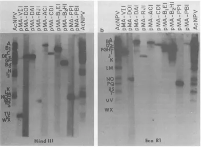
DNA.Figure
1shows
anautoradiogram
of AcNPV HindIII and EcoRI blots probed with
[32P]DNAs
isolated from 10 different cDNA
clones. These clones were designated pMA,
referring
toplasmids cloned by
oneof
us(M.J.A.),
followed by letters
todesignate the
AcNPV
HindIII, EcoRI, and, if
necessary, SstIfragments
towhich they
werehomologous. For
instance,
acDNA that
hybridized
toAcNPV
HindIII-V and EcoRI-I
wasdesignated pMA-VI.
All
hybridization locations
wereconsistent with
the
physical
mapof AcNPV
L-1 DNAshown
in
(I.0 4
-l
,> > fz C.Th z<< <
< CL, C.L 'a
b
e-2
.`%< ,4
-
t%
I I
Z
2-
22
c NL
Li<aacLcic.a
C! ICL4
Q) <
As
.._
'W v .; .:..:%'
WX'
Hind
Ill
Eco
RI
FIG. 1. Hybridization of32P-labeledAcNPV cDNAplasmids toHindIII-digested (a) orEcoRI-digested (b) AcNPVDNAblots. A 0.7% agarose slab gel containing fractionated
Hindlll-
orEcoRI-digested
AcNPV DNA wasblotted ontonitroceliulose
filter paper. Strips of theresulting blots were hybridized to32P-labeled
cDNA plasmids or32P-labeled
AcNPV DNA as a control. After hybridization, the blots wereautoradiographed. Thepositions offragments on the blots are shown on the left of each blot; the letters correspond to the recently accepted convention for AcNPV fragment designations. The first and last strips of each blot were hybridized to the entire AcNPV L-1 DNA. Other strips were hybridized to the clones designated.
on November 10, 2019 by guest
http://jvi.asm.org/
[image:4.491.54.447.326.612.2]F
VTN
D XJ LMR E OU I1111 III III I
A C WHS BS B2KOPG F
,, It I 1111 I HinD
M
I RO A J KTMN FVU C GW D QL E HSXP B
LIII I, ,, II,iI Ia I III EcoR I
C D F H E LM
I I I
A B G I JK C
I I I XhoI
B E C F GH D I A B
I I I 11 I
G
BF C A E D B
.11 IL
A D C B A
I-I
Sst
I
BamH
I
Sma
I
I I
0 10 20 30 40 50 60 70 80 90 tOO
% Genome
FIG. 2. Physicalmapof the L-1variant of AcNPV. The circularmapof AcNPVwaslinearized foreaseof presentation. The restrictionmapsitesare acombination of the sitesdetermined byMiller and Dawes for L-1
(14) and thesites determined by Cochranetal.for HR(2).Thefragment letter designationsaresimilartothose described by Cochranetal. (14),exceptthat theHindlll fragment in the40-to50-mapunitregion is designated HindIII-A and the two HindIll fragments in the 70- to 85-mapunit region are designated B1 and B2. The assignment of Bi and B2tospecific AcNPV L-1 HindIll fragments is shown in Fig. 1. Formerly (14), Bi was
designated B and B2 was designated H in the AcNPV L-1 physical map. The current designations now correspondtotheconvention recently adopted by workers in the AcNPV field.
Fig. 2. A summary of the map positions and
relativeamountsof the 45 clones is presented in Table 1. Some of the pMA-PP clones also hy-bridizedtoHindIII-Q, which adjoins HindIII-P
on the AcNPV physical map. When multiple
clones were available forasinglemapposition,
one was chosen for further study (e.g.,
pMA-VIl).
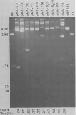
Sizes of the cDNA insertions. Figure 3 shows the sizes of the cDNA insertions obtained by PstI digestion of the recombinant DNA plas-mids. The insertion sizes ranged from 0.22 kb (for pMA-DO1) to1.5kb (for pMA-DA1).Two
of theplasmidDNAsshown inFig. 3wereonly
partially digested, and the sizes of theseDNAs
were estimated in other similar experiments.
PstIdigestions ofoneof the clones
(pMA-BlE1)
produced two small fragments in addition to
unit-length pBR322, indicatingthepresenceofa
PstI site within the insertion (inadditionto the vector PstI site[s] regenerated by the cloning procedure). Two PstI sites were located in the HindIII-B1/EcoRI-EI region ofthe AcNPV L-1
map(Fig. 2).
Protein translation of AcNPV mRNA. Cyto-plasmic RNA thatwasprepared from cells 27 h
after infection with AcNPV was translated in vitro by using a rabbit reticulocyte lysate
sys-tem. The [3H]leucine-labeled proteinswere
sep-arated on 10%o polyacrylamide gels and
autora-diographed (Fig. 4,lanes b andf0. Weobserved at least 25 peptides with molecularweights be-tween85,000and16,000andadditionalpeptides
that
migrated
atthe dye
front. Major peptides
were
observed
at63K,
42K, 35K, 32K, 31K,30K, 25K, and 18K. For
comparison,
nonoc-cluded virions
(NOV)
labeled with
[3HJleucine
in AcNPV
L-1-infected
S.
frugiperda cell
cul-tures were
purified
(9), and 21 proteins
wereresolved
on10%
SDS
gels (Fig. 4, lane a) and
15% SDS-urea
gels (see below).
[image:5.491.106.395.82.263.2]Hybridization selection
andprotein translation.
TABLE 1. Locations and abundance of cDNA clones
Clone Physicalmappositions
No.
of HindIlI EcoRI SstIclones'
pMA-VIb
V I 12pMA-DO D 0 1
pMA-DA D A 1
pMA-RJ R J 1
pMA-ACG A G G 4
pMA-ACD A C D 2
pMA-CD C D 2
pMA-BlE B1 E 1
pMA-B2H B2 H 3
pMA-PP P P 17
pMA-PB P B 1
a A total of 45 AcNPV-homologous cDNA clones werelocated on the AcNPV L-1 physicalmap.
bAll cDNAplasmidsaredesignatedpMA,
indicat-ing that one ofus (M.J.A.) constructed the clones,
and then distinguished by letters indicating their
HindlIl, EcoRI, and, in somecases, SstI map
posi-tions. For instance, the first clone listed is homologous toHindIII-VandEcoRI-Ifragments of AcNPVL-1.
J. VIROL.
on November 10, 2019 by guest
http://jvi.asm.org/
[image:5.491.253.445.464.625.2]cDNA CLONES OF LATE AcNPV mRNA 787
P- O <
--> CL 0 C
I I I I
Y a aCL Xa
4.36
2.96
.78
.35
.28
I nsert
Size
(Kb)
_ _n
IK I
I_ c-i mI
m-
cn
mL
wI
-ax CL Cl
i-
co
Nq
K) r- r- q oI N . . . vOQ Nr N ; 0 O
m
CL
(n
Q N
OD
FIG. 3. PstI fragments of AcNPV cDNA-containing plasmids. Samples (2
pug)
ofplasmid DNA were digested withPstI
and loaded into slots of a 1.4% agarose horizontal slab gel. As molecular weight markers, pBR322 wascodigestedwithHindIII,BamHI,SalI,andPstIand loaded into the outer slots. cDNA clones are indicated at the top, andinsertion sizes are indicated at the bottom in kilobase pairs (Kb). Digestion was not complete for pMA-CDI and
pMA-PB1,
and thesizes of these clones were determined in a similar experiment.RNAs
homologous
tothe
cloned
cDNA plasmidDNAs
werepurified from total
cytoplasmicRNA
isolated
from
S.
frugiperda
cells late
(27 h)in
infection by hybridization
to the plasmidcDNAs bound
tonitrocellulose.
ThecDNA-homologous RNAs
wereeluted and then
trans-lated
into
proteins in vitro.
As shown
in Table
1, 17clones
were isolatedfrom the HindIII-P/EcoRI-P region,
12 cloneswere
isolated
from theHindIII-V/EcoRI-I
re-gion,
and
4clones
wereisolated
from theHindIII-AIEcoRI-C/SstI-G
region of the
AcNPV genome.Hybrid
selections and translations
withrepresentatives of these
clones gave 10-, 4-, and3-fold
stimulation, respectively, compared
withendogenous protein synthesis
levels of the lysatesystem,
suggesting that the frequency
of isolatesfrom each region correlated with
the amounts ofVOL. 44,1982
on November 10, 2019 by guest
http://jvi.asm.org/
[image:6.491.103.395.65.506.2]%a d e
200O
93.
46-26i
z 0 CL
co 0
0
[image:7.491.108.392.79.406.2]C.
FIG. 4. SDS-polyacrylamide gel fluorogram of in vitro translationproducts ofAcNPV-infected cellRNAand AcNPVcDNA-specific RNA. For comparison, [3H]leucine-labeledNOV (lane a)wereincluded;NOV peptide molecular weights (x103)areindicatedontheleft.Lanebshows the resultsof translation ofAcNPV-infected
cell RNA (25 ,ug) in acell-free rabbit reticulocyte lysate system, and the corresponding peptide molecular weights (x103)areindicatedonthe right. Lane d, Translation products without added RNA; lanesethroughi, translation of RNA from mock-infectedcells, RNAfrom AcNPV-infected cells,VI1-selected RNA, pMA-ACG1-selected RNA, andpMA-PP1-selected RNA, respectively. Lanesaand band lanescthroughiarefrom
two separate 10%oSDS-polyacrylamide gels. Gels impregnated with En3Hance (New EnglandNuclearCorp.)
wereexposedtofilmfor7days. The molecularweights(x103)of14C-labeled proteinstandards (std) (lane c)are
indicatedonthe left of each gel.
therespectivemRNAs in thecytoplasmic pool. Less than twofold stimulationwasobserved for
those clonesfor whichonlyonetothree clones
were isolated. The 3H-labeled proteins
synthe-sized from the selected mRNAs were resolved
on 10%SDSgels (Fig. 4and5). The pMA-VI1-selected RNA encoded a major 32K protein (Fig. 4, lane g) thatmigrated atthe positionof
polyhedrin, the major structural protein of the
occluded form of AcNPV(see below). In
addi-tion to the 32K protein, we observed a faint
background ofpeptides and amajor 18K
pep-tide.
Twoproteins(31Kand30K)weresynthesized
in approximately equal quantities from pMA-ACG1-selected RNA (Fig. 4, lane h). Both of
these proteins comigrated with NOV structural
proteins (compare Fig. 4, lanesaandh).
Proteins synthesized from pMA-PPI moved
with the dye front (Fig. 4, lane i) and were
resolved by electrophoresis in 15% SDS-urea
gels (Fig. 6). In these gels, whichwere
specifi-cally designed foraccurate analysisof the sizes
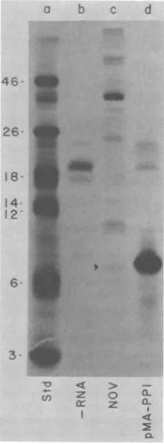
oflow-molecular-weight polypeptides,themajor polypeptide directed by pMA-PP1-selected RNAwas a7.2Kproteinthatcomigratedwitha
faintNOV structuralprotein (Fig. 6,lanescand
d). The larger 19K and 23K proteins in Fig. 6, laned, correspondedtoprotein products synthe-sized from endogenous mRNA in the lysate
system(Fig. 6, lane b).
Longer exposure times were required to
re-ai
b
105 95. D3. 85. 78
b68
6554. 49. 45. 42. 39.
26
18
3V. 30 24 23 22
aI-19. i6.
O
QLz z
u
c)
J.VIROL.
on November 10, 2019 by guest
http://jvi.asm.org/
VOL. 44, 1982
b
cd
eMh
200- : X
93 .
._,~~
..0.. 0 0 . .
[image:8.491.293.412.268.589.2]I31:4_ _U-.'.
FwIG|...5. In vir trnlto prdut of adiioa o
b-
enshZe o) tioae nswnR
e ld)anpMcR:(ln e)ddntirctth cythei of
Q C Q~ CL
FIG.
d. In vitro translationproducts of additional
AcNPV
cDNA-specific
RNAs.Laneacontained(aC-labeled
protein
standards(std);
themolecularweights
ofthese standards
(xl03)
are indicated on the left.Lanebshowsthetranslation
products
whennoRNA wasadded.Thepeptide
frompMA-D01-specific
RNA(lane
c)
isindicatedarrowhead.
by
anpMA-DA1 (lane
d)and
pMA-RJl (lane
e)didnotdirectthesynthesis
ofdetectable
peptides.
The translationproducts
fromspecific
RNAs forpMA-CDl (lane f), pMA-Bl
El(lane
g), pMA-B2H1 (lane h),
andpMA-PB31 (lane i)
areindicated
by
arrowheads. Thegel
was lOtoSDS-polyacrylamide,
andfluorographic
exposure was for26days.
veal
the
proteins encoded
by
the
remaining
cDNA-selected
RNAs. A
56K
protein
wassyn-thesized
from
pMA-DO1-selected
RNA
(Fig.
5,
lane c), and this
protein did not appear to
comigrate
with any NOV structural
protein.
Under the conditions
used,
noproteins
wereobserved
asproducts
of in
vitro translation of
RNA
selected
by either
pMA-DA1
orpMA-RJ1.
pMA-ACD1 was
nottested in the in
vitro
trans-lation
assay.
RNA
selected
by
pMA-CD1 directed the
syn-thesis
of
a25Kprotein which
comigrated
with
astructural NOV
protein.
A60K
protein was
synthesized from
pMA-BlE1-selected
RNA,
and
this
protein did
notcorrespond
to
viral
structural
proteins.
pMA-A2H-selected
RNAdirected the
synthe-sis
of
a37Kprotein
and,
to alesser
extent,
a26K
cDNA CLONES OF LATE AcNPV mRNA 789
protein.
Neither
of
these
proteins appeared
to
correspond
toviral structural
proteins.
The
RNA
selected
by
pMA-PB1 directed
the
synthe-sis of
twoproteins (31K
and
30K)
in
approxi-mately
equal quantities.
These
proteins
comi-grated
with
NOV
structural
proteins
and
with
the
proteins synthesized
from
pMA-ACG1
(Fig.
4,
lane
h).
A weak
band
at50K
wasalso
ob-served in
Fig. 5,
lane
i,
and
mayrepresent
anadditional
protein
product.
pMA-VIl
corresponds
tothe
polyhedrin
gene.To
demonstrate
that the 32K
protein
synthe-sized from
pMA-VI1-selected
RNA
waspolyhe-drin,
weperformed
immune
precipitations
(5)
with antisera raised
topurified
polyhedrin
(Fig.
7).
Immune
precipitation
of in
vitro-synthesized
46
26-I8
14-12
3.
a b C d
_. :
:Ej:.~~0
FIG. 6. TranslationofpMA-PPI-specificRNA and
peptideseparation onSDS-urea-15% polyacrylamide gels. Laneacontained "C-labeledprotein standards (std);themolecularweights(x103)of thesestandards
areindicated on the left. Lane b contained cell-free rabbitreticulocyte translation products with no added RNA. Forcomparison, lane c contained NOV with the 7.2K peptide(arrowhead). Lane d contained transla-tionproductsofpMA-PPl-specificRNA.
Fluorogra-phy
wasfor 5days.
on November 10, 2019 by guest
http://jvi.asm.org/
proteins directed by total RNA isolated late in
AcNPV-infected
S.frugiperda cells included
amajor
32Kprotein that
comigrated with purified
polyhedrin, an 18K protein, and a few faint polypeptides with molecular weights between 18,000 and 32,000
(Fig. 7, lanes
aand b).
Theseimmunoprecipitated products corresponded in
size and relative
intensity
tothe peptides
synthe-sized from pMA-VI1-selected RNA (Fig.
7, lanec). Immune
precipitation of the protein products
of
pMA-VI1-selected RNA resulted in the
pre-cipitation of the
32K, 18K,
and faintintermedi-CL
FIG. 7. ImmuneprecipitationofpMA-Vll-specific
RNAtranslationproducts byantiserumtopolyhedrin. Lanea, AcNPV-infected cell RNA in cell-freerabbit reticulocyte lysate translation reaction; lane b,
im-muneprecipitationof theproteinsin laneaby
antise-rum to polyhedrin; lane c, translation of pMA-VI1-specific RNA; lane d, precipitation with polyhedrin antiserum;lanee,[3H]leucine-labeled purified polyhe-drin; lane f, precipitation of 3H-labeled polyhedrin withantiserum topolyhedrin. Lanegcontained "C-labeledproteinstandards(std);the molecularweights (x103) of these standards areindicated ontheright. The gel was a 10o SDS-polyacrylamide gel, and fluorographywasfor7days.
ate proteins (Fig. 7, lane d). As a control, purified [3H]leucine-labeled polyhedrin (Fig. 7,
lane
e) wasalso immune
precipitated(Fig.
7,lane
f).
DISCUSSION
By 27 h postinfection, 20% or more of the poly(A)-containing mRNA found in
AcNPV-infected
S.frugiperda
cells is virusspecific.
Thisis demonstrated by the fact that 20% of the
ampicillin-sensitive, tetracycline-resistant
colo-nies,
obtained by cloning DNA complementary
to
poly(A)-containing
RNAisolated
late inAcNPV
infection, contain AcNPV-homologous
sequences.
The value 20%
represents amini-mum
since
someAcNPV
insertions
may be toosmall
torespond visibly
tothe AcNPV
probe ormay
be ampicillin sensitive but contain
noinser-tion.
A
total of
11different
DNAscomplementary
to
poly(A)-containing RNAs
present late inAcNPV infection
havebeen
successfully
syn-thesized
and cloned in
E.coli.
Manyof
thecDNA
insertions
arerelatively small (less than
0.5
kb
long),
indicating that only
aportion
of the
sequence
of each
mRNAhas
been cloned.
Deg-radation of
mRNAbefore
reversetranscription
does
not appear tobe
aproblem since
in vitro
translation
of the
sameRNA
results in
proteins
having molecular
weights
up to85,000.
Oligo-deoxythymidylic acid
was used as aprimer
forreverse
transcription,
and itis therefore
proba-ble that
only
the3'
ends of the mRNAs werecloned. Each cDNA
insertionprovides
avalu-able tool for
exploring
the
geneorganization
and
expression
of
AcNPV.
The
frequencies
of
appearanceof the different
cDNA
sequences(Table 1)
may correlate withthe
relative
amountsof the
sequences in themRNA
population.
The
mostabundantcDNA
isrepresented
by
pMA-PP1 and
may representmore
than
one-third of the
AcNPV-specific
poly(A)-containing
RNA. Avery smallprotein
(7.2K) is translated from this
mRNA.A
protein
of similar size
is foundas aminorcomponent ofextracellular NOV. The
7.2Kprotein does
notcorrespond in size
tothe
protamine-like
protein
found in AcNPV
nucleocapsids (22). The
direc-tion of
transcription of the
RNAinthe
HindIII-P/EcoRI-P region is probably from left
toright
on
the
physical
map(Fig. 2) since
thesmaller
pMA-PP
cloneshybridize only
toHindIII-P
andthe
larger clones
hybridize
toboth
HindIII-P
and-Q.
Thetranscript
mustlie
onthe left side
of
HindIII-P since another
clone, pMA-PB1,
hy-bridizes
toHindIII-P and EcoRI-B(Fig. 2).
The
second
mostabundant
cDNAfound
at27h
postinfection is homologous
tothe
mRNA thatdirects
thesynthesis
of
polyhedrin,
a 32Kpro-tein which is
themajor
structuralprotein of
theon November 10, 2019 by guest
http://jvi.asm.org/
[image:9.491.50.240.219.561.2]VOL. 44, 1982
occluded form of AcNPV. Vlak et al. (23) have
previously
implicated EcoRI-I as the location of
the polyhedrin gene by hybridization selection
with EcoRI-I isolated from gels of viral DNA
fragments, followed by in vitro translation of the
selected RNA. We have
further pursued the
location of polyhedrin by
defining HindIII-V as
the probable 3' end
of
the polyhedrin message
and
by demonstrating that the 32K protein
prod-uct, which is translated from
pMA-VI1-selected
mRNA, not
only
comigrates with purified
poly-hedrin but also
immunoprecipitates
specifically
with antisera to purified polyhedrin.
The
pMA-VI1 cDNA insertion also directs the
synthesis of a prominent 18K protein, as well
asa
few fainter bands
(Fig. 7). These proteins are
antigenically related to polyhedrin because they
also specifically immunoprecipitate with
polyhe-drin antibody.
There
areseveral
possible
expla-nations
for
the presence
of
the 18K
protein.
The
first involves
splicing of
AcNPV mRNA such
that two
different
transcripts
contain the
same3'
terminus but encode different
proteins.
This
explanation
is
unlikely
in
light
of
recenttran-scriptional studies
in
ourlaboratory
in which
pMA-VI1
DNA
wasused
as aprobe of
Northern
RNA
blots; this work indicated that
pMA-VI1
hybridizes to a
prominent
1.2-kb RNA
(D. W.
Miller,
and L. K.
Miller, manuscript
in
prepara-tion).
Although we have not excluded the
possi-bility
of splicing at the 5' end of the polyhedrin
mRNA, the
evidence suggests that there is only
one
major distinct
mRNA
homologous to
pMA-VII (the 3' end). Furthermore, a major 18K
pro-tein
has
not
been
observed
in
[35S]methionine-labeled infected
cells at 27 h
postinfection (L. K.
Miller
and R.
Trimarchi, unpublished data),
which argues
against
the
possibility
that two
different
proteins
aremade from the
samemRNA.
Based
onthese
considerations,
wecur-rently favor the
explanation
that the 18K
protein
and
the
other faint
proteins antigenically
related
to
polyhedrin
areprematurely terminated
poly-peptides (i.e.,
artifacts of the in vitro translation
system).
Specific prematurely
terminated
trans-lation products have been observed by
anumber
of
other workers
who used in vitro translation
systems
and may be due to
adeficiency of
some
tRNA
species
in the
translation
system.
Another abundant insertion found at 27 h
postinfection is represented by pMA-ACG1.
This clone is interesting because it selects an
RNA(s) that encodes two proteins (a 31K
pro-tein and a
30K protein), which are synthesized in
approximately equal quantities. We favor the
explanation of spliced RNA in this case since
there are
two
predominant 31K and 30K
pro-teins found in infected cells late in AcNPV
infection, as
well
as
two
NOV 31K and 30K
structural
proteins. Furthermore, hybridization
cDNA CLONES OF LATE AcNPV mRNA
791
of pMA-ACG1 to Northern blots of late RNA
has
indicated the presence of two predominant
RNA
species, which are approximately 1.1 and
1.3 kb long (Miller and Miller, unpublished
data). The use
of cDNA clones rather than
fragments
of viral genomic DNA is clearly
ad-vantageous in this work.
An unusual
observation is that
pMA-PB1-selected RNA also directs the synthesis of
ap-proximately equal quantities of two proteins
(31 K and 30K
proteins)
which
comigrate
with
the
pMA-ACG1-directed
proteins. The
pMA-ACG and pMA-PB DNAs
hybridize to regions at
map
positions 43 to 50 and 89.5 to 90.5 on the
AcNPV physical map,
respectively (almost
dia-metrically opposed
on acircular map), and there
is no apparent
cross-hybridization
between
these two
regions, even when the blots
areoverexposed. The pMA-PB1
insertion
reprodu-cibly selects for RNAs that direct the
synthesis
of the 31K and 30K
proteins,
but the level of
synthesis
of these
twoproteins
observed in
gels
of
the
translation
products
is
approximately
10-fold less than the level observed when
pMA-ACG1-selected
mRNAis used. It is
possible
that
the
31K and 30K
proteins synthesized by
pMA-ACG RNA and
pMA-PB RNA
aredifferent
proteins which
fortuitously comigrate.
Howev-er, this would be
anunusual
coincidence,
and
we are
investigating this phenomenon since it
may
reflect
anovel
transcriptional phenomenon.
RNA
selected
by
twoclones,
pMA-DA1 and
pMA-RJ1, did not direct the
synthesis of
any
observable
proteins
under the
conditions
used.
It
is
possible
that these
regions encode
very
large
proteins (>85K)
which
arenot
synthesized
efficiently
by the translation system.
However,
there
aremany other
explanations for
these
negative results,
including
the
possibility
that
the
homologous mRNAs
arepresent in very low
concentrations in the late mRNA
preparation.
Three
clones,
pMA-BlE1, pMA-DO1,
and
pMA-B2H1, direct the
synthesis
of
proteins
that
are
not
found in
NOV,
asjudged
by
alack
of
comigration
in
gels.
We cannotcategorically
state
that these
arenonstructural
proteins since
it is possible that the
proteins
areprocessed in
vivo (1). For instance, the 61K
protein of
pMA-BlEl may be related
tothe
NOV 64K to 65K
structural
protein,
which is somewhatdiffuse
and
may be
glycosylated in vivo. Both
the 56Kprotein of pMA-DO1 and the 37K protein of
pMA-B2H1
migrate
in close
proximity
toNOV
strutural
proteins.
Post-translational
processing
of AcNPV
proteins
invivo
resulting
in
major
size alterations of
theproteins does
notappear
to
bean extensive
phenomenon (2, 4, 25).
Even-tually,
genetic
mutants andmarker rescue
may
be
necessary
toconfirm
specific protein
assign-ments. The
pMA-CD1-selected
RNAdirects the
on November 10, 2019 by guest
http://jvi.asm.org/
FVN D XJ LMR E OU I A C WHS B B KQP G F
I"II I I I III I 11 11 I 1111 I
it wil I f 1 INI
m |
I If I I I I*, I III,, *.1 I I I, 1 I
I RO A J KTMN FVU C G W D QL E HSP E
tt
t
32K 56K 31Ka 30K 25K 61 K
PH
7.2 K
26 K a 37 K
HinDm
Eco R I
%31K a 30 K
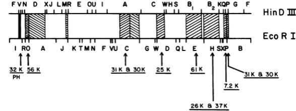
FIG. 8. Summary of cDNA map positions and selected proteins. The physical map of AcNPV is presented in linear form. The sizes of the proteins translated from AcNPV cDNA-selected RNA are indicated.
synthesis of
a25K
protein
whichcomigrates
with
astructural
protein
of NOV. In all cases ofcomigration
of
proteins,
thephysical identity
orantigenic
similarity of
theproteins
must bees-tablished before
it can be concluded that theproteins
areindeed
related.The
11different
regions
of DNA delineated
by
the
cDNAs isolated
in this work aredispersed
throughout
the viral genome. A summary of themap
positions
of the cDNAs and the
proteins
synthesized from cDNA-selected RNAs
isshown in
Fig.
8.
Transcriptional
studies with
these cDNA
clones
areunder
wayand should
reveal
considerable
information concerning
the
size(s)
of the
homologous transcript(s)
and
thetemporal control of each of the
transcripts.
All
of the cDNAs
represent RNAs found late ininfection,
but this does
not meanthat these
clones
arefound
exclusively
late in
infection.
ACKNOWLEDGMENT
This research was supported in part by Public Health Service grant Al 17338-05 from the National Institute of AllergyandInfectious Diseases.
LITERATURE CITED
1. Carstens, E. B., S. T. Tjia, and W. Doerfler. 1979. Infection ofSpodopterafrugiperdacells withAutographa californica nuclear polyhedrosis virus. I. Synthesis of intracellular proteins after virus infection. Virology 99:386-398.
2. Cochran, M. A., E. B. Carstens, B. T. Eaton, and P. Faulkner. 1982.Molecularcloningandphysicalmapping ofrestriction endonucleasefragmentsofAutographa cali-fornicanuclearpolyhedrosisvirus DNA. J. Virol.
41:940-946.
3. Craig,E.A.,B.J.McCarthy,andS.C.Wadsworth. 1979. Sequence organizationoftworecombinantplasmids
con-taininggenes forthemajorheatshock-inducedproteinof D.melanogaster. Cell 16:575-588.
4. Dobos, P., and M. Cochran. 1980. Protein synthesis in cells infectedbyAutographacalifornicanuclear polyhe-drosis virus(Ac-NPV): the effect ofcytosinearabinoside. Virology103:446-464.
5. Dougherty,W. G.,and E. Hiebert. 1980.Translation of potyvirusRNAinarabbitreticulocytelysate: identifica-tion of nuclear inclusionproteinsasproductsof tobacco etchvirusRNAtranslation andcylindricalinclusion pro-tein as a product of the potyvirus genome. Virology
104:174-182.
6. Holmes, D. S., and J. Bonner. 1973. Preparation, molecu-larweight, basecomposition,andsecondary structure of giant nuclear RNA.Biochemistry 12:2330-2338. 7. Kahn, M., R. Kolter, C. Thomas, D. Figurski, R. Meyer,
E. Remaut, and D. R. Helinski. 1979. Plasmidcloning vehicles derived from plasmids ColEl, R6K, and Rk2. MethodsEnzymol. 68:268-280.
8. Laemmli, U. K. 1970. Cleavage of structural proteins during the assembly of the head of bacteriophage T4. Nature (London)227:680-685.
9. Lee, H. H., and L. K.Miller. 1978. Isolation of genotypic variants of Autographa californica nuclear polyhedrosis virus. J. Virol. 27:754-767.
10. Manley, J. L., P. A.Sharp, and M. L. Gefter. 1979. RNA synthesisin isolated nuclei:identificationand comparison of adenovirus 2encoded transcripts synthesized in vitro and in vivo. J.Mol. Biol. 135:171-197.
11. McGrogan, M., D. J. Spector, C. J. Goldenberg, D. Halbert, and H. J. Raskas. 1979. Purification ofspecific adenovirus 2 RNAs by preparative hybridization and selective thermal elution.NucleicAcids Res. 6:593-07. 12. Miller, L. K. 1981. Construction of a genetic map of the baculovirus Autographacalifornicanuclearpolyhedrosis virus by marker rescueof temperature-sensitive mutants. J.Virol. 39:973-976.
13. Miller,L. K. 1981. Avirus vector for genetic engineering in invertebrates, p. 203-224. In N. Panapoulous (ed.), Genetic engineeringin the plant sciences. Praeger Pub-lishers,New York.
14. Miller, L. K., and K. P. Dawes. 1979.Physical mapof the DNA genome ofAutographa californica nuclear polyhe-drosis virus. J.Virol. 29:1044-1055.
15. Ricciardi, R. P., J. S. Miller, and B. E. Roberts. 1979. Purificationand mapping of specific mRNAs by hybrid-ization-selection and cell-free translation. Proc. Nati. Acad.Sci.U.S.A. 76:4927-4931.
16. Rigby, P. W. J., M.Dieckmann, C. Rhodes, and P. Berg. 1977. Labeling deoxyribonucleic acid to high specific activityinvitrobynick translation with DNA polymerase I. J.Mol.Biol. 113:237-251.
17. Roychoudury, R., and R. Wu. 1980. Terminal transferase-catalyzed addition of nucleotides to the 3' termini of DNA.Methods Enzymol.65:43-62.
18. Shapiro, A. L., E. Vinuela, and J. B. Maizel. 1967. Molecular weight estimation of polypeptide chains by electrophoresis in SDS-polyacrylamide gels. Biochem. Biophys. Res.Commun.28:815-820.
19. Smith, G. E., and M. D.Summers. 1980. The bidirectional transfer of DNA and RNA to nitrocellulose or diazoben-zyloxymethyl-paper. Anal. Biochem. 109:123-129. 20. Summers, M. D., R.Engler,L. A. Falcon, and P. Vail.
1975.Baculoviruses for insect pest control:safety consid-erations. American SocietyforMicrobiology, Washing-ton, D.C.
21. Thuring, R. W. J., J. P.M.Sanders, and P. Borst. 1975. A J. VIROL.
on November 10, 2019 by guest
http://jvi.asm.org/
[image:11.491.97.391.81.191.2]VOL. 44, 1982
freeze-squeeze method for recovering long DNA from agarose gels. Anal.Biochem. 66:213-220.
22. Tweeten, K. A., L. A.Bulla, Jr., and R. A. ConsIgi.1980. Characterization of an extremely basic protein derived from granulosis virus nucleocapsids. J.Virol. 33:866-876. 23. VIak, J. M., G. E. Smith, and M. D. Summers. 1981. Hybridization selection andin vitro translation of Auto-grapha californica nuclear polyhedrosis virus mRNA. J.
cDNA CLONES OF LATE AcNPV mRNA 793 Virol. 40:762-771.
24. Wickens, M. P., G. N. Bueli,and R. T.Schimke. 1978. Synthesis of double-stranded DNA complementary to lysozyme, ovomucoid, and ovalbumin mRNAs. J. Biol. Chem. 253:2483-2495.
25. Wood, H. A. 1980. Autographacalifornicanuclear poly-hedrosis virus-induced proteinsin tissue culture.Virology 102:21-27.