R E S E A R C H
Open Access
Identification of repressive and active
epigenetic marks and nuclear bodies in
Entamoeba histolytica
Daniela Lozano-Amado
1, Abril Marcela Herrera-Solorio
1, Jesús Valdés
2, Leticia Alemán-Lazarini
1,
Ma. de Jesús Almaraz-Barrera
1, Eva Luna-Rivera
1, Miguel Vargas
1and Rosaura Hernández-Rivas
1*Abstract
Background:In human hosts,Entamoeba histolyticacysts can develop into trophozoites, suggesting that the life cycle of this parasite are regulated by changes in gene expression. To date, some evidence has suggested that epigenetic mechanisms such as DNA methylation and histone modification are involved in the regulation of gene expression inEntamoeba. Some post–translational modifications (PTMs) at the N-terminus ofE. histolytica’shistones have been reported experimentally, including tri-methylation in the lysine 4 of histone H3 (H3K4me3) and dimethylation in the lysine 27 of histone H3 (H3K27me2), dimethylation of arginine 3 (H4R3me2) and the indirect acetylation of histone H4 in the N-terminal region. However, it is not known which residues of histone H4 are subject to acetylation and/or methylation or where in the nucleus these epigenetic marks are located.
Methods:Histones from trophozoites ofE. histolyticawere obtained and analyzed by LC-MS/MS. WB assays were performed using antibodies against epigenetic marks (acetylated lysines and methylated arginines). Immunofluorescence assays (IFA) were carried out to determine the distribution of PTMs and the localization of DNA methylation as a heterochromatin marker. Nuclear bodies such as the nucleolus were identified by using antibodies against fibrillarin and nucleolin and speckles by using anti-PRP6 antibody.
Results:Some new PTMs in histone H4 ofE. histolytica, such as the acetylation of lysines 5, 8, 12 and 16 and the monomethylation of arginine 3, were identified by WB. IFA demonstrated that some marks are associated with transcriptional activity (such as acetylation and/or methylation) and that these marks are distributed throughout theE. histolyticanucleus. Staining with antibodies against anti-pan-acetylated lysine H4 histone and 5-methyl cytosine showed that the activation and transcriptional repression marks converge. Additionally, two nuclear bodies, the nucleolus and speckles, were identified in this parasite.
Conclusions:This study provides the first evidence that the nucleus ofE. histolyticais not compartmentalized and contains two nuclear bodies, the nucleolus and speckles, the latter of which was not identified previously. The challenge is now to understand how these epigenetic marks and nuclear bodies work together to regulate gene expression inE. histolytica.
Keywords:Entamoeba histolytica, Transcriptional regulation, Histone post-translational modifications, Epigenetics, Nuclear architecture
* Correspondence:rohernan@cinvestav.mx
1Molecular Biomedicine Department, Centro de Investigación y de Estudios
Avanzados del Instituto Politécnico Nacional (IPN), Av. Instituto Politécnico Nacional # 2508, Apartado postal 14–740, 07360 D. F. Mexico, México Full list of author information is available at the end of the article
Background
The parasiteEntamoeba histolyticahas two morphologic-ally distinct life stages: the cyst, which is the infectious form that transmits disease from person to person, and the trophozoite, which is the invasive form that multiplies in the colon and can eventually invade the liver, brain and lungs. A total of 500 million people worldwide are affected by this parasite; resulting in 50 million cases of invasive disease and approx. 70,000 deaths annually [1].
Despite the medical relevance ofE. histolytica, very little is known about how gene expression is modulated in this parasite during the invasion of its human host or the en-cystation process. Changes in the abundance of transcripts inE. histolyticaare associated with human host invasion [2] and with conversion between the cyst and the tropho-zoite form. However, the molecular mechanisms that regulate gene expression in this parasite are poorly under-stood. A number of cis elements that function as gene promoters in this parasite and transcription factors that recognize these elements have been described [3]. Add-itionally, it has been shown that theE. histolyticagenome is organized into chromatin, whose fundamental unit is the nucleosome [4], and contains genes encoding histones H2A, H2B, H3 and H4. Thus, it is very likely that these histones form the nucleosomes of this parasite. However, the DNA that separates each nucleosome (the DNA linker) exhibits an irregular length compared with the 40 bp DNA linker found in metazoans [4]. Furthermore, it has been found that although the amino - terminus of the
E. histolyticahistones diverge from the primary sequence present in the metazoan histones, they are highly basic and contain several lysine and arginine residues that may be potential targets for post-translational modifica-tions such as acetylation and methylation, through the ac-tion of histone acetyltransferases (HATs) or lysine or arginine methyl transferases (HKMTs or PRMTs), respect-ively [5].In silicoanalysis of theE. histolyticagenome has revealed the presence of HAT enzymes belonging to the GNAT and MYST families as well as the presence of a pro-tein capable of removing acetyl groups present at the amino-terminus of histones, a class I histone deacetylase (HDAC) [6]. To date, the only post-translational modi-fications that have experimentally been shown to occur at the amino-terminus of histone H3 are the di- and tri-methylation of lysine 4 (H3K4me2/3) inEntamoeba
[7], which are associated with changes in transcriptional activity, as well as the di-methylation of lysine 27, which is highly enriched in genes silenced through RNA interfer-ence (RNAi) [8]. In the case of E. histolytica, histone H4 shares 71 % identity with the mammalian histone H4 [5]. The differences primarily occur in the amino-terminus of histone H4, where three insertions that do not exist in other eukaryotic histone H4 genes are found. One of these insertions is located at the beginning of the NH2-terminus,
while the second is located between amino acids 10 and 11, and the third is located after amino acid 14 [5]. This last insertion site merits particular attention due to the presence of three extra lysine residues, which could serve as targets for post-translational modifications such as acetylation or methylation. However, it is not known which residues of histone H4 are subject to acetylation and/or methylation, where in the nucleus these epigenetic marks are located, and what roles they play in the nuclear archi-tecture. To address these questions, western blot (WB) analyses and immunofluorescence assays (IFAs) were per-formed using commercial antibodies against histone H4. Our data suggest that histone H4 is acetylated at lysine residues (K) K5, K8, K12 and K16 and that arginine 3 is mono-methylated. However, antibodies that recognize tri-methylated K20 did not detect this epigenetic mark at histone H4. Furthermore, IFAs performed with antibodies directed against pan-acetyl histone H4 and monomethyl arginine 3 of histone H4 showed that these epigenetic marks associated with transcriptional activation are distrib-uted throughout the nucleus. A similar distribution pattern was found using an antibody that detects DNA methyla-tion (5-methyl cytosine). Taken together, these data indi-cate that unlike what has been reported in eukaryotes and other parasites, the nucleus of this parasite is not compart-mentalized and contains two types of nuclear bodies: the nucleolus and speckles.
Methods
Cell cultures ofE. histolytica
Trophozoites of E. histolytica strain HM1:IMSS were axenically cultured at 37 °C in TYI-S-33 medium and harvested from confluent cultures as described [9].
Nuclear acid protein extracts
discarded, whereas the pellet, corresponding to the nuclear fraction, was resuspended in 100 μl of Tris-EDTA buffer (28.5 mM Tris pH 7.4, 37 mM Tris-EDTA) with 0.4 M of HCl and incubating overnight at 4 °C. The acid extract was centrifuged for 10 min at 21,000 × g. 800 μL of ice-cold acetone was added to the supernatant, and the mixture was incubated overnight at−20 °C. Precipitated proteins were collected by centrifugation at 21,000 × g for 15 min, washed once with acetone and air dried. The pellet was resuspended in Tris–HCl pH 8.8 and stored at−20 °C. All buffers used in this protocol contained protease inhibi-tors (Complete, EDTA-free, Roche).
Nuclear proteins preparation
8 × 10⁷log phase cells were lysed as described above, and the nuclear fractions were purified by sucrose gradient centrifugation. The pellet was resuspended in 100 μl of RIPA buffer (50 mM Tris pH 7.4, 150 mM NaCl, 5 mM EDTA, 1 % NP-40, 0.5 % sodium deoxycholate, 0.1 % SDS) keeping it on ice for 30 min, mixing occasionally. The sample was centrifuged at 21,000 × g for 20 min at 4 ° C and the supernatant was recovered and stored at−20 ° C. All buffers used in this protocol contained protease in-hibitors (Complete, EDTA-free, Roche).
Mass spectrometry analysis ofE. histolyticahistones
Proteins were proteolytically digested in-gel after reduc-tion in 10 mM DTT and alkylareduc-tion in 55 mM iodoaceta-mide (both buffered in 50 mM ammonium bicarbonate). Sequencing grade trypsin (250 ng, Promega) in 50 mM ammonium bicarbonate was used to digest protein over-night at 37 °C. Digested peptides were analyzed by LC-MS/MS on a Thermo Scientific Exactive Plus Orbitrap Mass Spectrometer in conjunction with an EASY-nLC II nano UHPLC and Proxeon nanospray source. The digested peptides were loaded on a 100 micron × 25 mm Magic C18 100 Å 5U reverse phase trap where they were desalted online before being separated using a 75 mi-cron × 150 mm Magic C18 200 Å 3U reverse phase column. Peptides were eluted with an increasing per-centage of acetonitrile over the course of a 60 min gradi-ent with a flow rate of 300 nl/min. An MS survey scan was obtained for the m/z range 300–1600 and acquired with a resolution of 70,000 and a target of 1 × 10⁶ions or a maximum injection time of 30 msec. MS/MS spec-tra were acquired using a top 15 method where the top 15 ions in the MS spectra were subjected to HCD (High Energy Collisional Dissociation). MS/MS spectra were acquired with a resolution of 17,500 and a target of 5 × 10^4 or a maximum injection time of 50 msec. An isola-tion mass window of 1.6 m/z was used for precursor ion selection, charge states 2–4 were accepted, and a nor-malized collision energy of 27 % was used for fragmenta-tion. A 5 s duration was used for dynamic exclusion.
Tandem mass spectra were extracted and charge state deconvoluted with Proteome Discoverer (Thermo Scientific) and searched using X! Tandem (The GPM, thegpm.org; version Sledgehammer 2013.09.01.2)). X! Tandem was set to search all proteins in the Uniprot.orgE. histolyticadatabase (April 21 2015) plus the cRAP database of common laboratory contaminants (www.thegpm.org/ crap/; 114 entries), and an equal number of reverse protein sequences (16,138 entries total). X! Tandem was searched with a parent ion mass tolerance of 20 PPM, a fragment ion tolerance of 20 PPM, and trypsin as the digestion enzyme with 1 maximum missed cleavage. Carbamido-methylation of cysteine was specified as a fixed modifica-tion. Deamidation of asparagine and glutamine, oxidation of methionine and tryptophan, and Glu- > pyro-Glu, Gln- > pyro-Glu, and ammonia loss of the n-terminus were specified as variable modifications.
Scaffold (version 4.4.0, Proteome Software Inc., Portland, OR) was used to validate MS/MS based protein and pep-tide identifications. Peppep-tide identifications were accepted if they could be established at greater than 95.0 % probability by the Scaffold Local FDR algorithm. Protein identifica-tions were accepted if they could be established at greater than 79.0 % probability to achieve an FDR less than 2.0 % and contained at least 1 identified peptide. Actual protein and peptides FDRs were 0 %. Protein probabilities were assigned by the Protein Prophet algorithm (Nesvizhskii, Al et al., 2003) [11]. Proteins that contained similar peptides and could not be differentiated based on MS/MS analysis alone were grouped to satisfy the principles of parsimony. Proteins sharing significant peptide evidence were grouped into clusters.
Immunofluorescence assays
Trophozoites in a logarithmic growth phase were har-vested and transferred on glass coverslips coating with poly L - lysine and incubated for 3 h at 37 °C to let them attach to the glass surface. An indirect immuno-fluorescence assay was performed as follows. Amoebas were fixed and permeabilized with cold methanol-acetone 50:50 for 10 min at room temperature and washed twice with PBS buffer. After, trophozoites were incubated for 1 h with 1 % bovine serum albumin in PBS buffer and samples were reacted with anti-acetyl-histone H4 (Millipore 06-866) 1:1500, anti-histone H4 acetyl K12 antibody (Abcam ab61238) 1:300, anti-histone H4 mono methyl R3 antibody (Abcam ab17339) 1:200, anti-5-methylcytosine antibody
(Abcam ab10805) 1:25, anti-lamin B1 antibody
A-11001) 1:100. Nuclei were stained with 4,6-Diamidino-2-Phenylindole (DAPI) Vectashield Mounting (Vector H-1200) and samples were observed through a confocal microscope Fluoview Olympus FV300. Software 4.3.
Western blot
Nuclear acid proteins were separated on 18 % polyacryl-amide SDS-PAGE gel while nuclear extract were sepa-rated on 10 % polyacrylamide SDS-PAGE gel and transferred to a nitrocellulose membrane according to the protocol described by Towbin et al., 1979 [12]. The membrane was exposed to Ponceau S to verify the effi-ciency of the transfer. The membrane was blocked with 5 % milk in PBS buffer- Tween-20 0.05 % (PBS-T) for 2 h at room temperature and then incubated with the histone H3 antibody (Abcam ab1791) 1:5000, anti-histone H4 antibody (Santa Cruz sc-8658-R) 1:2000, acetyl-histone H4 (Millipore 06-866) 1:4000, anti-histone H4 acetyl K12 antibody (Abcam ab61238) 1:2000, anti-histone H4 mono methyl R3 antibody (Abcam ab17339) 1:500, anti-trimethyl-histone H4 (Lys20) antibody (Millipore 07-463) 1:500, anti-lamin B1 antibody (Abcam ab16048) 1:5,000, anti-fibrillarin 1:3,000, anti-PRP6 1:500, all diluted with 2 % milk PBS-T overnight at 4 °C. PBS-The membranes were rinsed 3 times with PBS-T and incubated for 2 h with a horseradish peroxidase (HRP)-conjugated goat rabbit IgG anti-body (Thermo Fisher Scientific G-21234) 1:7,500 or goat anti-mouse IgG antibody (Thermo Fisher Scientific G-21040) 1:7,500 diluted in 2 % milk PBS-T. The antibody staining reaction on the membranes were developed by SuperSignal™ West Femto Maximum Sensitivity Sub-strate (Thermo Scientific 34095).
Results
In eukaryotes, the NH2-terminus of histone H4 can undergo various post-translational modifications (PTMs), such as the acetylation of lysine residues 5, 8, 12 and 16, which are associated with transcriptional activation, and the trimethylation of lysine 20 of histone H4 (H4K20me3), which is associated with transcriptional repression [13]. To determine whether the amino-terminus of Entamoeba
histone H4 also exhibits these PTMs, we initially obtained a crude preparation of histones. For this purpose, the nuclei of trophozoites were obtained, and an acid extraction was performed to obtain basic proteins, including histones. This preparation was separated using 18 % SDS-PAGE, and an enrichment of the fraction from 10 to 20 kDa was observed (Fig. 1a), suggesting that histones ofE. histolyticawere most likely present in this region. To confirm this assumption, WB assays were performed using commercial antibodies recognizing the COOH-terminus of histones H3 and H4. In the case of histone H3, a signal of approximately 15 kDa was observed, and in the case of histone H4, the antibody
identified a protein of approximately 13 kDa. As a positive control for both antibodies, they were incubated withBos taurusthymus histones. As expected, the antibodies recog-nized a 15 kDa band for histone H3 and band of 11.33 kDa for histone H4. These data indicate that the histones of this parasite are indeed present in the obtained preparation of basic nuclear proteins. To confirm that this preparation contained not only E. histolytica histones H3 and H4 but also histones H2A and H2B, we proceeded to recover pro-teins in the range of 10 to 17 kDa from the SDS-PAGE gel, which were then analyzed via mass spectrometry (Fig. 1b). The results revealed that the sample did include histones H2A, H2B, H3 and H4 (Fig. 1c). All of these data indicated that this basic nuclear preparation contained the four ca-nonical histones [14]; none of the histone variants previ-ously identified in other eukaryotes and protozoans were observed in this analysis [14, 15].
As stated previously, the amino-terminus of histone H4 exhibits three insertions compared with the primary structure of eukaryotic histone H4 (Additional file 1: Figure S1A). However, when these inserts are removed, the amino-terminus of E. histolytica shows a high hom-ology (~81 %) with the amino-terminus of yeast and hu-man histone H4 (Additional file 1: Figure S1B). Taking this into account, we decided to identify post-translational modifications occurring on histone H4 of this parasite using commercial antibodies. Alignment (Additional file 1: Figure S1C) of the peptides used to obtain a pan-acetyl antibody for Tetrahymena thermophilus with the N-terminal region of histone H4 from E. histolytica
and Homo sapiens showed that lysines 5, 8, 12 and 16 are conserved at the amino-terminus of histone H4 of E. histolytica (Additional file 1: Figure S1C).
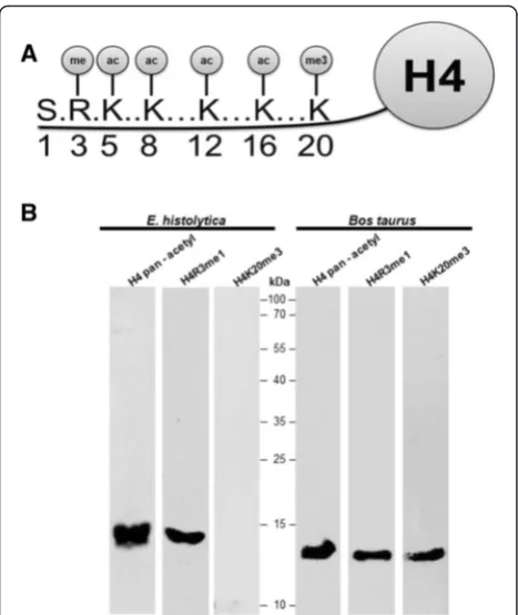
Thus, we decided to use this antibody to perform WB assays. The basic nuclear preparation was incu-bated with the pan-acetyl antibody, and a 13 kDa band was identified (Fig. 2b). Bos taurus histones were used as a positive control for the pan-acetyl histone antibody, which recognized an 11 kDa band (Fig. 2b). These results suggest that E. histolytica
histone H4 is acetylated on one or more of the ly-sines located at positions 5, 8, 12 and 16. Another modification that occurs at the amino-terminus of histone H4 is the monomethylation of arginine 3 (H4R3me1) (Fig. 2a). Thus, the basic nuclear proteins obtained from
is detected in the N-terminal tail on lysine 20 (H4K20) (Fig. 2a). This methylation mark is evolu-tionarily conserved from yeast to human and exists in three distinct states as mono-, di- and trimethylation. Each of these states results in distinct biological out-puts: Mono- (H4K20me1) and dimethylated H4K20 (H4K20me2) are involved in DNA replication and DNA damage repair, whereas trimethylated H4K20 (H4K20me3) is a hallmark of silenced heterochro-matic regions [16]. Because we are interested in iden-tifying one heterochromatin marker in E. histolytica
we proceeded to determine the presence of this PTM in the histone H4 of this parasite. Unexpectedly, no signal was found in extracts fromE. histolytica (Fig. 2b), but there was a signal in the histones from Bos taurus. This result suggests that the H4K20me3 epigenetic repres-sive mark is likely not present in this parasite [17], because this epigenetic mark is enriched in telomeric region in eukaryotic cells and up to now the telomere in E. histo-lytica has not been identified. In conclusion, taking into account all of these data, we propose that the amino-terminus of histone H4 of E. histolytica har-bors epigenetic marks associated with transcriptional activity, whereas no post-translational modification previously associated with transcriptional repression was identified.
In theE. histolyticanucleus the activation and transcriptional repression marks converge
To determine if the previously identified lysine acetyl-ation or mono-methylacetyl-ation of arginine 3 in histone H4 were located in different regions within the nucleus, im-munofluorescence assays were performed using the pan-acetyl histone H4 antibody. The results show that this signal is present in all trophozoites, and when merged with DAPI, we found that this signal was located in the nucleus of all trophozoites (Fig. 3a). The amplification (300×) of one trophozoite image revealed that the acety-lated lysines of histone H4 were distributed throughout the nucleus (Fig. 3a). Subsequently, an immunofluores-cence assay was performed to determine whether each of the lysines identified by the pan-acetyl antibody (K5, K8, K12, and K16) was located in a specific region of the nucleus, such that the observed signals corresponded to the addition of each of the acetylated lysines. For this immunofluorescence assay, an antibody that specifically recognizes K12 of histone H4 was used. We again found that the signal for the acetylated K12 of histone H4 is present in all trophozoites. However, similar to the re-sults obtained with the pan-acetyl H4 antibody, it was located throughout the nucleus (Fig. 3a). To assess whether the other epigenetic mark identified through WB (the mono-methylation of arginine 3 of histone H4,
[image:5.595.55.540.90.339.2]which has been shown to be associated with transcrip-tional activation) was also distributed throughout the nucleus or was included among the acetylated lysines, IFA was performed (Fig. 3a). Once again, the signal obtained with this antibody was present in all nuclei, showing a distribution throughout the nucleus when ob-served at a higher resolution (300×) (Fig. 3a). All of these data suggest that activation marks (such as acetyl-ation and/or methylacetyl-ation) present in this parasite are distributed throughout theE. histolyticanucleus.
Because our WB assays with the H4K20me3 anti-body resulted in no signal, we were not able to use this antibody to identify heterochromatin regions in this parasite [17]. For this reason and considering that his-tone methylation and DNA methylation are mechanisms that act in concert and that the presence of DNA methy-lation has previously been demonstrated in this parasite [18], we decided to use an antibody that recognizes methylated cytosines (anti-5-methyl cytosine) to indir-ectly locate transcriptionally inactive regions in amoebas (Fig. 3a). Our data indicated that this mark was present in most trophozoites, and merging with DAPI showed that it was also present in the nucleus. Unexpectedly,
the amplification of some of these signals (300×) from three independent experiments showed that the methyl-ated DNA was distributed throughout the nucleus (Fig. 3a). In order to establish if the euchromatin marks (pan-acetyl histone H4) and DNA methylation marks overlapped, IFAs were performed with anti-pan-acetyl histone H4 and 5-methyl-cytosine histone H4 anti-bodies. The results of the immunofluorescence analyses indicated that the activation and transcriptional repres-sion marks co-localize inE. histolytica(Fig. 3b).
TheE. histolyticanucleus contains at least two types of nuclear bodies
Studies aimed at determining how the nucleus is phys-ically and functionally organized have revealed that it is very organized and highly dynamic. A prominent fea-ture of the nuclear landscape is its ability to harbor a variety of discrete subnuclear organelles, collectively re-ferred to as nuclear bodies. Nuclear bodies spatially compartmentalize the nuclear environment and create different sites where proteins and RNAs concentrate, which streamlines biological processes such as replica-tion, DNA repair and messenger RNA maturation [19].
To determine whether there are also nuclear bodies in theE. histolyticanucleus, we employed antibodies against two proteins specific to the nucleolus: fibrillarin (which was kindly provided by Dr. Miguel Ángel Vargas) and nucleolin. Initially, nuclear E. histolytica trophozoite ex-tracts were assessed via WB to validate the anti-fibrillarin antibody. A protein of the expected size of approximately 35 kDa was identified (Fig. 4a). Based on these results, both antibodies were used to perform IFAs. Figure 4b and c show that the anti-nucleolin and anti-fibrillarin antibodies produced three types of signals. One of the signal patterns, identified in 50 of 100 parasite cells, showed fibrillarin and nucleolin to be largely located at the periphery of the nucleus. In 35 of the cells, the two proteins were instead located at both poles of the nucleus, and in 15 of the cells, the proteins were located at just one end of the nucleus (Fig. 4b and c). For nucleolin, in addition to producing three types of signals, as was observed for fibrillarin (Fig. 4c), other signals could also be identified because this protein is not only a constituent of the nucleolus in eukaryotic organisms but also performs other functions in the nucleus [20]. Therefore, IFAs were performed to deter-mine if nucleolin and fibrillarin co-localize. The overlap of the two proteins corroborated the existence of a nucleolus not only at the periphery of the nucleus of Entamoeba
(Fig. 4d), but also in one or two poles of the nucleus, two patterns not described previously (Fig. 4d) [21]. Finally, a mouse anti-lamin B1 antibody was used to demonstrate that the nucleolus is located at the nuclear periphery. Ini-tially, we decided to determine whether the mouse anti-lamin B1 antibody recognized a anti-lamin-like protein in E. Fig. 2The N-terminal region of histone H4 fromE. histolyticais
[image:6.595.57.291.86.364.2]histolytica nuclear extracts via WB assays (Fig. 4e). The mouse anti-lamin B1 antibody recognized a protein of ap-proximately 78 kDa in the nuclear extracts ofE. histolytica
(Fig. 4e). Thus, we proceeded to use this antibody in fur-ther IFAs: the signal obtained with the lamin B1 anti-body was located across the entire surface of the nucleus, and when superimposed on the image of nuclei stained with DAPI, we could clearly distinguish the nucleus from the cytoplasm (Fig. 4f). More importantly, the signal sug-gested the existence of a lamin-B-like protein in the amoeba. Finally, a co-localization assay was performed with anti-lamin B1 and anti-fibrillarin, which revealed that the two signals co-localized at the periphery of theE. histolytica
trophozoite nucleus (Fig. 4g). This finding confirmed that the nucleolus shows the perinuclear localization in the amoeba.
TheE. histolyticanucleus contains at least two types of nuclear bodies
To identify other common types of non-membranous nuclear bodies that are present in many eukaryotic organisms, such as speckles which are nuclear domains enriched in pre-mRNA splicing factors, we decided to use RNA processing proteins (PRPs), a type of organelle-specific protein. TheE. histolytica PRP6 protein was de-scribed in 2000 by the group of Dr. Vargas Mejia, and we employed the antibody obtained in that study to perform WB assays and verify its functionality [22]. The anti-PRP6 antibody recognized a protein of the expected size of approximately 105 kDa (Fig. 5a), as previously described [22]. Thus, this antibody was used in further immuno-fluorescence assays. The signal corresponding to anti-PRP6 (speckled structures) was present in all trophozoite nuclei. However, amplification of some of the obtained images (300×) showed that this protein presented three types of signals. One signal type occurred both within the nucleus and at the periphery (40 %), the second sig-nal type was observed only at the periphery (40 %), and the third signal type was only present within the nu-cleus (20 %), (Fig. 5b). To demonstrate that the signal was effectively both inside and at the periphery of the nucleus, but not in the cytoplasm, another immuno-fluorescence assay was performed using the anti-PRP6 and anti-lamin B1 antibodies. The result indicated that the PRP6 signal was both within the nucleus and at its periphery (Fig. 5c).
Lastly, to establish whether the nucleolus and speckles occupied different locations within the nucleus, IFAs using the anti-PRP6 and anti-nucleolin antibodies were performed. Figure 5d shows that PRP6 and nucleolin oc-cupied a different site within the nucleus only when PRP6 was located inside the nucleus (Fig. 5f ). However, when PRP6 was located in the nuclear periphery and also in the nucleus (Fig. 5d) or only in the nuclear per-iphery Fig. 5e), it co-localized with nucleolin. Taking these data together, we can suggest that there are at least two dynamic nuclear bodies in theE. histolyticanucleus: the nucleolus and speckles.
Discussion
To complete its life cycle, E. histolytica must differenti-ate into a mobile and invasive form, the trophozoite, and an infectious form, the cyst. During its differentiation process, the parasite must regulate genes in a phase-specific manner to allow it to complete its life cycle. However, the molecular mechanisms that regulateE. his-tolyticagene expression have been poorly studied. Tran-scription has been shown to represent a significant control point in higher eukaryotes, where chromatin is involved in the regulation of gene expression [23]. Ex-perimental evidence indicates that chromatin may also regulate gene expression in E. histolytica, as it has been demonstrated that E. histolytica DNA is organized into chromatin and that the nucleosome (the fundamental unit of chromatin) consists of four canonical histone dimers. In addition,E. histolyticaproduces proteins that acetylate, deacetylate and methylate histones [6, 8, 24], which are responsible for chromatin structure modifica-tions. Post-translational modifications of histones and DNA methylation are two of the epigenetic mechanisms that have been best studied in eukaryotic organisms. Both mechanisms have already been described in E. histolytica. However, the residues of histones H3 or H4 upon which these post-translational modifications occur as well as their localization in the nucleus of this parasite are unknown. To answer these questions, a crude preparation of histones from this parasite was ob-tained, but despite using protocols and resins previously employed in other eukaryotes and other parasites, a pure preparation of histones that would allow us to perform mass spectrometry assays and to identify post-translational modifications of amoeba histones could not be recovered.
(See figure on previous page.)
We believe that the differences in the isoelectric point of theEntamoebahistones (as they are less basic, according to the Compute Pi/Mw tool program) compared with those of humans could have interfered with the purification of the
E. histolyticahistones. Another unexpected result was that only peptides corresponding to the 4 canonical histones were identified through the mass spectrometry analysis of the crude preparation of E. histolytica histones, and no histone variants were identified. In addition, an in silico
analysis of the data bank for this parasite did not return any results. Thus, it is necessary to establish an improved meth-odology for purifying the histones of this parasite, which would allow not only the identification of post-translational modifications occurring at the terminal ends ofEntamoeba
histones but also the enrichment of histone variants from this parasite (if any exist). If no such variants are found, it would indicate that E. histolytica only exhibits canonical histones. This contrasts with previous reports on other parasites, such as Plasmodium falciparum and Trypano-soma brucei, in which histone variants have been identified (e.g., H.3.3, CenH3, H2AZ and histone H2AX) and their role in regulating gene expression subsequently demon-strated [15, 25–28].
Interestingly, in this study, we found that when the insertions present at the amino-terminus of histone H4 ofE. histolyticawere removed, this histone was virtually identical (81 %) to that of Saccharomyces cerevisiae. Considering that this histone exhibits lysine residues at positions 5, 8, 12 and 16 that can be acetylated [13], we decided to use a pan-acetyl antibody. This demonstrated for the first time that the amino-terminus of histone H4 is acetylated at lysines 5, 8, 12 and 16 and monomethy-lated at arginine 3. In contrast, commercial antibodies recognizing tri-methylated lysine 20 failed to identify this mark. However, the absence of this PTM could be due to the enrichment of H4K20me3 in telomeric hetero-chromatin [29, 30]. The fact that E. histolytica chromo-somes are circular and linear and that telomeric sequences have not been identified in the linear chromosomes could explain the absence of this epigenetic mark.
In this study, the nuclear localization of the identified epi-genetic marks was also determined. The obtained data on the E. histolytica marks suggest that the activation and repression signals converge. This finding is in contrast to previous descriptions in many organisms, from yeast to humans, as well as in two of the best-studied parasites, P. falciparum and T. brucei, and suggests that there is no compartmentalization in the nucleus ofE. histolytica. This finding is very interesting because in the case ofP. falcip-arum, it has been reported that the nucleus is compartmen-talized into a central region rich in epigenetic marks associated with transcriptional activation (H3K4me3 and pan-acetyl histone H4), a perinuclear repression center region enriched in marks of repression such as H3K9me3 and histone deacetylasePfSir2, and the site of expression of
vargenes and the nucleolus [31, 32]. A similar situation has been established in T. brucei, which also exhibits a com-partmentalized nucleus [33]. This compartmentalization is involved in regulating the expression of genes involved in antigenic variation, as observed in P. falciparum; these genes are VSG forT. bruceiand PfEMP-1 forP. falciparum. Thus, if these data are corroborated, the nuclear architec-ture may not function as another epigenetic mechanism that regulates gene expression in E. histolytica. This could be because E. histolytica diverged very early and nuclear compartmentalization may have arisen later as parasite life cycles became more complex, requiring more finely regulated expression of their genes in a host, organ-specific manner.
Another interesting finding involved the identifica-tion of a possible lamin B1 in E. histolytica. Lamin plays important roles in many nuclear processes, includ-ing transcription, DNA replication, cell cycle control and DNA repair [23]. For quite some time, it was believed that lamin was an exclusive metazoan compartment. However, a protein similar to lamin, designated Nup-1, was recently identified in T. brucei [34], E. invadens,
Gregarina melanopli, Euglena gracilis, Giardia and
Trichomonas, indicating that lamin is not exclusive to metazoans [35]. Thus, the 78 kDa protein identified inE. (See figure on previous page.)
[image:10.595.63.539.87.102.2]histolytica in this study that might be lamin-like may also be involved in regulating gene expression by inter-acting with chromatin, as reported in eukaryotes andT. brucei. Therefore, the development of parasites in which this protein is knocked down will be necessary to allow us to elucidate their participation in genome organization and, thus, in regulating gene expression inE. histolytica. Additionally, techniques such as chromatin conform-ation capture (3C) must be implemented to map the contact between chromatin and this lamin B-like protein.
In this study, two functionally distinct types of nuclear bodies were also visualized: the nucleolus and speckles. The presence of a nucleolus was previously demon-strated at the nuclear periphery inE. histolytica [21]. In the present study, two signals, plus one signal located at one end of the nucleus and another located at both ends of the nucleus, were identified. Cell cycle-dependent dy-namic organization of the nucleolus has been demon-strated inP. falciparum, in which the rDNA is located at one end of the nucleus during the ring stage. However, during the replication stages, the rDNA disintegrates into individual units and is observed as multiple foci in the nucleolus. This demonstrates that rDNA clustering is cell cycle-dependent [36]. Thus, we propose that the nucleolus of E. histolytica is also a highly dynamic cell-and cycle-dependent type of nuclear body. Speckles are another highly conserved type of nuclear body. In eukaryotic organisms, speckles are subnuclear structures that act as compartments that can provide splicing fac-tors to active transcription sites, which are observed in the nucleus as irregular dotted structures that vary in size and shape [37]. Considering that speckles are highly conserved nuclear bodies, we decided to determine whether speckles might be present in the nucleus of Ent-amoeba, as it was previously found that E. histolytica
has 3000 introns among 9938 identified genes [38]. In addition, some of the factors that constitute the spliceo-some of this parasite have been recently reported [39]. These results suggest the existence of this nuclear body, which could provide the splicing factors needed to remove introns from E. histolytica mRNA. We are currently attempting to demonstrate the relationship
between this nuclear body and the RNA pol II transcrip-tion machinery using 5-bromouridine 5-triphosphate (BrUTP) incorporation assays. Finally co-localization assays between nucleolin and PRP6 indicate that when PRP6 is inside the nucleus, it is not co-localized with nucleolin (nucleolus). However, when the PRP6 signal is located in the nuclear periphery or in a pole of the nucleus, both proteins co-localize. These results suggest that while the nucleolus and speckles identified through these proteins are two nuclear bodies, the proteins that constituted both nuclear bodies are highly dynamic and may follow specific pathways between different nuclear bodies prior to activation. Indeed, recent experimental evi-dence suggests that specific proteins of an organelle may not only reside in the nuclear body in which they work but also travel between different nuclear bodies [40, 41].
Conclusions
In this work, we demonstrated for first time that lysines 5, 8, 12 and 16 ofE. histolyticahistone H4 are acetylated and that arginine 3 is monomethylated. The location of these marks in the nucleus of this parasite suggest (but are not conclusive), that unlike other protozoans and eu-karyotes, the activation and repression marks co-localized. Finally, the presence of at least two types of nuclear bodies, nucleolus and speckles, was demon-strated. Thus, this study provides new tools that can be used in various tests, such as chromatin-immunoprecipitation (ChIP), to determine which genes are regulated by pan-acetyl histone H4 and to study the role of lamin in processes such as replication, cell division and the organization of chromatin via 3C assays. The challenge is now to understand how these epigenetic marks affect chromatin and how the nuclear bodies work together to regulate gene expression in this parasite.
Additional file
Additional file 1: Figure S1.The N-terminal region of histone H4 of E. histolytica is conserved among different eukaryotic cells. A) Alignment of the N-terminal region of histone H4 fromE. histolytica(Eh),P. falciparum (Pf ),Saccharomyces cerevisiae(Sc),Drosophila melanogaster(Dm), and (See figure on previous page.)
[image:12.595.62.541.87.102.2]Homo sapiens(Hs). (*). Identical residues; (:) compensatory changes. Three insertions present in the N-terminal region of histone H4 ofE. histolytica are indicated in red letters. B) Elimination of the three insertions present in the N-terminal region of histone H4 ofEh(indicated by lines) generates a highly conserved N-terminal region amongEh,Pf,Sc,DmandHs. Alignment of the H4 N-terminal region ofEh,Pf,Sc,DmandHswith H4 pan-acetylated peptide fromTetrahymena thermophilesand a H4 arginine 3 mono-methylated peptide are shown. Rectangle indicated the most conserved residues identified for both peptides. Lysines and arginine amino acids susceptible to be acetylated are indicated in bold. (DOCX 27 kb)
Abbreviations
ChIP:Chromatin-Immunoprecipitation; BrUTP: 5-bromouridine 5-triphosphate; H3K4me3: Histone 3 lysine 4 trimethylation; H3K27me2: Histone 3 lysine 27 dimethylation; H4R3me2: Histone 4 arginine 3 dimethylation; H4K20me3: Histone 4 lysine 20 trimethylation; HATs: histone acetyltransferases; HKMTs: lysine methyl transferases; PRMTs: arginine methyl transferases.
Competing interests
The authors declare that they have no competing interests.
Authors’contributions
DLA conceived and carried out the experiment, analyzed the data and drafted the manuscript; AMHS analyzed the data and drafted the manuscript; JV analyzed the data and drafted the manuscript; LAL captured the microscopy images and analyzed the data; MJIB carried out some IFAs; ELR produced and provided us with the fibrillarin antibodies; MAV analyzed the data and drafted the manuscript; RHR conceived and designed the study, analyzed the data and drafted the manuscript. All authors read and approved the final version of the manuscript.
Acknowledgments
This work was supported by the Consejo Nacional de Ciencia y Tecnología [45687/A-1] and the French-Mexican collaborative program [ANR-CONACyT Paractin 140364]. Daniela Lozano Amado is a recipient of a CONACyT fellowship (397342).
Author details 1
Molecular Biomedicine Department, Centro de Investigación y de Estudios Avanzados del Instituto Politécnico Nacional (IPN), Av. Instituto Politécnico Nacional # 2508, Apartado postal 14–740, 07360 D. F. Mexico, México.
2Biochemistry Department, Centro de Investigación y de Estudios Avanzados
del Instituto Politécnico Nacional (IPN), Av. Instituto Politécnico Nacional # 2508, Apartado postal 14–740, 07360 D. F. Mexico, México.
Received: 25 September 2015 Accepted: 6 January 2016
References
1. Debnath A, Parsonage D, Andrade RM, He C, Cobo ER, Hirata K, et al. A high-throughput drug screen for Entamoeba histolytica identifies a new lead and target. Nat Med. 2012;18(6):956–60.
2. Ehrenkaufer GM, Haque R, Hackney JA, Eichinger DJ, Singh U. Identification of developmentally regulated genes in Entamoeba histolytica: insights into mechanisms of stage conversion in a protozoan parasite. Cell Microbiol. 2007;9(6):1426–44.
3. Gomez C, Esther Ramirez M, Calixto-Galvez M, Medel O, Rodriguez MA. Regulation of gene expression in protozoa parasites. J Biomed Biotechnol. 2010;2010:726045.
4. Torres-Guerrero H, Peattie DA, Meza I. Chromatin organization in Entamoeba histolytica. Mol Biochem Parasitol. 1991;45(1):121–30. 5. Binder M, Ortner S, Plaimauer B, Fodinger M, Wiedermann G, Scheiner O,
et al. Sequence and organization of an unusual histone H4 gene in the human parasite Entamoeba histolytica. Mol Biochem Parasitol. 1995;71(2): 243–7.
6. Ramakrishnan G, Gilchrist CA, Musa H, Torok MS, Grant PA, Mann BJ, et al. Histone acetyltransferases and deacetylase in Entamoeba histolytica. Mol Biochem Parasitol. 2004;138(2):205–16.
7. Mirelman D, Anbar M, Bracha R. Epigenetic transcriptional gene silencing in Entamoeba histolytica. IUBMB Life. 2008;60(9):598–604.
8. Foda BM, Singh U. Dimethylated H3K27 is a repressive epigenetic histone mark in the protist entamoeba histolytica and is significantly enriched in genes silenced via the RNAi pathway. J Biol Chem. 2015;290(34):21114–30. 9. Diamond LS, Harlow DR, Cunnick CC. A new medium for the axenic
cultivation of Entamoeba histolytica and other Entamoeba. Trans R Soc Trop Med Hyg. 1978;72(4):431–2.
10. Byers J, Faigle W, Eichinger D. Colonic short-chain fatty acids inhibit encystation of Entamoeba invadens. Cell Microbiol. 2005;7(2):269–79. 11. Nesvizhskii AI, Keller A, Kolker E, Aebersold R. A statistical model for identifying
proteins by tandem mass spectrometry. Anal Chem. 2003;75(17):4646–58. 12. Towbin H, Staehelin T, Gordon J. Electrophoretic transfer of proteins from
polyacrylamide gels to nitrocellulose sheets: procedure and some applications. Proc Natl Acad Sci U S A. 1979;76(9):4350–4.
13. Kouzarides T. SnapShot: Histone-modifying enzymes. Cell. 2007;131(4):822. 14. Dalmasso MC, Sullivan Jr WJ, Angel SO. Canonical and variant histones of
protozoan parasites. Front Biosci. 2011;16:2086–105.
15. Sullivan Jr WJ, Naguleswaran A, Angel SO. Histones and histone modifications in protozoan parasites. Cell Microbiol. 2006;8(12):1850–61. 16. Jorgensen S, Schotta G, Sorensen CS. Histone H4 lysine 20 methylation: key
player in epigenetic regulation of genomic integrity. Nucleic Acids Res. 2013;41(5):2797–806.
17. Kourmouli N, Jeppesen P, Mahadevhaiah S, Burgoyne P, Wu R, Gilbert DM, et al. Heterochromatin and tri-methylated lysine 20 of histone H4 in animals. J Cell Sci. 2004;117(Pt 12):2491–501.
18. Fisher O, Siman-Tov R, Ankri S. Characterization of cytosine methylated regions and 5-cytosine DNA methyltransferase (Ehmeth) in the protozoan parasite Entamoeba histolytica. Nucleic Acids Res. 2004;32(1):287–97. 19. Mao YS, Zhang B, Spector DL. Biogenesis and function of nuclear bodies.
Trends Genet. 2011;27(8):295–306.
20. Durut N, Saez-Vasquez J. Nucleolin: dual roles in rDNA chromatin transcription. Gene. 2015;556(1):7–12.
21. Jhingan GD, Panigrahi SK, Bhattacharya A, Bhattacharya S. The nucleolus in Entamoeba histolytica and Entamoeba invadens is located at the nuclear periphery. Mol Biochem Parasitol. 2009;167(1):72–80.
22. Hernandez-Rivas R, Ramirez C, Guillen N, Vargas M. DNA cloning of the Entamoeba histolytica PRP6 gene: a putative U4/U6 small nuclear ribonucleoprotein particle (snRNP). Arch Med Res. 2000;31(4 Suppl):S294–5. 23. Meier K, Brehm A. Chromatin regulation: how complex does it get?
Epigenetics. 2014;9(11):1485–95.
24. Borbolla-Vazquez J, Orozco E, Betanzos A, Rodriguez MA. Entamoeba histolytica: protein arginine transferase 1a methylates arginine residues and potentially modify the H4 histone. Parasit Vectors. 2015;8:219.
25. Siegel TN, Hekstra DR, Kemp LE, Figueiredo LM, Lowell JE, Fenyo D, et al. Four histone variants mark the boundaries of polycistronic transcription units in Trypanosoma brucei. Genes Dev. 2009;23(9):1063–76. 26. Lowell JE, Kaiser F, Janzen CJ, Cross GA. Histone H2AZ dimerizes with a
novel variant H2B and is enriched at repetitive DNA in Trypanosoma brucei. J Cell Sci. 2005;118(Pt 24):5721–30.
27. Petter M, Selvarajah SA, Lee CC, Chin WH, Gupta AP, Bozdech Z, et al. H2A.Z and H2B.Z double-variant nucleosomes define intergenic regions and dynamically occupy var gene promoters in the malaria parasite Plasmodium falciparum. Mol Microbiol. 2013;87(6):1167–82.
28. Petter M, Lee CC, Byrne TJ, Boysen KE, Volz J, Ralph SA, et al. Expression of P. falciparum var genes involves exchange of the histone variant H2A.Z at the promoter. PLoS Pathog. 2011;7(2):e1001292.
29. Nishioka K, Rice JC, Sarma K, Erdjument-Bromage H, Werner J, Wang Y, et al. PR-Set7 is a nucleosome-specific methyltransferase that modifies lysine 20 of histone H4 and is associated with silent chromatin. Mol Cell. 2002;9(6): 1201–13.
30. Sarg B, Koutzamani E, Helliger W, Rundquist I, Lindner HH. Postsynthetic trimethylation of histone H4 at lysine 20 in mammalian tissues is associated with aging. J Biol Chem. 2002;277(42):39195–201.
31. Lopez-Rubio JJ, Mancio-Silva L, Scherf A. Genome-wide analysis of heterochromatin associates clonally variant gene regulation with perinuclear repressive centers in malaria parasites. Cell Host Microbe. 2009; 5(2):179–90.
32. Issar N, Ralph SA, Mancio-Silva L, Keeling C, Scherf A. Differential sub-nuclear localisation of repressive and activating histone methyl modifications in P. falciparum. Microbes Infect. 2009;11(3):403–7.
34. DuBois KN, Alsford S, Holden JM, Buisson J, Swiderski M, Bart JM, et al. NUP-1 Is a large coiled-coil nucleoskeletal protein in trypanosomes with lamin-like functions. PLoS Biol. 2012;10(3):e1001287.
35. Kollmar M. Polyphyly of nuclear lamin genes indicates an early eukaryotic origin of the metazoan-type intermediate filament proteins. Sci Rep. 2015;5: 10652.
36. Mancio-Silva L, Zhang Q, Scheidig-Benatar C, Scherf A. Clustering of dispersed ribosomal DNA and its role in gene regulation and chromosome-end associations in malaria parasites. Proc Natl Acad Sci U S A. 2010;107(34):15117–22. 37. Spector DL, Lamond AI. Nuclear speckles. Cold Spring Harb Perspect Biol.
2011;3:2.
38. Loftus B, Anderson I, Davies R, Alsmark UC, Samuelson J, Amedeo P, et al. The genome of the protist parasite Entamoeba histolytica. Nature. 2005; 433(7028):865–8.
39. Valdes J, Nozaki T, Sato E, Chiba Y, Nakada-Tsukui K, Villegas-Sepulveda N, et al. Proteomic analysis of Entamoeba histolytica in vivo assembled pre-mRNA splicing complexes. J Proteomics. 2014;111:30–45.
40. Pederson T. Diffusional protein transport within the nucleus: a message in the medium. Nat Cell Biol. 2000;2(5):E73–4.
41. Carmo-Fonseca M, Platani M, Swedlow JR. Macromolecular mobility inside the cell nucleus. Trends Cell Biol. 2002;12(11):491–5.
• We accept pre-submission inquiries
• Our selector tool helps you to find the most relevant journal
• We provide round the clock customer support
• Convenient online submission
• Thorough peer review
• Inclusion in PubMed and all major indexing services
• Maximum visibility for your research
Submit your manuscript at www.biomedcentral.com/submit