M E T H O D O L O G Y
Open Access
A rapid sensitive, flow cytometry-based method
for the detection of
Plasmodium vivax-infected
blood cells
Wanlapa Roobsoong
1,2, Steven P Maher
1, Nattawan Rachaphaew
2, Samantha J Barnes
1, Kim C Williamson
3,
Jetsumon Sattabongkot
2and John H Adams
1*Abstract
Background:Plasmodium vivaxpreferentially infects Duffy-positive reticulocytes and infections typically have few parasite-infected cells in the peripheral circulation. These features complicate detection and quantification by flow cytometry (FC) using standard nucleic acid-based staining methods. A simple antibody-based FC method was developed for rapid parasite detection along with simultaneous detection of other parasite and erythrocyte markers. Methods:Clinical samples were collected from patients diagnosed withP. vivaxat a district Malaria Clinic in
Kanchanaburi, Thailand. OneμL of infected blood was washed, fixed, stained with aPlasmodiumpan-specific anti-PfBiP antibody conjugated with Alexa Fluor 660, and analysed by FC. Additional primary conjugated antibodies for stage-specific markers ofP. vivaxfor late trophozoite-early schizonts (MSP1-Alexa Fluor 660), late-stage schizonts (DBP-Alexa Fluor 555), and sexual stages (Pvs16) were used to differentiate intra-erythrocytic developmental stages. Results:The percentages ofP. vivax-infected cells determined by the FC method and manually by microscopic examination of Giemsa-stained thick blood smears were positively correlated by Spearman’s rank correlation coefficient (R2= 0.93843) from 0.001 to 1.00%P. vivax-infected reticulocytes.
Conclusions:The FC-based method is a simple, robust, and efficient method for detectingP. vivax-infected reticulocytes.
Keywords:Malaria, Vivax malaria,Plasmodium vivax, Flow cytometry, Diagnosis
Background
Diagnosis of malaria parasites in field isolates typically relies on light microscopic detection of Plasmodium -infected blood cells in Giemsa-stained smears. Infection levels are often low and accurate determination of a para-sitaemia requires the use of thick Giemsa-stained blood smears instead of thin blood smears used in acute infec-tions or for monitoring in vitro cultures. Direct light microscopic observationPlasmodium-infected reticulo-cytes in thick smears is a slow procedure that is reliant on a skilled microscopist to manually count infected reticulocytes, since the parasites’ morphologic features and staining patterns are distorted in thick smears. The
difficulty of accurate identification and quantification is exacerbated with Plasmodium vivax, which is a leading cause of malaria in many countries [1], because parasi-taemias are relatively low and the variable properties of the host reticulocytes [2,3].
In recent years numerous flow cytometric-based methods have been developed to detect and quantify Plasmodium falciparumin the laboratory [4-9]. Many of these methods use nucleic acid staining to detect parasite-infected cells, since nucleated white blood cells (WBCs) are removed for culture and mature erythrocytes retain virtually no nucleic acid. Gradually these methods are being adapted for use in field laboratories and clinics. However, blood samples directly from malaria patients frequently have many contaminating WBCs and anaemia in chronic infections can enhance reticulocytaemia. Therefore, analysis of clinical isolates and the preference of P. vivax for reticulocytes, * Correspondence:[email protected]
1
Department of Global Health, College of Public Health, University of South Florida, Tampa, FL, USA
Full list of author information is available at the end of the article
which still retain abundant amounts of nucleic acids in the cytosol, complicates the use of the nucleic acid stain-based flow cytometry (FC) methods. This has led to investigation of alternative staining strategies to identify the parasite-infected blood cells from field isolates. The objective of the study was to develop a strategy for rapid antibody-based staining to identifyP. vivax-infected red blood cells as well as determine P. vivax stages of development. To identify parasite-infected blood cells, antibody to a C-terminal peptide epitope of PfBiP was used, which is a conserved cytoplasmic protein involved in endoplasmic reticulum (ER) retrograde trafficking [10,11]. Also known as the 78-kDa glucose regulated protein and heat shock protein 70–2, it is highly conserved amongst eukaryotic organisms [12]. Constitutive expression of BiP through asexual blood-stage development coinciding with con-tinued ER development has led to the common use of anti-PfBiP as reference antigen inP. falciparum studies [13,14]. The antibodies to this conserved ER-resident protein were reactive withP. vivaxand quantification of parasite-infected reticulocytes correlated accurately with light microscopic calculations.
Methods
Fresh isolates human malaria parasites
The Ethical Review Committee of Faculty of Tropical Medicine, Mahidol University, approved a Human Sub-jects protocol for this study. Fresh isolates of P. vivax
were collected from symptomatic patients attending the malaria clinics in Kanchanaburi Province in districts near the western border of Thailand. After informed con-sent was obtained, a 20 ml sample of P. vivax-infected blood was drawn by venipuncture into a 50 ml tube con-taining heparin. Thick and thin smears were made from 1 μl of packed blood cells before and after removal of WBCs. After being completely dried, thin blood film was fixed with absolute methanol for 30 sec. Both thick and thin blood films were stained with Giemsa at 1:10 dilution for 15 min, rinsed with water, allowed to dry completely, and the sample was blinded when counting by light microscopy.
Short-termin vitroculture ofPlasmodium vivax
WBCs were removed from P. vivax-infected blood by passage through Plasmodipur filter (EuroProxima) [15]. For short-term culture, filtered P. vivax-infected blood was incubated for 20–24 hr with McCoy’s 5A medium (Sigma) supplemented with 25% heat inactivated human AB serum (Interstate Blood Bank) in T75 cm2 tissue culture flask. The culture flask was placed in a sealed container purged under mixed gas 90:5:5% N:O:CO2 and maintained at 37°C [16].
Indirect immunofluorescence assay (IFA) Preparing IFA microscope slides
Field isolates of P. vivaxand a laboratory line ofP. fal-ciparum strain NF54 were used to prepare thin blood smears on microscope slides for IFA. Briefly, after 20-24-hr culture P. vivax-infected blood was separated by 47% Percoll (Sigma) gradient centrifugation and an enriched fraction of schizont-stage parasites were col-lected at the gradient interface [16]. After three washes with PBS the enriched parasite fraction was diluted to 1% (v/v) with PBS and 1μl of diluted parasite was spotted on multi-well slides. Plasmodium falciparum NF54 IFA microscope slides were prepared from cultures (RPMI1640 medium supplemented with 5% Albumax to a 10% parasit-aemia) concentrated by centrifugation, washed extensively with PBS, and suspended to 1% (v/v) with PBS. Oneμl aliquot of diluted parasite-infected reticulocyte suspen-sion was spotted on multi-well slides, air dried at room temperature (RT), hermetically sealed, and stored at -20°C until used.
IFA staining
Parasites were fixed with 4% paraformaldehyde for 20 min at RT, treated with 1% Triton X-100 (TX-100) for 20 min at RT, and washed three times with PBS con-taining Tween-20 (PBST). To minimize non-specific binding, IFA slides were treated with 3% BSA for 30 min at 37°C and washed 3 times with PBST. Finally, parasites were stained for 30 min at RT in a dark moist chamber with anti-PfBiP (MRA28) Alexa Fluor 660 at a 1:100 di-lution alone and in combination with anti-DBP (3D10) Alexa Fluor 488 at 1:100 dilution or with anti-PvMSP1-19 Alexa Fluor555 at 1:100 dilution. For staining of gametocytes, parasites were stained with anti-pvs16 for 30 min at RT. After washing with PBST a combination of anti-PfBiP Alexa Fluor 660 at 1:100 diluion and Goat anti-mouse Alexa Fluor 488 at 1:500 dilution was added. After staining, microscope slides were washed three times, DAPI plus anti-fade was applied to the slide, and the slide was covered with a cover slip. Parasites were observed by epifluorescence and phase contrast micros-copy (Olympus IX71 and DeltaVision CORE) and confocal microscopy (Zeiss LSM700). Micrograph images were pre-pared for publication using SoftWorx (Applied Precision) and ZEN (Zeiss).
Direct conjugation of antibodies
Fluor 660, anti-PvMSP1-19 purified rabbit IgG was con-jugated with Alexa Fluor 555, and anti-DBP (3D10) monoclonal antibody was conjugated with Alexa Fluor 488. All direct conjugated antibodies were aliquot and kept at -20°C.
Sample preparation for flow cytometry
One μL of infected blood was suspended in 100 μL of wash buffer PBS-B (PBS + 0.05% BSA) and fixed with 100μL of 0.05% glutaraldehyde for 5 min at RT. Post fix-ation, cells were washed in PBS-B, treated with 100μL of 0.3% TX-100 for 5 min at RT, washed twice in PBS-B, and blocked with 3% BSA for 5 min at RT. After washing in PBS-B, samples were processed for antibody staining at RT for 10 min in the dark, using 100μL of the following: (i) anti-PfBiP Alexa Fluor 660 (1:100); (ii) anti-PvMSP1-19 Alexa Fluor 555 (1:100) (iii) anti-DBP (3D10) [17] Alexa Fluor 488; and, (iv) anti-Pvs16 (1:100) (mouse antiserum prepared against bacterial-expressed product) followed by secondary antibody staining (washed 1X PBS-B and then incubated with 100 μL of goat anti-Mouse IgG FITC (1:1,000) (DAKO)) for 10 min at RT in the dark). After washing with 100μL of PBS + 0.05% BSA, the sample was suspended with 400μL of PBS-B. Samples (ii), (iii) and (iv) were co-stained with anti-PfBiP Alexa Fluor 660 (1:100). As a control, 1μl of packed uninfected blood, without re-moval of WBCs, was stained along with the infected blood and used as the control. The samples were kept in the dark at 4°C until FC analysis.
Flow cytometry
The analysis by FC was performed on an Accuri C6 (BD Biosciences) with standard optic configuration (488 nm blue laser and 633 nm red laser). The threshold was set at 80,000 on forward scatter (FSC-A). The sample was transferred to 12*75 mm round bottom tube. One million events were acquired for each sample. Data analysis was performed with CFlow Sampler version 1.0.227.4 (BD Biosciences).
Statistical analysis
A Spearman correlation was performed, using SAS 9.2 (Cary, NC, USA released 2008), to determine if there was a correlation between the parasitaemia calculated by microscopy and the parasitaemia calculated by flow cytometry.
Results
PlasmodiumBiP is conserved
Plasmodium falciparum BiP is an abundantly expressed protein of the ER involved in retrograde transport. Previ-ous studies identified PfBiP as a useful localization marker and as a reference standard loading control for analysis of relative levels of parasite protein expression [11,18,19].
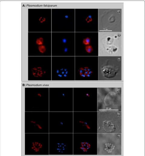
The IFA results in this study reconfirmed these observa-tions demonstrating this anti-PfBiP reacted with all intra-erythrocytic developmental stages ofP. falciparum
(Figure 1A). Localization patterns reflected temporal and spatial changes in localization consistent [with] morphology of the ER during asexual stage growth and development. The fluorescence signal was generally dis-persed in the cytoplasm of ring-stage parasites, increas-ing in abundance durincreas-ing trophozoite and early schizont stages before concentrating in the perinuclear apical end of forming merozoites in late segmenting schizonts. A comparison of theP. falciparumBiP peptide sequence (SGDEDVDSDEL of PF3D7_0917900) that was used to produce the anti-PfBiP antibodies to the orthologous
P. vivax sequence (SADEDVDSDEL of PVX_099315) indicates that this sequence is highly conserved (Table 1). To investigate usefulness of anti-PfBiP as a marker for
P. vivax, this antibody was tested for IFA reactivity to intra-erythrocytic developmental stages ofP. vivax. The fluorescence signal was similar to the dispersed patterns in the cytoplasm of ring, trophozoite and early schizont when it is concentrated at the apical end of daughter mer-ozoites in segmented schizont ofP. vivax(Figure 1B). The anti-PfBiP was specific for the cytoplasm of the P. vivax
and did not react to uninfected reticulocytes.
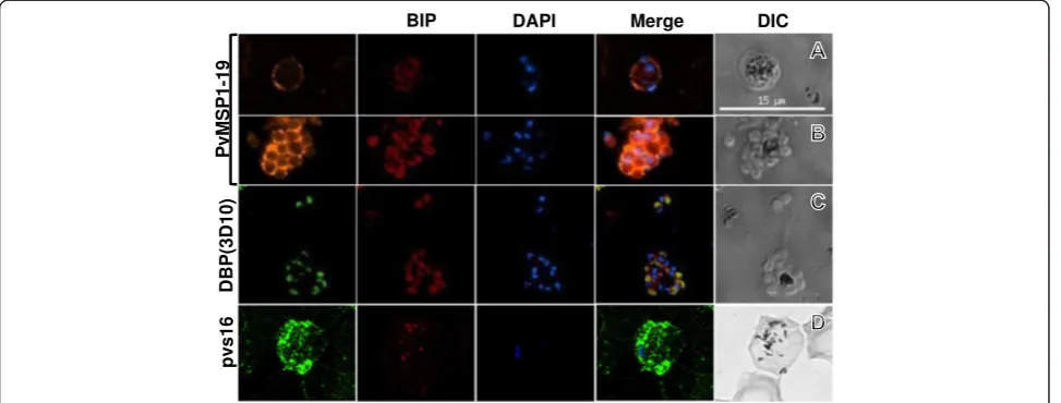
Stage-specific antibodies anti-PvMSP1 and anti-DBP (3D10) were evaluated in combination with anti-PfBiP to distinguishP. vivaxdevelopmental stages. In trophozoite/ early schizont stages, the anti-PvMSP1 showed the cir-cular fluorescence pattern in close proximity to the parasite plasma membrane while anti-PfBiP showed the diffuse fluorescence pattern in the cytoplasm of the para-site (Figure 2). In late-stage segmented schizonts, anti-PfBiP showed fluorescence signal at the apical end in close proximity to the nucleus of the merozoite while the anti-PvMSP1 fluorescence pattern localized at the membrane surrounding the daughter merozoites. The anti-DBP (3D10) fluorescence was detected only in late schizonts and localized at the apical end immediately adjacent to BiP localization (Figure 2C). In P. vivax gametocytes, the anti-Pvs16 was widely distributed in the peripheral cytoplasm while anti-BiP staining was relatively less in a dispersed punctate pattern (Figure 2D).
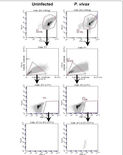
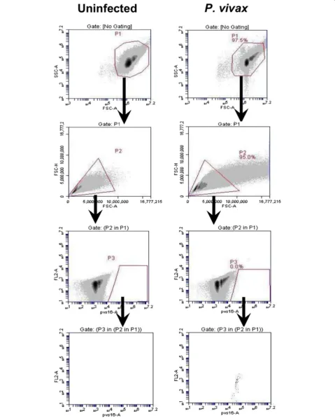
Flow cytometry gating parameters
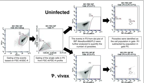
The rapid antibody IFA staining method using anti-PfBiP Alexa Fluor 660 was adapted to blood cells in sus-pension and evaluated by FC for specificity in detecting
The events in gate P1 were viewed in FSC-A/FSC-H profile to observe cell duplet. The single cells from gate P2 were selected to view in the anti-BiP Alexa 660-A/ FL2-A dot plot. The events in gate P3 were selected and viewed in anti-BiP Alexa 660-A/ FL2-A dot plot. In this plot, the events in gate P5 of the uninfected sample were
considered as the background and were excluded from R1. In P. vivax-infected samples, parasites were identi-fied as anti-BiP + events in gate R1 subtracted by the background in gate P5 (Figure 3). For identification of
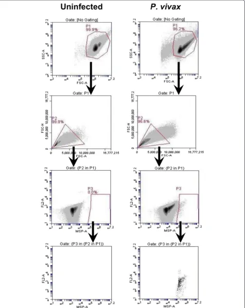
[image:4.595.57.539.88.604.2]events were viewed in anti-MSP1 Alexa Fluor 555-A/ FL2-A (Figure 4), anti-DBP Alexa Fluor 488-A/ FL2-A (Figure 5) and anti-Pvs16-A/ FL2-A dot plot (Figure 6) for anti-PvMSP1-19, anti-DBP (3D10) and Pvs16 stain-ing, respectively.
Determination of parasitaemia and staging of the parasite
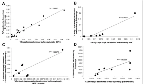
The FC parameters for anti-PfBiP Alexa Fluor 660 detec-tion was used to estimate the total number ofP. vivax -in-fected reticulocytes in all samples. In 18 clinical samples, parasitaemias were determined both by anti-PfBiP FC and by direct light microscopic observation of Giemsa-stained thick blood smears. There was a significant correlation of the parasitaemias determined by FC and microscopy (R2= 0.94, P < 0.001, n = 18) (Figure 7A). To differenti-ate asexual developmental stages, anti-PfBiP was com-bined with stage-specific antibodies, anti-PvMSP1-19
and anti-DBP. MSP1-positive events identified stages beginning with mid-trophozoite/early schizonts through to completion of schizont development (MSP+). DBP-positive events were identified as beginning mid-schizont through to late-stage segmented schizonts (DBP+). Para-site stages positive for Pvs16 were identified as gameto-cytes (Pvs16+). Differential staining patterns were used to define the stages of development within each sample (Table 2). The number of ring and trophozoite stages was obtained by subtracting the number of BiP + events by the number of MSP1+ and Pvs16 + events while the number of early schizonts was obtained by subtracting the number of MSP1+ events by the number of DBP + events. The number of segmented schizonts and gametocytes were ob-tained directly from DBP + and Pvs16+ events, respect-ively. The parasitaemia of the individual asexual blood stages and the gametocyte-infected reticulocytes obtained from FC were compared with the corresponding values obtained from microscopy (Figure 7). There was a positive correlation of the ring stage parasitaemia (Figure 7B), schizont stage parasitaemia (Figure 7C) and gametocy-taemia (Figure 7D) determined by FC and microscopy (R2= 0.8847 (N = 8), 0.6628 (N = 8) and 0.5293 (N = 8), respectively).
[image:5.595.57.291.112.195.2]To monitor the maturation of the parasites by flow cy-tometry, aliquots ofP. vivax-infected blood was collected from short-term cultures at 12-hr and 24-hr intervals. The positive events from each stage-specific antibody were translated to maturation stage of the parasite (Table 2), using the strategy described above, and compared with those obtained from microscopic examination. There was an increase of MSP1+ and DBP + events while the number Table 1 Peptide sequences of BiP amongPlasmodium
species
Plasmodiumspecies Gene ID Amino acid sequence
P. falciparum PF3D7_0917900 SGDEDVDSDEL
P. vivax PVX_099315 SaDEDVeSDEL
P. knowlesi PKH_071520 SGDEDVeSDEL
P. yoelii PY05001 pGDEDVDSDEL
Consensus **DEDV*SDEL
BiP peptide sequences fromP. falciparum, P. vivax, P. knowlesiandP. yoelii
were compared. The concensus peptide sequence of BiP (**DEDV*SDEL) was identified fromP. falciprumBiP orthologs. Sequences were obtained from PlasmoDB (version 10.0) [20].
[image:5.595.57.544.490.675.2]of BiP + events was similar during the examination period as determined by FC (Figure 8). Similar results were obtained by microscopic examination.
Discussion
Plasmodium falciparum BiP is defined as an endoplas-mic reticulum resident protein involved in retrograde transport. Important for this study, the peptide sequence of PfBiP used to prepare an anti-PfBiP peptide serum is very similar among Plasmodium orthologues. Previous studies have demonstrated that BiP is constitutively expressed during asexual blood-stage development and the sera reacted specifically to the ER compartment of
P. falciparum, Plasmodium berghei and Plasmodium yoelii[11,21,22]. In this study, the broad pan-species re-activity of the anti-PfBiP was used to quantifyP. vivax
blood stages in clinical isolates and short-term in vitro
cultures, demonstrating that the anti-PfBiP can be used as a universal antibody to detect developing blood stages of bothP. falciparumandP. vivax.
Counting the parasitaemia by light microscopy is still the gold standard, but it is a difficult, time consuming method requiring a high degree of training to accurately identify P. vivax in thick Giemsa-stained blood smears.
Flow cytometry is becoming a more accessible tool for many malaria research laboratories as the equipment becomes less expensive, more reliable and easier to use. Greater accessibility to FC has translated into more ap-plications developed forP. falciparumresearch [23-25]. Most of the established methods for P. falciparum are based at least partly on nucleic acid staining. While these methods may be very useful for P. falciparum
studies, they have been less valuable forP. vivaxdue to its preferential infection of reticulocytes that contain high levels of nucleic acids. The problem is com-pounded for analysis ofin vitro andex vivocultures of
P. vivaxwhen reticulocytes derived fromin vitro differ-entiation of hematopoietic stem cells are used and are contaminated with nucleated erythroid precursor cells.
Conclusion
The antibody-based staining that has been developed in this study offers the opportunity for a robust, simple method for sensitive real-time detection of theP. vivax -infected cells in a standardized procedure that should re-duce technical variation among researchers and labora-tories. The reactivity of anti-PfBiP to bothP. falciparum
andP. vivaxand any stages of blood-stage parasite offers Gating of the events
based on FSC-A/SSC-A
Gating of the single cells in P2 from FSC-A/FSC-H profile
Parasites were identified as the cell population in gate R1
subtracted by population in gate P5. The events in P3 from dot plot of
BIP Alexafluor660/FL3 were further analyzed to quantify the
number of parasites.
Uninfected
[image:6.595.61.539.89.364.2]P. vivax
an advantage of using this antibody for determining the parasitaemia by flow cytometry. With an optimized protocol, the antibody-based FC can detect one para-site per one million cells count (ten parapara-sites/1 μl PRBC) and generates almost no background. When combined the PfBiP with other stage specific anti-bodies, anti-MSP1-19, anti-DBP and anti-Pvs16, the antibody-based FC can be used to monitor the matur-ation of P. vivax parasite in the culture. The antibody based-FC using anti-PfBiP is a very sensitive and highly accurate method, which offers the new way to detect parasite in vivax malaria research.
[image:10.595.304.540.491.647.2]Figure 7Correlation of total parasitaemia (A), ring/trophozoite stage parasitaemia (B), schizont stage parasitaemia (C), and gametocytaemia (D) ofPlasmodium vivaxdetermined by flow cytometry and counting of the Giemsa-stained blood smear by light microscopy.The total parasitaemia, ring/trophozoite stage parasitaemia, schizont stage parasitaemia and gametocytaemia obtained from flow cytometry shown positive correlation with the one obtained from counting of the Giemsa-stained blood smear by light microscopy (Spearman correlation coefficient = 0.9348 (N = 18), 0.8847 (N = 8), 0.6628 (N = 8) and 0.5293 (N = 8), respectively).
Table 2 Differential staining pattern ofP. vivax
Stage BiP MSP1 DBP PVS16
Ring to Mid-stage Trophozoite + - -
-Late-Trophozoite-early schizont + + -
-Mid-late Schizont + + +
-Gametocyte + - - +
Ring to mid trophozoite were stained with BiP, late trophozoite to early schizont were stained with BiP and MSP1, mid to late schizont were stained with BiP, MSP1 and DBP, and gametocyte was stained with BiP and pvs16.
Figure 8Monitoring the development of the blood-stage
[image:10.595.57.293.637.708.2]Abbreviations
BiP:Binding immunoglobulin protein of endoplasmic reticulum; DBP: Duffy binding protein; FC: Flow cytometry; IFA: Immunofluorescence assay; PvMSP1:Plasmodium vivaxmerozoite surface protein-1; PBS: Phosphate-buffered saline; PBST: PBS containing Tween-20; PfBiP:Plasmodium falciparum
BiP; Pvs16:Plasmodium vivaxsexual antigen 16; WBC: White blood cells.
Competing interests
The authors declare that they have no competing interests.
Authors’contributions
WR and JHA conceived of the study. WR, SPM, JS and JHA designed the experiments. WR, SPM, NR, and SJB carried out the experiments. WR, SJB and JHA drafted the manuscript. All authors read and approved the final manuscript.
Acknowledgements
This work was supported by grants NIH R01AI064478 (JHA), NIH R01AI069314 (KCW), and Bill & Melinda Gates Foundation (JHA). We thank theP. vivax
patients and residents of Kanchanaburi Province, Thailand for their generosity for participation in the study.
Author details
1Department of Global Health, College of Public Health, University of South
Florida, Tampa, FL, USA.2Mahidol Vivax Research Unit, Faculty of Tropical
Medicine, Mahidol University, Bangkok, Thailand.3Department of Biology, Loyola University, Chicago, IL 60660, USA.
Received: 2 October 2013 Accepted: 22 January 2014 Published: 14 February 2014
References
1. Guerra CA, Howes RE, Patil AP, Gething PW, Van Boeckel TP, Temperley WH, Kabaria CW, Tatem AJ, Manh BH, Elyazar IR, Baird JK, Snow RW, Hay SI:The international limits and population at risk ofPlasmodium vivax transmission in 2009.PLoS Negl Trop Dis2010,4:e774.
2. Kitchen SK:The infection of reticulocytes byPlasmodium vivax.Am J Trop Med Hyg1938,18:347–353.
3. Malleret B, Xu F, Mohandas N, Suwanarusk R, Chu C, Leite JA, Low K, Turner C, Sriprawat K, Zhang R, Bertrand O, Colin Y, Costa FT, Ong CN, Ng ML, Lim CT, Nosten F, Rénia L, Russell B:Significant biochemical, biophysical and metabolic diversity in circulating human cord blood reticulocytes.
PLoS One2013,8:e76062.
4. Grimberg BT, Erickson JJ, Sramkoski RM, Jacobberger JW, Zimmerman PA:
MonitoringPlasmodium falciparumgrowth and development by UV flow cytometry using an optimized Hoechst-thiazole orange staining strategy.
Cytometry A2008,73:546–554.
5. Hare JD, Bahler DW:Analysis ofPlasmodium falciparumgrowth in culture using acridine orange and flow cytometry.J Histochem Cytochem1986,
34:215–220.
6. Kosaisavee V, Suwanarusk R, Nosten F, Kyle DE, Barrends M, Jones J, Price R, Russell B, Lek-Uthai U:Plasmodium vivax: isotopic, PicoGreen, and microscopic assays for measuring chloroquine sensitivity in fresh and cryopreserved isolates.Exp Parasitol2006,114:34–39.
7. Pattanapanyasat K, Yongvanitchit K, Tongtawe P, Tachavanich K, Wanachiwanawin W, Fucharoen S, Walsh DS:Impairment ofPlasmodium falciparumgrowth in thalassemic red blood cells: further evidence by using biotin labeling and flow cytometry.Blood1999,93:3116–3119. 8. Staalsoe T, Giha HA, Dodoo D, Theander TG, Hviid L:Detection of
antibodies to variant antigens onPlasmodium falciparum-infected erythrocytes by flow cytometry.Cytometry1999,35:329–336. 9. Theron M, Hesketh R, Subramanian S, Rayner J:An adaptable two-color
flow cytometric assay to quantitate the invasion of erythrocytes by Plasmodium falciparumparasites.Cytometry A2010,77:1067–1074. 10. Kumar N, Syin C, Carter R, Quakyi I, Miller LH:Plasmodium falciparumgene
encoding a protein similar to the 78-kDa rat glucose-regulated stress protein.Proc Natl Acad Sci U S A1988,85:6277–6281.
11. Noe AR, Fishkind DJ, Adams JH:Spatial and temporal dynamics of the secretory pathway during differentiation of thePlasmodium yoelii schizont.Mol Biochem Parasitol2000,108:169–185.
12. Hager KM, Striepen B, Tilney LG, Roos DS:The nuclear envelope serves as an intermediary between the ER and golgi complex in the intracellular parasiteToxoplasma gondii.J Cell Sci1999,112:2631–2638.
13. Russo I, Oksman A, Vaupel B, Goldberg DE:A calpain unique to alveolates is essential inPlasmodium falciparumand its knockdown reveals an involvement in pre-S-phase development.Proc Natl Acad Sci U S A2009,
106:1554–1559.
14. van Dooren GG, Marti M, Tonkin CJ, Stimmler LM, Cowman AF, McFadden GI:Development of the endoplasmic reticulum, mitochondrion and apicoplast during the asexual life cycle ofPlasmodium falciparum.Mol Microbiol2005,57:405–419.
15. Janse CJ, Camargo A, Delportillo HA, Herrera S, Waters AP, Kumlien S, Mons B, Thomas A:Removal of leucocytes fromPlasmodium vivax-infected blood.Ann Trop Med Parasit1994,88:213–216.
16. Udomsangpetch R, Somsri S, Panichakul T, Chotivanich K, Sirichaisinthop J, Yang Z, Cui L, Sattabongkot J:Short-term in vitro culture of field isolates of Plasmodium vivaxusing umbilical cord blood.Parasitol Int2007,56:65–69. 17. Ntumngia FB, Schloegel J, Barnes SJ, McHenry AM, Singh S, King CL, Adams
JH:Conserved and variant epitopes ofPlasmodium vivaxDuffy binding protein as targets of inhibitory monoclonal antibodies.Infect Immun
2012,80:1203–1208.
18. Fennell C, Babbitt S, Russo I, Wilkes J, Ranford-Cartwright L, Goldberg DE, Doerig C:PfeIK1, a eukaryotic initiation factor 2alpha kinase of the human malaria parasitePlasmodium falciparum, regulates stress-response to amino-acid starvation.Malar J2009,8:99.
19. Kumar N, Koski G, Harada M, Aikawa M, Zheng H:Induction and localization ofPlasmodium falciparumstress proteins related to the heat shock protein 70 family.Mol Biochem Parasitol1991,48:47–58.
20. Aurrecoechea C, Brestelli J, Brunk BP, Dommer J, Fischer S, Gajria B, Gao X, Gingle A, Grant G, Harb OS, Heiges M, Innamorato F, Iodice J, Kissinger JC, Kraemer E, Li W, Miller JA, Nayak V, Pennington C, Pinney DF, Roos DS, Ross C, Stoeckert CJ Jr, Treatman C, Wang H:PlasmoDB: a functional genomic database for malaria parasites.Nucleic Acids Res2009,37:D539–D543. 21. Klemba M, Gluzman I, Goldberg DE:APlasmodium falciparumdipeptidyl
aminopeptidase I participates in vacuolar hemoglobin degradation.J Biol Chem2004,279:43000–43007.
22. Kumar N, Nagasawa H, Sacci JB, Sina BJ, Aikawa M, Atkinson C, Uparanukraw P, Kubiak LB, Azad AF, Hollingdale MR:Expression of members of the heat-shock protein-70 family in the exoerythrocytic stages of Plasmodium-bergheiandPlasmodium-falciparum.Parasitol Res1993,
79:109–113.
23. Apte SH, Groves PL, Roddick JS, PdH V, Doolan DL:High-throughput multi-parameter flow-cytometric analysis from micro-quantities of Plasmodium-infected blood.Int J Parasitol2011,41:1285–1294.
24. Fu Y, Tilley L, Kenny S, Klonis N:Dual labeling with a far red probe permits analysis of growth and oxidative stress inP. falciparum-infected erythrocytes.Cytometry A2010,77:253–263.
25. Russell B, Suwanarusk R, Borlon C, Costa FT, Chu CS, Rijken MJ, Sriprawat K, Warter L, Koh EG, Malleret B, Colin Y, Bertrand O, Adams JH, D'Alessandro U, Snounou G, Nosten F, Rénia L:A reliable ex vivo invasion assay of human reticulocytes byPlasmodium vivax.Blood2011,118:e74–e81.
doi:10.1186/1475-2875-13-55
Cite this article as:Roobsoonget al.:A rapid sensitive, flow