S H O R T R E P O R T
Open Access
Experimental validation of predicted subcellular
localizations of human proteins
Nagendra K Chaturvedi
1, Riyaz A Mir
1, Vimla Band
1,2, Shantaram S Joshi
1and Chittibabu Guda
1,2,3,4*Abstract
Background:Computational methods have been widely used for the prediction of protein subcellular localization. However, these predictions are rarely validated experimentally and as a result remain questionable. Therefore, experimental validation of the predicted localizations is needed to assess the accuracy of predictions so that such methods can be confidently used to annotate the proteins of unknown localization. Previously, we published a method called ngLOC that predicts the localization of proteins targeted to ten different subcellular organelles. In this short report, we describe the accuracy of these predictions using experimental validations.
Findings:We have experimentally validated the predicted subcellular localizations of 114 human proteins corresponding to nine different organelles in normal breast and breast cancer cell lines using live cell imaging/confocal microscopy. Target genes were cloned into expression vectors as GFP fusions and cotransfected with RFP-tagged organelle-specific gene marker into normal breast epithelial and breast cancer cell lines. Subcellular localization of each target protein is confirmed by colocalization with a co-expressed organelle-specific protein marker. Our results showed that about 82.5% of the predicted subcellular localizations coincided with the experimentally validated localizations. The highest agreement was found in the endoplasmic reticulum proteins, while the cytoplasmic location showed the least concordance. With the exclusion of cytoplasmic location, the average prediction accuracy increased to 90.4%. In addition, there was no difference observed in the protein subcellular localization between normal and cancer breast cell lines. Conclusions:The experimentally validated accuracy of ngLOC method with (82.5%) or without cytoplasmic location (90.4%) nears the prediction accuracy of 89%. These results demonstrate that the ngLOC method can be very useful for large-scale annotation of the unknown subcellular localization of proteins.
Keywords:Protein subcellular localization, ngLOC prediction, Gene cloning, Experimental validation, GFP fusion, Live cell imaging/confocal microscopy
Findings Background
Subcellular localization of proteins to specific compart-ments is fundamental to the structural organization and functioning of all living cells. Proteins that are localized to unintended organelles have been implicated in the devel-opment of many human diseases; therefore, knowledge of the protein subcellular localization can benefit target identification in the drug discovery process [1].
Protein subcellular localization is an important attribute of protein function; thus, prediction of the same aids in
genome annotation of high-throughput studies. Numerous computational methods have been used for the prediction of proteins subcellular localization [2]. Among these, some are limited by predicting only a small number of organelles in the cell [3,4] while some others exhibit lack of a balance between sensitivity and specificity [5,6]. Previously, we have developed a method called ngLOC, an n-gram based Bayesian method that can predict a wide range of subcellular locations including multiple localizations of proteins [7,8]. This method makes its predictions solely based on the protein sequence information without the need for any extraneous information; therefore ngLOC is highly favorable for proteome-wide prediction of sub-cellular localizations.
The ngLOC method predicts subcellular locations at a high overall accuracy of 89%, while the accuracy is much * Correspondence:[email protected]
1Department of Genetics, Cell Biology and Anatomy, University of Nebraska
Medical Center, 985870 Nebraska Medical Center, Omaha, NE 68198, USA 2Fred and Pamela Buffet Cancer Center, Omaha, USA
Full list of author information is available at the end of the article
higher (93-96%) in organelles with smaller proteomes such as lysosomes, peroxisomes, Golgi, etc., [7] that are typically difficult to predict due to lack of sufficient size datasets. Although computational predictions provide wealth of in-formation for the subcellular localization of proteins, these predictions remain questionable unless they are validated by experimental methods. In the present study, we report experimental validations for ngLOC predicted subcellular localizations of human proteins. Our results corroborated the predicted results; thus ngLOC method can be used for proteome-wide annotation of protein localizations.
Materials and methods
Reagents and materials
Restriction enzymes and DH5α-competent cells were pur-chased from New England Biolabs (MA, USA). Trizol™, transfection reagent Lipofectamine2000TM, and red fluor-escent tagged-subcellular markers including Mitotracker™ Red FM, Lysotracker™ Red, ER-Tracker™ Red, BODIPY® TR ceramide, Hoechst 33342 and Alexa Fluor® 594 WGA were obtained from Invitrogen (CA, USA). A cDNA syn-thesis and ligation kit was purchased from Promega (WI, USA). Primers of all cloned genes for the PCR amplifica-tion were obtained from Integrated DNA Technologies Inc. (Coralville, IA). A 2X PCR amplification kit was pur-chased from Applied Biological Materials Inc. (Richmond, Canada). Plasmid and DNA gel extraction kits were obtained from Qiagen Inc. (Valencia, CA). Fluorodish 35 mm petriplates for live cell imaging were purchased from World Precision Instruments (Sarasota, FL). All plastic wares for mammalian cell culture were pur-chased from Corning Costar Corp. (NY, USA).
Plasmids and constructs
pEGFP-N1 vector was kindly provided by Dr. Hamid Band (UNMC). Ten GFP-tagged full-length human gene constructs (ngLOC predicted), which include: SACM1L, ST13, TUBAL3, USMG5, DECR2, AMY2B, UXS1, LGMN, NR2F1 and NAPB were obtained from Origene Technologies Inc. (Rockville, MD). Six RFP-tagged subcellular specific human gene constructs (positive markers) which include: endoplasmic reticulum specific ETS, Golgi specific TGOLN, peroxisome specific PXPM2, mitochondria specific PDHA1, plasma membrane specific LCK and cytoskeleton specific β-ACTIN were also pur-chased from Origene Technologies Inc. (Rockville, MD).
Isolation of RNA and cDNA preparation
Total RNA was extracted from HEK-293 T cells using the TRIzol™ method according to the manufacturer’s instruc-tions. RNA quantity and purity were determined by UV spectrophotometry and by electrophoresis on a 2% agarose gel. Two micrograms of RNA was then reverse transcribed using random hexamer primers and the superscript RT
enzyme according to the manufacturer’s instructions (Invitrogen, CA).
PCR amplification and gene cloning
PCR Amplification was achieved with the 2X PCR master mix kit containing Taq DNA polymerase using 30–35 cycles according to the manufacturer’s protocols. For amplification, the two sets of primers with appropriate restriction enzymes were used against full-length ORF of each human gene. The primers used for the genes cloning of this study have been tabulated in Additional file 1. Each PCR amplified gene product was separated on 1% agarose gel in 1X TAE buffer (pH 8.0) and visu-alized by ethidium bromide staining. The gel extraction of PCR amplified gene products were purified using a gel extraction kit; then these purified genes products were double digested with restriction enzymes using the combin-ation of either NheI/XhoI, NheI/HindIII or BglII/BamHI. Following restriction digestions, the full-length genes were cloned into a pEGFP-N1 vector using a LigaFast ligation kit and were transformed into E.coli (DH5α) bacterial strain. The positive clones of the genes were screened and con-firmed, following appropriate restriction digestion.
Cell lines and culture conditions
The normal breast epithelial cell lines MCF-10A and MCF-12 F were obtained from the American Type of Culture Collection (Rockville, MD). These cell lines were maintained in D-media described previously [9]. The breast cancer epithelial cell lines MCF-7 and MDA-MB-231 were kindly provided by Dr. Vimla band (UNMC). These cells were cultured in α-MEM media supplemented with 10% FBS (Invitrogen, CA), 2 mM glutamine (Invitrogen, CA), 50μg/ml gentamicin (Invitrogen CA), 1x sodium pyruvate (Invitrogen CA), 1x MEM non-essential amino acid (Invitrogen, CA), 1x HEPES (Invitrogen, CA) and 1μg/ml insulin (Sigma). The cultures were maintained in a humidi-fied incubator adjusted at 5% CO2 and 95% air atmosphere at 37°C. All cultures were passaged twice a week and main-tained at a concentration no greater than 1 × 10⁶/ml.
Transient transfections and confocal microscopy
supplemented with a complete respective media and given another 12 hours of incubation for the protein expression. Following protein expression, subcellular distribution and co-localization of proteins were assessed under the con-focal microscope. Alternatively, other red fluorescent subcellular specific markers (dye) were also used with live cells to validate the each localization. Each predicted localization was confirmed and validated when the co-localization produces a yellow color upon merging the images of specific subcellular markers. Nuclear stain Hoechst-33342 (1μg/ml) was added to live cells for the visualization of nucleus. Fluorescence images of live cells were recorded through Zeiss LSM 710 confocal microscope (Jena, Germany) with 40X objective lens. Images were cap-tured and analyzed with LSM software (Jena, Germany) and processed using standard software programs.
Results and discussion
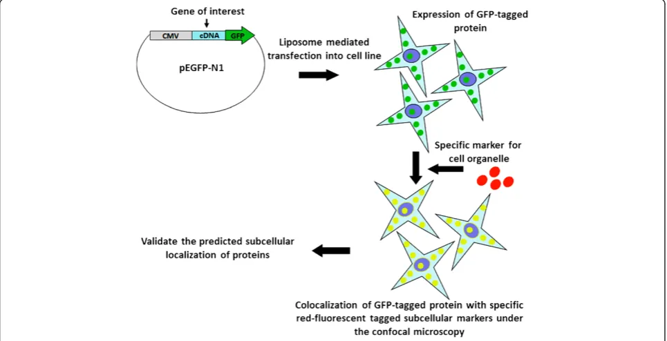
The research strategy used for experimental validation of ngLOC predicted protein subcellular localizations is described in Figure 1. cDNA was synthesized from HEK-293 T cells; with the use of cDNA, the genes of 105 target proteins of human origin were PCR amplified and then cloned into a GFP expression vector (pEGFP-N1) with GFP at the N-terminus as a fusion gene. Using the ngLOC method, 114 target proteins with predicted subcellular
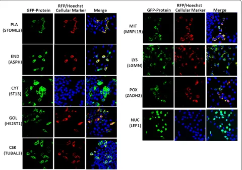
localization (includes 105 locally cloned and nine com-mercially obtained) were selected for this validation study (Additional file 2). GFP expressing fusion genes along with corresponding location-specific RFP-tagged protein markers, were transiently co-expressed following gene transfection into two normal breast (MCF-10A, MCF-12 F) and two breast cancer (MCF-7, MDA-231) cell lines; then their subcellular localization was determined using live cell imaging/confocal microscopy. In the present study, nine different subcellular compartments were selected for val-idating the predicted subcellular localization of proteins. The images in Figure 2 show a representation of validated localizations for predicted proteins in each compartment. The localizations for each compartment (except for nu-cleus and cytoplasm) were determined by observing the colocalization of GFP- and RFP-tagged proteins, which produced a yellow color upon merging the images. For the nucleus and cytoplasm, we used a nuclear (Hoechst) stain to validate the protein subcellular localization in either location (Figure 2).
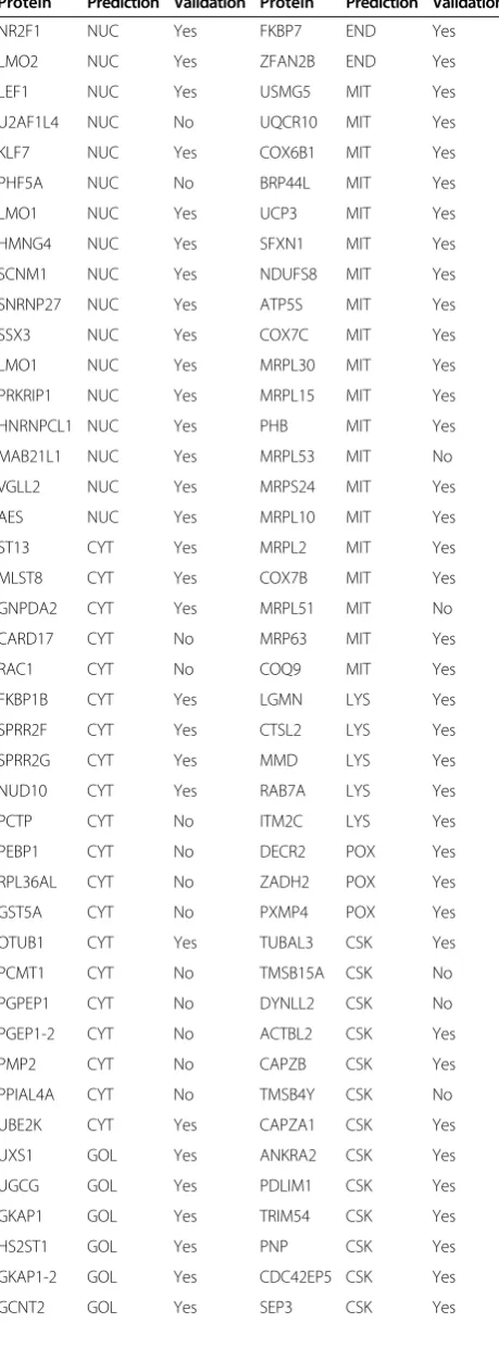
[image:3.595.57.540.420.667.2]Table 1 lists the prediction for each gene tested, along with the outcome of the validation experiment. Similarly, Figure 3 shows the number of tested and succeeded pro-teins in the validation experiments. Our live cell imaging results showed that overall about 82.5% (94 out of 114) of proteins validated in this study agreed with the ngLOC
predicted localizations; these results were consistent in all four cell lines tested. However, with the exclusion of the cytoplasm location that shows the lowest accuracy (45%), the average prediction rate increases to 90.4% (85 out of 94). ngLOC method outputs the predictions in a ranked order by using the associated confidence score (probability) for each location. The top two locations can be predicted within a close confidence range, suggesting that either or both of the predictions can be true. It is known that a number of proteins are localized to multiple organelles in eukaryotic cells (7). To test the accuracy of the second choice we also validated the second predictions of 30 proteins, which included 17 proteins (Set I) whose first choice predictions were proven wrong and 13 proteins (Set II) whose first choice predictions were accurate in the above experiments. From Set I, 10 proteins have shown homogenous distribution in cells, suggesting their localization both in the cytoplasm and nucleus (Table 2). For seven of these 10 proteins, the top two ngLOC predic-tions were cytoplasm or nucleus, which support our results
that these proteins are localized in both nucleus and cytoplasm. From the other 7 proteins in Set I, the second choice predictions were validated as correct only for 2 proteins (Table 2). Validation results on Set II showed that about 46% (6 out of 13) of the proteins tested have also agreed with the second prediction (Table 2), indicating that these proteins are dual localized. With the inclusion of the second prediction validations, we have experimen-tally validated the subcellular localization of 144 ngLOC predictions.
[image:4.595.59.540.90.427.2]for the proteins validated. Our validation results showed that 88% (50 out of 57) of the low CS group proteins were predicted accurately, compared to that of the high CS group proteins, which was 77% (44 out of 57). While these results are counter-intuitive, the high CS group contains a number of proteins that are predicted to be localized to cytoplasm, which has the highest false positive rate. With-out counting the cytoplasmic proteins, the accuracies would be 92% for low CS group and 89% for the high CS group. These results demonstrate that there is no significant correlation between the CS and prediction accuracy. We presume that the lack of correlation is due to the unbalanced selection of validated proteins from a narrow range of confidence scores (see Additional file 3: Table S1), which in turn is due to feasibility (project costs limiting the sample size) and technical (PCR amplification of longer genes) issues that limited our ability to select proteins from a wider CS range for validation.
Table 1 Experimental validation for ngLOC predicted proteins subcellular localization
Protein Prediction Validation Protein Prediction Validation
NR2F1 NUC Yes FKBP7 END Yes
LMO2 NUC Yes ZFAN2B END Yes
LEF1 NUC Yes USMG5 MIT Yes
U2AF1L4 NUC No UQCR10 MIT Yes
KLF7 NUC Yes COX6B1 MIT Yes
PHF5A NUC No BRP44L MIT Yes
LMO1 NUC Yes UCP3 MIT Yes
HMNG4 NUC Yes SFXN1 MIT Yes
SCNM1 NUC Yes NDUFS8 MIT Yes
SNRNP27 NUC Yes ATP5S MIT Yes
SSX3 NUC Yes COX7C MIT Yes
LMO1 NUC Yes MRPL30 MIT Yes
PRKRIP1 NUC Yes MRPL15 MIT Yes
HNRNPCL1 NUC Yes PHB MIT Yes
MAB21L1 NUC Yes MRPL53 MIT No
VGLL2 NUC Yes MRPS24 MIT Yes
AES NUC Yes MRPL10 MIT Yes
ST13 CYT Yes MRPL2 MIT Yes
MLST8 CYT Yes COX7B MIT Yes
GNPDA2 CYT Yes MRPL51 MIT No
CARD17 CYT No MRP63 MIT Yes
RAC1 CYT No COQ9 MIT Yes
FKBP1B CYT Yes LGMN LYS Yes
SPRR2F CYT Yes CTSL2 LYS Yes
SPRR2G CYT Yes MMD LYS Yes
NUD10 CYT Yes RAB7A LYS Yes
PCTP CYT No ITM2C LYS Yes
PEBP1 CYT No DECR2 POX Yes
RPL36AL CYT No ZADH2 POX Yes
GST5A CYT No PXMP4 POX Yes
OTUB1 CYT Yes TUBAL3 CSK Yes
PCMT1 CYT No TMSB15A CSK No
PGPEP1 CYT No DYNLL2 CSK No
PGEP1-2 CYT No ACTBL2 CSK Yes
PMP2 CYT No CAPZB CSK Yes
PPIAL4A CYT No TMSB4Y CSK No
UBE2K CYT Yes CAPZA1 CSK Yes
UXS1 GOL Yes ANKRA2 CSK Yes
UGCG GOL Yes PDLIM1 CSK Yes
GKAP1 GOL Yes TRIM54 CSK Yes
HS2ST1 GOL Yes PNP CSK Yes
GKAP1-2 GOL Yes CDC42EP5 CSK Yes
[image:5.595.58.288.117.738.2]GCNT2 GOL Yes SEP3 CSK Yes
Table 1 Experimental validation for ngLOC predicted proteins subcellular localization(Continued)
GABRAPL2 GOL No ACTRT3 CSK Yes
ZADHHC3 GOL Yes NABP PLA Yes
ST6SIA1 GOL Yes GNAS PLA Yes
SACM1L END Yes KCNIP2 PLA Yes
SEC11A END Yes MOG PLA Yes
SEC11C END Yes CD8B PLA Yes
CNPY3 END Yes CACNG4 PLA Yes
CNPY3-2 END Yes IFITM2 PLA No
SEC61G END Yes STOML3 PLA Yes
DGAT2 END Yes RTP1 PLA Yes
ASPH END Yes ABHD6 PLA Yes
MEST END Yes RASD2 PLA Yes
DOLPP1 END Yes TMEM68 PLA Yes
POFUT1 END Yes RHOV PLA Yes
[image:5.595.303.539.562.688.2]Despite the lower CS range predictions for proteins localized to ER (33-65%), lysosomal (38-50%) and per-oxisomal (23-39%), the validation accuracy is 100% at these locations. Similarly, plasma membrane, cytoskeletal, mitochondrial, Golgi and nuclear proteins recorded about 85% accuracy (Figure 3). Conversely, cytoplasmic proteins scored the lowest with only 45% prediction accuracy. The high false positives in this location can be attributed to the fact that cytoplasm location, being the default loca-tion for protein synthesis, lacks specific targeting sig-nals that makes it difficult to predict. Another reason could be the dual- or multi-localization of about one-third
of cytoplasmic proteins to other locations (7); where, the machine learning methods face difficulty in discriminating the cytoplasmic proteins compared to those from other locations.
Overall, the experimental validations in this study prove that the ngLOC method can predict the subcellular localization of proteins at an accuracy of 82.5%, contrary to the reported accuracy of 89% (7). However, with the ex-clusion of the low performing cytoplasmic location (45%), the average accuracy rate jumped to 90.4% (85 out of 94). As shown in Figure 3, the accuracy is especially notable for the locations with smaller proteomes (ER, Golgi, Lyso-some and PeroxiLyso-some), which are typically difficult to predict by machine learning methods. These results demonstrate the robustness, accuracy, and application in annotating the unknown subcellular localization of pro-teomes of eukaryotic species using the ngLOC method.
Conclusion
This study experimentally validates and reports the accur-acy of a computational method called ngLOC that predicts the subcellular localization of protein sequences in eukaryotic cells. We validated 114 human proteins that were predicted to be localized to nine distinct subcellular locations in eukaryotic cells. The overall validation accur-acy rate of ngLOC method is at 82.5%, while the rate improved to 90.4% just by excluding the cytoplasmic lo-cation, compared to the overall prediction accuracy of 89%. Thus, this validation study demonstrates that ngLOC can be reliably used (with the exception of cyto-plasmic location) to annotate the subcellular localization of proteins and affirms the utility of this method in large-scale annotation of newly sequenced proteomes.
Additional files
Additional file 1:Primer list with the restriction sites used for gene cloning.
Additional file 2:Predictions by ngLOC method for proteins without a known subcellular localization.
Additional file 3: Table S1.Statistics showing the spread and range of confidence scores (CS) in the predicted and validated proteins in each subcellular location.
Abbreviations
GFP:Green fluorescent protein; RFP: Red fluorescent protein; PLA: Plasma membrane; CSK: Cytoskeleton; CYT: Cytoplasm; END: Endoplasmic reticulum; GOL: Golgi complex; MIT: Mitochondria; LYS: Lysosome; POX: Peroxisome; NUC: Nucleus; UNMC: University of Nebraska Medical Center.
Competing interests
The authors declare that they have no competing interests.
Authors’contributions
[image:6.595.58.289.114.564.2]NKC and CG conceived and designed the research study. NKC, RAM, and CG performed the experiments. NKC and CG analyzed the data, wrote the paper, and revised the paper critically. RAM, VB and SSJ revised the paper and
Table 2 Experimental validation of ngLOC top second predicted proteins subcellular localization
Protein First Prediction
Second Prediction
Validation for First prediction
Validation for Second prediction
U2AF1L4 NUC CYT * *
CARD17 CYT NUC * *
RAC1 CYT PLA No No
PCTP CYT NUC * *
PEBP1 CYT PLA No No
RPL36AL CYT NUC * *
GST5A CYT NUC * *
PCMT CYT NUC * *
PGPEP1 CYT NUC * *
PMP2 CYT NUC * *
PPIAL4A CYT MIT No Yes
GABRAPL2 GOL CSK No Yes
MRPL51 MIT CYT No No
MRPL53 MIT CYT No No
DYNLL2 CSK NUC * *
TMSB4Y CSK CYT * *
TMSB15A CSK NUC * *
PXMP4 POX PLA Yes No
MMD LYS PLA Yes No
RAB7A LYS PLA Yes No
SEC61G END MIT Yes Yes
DGAT2 END PLA Yes No
DOLPP1 END PLA Yes Yes
SEC11A END PLA Yes No
CNPY3 END PLA Yes No
LMO1 NUC MIT Yes Yes
LMO2 NUC PLA Yes Yes
NUDT10 CYT PLA Yes No
CDC42EP5 CSK PLA Yes Yes
ZDHHC3 GOL PLA Yes Yes
contributed reagents and materials. All authors read and approved the final manuscript.
Acknowledgements
This research is fully supported by National Institutes of Health [1R01GM086533-01A1 to CG]. The authors thank the confocal core and the bioinformatics and systems biology core at UNMC for their help in this study. The authors also thank Dr. Hamid Band (UNMC) for providing the pEGFP-N1 vector for our cloning experiments, and Mrs. Megan Brown for proofreading the manuscript.
Author details 1
Department of Genetics, Cell Biology and Anatomy, University of Nebraska Medical Center, 985870 Nebraska Medical Center, Omaha, NE 68198, USA. 2
Fred and Pamela Buffet Cancer Center, Omaha, USA.3Eppley Institute for Cancer Research, Omaha, USA.4Bioinformatics and Systems Biology Core, University of Nebraska Medical Center, Omaha, NE 68198-5805, USA.
Received: 1 October 2014 Accepted: 10 December 2014 Published: 15 December 2014
References
1. Donnes P, Hoglund A:Predicting protein subcellular localization: past, present, and future.Genomics Proteomics Bioinformatics2004,2:209–215. 2. Sprenger J, Fink JL, Teasdale RD:Evaluation and comparison of
mammalian subcellular localization prediction methods.BMC Bioinformatics2006,7(Suppl 5):S3.
3. Emanuelsson O, Nielsen H, Brunak S, von Heijne G:Predicting subcellular localization of proteins based on their N-terminal amino acid sequence.
J Mol Biol2000,300:1005–1016.
4. Guda C, Fahy E, Subramaniam S:MITOPRED: a genome-scale method for prediction of nucleus-encoded mitochondrial proteins.Bioinformatics
2004,20:1785–1794.
5. Nakai K, Kanehisa M:A knowledge base for predicting protein localization sites in eukaryotic cells.Genomics1992,14:897–911.
6. Park KJ, Kanehisa M:Prediction of protein subcellular locations by support vector machines using compositions of amino acids and amino acid pairs.Bioinformatics2003,19:1656–1663.
7. King BR, Guda C:ngLOC: an n-gram-based Bayesian method for estimating the subcellular proteomes of eukaryotes.Genome Biol2007,8:R68. 8. King BR, Vural S, Pandey S, Barteau A, Guda C:ngLOC: software and web
server for predicting protein subcellular localization in prokaryotes and eukaryotes.BMC Research Notes2012,5:351.
9. Band V, Zajchowski D, Kulesa V, Sager R:Human papilloma virus DNAs immortalize normal human mammary epithelial cells and reduce their growth factor requirements.Proc Natl Acad Sci U S A1990,87:463–467.
doi:10.1186/1756-0500-7-912
Cite this article as:Chaturvediet al.:Experimental validation of predicted subcellular localizations of human proteins.BMC Research Notes20147:912.
Submit your next manuscript to BioMed Central and take full advantage of:
• Convenient online submission
• Thorough peer review
• No space constraints or color figure charges
• Immediate publication on acceptance
• Inclusion in PubMed, CAS, Scopus and Google Scholar
• Research which is freely available for redistribution