Open Access
Research
Uptake of proteins and degradation of human serum albumin by
Plasmodium falciparum
– infected human erythrocytes
Ahmed EL Tahir
1,2
, Pawan Malhotra
2
and Virander S Chauhan*
2
Address: 1Department of Biochemistry and Nutrition, Faculty of Medicine, University of Gezira, P.O.Box 20, Sudan and 2Malaria Group, International Centre for Genetic Engineering and Biotechnology, P.O.Box 10504, Aruna Asaf Ali Marg, New Delhi 110067, India
Email: Ahmed EL Tahir - ahmedeltahirm@yahoo.com; Pawan Malhotra - pawanm@icgeb.res.in; Virander S Chauhan* - virander@icgeb.res.in * Corresponding author
Abstract
Background: Intraerythrocytic malaria parasites actively import obligate nutrients from serum and export proteins and lipids to erythrocyte cytoplasm and membrane. The import of macromolecules in the malaria parasite has been the subject of many debates. To understand the import of macromolecules by the parasite, we studied the uptake of proteins by Plasmodium falciparum infected human erythrocyte.
Methods: Proteins were biotin labelled individually, purified on a gel filtration column and added to uninfected and infected asynchronized culture. The uptake of these proteins by malaria parasites was determined by western blot analysis of parasite pellet and their different fractions using streptavidin-horseradish conjugate. To further confirm this import, we studied the uptake of 125
I-labelled proteins by western blot analysis as well as used direct immunofluorescence method.
Results: Here we show that biotin labelled and radio-iodinated polypeptides of molecular sizes in the range of 45 to 206 kDa, when added in the culture medium, get direct access to the parasite membrane through a membrane network by by-passing the erythrocyte cytosol. The import of these polypeptides is ATP-dependent as sodium azide treatment blocks this uptake. We also show that malaria parasites have the ability to take up and degrade biotin labelled human serum albumin, which has been shown to be essential for the parasite growth.
Conclusions: These results can be used, as a basis to explore the role of human serum albumin in the intraerythrocytic development of parasites, and this in turn can be an important adjunct to the development of novel antimalarial drugs.
Background
During the asexual erythrocytic stage of their life cycle, the malaria parasite Plasmodium falciparum grows and propa-gates within the red blood cells (RBCs) of their host. With-in RBC, a sWith-ingle With-intraerythrocytic parasite reproduces asexually to produce 16 to 32 progeny within 48 h. The high rate of multiplication necessitates efficient trafficking of solute and macromolecules between the external
medi-um and the parasites. Trafficking pathways in malaria-in-fected erythrocytes are complex and the solute passing between the parasite and plasma must traverse a series of three membranes, those of RBC, the parasitophorous vac-uole and the parasite [1]. It has largely been recognized that malaria parasites usually import low molecular weight nutrients such as polyols, amino acids, lipids, nu-cleosides, inorganic anions and cations from the plasma
Published: 7 May 2003
Malaria Journal 2003, 2:11
Received: 3 February 2003 Accepted: 7 May 2003
This article is available from: http://www.malariajournal.com/content/2/1/11
[2–4]. During the last decade, macromolecule uptake by malaria-infected erythrocytes has been the subject of con-tention among different groups. Using fluorescent macro-molecules, Pouvelle and co-workers [5] have shown that intraerythrocytic P. falciparum can endocytose dextran, protein A and an IgG2 antibody. It was shown that these molecules do not cross the erythrocyte or parasitophorous vacuole membranes, but rather gain direct access from the external medium to the parasite through a duct. Based on their findings, they proposed a parasitophorous duct pathway for the direct access of macromolecules through the formation of aqueous channels. These findings were further supported by Loyevsky et al. [6] who showed that desferrioxamine and phloridzin drugs that inhibit para-site growth in culture find direct access to the parapara-site from external medium. In another report, Goodyer et al. [7] proposed two distinct pathways for the macromolecu-lar transport. Recently, Bonday et al [8] showed how the different recombinant fragments of RBC δ – aminonle-vulinate dehydratase (ALAD) were imported into the par-asite from the external medium. However, the concept of direct access of macromolecules by the malaria parasite has been refuted by a number of groups who reported the inability of P. falciparum – infected erythrocytes to take up either macromolecules or small latex beads [9–12]. These groups suggested that the manifestation of the duct might be due to experimental artefacts. One of these groups showed the existence of an interconnected network of tubovesicular membranes (TVM) and suggested that these membrane can only import small molecules whereas the secretary cleft and lipid rafts may play a role in endovacu-lation and macromolecules transport [12–14]. We antici-pate that at least some aspect of these pathways may be biologically unique and therefore potentially targets for chemotherapeutic intervention. Here we show that biotin labelled and radio-labelled proteins of different molecu-lar weights gain access to the parasite, when added in the external medium. These observations were based on the western blot analysis of these proteins in the purified par-asite extract, unlike earlier studies, which were based mostly on immunofluorescence localization. We also show that these imported biotinylated proteins in the par-asite existed in the Trixton X100 insoluble parpar-asite mem-brane fraction and their uptake by the parasite was ATP dependent. We extend our analysis to show that, when bi-otinylated human serum albumin is taken up by the par-asite, it undergoes proteolysis, suggesting that the parasite has the ability to take up human serum albumin which is important for intraerythrocytic growth and differentiation.
Materials and Methods
Materials
Human serum albumin (HSA), egg albumin, β amylase, β galactosidase, sodium azide, Brefeldin A (BFA),
Mon-ensin, Isopropyl-β-D-thiogalactopyranoside (IPTG) and Luria Bertani Broth (LB) were obtained from Sigma Chemical Co. (St. Louis, Mo, USA). A protein biotinyla-tion kit was obtained from (Amersham Pharmacia Bio-tech, U.K.). Na 125I (1 mCi/µl) was purchased from
Amersham Life Science. Bicinchoninic acid (BCA) protein assay kit and iodobeads were obtained from Pierce. A C8 HPLC column and C18 Sep-Pak were from Waters. Vector pET-3d was purchased from Novagen (Madison, WI, USA).
Parasite Culture
P. falciparum culture (3D7 strain) was grown essentially by the method of Trager and Jensen [15] in RPMI 1640 me-dium, supplemented with 10% heat inactivated pooled human serum and O +ve washed human RBC. The culture flasks were incubated at 37°C in a CO2 (5%) incubator. Parasite cultures were synchronised by treating the culture with 5% sorbitol [16]. Parasitemia was measured from methanol fixed and Giemsa-stained smears.
Isolation of parasite and parasite membranes from infect-ed rinfect-ed blood cells
The parasitized RBCs were harvested by low speed centrif-ugation (800 × g, 5 min) and washed five times with in-complete RPMI medium. The pellet solutions were suspended in an equal volume of 0.15% saponin in PBS (weight/volume), the final concentration of saponin was 0.075% and incubated at 37°C in a shaking water bath for 20 min to allow complete lysis of the RBC. The lysate was centrifuged at 1000 × g for 10 min at 4°C. The superna-tant was collected and the parasite pellet was washed five times with cold PBS at 4°C [8,17]. To isolate the parasite membrane fraction, the parasite was lysed for 1 h at 4°C in 20 mM Tris-HCl buffer, pH 7.5 containing 0.2% (weight/volume) Triton X-100, and was sonicated briefly. The lysate was spun at 12000 × g for 30 min and the mem-brane pellet (Triton X-100 insoluble fraction) and the par-asite cytoplasm were used for further analysis. [8].
Expression and purification of recombinant P. falciparum
histidine rich protein-2 (PfHRP-2)
Plasmid containing the gene encoding for PfHRP-2 in a pET-3d vector was transformed into E. coli BL21 (DE3) cells and the transformed E.coli were grown in LB media containing 100 µg ampicillin/ml. Expression of the pro-teins was induced by the addition of 0.4 mM IPTG. For the large-scale purification of proteins, one litre of the cul-tures was grown and induced by IPTG [18]. The protein was purified by metal-chelate chromatography on Ni+2
-nitroacetate column using imidazole for elution [19].
Biotinylation and radio-iodination of proteins
protocol. Briefly, the protein was diluted to 1 mg/ml in the 40 mM bicarbonate buffer pH 8.6 and 40 µl of bioti-nylation reagent was added for each milligram of protein. The reaction was incubated at room temperature for 1 h with constant agitation. The biotinylated protein was then purified by gel filtration chromatography using Sephadex G-25 column.
Radio-iodination of protein was done using Iodo-Beads according to the manufacturer's instructions. Iodinated proteins were purified using Sep-Pak Plus C18 cartridge.
Uptake of proteins and their analysis
To study the uptake of proteins, biotin labelled or radio-iodinated proteins were diluted to the appropriate con-centration using 1X RPMI 1640 to a final pH 7.4. P. falci-parum cultures (5% parasitaemia; 10 ml) were incubated for different times with labelled polypeptides. After the in-cubation, parasite pellets and their different fractions were purified as described above and the labelled proteins in these fractions were analysed by SDS-PAGE and western blot analysis.
For SDS-PAGE analysis, purified parasite pellets or mem-brane fractions were solubilized in SDS sample buffer with (5%) β – mercaptoethanol, boiled and electro-phoresed on 7.5% or 10% SDS-polyacrylamide gels. In case of radio-iodinated proteins, the gels were dried and exposed directly to X-ray film. To detect biotin labelled proteins, proteins were electrotransferred on nitro-cellu-lose membrane. The membranes were subsequently blocked with 5% milk or casein, incubated with streptavi-din-conjugated horseradish peroxidase and detected by enhanced chemiluminescence.
Treatment of infected cultures with sodium azide, BFA and Monensin
Treatment of parasitised erythrocytes with sodium azide was carried out as described earlier [20]. Briefly, 2x10⁷
cells in the infected cultures were incubated with 1 mM sodium azide for 6 min at 37°C. The cells were subse-quently incubated with labelled proteins and cultures were continued for another 30 min. BFA and Monensin treatments were performed as described previously [21,22]. Briefly, 2x10⁸ infected erythrocytes were
resus-pended at a haematocrit of 5% in RPMI 1640 medium containing 10% human serum and incubated with BFA at 10 µg/ml or with monensin at 5 µg/ml. After the treatment with BFA or monensin for 10 min, labelled proteins were added and the cultures were continued for another 6 h.
Incubation of human serum albumin (HSA) with tropho-zoite extract
Infected erythrocytes (4.8x10⁸ trophozoites) were
harvest-ed by low speharvest-ed centrifugation (800 × g, 5 min) and
washed five times with incomplete RPMI medium. They were the suspended in 200 µl of 10 mM Tris pH 7.5, sub-jected to 4 freeze/thaw cycles and centrifuged at 15,000 × g for 30 min at 4°C. The supernatants thus obtained were collected and used as a trophozoite extracts. 40 µl of tro-phozoite extract were incubated with 130 µg of HSA. Two control reactions, one containing only parasite extracts and the other containing HSA were set alongside the ex-perimental reaction. All the three reactions were brought to a 200 µl of volume with 10 mM tris-HCl (pH 7.0), in-cubated overnight at 37°C and then centrifuged at 15,000 × g for 30 min at 4°C. These samples were later analysed on a C8 reverse phase column and chromatography was performed with a linear gradient of acetonitrile buffer A (0.05 TFA in H2O) to buffer B (70% CH3CN). Products
were detected at 214 nm.
Direct immunofluorescence
Samples from parasite cultures incubated with biotinylat-ed proteins were taken and processbiotinylat-ed for immunofluores-cence. Smears of infected red blood cells were made on glass slides and allowed to dry for 10 min then fixed with acetone: methanol (9:1 vol/vol) mixture at -20°C and dried again. Slides were washed with PBS several times, blocked with 1% bovine serum albumin in 0.5% Tween 20 and subsequently, incubated in 1:100 diluted strepta-vidin-fluorescein isothiocyanate (FITC) for 2 h at room temperature. The slides were washed with PBS several times before visualization by fluorescence microscopy.
Results
Uptake of proteins by P. falciparum-infected human erythrocytes
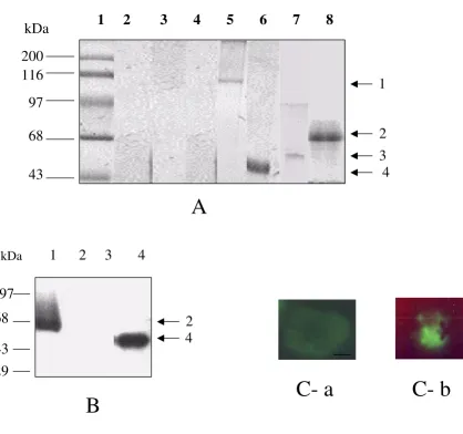
The uptake of macromolecules by infected human RBC has been mostly studied using fluorescent labelled mole-cules and microscopic examination of the treated para-sites [5,7,10], However, the existence of macromolecular import has been the subject of debate because of the ex-perimental designs [9–11]. To shed more light on macro-molecular import into the malaria infected human RBCs; we used biotin labelled as well as radio-iodinated proteins of different molecular weights. Five proteins, egg albumin (45 kDa), recombinant PfHRP-2 (66 kDa), HSA (68 kDa),
the infected RBC cytoplasm after saponin lysis also did not show the presence of biotin labelled – polypeptides (Fig. 1A, lane 4). To further confirm this import, we stud-ied the uptake of 125I labelled recombinant PfHRP-II and
egg albumin. Both these proteins were also taken up by the infected RBC as shown in Fig. 1B (lanes 1 and 4).
Up-take of proteins by malaria parasite was also confirmed by direct immunofluorescence (Fig. 1C).
[image:4.612.82.501.87.480.2]We next studied the location of biotin labelled proteins in different compartments of parasite. Analysis of parasite cytoplasm and parasite membrane fractions using strepta-vidin-horseradish conjugate showed that the labelled Figure 1
Uptake of high molecular weight proteins by infected human erythrocytes (A) Western blot analysis of biotin labelled proteins in different fractions of saponin lysed parasitized RBC and uninfected RBCs using streptavidin horseradish peroxidate conju-gate. Protein markers (lane 1), saponin treatment lysate of uninfected RBCs incubated with labelled PfHRP-2 (lane 2), intact parasite pellet of infected RBCs incubated with unlabelled recombinant PfHRP-2 (lane 3), supernatant of infected RBCs treated with labelled PfHRP-2 (lane 4), parasite pellet of infected RBC incubated with labelled β galactosidase (lane 5), egg albumin (lane 6), β amylase (tetramer) (lane 7) and recombinant PfHRP-2 (lane 8). (B) SDS-PAGE analysis of radio-iodinated proteins imported by the parasite from the culture. Saponin lysed intact parasite pellet incubated with recombinant 125I PfHRP-2 (lane
1) and 125I egg albumin (lane 4). Supernatant of saponin lysed infected RBC incubated with 125I PfHRP-2 (lane 2) and 125I egg
albumin (lane 3). (C) Direct immunofluorescence analysis to show the uptake of HAS by infected human erythrocytes. (A) uninfected RBCs incubated with biotin labelled protein and (B) infected RBCs incubated with biotin labelled protein.
1 2 3 4 5 6 7 8
200
116
97
68
43
68
A
B
97
43
29
1 2 3 4
kDa
kDa
C- a
C- b
1
2
3
4
proteins were localized only in Triton X-100 insoluble fraction of the parasite (Fig. 2 lane 3). Localization of bi-otin labelled polypeptide in Triton X-100 insoluble frac-tions indicated that import of labelled protein may takes place through membranes.
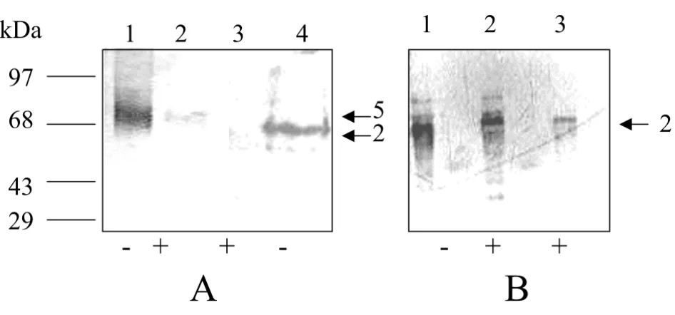
Effect of inhibitors on protein import to the infected hu-man red blood cells
To further understand the mechanism of import of pro-teins into infected RBCs we investigated the effect of sodi-um azide on the uptake of biotin labelled HSA and recombinant PfHRP-2. Sodium azide treatment of malar-ia parasite has been shown to considerably deplete ATP levels in the parasite [20]. As shown in Fig. 3A there was a significant decrease in the uptake of these proteins by the parasites after sodium azide treatment (lanes 2 and 3), compared to the uptake shown by untreated parasites (lanes 1 and 4). We also investigated the effect of BFA and monensin on the import of biotin labelled recombinant PfHRP-2. Both BFA and monensin have been previously used effectively in eukaryotic cells as well as in P. falci-parum to study the export of different proteins [20,21]. Both these inhibitors did not affect the uptake of recom-binant PfHRP-2 (Fig. 3B, lanes 2 and 3). These studies thus suggested that the import of proteins into the parasites is insensitive to BFA and monensin but depend-ent on ATP.
Uptake of human serum albumin and its degradation by P. falciparum
To investigate the fate of proteins imported into the para-site, we carried out a time course study. The uptake of three different biotin labelled proteins, namely egg
albu-min, recombinant PfHRP-2 and HSA were studied up to 12 h. As shown in Fig. 4 we could detect both egg albumin and PfHRP-2 in the intact parasite up to 12 h (lanes 1,2,3 and 4). However, to our surprise, we could detect biotin labelled HSA in the parasite pellets only up to 30 min (lane 5). At 12 h we could not detect any biotinylated HSA in the intact parasite pellets (lane 6). These results suggest-ed that the parasite has the ability to take up HSA and this HSA gets slowly processed in the parasite.
P. falciparum trophozoite extract generate discrete pep-tide fragments of HSA
It has been shown that serum is essential for the growth and progression of malaria parasites. Fractionation and analysis of serum components have shown that only frac-tions containing serum albumin have the ability to sus-tain growth and development of malaria parasite [23]. To further explore whether peptides/amino acids are pro-duced from HSA by the enzymatic activity of the trophozoite, HSA was incubated with P. falciparum tro-phozoite extract. As a control, HSA alone and trotro-phozoite extract alone were incubated separately overnight under similar conditions. Reverse phase chromatography analy-sis was performed on the product of overnight incubation of the parasite lysate with HSA (Fig. 5B,5a). The action of trophozoite extract on HSA resulted in a substantial de-crease in the optical density (O.D.) value of HSA and gen-erated numerous peaks on RP-HPLC which were neither present in lysate alone (Fig. 5B,5b) nor in HSA alone (Fig. 5A). However, the trophozoite extract produced very little degradation of egg albumin (data not shown). This result and the earlier in vitro study showing the degradation of HSA, clearly indicated that parasitized RBC has the ability to degrade HAS at neutral pH.
Discussion
[image:5.612.72.283.94.212.2]P. falciparum, an intraerythrocytic protozoan parasite ex-ports antigens and imex-ports extracellular nutrients to survive. It is now well established that a number of para-site proteins such as PfEMP-2, PfHRP-1 and PfHRP-2 are exported to the host cell or the extracellular medium by the parasites [12,24,25]. While the export of proteins has been accepted the import of proteins and macromolecules by malaria parasite has not been equivocally accepted. Studies showing the uptake of large macromolecules by P. falciparum has been questioned [10–12]. However, the de-pendence of parasite growth and intraerythrocytic devel-opment on serum and trafficking of large molecular weights compounds such as desferrioxamine and phlo-ridzin that inhibit parasite growth suggest that macromo-lecular import does take place in this parasite [5–7,22]. While investigating the export and import of PfHRP-2, a histidine rich protein being secreted in large amounts throughout the erythrocytic stage of the malaria parasite, we found that biotin labelled PfHRP-2 was also taken up Figure 2
Western blot analysis to show location of imported biotin labelled PfHRP-2 in different compartments of parasite. Supernatant after saponin lysis of infected RBC (lane 1), puri-fied intact parasite pellet (lane 2), Triton X-100 insoluble par-asite membrane fraction (lane 3), parpar-asite cytoplasm (lane 4).
1 2
3 4 5
97
68
43
29
kDa
by the parasite from the culture medium. We wondered if this uptake is specific for PfHRP-2 and extended this study to other polypeptides of different molecular weights. Sur-prisingly, we found that all biotin labelled/radio-iodinat-ed proteins, when addlabelled/radio-iodinat-ed in the culture mlabelled/radio-iodinat-edium were, found to be localized in the parasite pellets and in partic-ular in Triton X-100 insoluble fraction regardless of their size. We did not observe any of the labelled protein in host cell cytoplasm. Their results indicated that polypep-tides, when added to the culture medium, gain direct ac-cess to the parasite regardless of their molecular weights and primary sequences. This situation is however, differ-ent to the process of translocation of malaria proteins in different compartments of the parasite which seem specif-ic to signal sequences [26]. In comparison to earlier ap-proaches where uptake of macromolecules was studied using confocal/electron microscopy, we studied the up-take by SDS-PAGE and western blot analysis. While we were studying this import of polypeptides, Bonday et al.
[8] demonstrated the import of different polypeptide frag-ments of ALAD into the parasite. Nyalwidhe et al. [27] re-ported that nonpermeant biotin derivatives gain access to the parasitophorous vacuole in P. falciparum-infected erythrocytes permeabilized with streptolysin O, where the derivatives gain access to the vacuole lumen but not to the
parasite cytosol. These results thus supported the previous observation of Pouvelle et al. [5], which suggested that macromolecules from the external medium get access into the intracellular parasite by-passing the host cell cyto-plasm. Since we performed the experiments under normal culture conditions, carefully checked for the purity of macromolecules, ruled out the binding of biotin labelled molecules uninfected RBC membranes and localized this labelled molecules by SDS-PAGE in infected RBCs, we be-lieve that these results and their interpretation are valid and cannot be attributed to contamination of the tracers by low molecular weight impurities or due to the degrada-tion of labelled macromolecules into smaller molecules. Recently, we demonstrated the inhibitory effect of falci-pain's dsRNAs to the parasites suggesting the permeability of these macromolecules to the infected human erythro-cytes [28].
[image:6.612.62.535.99.320.2]The export of several parasite proteins (PfEMP2 and PfHRP2) has been shown to be mostly dependent on a BFA dependent pathway [24]. Low levels of BFA insensi-tive export have also been shown [25]. To understand the mechanism of import or uptake of protein by the parasite, in the present study, we treated the parasite cultures with BFA, monensin or sodium azide. BFA and monensin, Figure 3
(A), Western blot analysis to show the effect of sodium azide on the import of biotinylated HSA and PfHRP-2 to the infected human red blood cells. (A), Uptake of HSA by untreated parasite culture (lane 1) uptake of HSA after sodium azide treatment (lane 2). Uptake of PfHRP-2 by sodium azide treated parasite culture (lane 3), uptake of PfHRP-2 by untreated parasite culture (lane 4). (B), Western blot analysis to show the effects of BFA and monensin to the uptake of biotinylated PfHRP-2. Parasite pellets of untreated culture (lane 1), BFA treated culture (lane 2) and monesnin treated culture (lane 3).
1 2 3
kDa
1 2 3 4
+ +
-2
5
- + +
97
68
2
43
29
which inhibit intracellular transport of protein through the endoplasmic reticulum/Golgi complex, did not affect the uptake of different proteins, while sodium azide, which is known to deplete ATP in the parasite [20], re-duced the uptake of proteins. These results were in agreement with the earlier observation by Pouvelle et al.
[5] where uptake of rhodamine-dextran was shown to be inhibited by depletion of ATP.
It has been shown that in vitro serum albumin and its as-sociated fatty acids are essential for intraerythrocytic de-velopment and cell cycle progression of P. falciparum [23]. We extended our studies to understand the fate of HSA taken up by P. falciparum. Surprisingly, our experiments provided evidence that HSA is the only protein of those investigated here that undergoes proteolysis inside the parasite in a time-dependent manner. To further explore the proteolysis of HSA by the parasite, HSA was incubated with parasite extract overnight and analysed on a C18 col-umn. A similar proteolytic study has been earlier carried out on haemoglobin by Kolakovich et al. [29]. As shown in Fig. 5B, most of the HSA was found to be cleaved at neutral pH by the parasite extract. Taken together, our data suggest the uptake and breakdown of HSA by the ma-laria parasite. Serum albumin is known as a lipid carrier protein in blood, and it has been shown earlier that both lipids as well as serum albumin are essential for optimum parasite growth in vitro [23,30]. It may be that the sole role of HSA is to provide lipids to the parasite for its growth, but it is tempting to speculate, based on the results of the present study, that degradation of HSA inside the parasite
may serve as an additional source, along with haemoglob-in, for the amino acid pool required by the parasite for its growth. Further investigations to elucidate the role of HSA will extend out understanding of lipid and protein uptake by malaria parasites and may help identify additional chemotherapeutic targets to combat the persistent and deadly malaria parasite P. falciparum.
Authors' contributions
[image:7.612.326.544.88.449.2]AE carried the practical work, PM supervise and participat-ed in the biotinylation and immunoflourescence works, VSC the group leader participated in its design and co-or-dination. All authors read and approved the manuscript. Figure 4
Western blot analysis of saponin lysed parasite pellet to show the uptake of biotinylated egg albumin after 30 min (lane 1) and after 12 h (lane 2), PfHRP-2 after 30 min (lane 3) and 12 h (lane 4) and HSA after 30 min (lane 5) and 12 h (lane 6) of incubation.
kDa 97
1 2 3 4 5 6
5
2
684
4329
Figure 5
Incubation of HSA with trophozoite lysate generates discrete fragments. Reverse phase chromatography profile of HSA[A] trophozoite lysate alone [B (b)] and HSA plus trophozoite lysate incubated overnight [B (a)].
A
B
b a
[image:7.612.57.289.105.218.2]Publish with BioMed Central and every scientist can read your work free of charge "BioMed Central will be the most significant development for disseminating the results of biomedical researc h in our lifetime."
Sir Paul Nurse, Cancer Research UK
Your research papers will be:
available free of charge to the entire biomedical community
peer reviewed and published immediately upon acceptance
cited in PubMed and archived on PubMed Central
yours — you keep the copyright
Submit your manuscript here:
http://www.biomedcentral.com/info/publishing_adv.asp
BioMedcentral
Acknowledgements
We thank Dr. J. S. Grewal and Dr. Ashima Bhardwaj for their help in radio-labelling and HPLC purification. We also thank Dr. Shahid Jameel for his critical comments and suggestion on the manuscript. Dr. Ahmed EL tahir was supported by ICGEB postdoctoral fellowship.
References
1. Ginsburg H and Kirk K Membrane transport in the malaria – in-fected erythrocytes. In "Malaria" ASM Press, Washington DC
1998, 219-232
2. Sherman IW Membrane structure and function of malaria par-asites and the infected erythrocyteParasitology 1985, 91: 606-645
3. Sherman IW Mechanism of molecular trafficking in malaria
Parasitology 1988, 96:857-881
4. Elford BC, Cowan GM and Ferguson DSP Transport and traffick-ing in malaria-infected erythrocytes Trends Microbiol 1997,
5:463-465
5. Pouvelle B, Spiegel R, Hsiao L, Howard RJ, Morris RL, Thomas AP and Taraschi TF Direct access to serum macromolecules by in-traerythrocytic malaria parasitesNature 1991, 353:73-75 6. Loyevsky M, Lytton SD, Mester B, Libman J, Shanzer A and
Caban-tchik ZI The antimalarial action of desferal involves a direct access route to erythrocyte (Plasmodium falciparum) parasite
J Clin Invest 1993, 91:218-224
7. Goodyer ID, Pouvelle B, Schneider TG, Trelka DP and Taraschi TF
Characterization of macromolecular transport pathways in malaria – infected erythrocytes Mol Biochem Parasitol 1997,
87:13-28
8. Bonday ZQ, Dhanasekaran S, Rangarajan PN and Padmanaban G Im-port of host δ – amino-levulinate dehydratase into the malarial parasite: Identification of new drug targetNature
2000, 6:898-903
9. Fujioka H and Aikawa M Morphological changes of clefts in Plas-modium-infected erythrocytes under adverse conditionsExp Parasitol 1993, 76:302-307
10. Haldar K and Uyetake L The movement of fluorescent endo-cytic tracers in Plasmodium falciparum infected erythrocytes
Mol Biochem Parasitol 1992, 50:161-178
11. Hibbs AR, Stenzel DJ and Saul A Macromolecular transport in malaria – does the duct exist?Eur J Cell Biol 1997, 72:182-188 12. Lauer SA, Rathod PK, Ghori N and Halder K A membrane net
work for nutrient import in red cells infected with the malar-ia parasiteScience 1997, 276:122-1225
13. Haldar K, Mohandas N, Samuel BU, Harrison T, Hiller NL, Akompong T and Cheresh P Protein and lipid trafficking induced in eryth-rocytes infected by malaria parasitesCell Microbiol 2002, 4: 383-395
14. Haldar K, Samuel BU, Mohandas N, Harrison T and Hiller NL Trans-port mechanisms in Plasmodium infected erythrocytes: Lip-id rafts and a tubovesicular net work Int J Parasitol 2001,
31:1393-1340
15. Trager W and Jensen JB Human malaria parasites in continuous cultureScience 1976, 193:673-675
16. Lambros C and Vanderberg JB Synchronization of Plasmodium falciparum erythrocytic stages in culture J Parasitol 1979,
65:428-420
17. Siddiqui WA, Kan SC, Kramer K and Richmond-Cru SM In vitro pro-duction and partial purification of Plasmodium falciparum an-tigenBull World Health Organ 1979, 57:75-82
18. Sullivan DJ, Gluzman IY and Goldberg DE Plasmodium falciparum hemoglobin mediated by histidine rich proteinsScience 1996,
271:219-221
19. Pandey AV, Bisht H, Babbarwal VK, Srivastava J, Pandey KC and Chau-han VS Mechanism of malarial haem detoxification by chloroquineBiochem J 2001, 335:333-338
20. Haldar K, de Amorin AF and Cross GAM Transport of fluorescent phospholipid analogues from the erythrocytic membrane to the parasite in Plasmodium falciparum-infected cellsJ Cell Biol
1988, 108:2183-2192
21. Wiser MF, Lanners HN, Bafford RA and Favaloro JM A novel alter-native secretory pathway for the export of Plasmodium pro-teins into the host erythrocyteProc Natl Acad Sci USA 1997,
94:9108-9113
22. Qui Z, Tufaro F and Gillam S Brefeldin A and monensin arrest cell surface expression of membrane glycoproteins and re-lease of rubella virusJ Gen Virol 1995, 76:855-863
23. Mitamura T, Hanad K, Ko-Mitamura P, Nishijima M and Honi T Se-rum factors governing intraerythocytic development and cell cycle progression of Plasmodium falciparum Parasitol Int
2000, 49:219-229
24. Howard RJ, Lyon JA, Uni S, Aikawa M, Aley SB, Leech JH, Lew AM, Wellems TE, Rener J and Taylor DW Secretion of a malarial his-tidine-rich proteins (PfHRP-II) from Plasmodium falciparum infected erythrocytesJ Cell Biol 1986, 103:1269-1277
25. Taylor DW, Parra M, Chapman GB, Atearns ME, Rener J, Aikawa M, Uni S, Aley SB, Panton LJ and Howard RJ Localization of Plasmodi-um falciparPlasmodi-um histidine rich protein 1 in the erythrocyte skel-eton under knobsMol Biochem Parasitol 1987, 25:165-174 26. Burghaus PA and Lingelbach K Luciferase, when fused to an
N-terminal signal peptide, is secreted from transfected Plasmo-dium falciparum and transported to the cytosol of infected erythrocytesJ Biol Chem 2001, 276:26838-26845
27. Nyalwidhe J, Baumeister S, Hibbs AR, Tawill S, Papakrivos J, Volker U and Lingelbach KA Nonpermeant biotin derivatives gain access to the parasitophorous vacuole in Plasmodium falciparum -in-fected erythrocytes permeabilized with streptolysin OJ Biol Chem 2002, 277:40005-40011
28. Malhotra P, Dasaradhi PV, Kumar A, Mohmmed A, Agrawal N, Bha-tangar PK and Chauhan VS Double-stranded RNA-mediated gene silencing of cysteine proteases (falcipain-1 and -2) of Plasmodium falciparumMol Microbiol 2002, 45:1245-1254 29. Kolakovich KA, Gluzman IY, Duffin KL and Goldberg DE
Genera-tion of hemoglobin peptides in the acidic digestive vacuole of Plasmodium falciparum implicates peptide transport in amino acid productionMol Biochem Parasitol 1997, 87:123-135