City, University of London Institutional Repository
Citation
:
Wang, Z., Slabaugh, G. G., Unal, G. B. and Fang, T. (2007). Registration of
Ultrasound Images Using an Information-Theoretic Feature Detector. Paper presented at the
The Fourth IEEE International Symposium on Biomedical Imaging (ISBI ’07), 12-04-2007 -
15-04-2007, Washingon D.C., USA.
This is the accepted version of the paper.
This version of the publication may differ from the final published
version.
Permanent repository link:
http://openaccess.city.ac.uk/4411/
Link to published version
:
http://dx.doi.org/10.1109/ISBI.2007.356957
Copyright and reuse:
City Research Online aims to make research
outputs of City, University of London available to a wider audience.
Copyright and Moral Rights remain with the author(s) and/or copyright
holders. URLs from City Research Online may be freely distributed and
linked to.
REGISTRATION OF ULTRASOUND IMAGES USING AN INFORMATION-THEORETIC
FEATURE DETECTOR
Zhe Wang
New Jersey Institute of Technology
Department of Electrical and Computer Engineering
Newark, NJ 07102
Greg Slabaugh, Gozde Unal, Tong Fang
Siemens Corporate Research
Intelligent Vision and Reasoning Department
Princeton, NJ 08540
ABSTRACT
In this paper, we present a new method for ultrasound image registration. For each image to be registered, our method first applies an ultrasound-specific information-theoretic feature detector, which is based on statistical modeling of speckle and provides a feature image that robustly delineates impor-tant edges in the image. These feature images are then regis-tered using differential equations, the solution of which pro-vides a locally optimal transformation that brings the images into alignment. We describe our method and present exper-imental results demonstrating its effectiveness, particularly for low contrast, speckled images. Furthermore, we compare our method to standard gradient-based techniques, which we show are more susceptible to misregistration.
Index Terms— Image registration, information theory, biomedical image processing
1. INTRODUCTION
Image registration is fundamental problem in medical imag-ing and has numerous clinical applications, includimag-ing disease detection, analysis, and treatment. The images may be taken at different times, from different viewpoints, from different sensors, etc., and the goal is to recover the geometric trans-formation that brings the images into alignment.
There has been much literature devoted to the general prob-lem of image registration and several good surveys [1, 2] and books [3, 4, 5] exist on the subject. A recent trend in the lit-erature is the use of information theory to statistically model or compare images/image regions as well as differential equa-tions [6, 7] to solve for the registration parameters. We adopt such an approach in this paper. In addition, we note that re-cently there has been a renewed interest in gradient-based reg-istration techniques [8], which are simple to implement and have numerous advantages over mutual-information based meth-ods.
While generic image registration methods can be used to align ultrasound images, better results are attainable when one incorporates domain-specific knowledge into the registra-tion algorithm. Ultrasound images are corrupted by speckle,
which is an interference process resulting from random back-scattering in a resolution cell of the ultrasound beam. Speckle appears as a spatially correlated noise pattern, and its inten-sity distribution is well-known to be non-Gaussian. Indeed, fully formed speckle is known to have a Rayleigh distribution in the envelope detected image and Fisher-Tippett (doubly ex-ponential) distribution in the log-compressed image [9].
For a fixed position of scatterers relative to the ultrasound beam, speckle is deterministic. Therefore, for small displace-ments, the speckle is correlated in one image to the next, and this fact has been used in speckle tracking methods [10]. However, if the displacement is larger, or images are taken of the same region from different scans, from different trans-ducers, etc., the correlation of the speckle will no longer ex-ist. In such cases, registration algorithms that are based on comparing images on a pixel-to-pixel basis will have diffi-culty, since two corresponding pixels from the same anatomic structure can have very different intensity levels due to inten-sity variations of the speckle. Instead of comparingsamples of Fisher-Tippett distributions from one image to another, it would be preferable to compare estimates coming from the distributionsinstead. This is the method we take in our ap-proach.
While papers on ultrasound registration appear in the liter-ature [11], many of these papers use generic registration algo-rithms. However, ultrasound-specific registration algorithms that have appeared in the literature include [12, 13], which are based on probability distributions that come from theoretical speckle models. The similarity metrics used in these papers rely on pixel-to-pixel intensity comparisons, which, for rea-sons given above, may not be desirable in many applications, given then randomness of speckle noise. Unlike such previous work, our method is distribution-based, improving robustness to the noise.
Gaussian filter or the Canny edge detector. In this paper, we use these feature detected images for registration. For each image to be registered, we first apply our information-theoretic feature detector to the image, producing a feature map that is robust to noise but still captures the significant edges in the image. We then register these feature maps using a sum of squared differences (SSD) similarity metric, which is used to guide differential equations that update the registra-tion.
2. METHOD
Our method has two major steps: first, using a feature detec-tion method, we compute an edge map for each image to be registered. Next, we register the edge maps using differential equations.
2.1. Feature detection
Our feature detector is fully described in [14]; however, for completeness we briefly review it here.
The feature detector we employ is based on a statisti-cal comparison of regions in an ultrasound image. As men-tioned above, fully formed speckle in the log-compressed im-age (also called thedisplay image) can be modeled using a Fisher-Tippett (FT) distribution. The FT distribution has the form
p(I(x, y)) = 2e
“
2I(x,y)−ln 2σ2−e2I(x,y)−ln 2σ2”
, (1)
whereσ2denotes the Fisher-Tippett parameter of the reflec-tivity samples. Note that this distribution is fully described by this one parameter.
Given a regionΩinside an ultrasound image, we can sta-tistically estimate the FT distribution using the maximum like-lihood estimator,σ2= 12
R
ΩeR2I(x,y)dxdy Ωdxdy , where
Ωdxdyis the
area inside the region.
Our feature detector is based on information-theoretic com-parison of two regions in an ultrasound image. That is, given two FT distributions coming from different regions in the im-age, one parameterized by σ1 and other parameterized by σ2, we compute the J-divergence, or symmetrized
Kullback-Liebler distance, as a measure of how “different” the distribu-tions are. The J-divergence of two Fisher-Tippett distributed variables was derived in [14] as
J = 12e−
1 2σ2
1 −ln 2σ12+ ln 2σ22−1 −21
σ12+ σ21 σ22 +
1 2σ22
+12e−
1
2σ22 −ln 2σ2
2+ ln 2σ12−1
− 1
2σ22+ σ22 σ21 +
1 2σ21
(2)
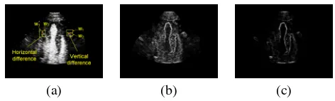
Our feature detector forms sliding windows, which are placed on either side of a pixel, as shown for two windowsw1
andw2in Figure 1 (a). Given the set of pixels inw1, we de-termine a FT parameterσ21(using the estimator give above), and likewise, we estimateσ22 inw2. Then, we compute
J-divergence between these two distributions using Equation (2) as a measure of how different the regions are. When the win-dows are placed to the left and to the right of the pixel, this gives a horizontal distance mapJx(x, y)that is functionally similar to the gradient operator in thexdirection, except that the values are non-negative. This can be repeated in they
direction. Then, we define a feature mapF(x, y)as
F(x, y) =
Jx(x, y)2+Jy(x, y)2. (3) Figure 1 (c) shows an example of a cardiac ultrasound image and its feature mapF(x, y). Note that this feature detector is much less distracted by the speckle compared to the gradient estimator shown in (b), yet still detects the important edges in the image. The robustness of the feature detector comes from two sources: first, it compares distributions to distributions, rather than pixels to pixels, and second, it is based on inte-grals of the image and not derivatives. Taking derivatives of noisy data is often undesirable in image processing, as doing so emphasizes the noise.
We apply this feature detector to each image to be reg-istered. This transforms the image into a feature image that contains the important edges needed for registration while si-multaneously mitigating false responses due to the speckle. These feature-detected images are then passed to the registra-tion algorithm, described next.
(a) (b) (c)
Fig. 1. Feature detection in a cardiac ultrasound image. Im-age (a), gradient (b), and J-divergence feature map (c) com-puted on the display image using the Fisher-Tippett method. Please see the digital version of the images for maximal qual-ity.
2.2. Registration
LetT(x, y)be the transformation between the two feature de-tected images,F1and andF2. Our objective is to estimate the
[image:3.612.318.554.461.532.2]two feature maps,
E(T(x, y)) =
[F1(x, y)−F2(T(x, y))]2dxdy, (4)
where the transformation is applied to the second image. We note thatT(x, y)can include rigid, affine, projective, or non-rigid transformations; in this paper we consider non-rigid trans-forms. Starting with an initial guess, we can iteratively up-date the transformation using differential equations based on a Gauss-Newton optimization [15] to minimize the energy functional in Equation 4. Upon convergence, the transforma-tion will be a local optimum of the energy.
3. EXPERIMENTAL RESULTS
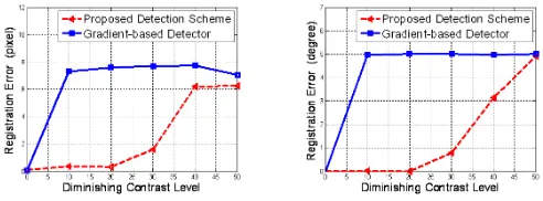
We begin with some experiments with synthetically generated data, designed to study the registration performance as the im-age contrast is diminished. The imim-ages and their feature de-tection results are shown in Figure 2. For comparison, we also produce results using a standard edge map formed using a dif-ference of Gaussian filter, which provides a smoothed imple-mentation of the gradient. Notice that the Fisher-Tippett fea-ture detector robustly identifies the important feafea-tures with-out many false detections due to speckle. In these experi-ments, the ground truth registration parameters are(5,5)for the translation and 5◦ for the rotation. The registration er-ror is denoted as the squared erer-ror of the estimated parameter compared to the ground truth value, and plotted in Figure 3. Notice that the registration error of the gradient-based edge maps (blue solid curves) quickly increases as the contrast is diminished, while the registration of the proposed method (red dashed curves) is significantly lower.
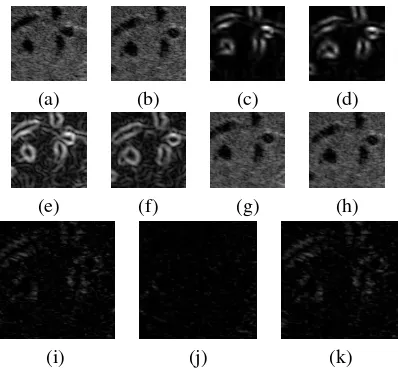
We examined the effectiveness of the proposed scheme for large regions extracted from abdominal ultrasound ages (which is more challenging that registering the full im-ages), depicted in Figures 4 and 5. While ground truth is not available, we do compute difference images and the sum of squared differences (SSD) between the original and regis-tered images. Notice that the difference images for the Fisher-Tippett technique, image (j) in Figures 4 and 5, is significantly lower than those of the gradient method, shown in (k) of the two figures. Quantitatively, the SSD decreased by51.4%and
56.3%for the proposed method; while with gradient-based method, the SSD was decreased only by0%and21.2%.
4. CONCLUSIONS
In this paper, we presented an ultrasound image registration method based on matching edge maps generated by a statisti-cal feature detector based on theoretistatisti-cal distributions of fully formed speckle in an ultrasound image. We presented the de-tails of our method, and demonstrated its ability to accurately register ultrasound images, even for low contrast, speckled
Fig. 3. Registration error as a function of diminishing con-trast. We show both the translational error (left) and rotational error (right) for the feature images generated by the gradient (blue) and Fisher-Tippett (red) detectors.
data, and showed how this method is more robust to noise than standard gradient-based methods.
For future work, we are interested in fully validating the method by testing it with more data. However, the prelimi-nary results presented in the paper indicate the proposed method has much promise in robust registration of ultrasound data.
5. REFERENCES
[1] B. Zitova and J. Flusser, “Image registration methods: A survey,”Image and Vision Computing, vol. 21, no. 11, pp. 977–1000, 2003.
[2] J.B. Maintz and M.A. Viergever, “A Survey of Medical Image Registration,” Medical Image Analysis, vol. 2, no. 1, pp. 1–36, 1998.
[3] Hajnal J., D. Hill, and D. Hawkes, Medical Image Reg-istration, CRC Press, first edition, 2001.
[4] A. Goshtasby,2-D and 3-D image registration for med-ical, remote sensing, and industrial applications, J. Wi-ley and Sons, 2005.
[5] J. Modersitzki, Numerical Methods for Image Registra-tion, Oxford Science Publications, Oxford, 2004.
[6] G. Hermosillo, C. Chefd’Hotel, and O. Faugeras, “Vari-ational Methods for Multimodal Image Matching,” In-ternational Journal of Computer Vision, vol. 50, no. 3, pp. 329 – 343, 2002.
[7] E. DAgostino, F. Maes, D. Vandermeulen, and P. Suetens, “A viscous fluid model for multimodal non-rigid image registration using mutual information,” in Proc. MICCAI, 2002.
[image:4.612.325.571.77.168.2]Fig. 2. Feature detection in synthetic ultrasound images. The top row shows the original synthetic ultrasound images with diminishing contrast. There are two images at each contrast level. The middle row shows the feature detection with Fisher-Tippett method, while the bottom row shows the feature detection with the difference of Gaussian filter. Notice the cleaner edge detection result of the Fisher-Tippett method, which has fewer false detections due to the speckle.
(a) (b) (c) (d)
(e) (f) (g) (h)
(i) (j) (k)
Fig. 4. Registration results of ultrasound images. Original (a)(b), feature detection with Fisher-Tippett (c)(d), gradient-based feature detection (e)(f), registered image with Fisher-Tippett and gradient detector (g)(h), original residue image (i), residue image with Fisher-Tippett detector (j), residue im-age with gradient detector (k).
[9] V. Dutt and J. Greenleaf, “Statistics of the Log-Compressed Envelope,” Journal of the Acoustical So-ciety of America, vol. 99, no. 6, pp. 3817–3825, 1996.
[10] M. O’Donnell, A. R. Skovoroda, B. M. Shapo, and S. Y. Emelianov, “Internal Displacement and Strain Imaging Using Ultrasonic Speckle Tracking,”IEEE Transactions on Ultrasonics, Ferroelectrics and Frequency Control, vol. 41, no. 3, pp. 314–325, 1994.
[11] M.J. Ledesma-Carbayo, J. Kybic, M. Desco, A. San-tos, M. S¨uhling, P. Hunziker, and M. Unser, “Spatio-Temporal Nonrigid Registration for Ultrasound Cardiac Motion Estimation,” IEEE Transactions on Medical Imaging, vol. 24, no. 9, pp. 1113–1126, 2005.
(a) (b) (c) (d)
(e) (f) (g) (h)
(i) (j) (k)
Fig. 5. Registration results of ultrasound images. Original (a)(b), feature detection with Fisher-Tippett (c)(d), gradient-based feature detection (e)(f), registered image with Fisher-Tippett and gradient detector (g)(h), original residue image (i), residue image with Fisher-Tippett detector (j), residue im-age with gradient detector (k).
[12] B. Cohen and I. Dinstein, “New Maximum Likelihood Motion Estimation Schemes for Noisy Ultrasound Im-ages,”Pattern Rec., vol. 35, no. 2, pp. 455–463, 2002.
[13] D. Boukerroui, J.A. Noble, and M. Brady, “Velocity Es-timation in Ultrasound Images: a Block Matching Ap-proach,” inProc. IPMI, 2003, pp. 586 –598.
[14] G. Slabaugh, G. Unal, and T. Chang, “Information-Theoretic Feature Detection in Ultrasound Images,” in IEEE 2006 International Conference of the Engineering in Medicine and Biology Society, 2006.
[image:5.612.71.560.71.177.2] [image:5.612.338.537.259.443.2] [image:5.612.76.276.259.445.2]