CLONIDINE AS AN ADJUVANT FOR ULTRASOUND GUIDED SUPRACLAVICULAR BRACHIAL PLEXUS BLOCK FOR UPPER
EXTREMITY SURGERIES UNDER TOURNIQUET
Dissertation submitted to
THE TAMILNADU DR.M.G.R MEDICAL UNIVERSITY
In partial fulfilment for the award of the degree of
DOCTOR OF MEDICINE
IN
ANAESTHESIOLOGY
BRANCH X
DEPARTMENT OF ANAESTHESIOLOGY, THANJAVUR MEDICAL COLLEGE,
CERTIFICATE
This is to certify that the dissertation entitled “CLONIDINE AS AN ADJUVANT FOR ULTRASOUND GUIDED SUPRACLAVICULAR BRACHIAL PLEXUS BLOCK FOR UPPER EXTREMITY SURGERIES UNDER TOURNIQUET” submitted by Dr. JOSEPH SELVARAJ DENISWINSTON in partial fulfilment for the award of the degree of Doctor of Medicine in Anaesthesiology by the Tamilnadu Dr.M.G.R Medical University, Chennai is a bonafide record of the work done by him in the Department of Anaesthesiology, Government Thanjavur Medical College, during the academic year 2015 -2018.
Dr.C.Kumaran M D(Anaes)., Prof.Dr.ShanthiPaulrajM.D(Anaes)., Associate Professor, Head of the Department, Department of Anaesthesiology, Department of Anaesthesiology, Thanjavur Medical College, Thanjavur Medical College,
Thanjavur. Thanjavur.
Prof.Dr.S.Jeyakumar,M.S.,M.Ch.,Vascular Surgery
DECLARATION
I, Dr. JOSEPH SELVARAJ DENISWINSTON solemnly declare that the dissertation titled “CLONIDINE AS AN ADJUVANT FOR ULTRASOUND GUIDED SUPRACLAVICULAR BRACHIAL PLEXUS
BLOCK FOR UPPER EXTREMITY SURGERIES UNDER
TOURNIQUET” is a bonafide work done by me at Thanjavur Medical College Hospital, Thanjavur, during 2015 – 2018.
The dissertation is submitted to “The Tamilnadu Dr.M.G.R Medical University, Chennai” Tamilnadu as a partial fulfilment for the requirement of M.D Degree examinations – Branch –X (Anaesthesiology) to be held in May 2018.
Place: Thanjavur
Date:
CERTIFICATE – ІІ
This is to certify that this dissertation work titled “CLONIDINE AS AN ADJUVANT FOR ULTRASOUND GUIDED SUPRACLAVICULAR
BRACHIAL PLEXUS BLOCK FOR UPPER EXTREMITY
SURGERIES UNDER TOURNIQUET” of the candidate Dr. JOSEPH
SELVARAJ DENISWINSTON with registration number 201520207 for the
award of DOCTOR OF MEDICINE in the branch of ANAESTHESIOLOGY
(Branch X). I personally verified the urkund.com website for the purpose of
plagiarism check.I found that uploaded thesis file contains from introduction to conclusion pages and result shows 20 percentage of plagiarism in the dissertation.
Dr.C.Kumaran M D(Anaes).,
Associate Professor,
ACKNOWLEDGEMENT
I am extremely thankful to Prof.Dr.S.Jeyakumar M.S,M.Ch.,
Vascular Surgery, Dean, Thanjavur Medical College , for his kind
permission to carry out this study.
I am extremely grateful to Prof.Dr.ShanthiPaulraj,M.D.,
Anaesthesiology,Associate Professor and Head of the Department of
Anaesthesiology,for her concern and support in conducting the study.
I am immensely grateful to my guide Prof.Dr.C.Kumaran M.D.,
Anaesthesiology, Associate Professor, Department of Anaesthesiology
for his concern and support in conducting the study.
I express my gratitude to my respected co guide
Dr.R.HARIBASKAR M.D., Anaesthesiology, Assistant Professor,
Department of Anaesthesiology, for his inspiration,guidance and
comments at all stages of this study.
I am thankful to all Assistant Professor of the Department of
Anaesthesiology, for their guidance and help.
I am thankful to all my colleagues for the help rendered in
carrying out this dissertation.
Special thank all the patients for willingly submitting themselves
INDEX
SI.NO TITLE PAGE NO.
1 INTRODUCTION 1
2 AIM OF THE STUDY 3
3 ANATOMICAL CONSIDERATIONS 4
4 PHYSIOLOGICAL CONSIDERATIONS 17
5 PHARMACOLOGY OF ROPIVACAINE 24
6 PHARMACOLOGY OF CLONIDINE 33
7 REVIEW OF LITERATURE 45
8 MATERIALS AND METHODS 57
9 OBSERVATION AND RESULTS 64
10 DISCUSSION 88
11 SUMMARY 97
12 CONCLUSION 99
13 BIBLIOGRAPHY 100
14 PROFORMA 104
1
INTRODUCTION
“Pain, like pleasure is passion of the soul,
That is an emotion and not one of the senses”
- PLATO and ARISTOTLE (375 B.C)
Pain is a fundamental biological phenomenon.The International
Association for the Study of Pain has defined pain as “an unpleasant
sensory and emotional experience associated with actual or potential
tissue damage, or described in terms of such damage.” Pain is always
underestimated and undertreated. The relief of pain during surgery is
the main part of anaesthesia.
In 1784 James Moore used mechanistic concepts to promote
neural compression as a useful technique for the provision of surgical
anaesthesia.
In 1855, neurologic pain can be treated by circum-neural injection
of pain relieving drug. At the same year Gadecke (German) isolated
alkaloid from leaves of coca plant. In 1860 Albert Nieemann was
successful in isolating and naming the alkaloid from the leaves of
erythroxylon coca.
In 1911, Kullenkampff introduced the classic supraclavicular
2
subclavian perivascular approach of brachial plexus block. After
introduction of barbiturate and cyclopropane, the enthusiasm for block
anaesthesia waned in early 1940s.
In current recent years however, the technique has had resurgence,
due in large part to increased understanding of neural plasticity and the
possibility of minimizing hospital stay by effective use of regional block.
Several experimental and clinical studies have shown that alpha-2
adrenergic agonists like clonidine were able to prolong sensory and
motor blockade. The conventional parasthesia technique being a blind
technique may be associated with higher failure rate, injury to nerves &
vascular structures. To minimize these drawbacks, various techniques
and approaches were described. Among them, ultrasound visualization
of anatomical structure is the only method offering safe blocks of
superior quality by optimal needle positioning. Ultrasound has improved
success rate with excellent localization & improved safety margin..
This study was aimed to evaluate the clinical efficacy of clonidine
as an adjuvant to 0.75% ropivacaine for enhancing the quality and
duration of motor and sensory block along with duration of analgesia of
ultrasound guided supraclavicular brachial plexus block in upper limb
3
AIM OF THE STUDY
To evaluate the effectiveness of CLONIDINE AS AN
ADJUVANT FOR ULTRASOUND GUIDED SUPRACLAVICULAR
BRACHIAL PLEXUS BLOCK FOR UPPER EXTREMITY
SURGERIES UNDER TOURNIQUET for prolonging the
1. Onset of sensory blockade
2. Onset of motor blockade
3. Duration of sensory blockade
4. Duration of motor blockade
4
ANATOMICAL CONSIDERTIONS
Knowledge of the formation of brachial plexus and of its
distribution is absolutely essential to the intelligent and effective use of
brachial plexus anaesthesia for surgeries of the upper limb. Close
familiarity with the vascular, muscular and fascial relationships of the
plexus throughout its formation and distribution is equally essential to
the mastery of the various techniques of brachial plexus blockade.
FORMATION OF PLEXUS 21
Roots
The plexus is formed by the anterior primary rami of the 5th to 8th
cervical nerves, together with the bulk of the 1st thoracic nerve (C -8 and
T1). In addition there is frequently a contribution above from C4 to the
5th cervical root and another below from T2 to the 1st thoracic nerve.
Occasionally the plexus is mainly derived from C4 - 8 (Pre -fixed
plexus) or from C6 - T2 (post - fixed plexus).
Trunks
The five roots of the plexus emerge from the intervertebral
5
tubercles of the corresponding transverse process. All five roots then
become sandwiched between scalenus anterior and medius.
Here the roots of C5 and C6 unite into the upper trunk. The
root of C7 continues as the middle trunk and those of C8 and T1 into
the lower trunk. Each trunk divides behind the clavicle, into anterior and
posterior divisions, which unite in the axilla to form the cords.
Cords
The six divisions stream into the axilla and there unite up into
three cords. Lateral, medial and posterior. These cords are composed as
follows:
The union of the anterior divisions of the upper and middle trunks
forms the lateral cord. The medial cord represents the continuation of
the anterior division of the lower trunks. The posterior cord comprises
of all three posterior divisions.
The composition of the brachial plexus can be summarized as follows:
1. Five roots - the anterior primary rami of C5 - 8 and T1
2. Three trunks.
6
b) Middle trunk, C7 alone and
c) Lower trunk, C8 and T1
3. Six divisions - each trunk divides into an anterior and posterior
division.
4. Three cords
a) Lateral cord, the fused anterior divisions of upper and middle
trunks (C5 - C7)
b) Medial cord, the anterior division of the lower trunk (C8, T1)
c) Posterior cord, formed by the union of the posterior division
7
8 Roots
Lie between the scalenus anterior and medius. The roots of the
plexus lie above the second part of the subclavian artery.
Trunks
The upper and middle trunks lie above the subclavian artery as the
stream across the 1st rib, but the lower trunk lies behind the artery and
may groove the rib immediately posterior to the subclavian groove.
Division
At the lateral border of the 1st rib the trunks bifurcate into
divisions, which are situated behind the clavicle.
Cords
The cords are formed at the apex of the axilla and become
9
10 BRANCHES
Branches are given off from
1. Roots
2. Trunks and
3. Cords
3.1 Branches from the Roots
3.1.1. Nerve to the serratus anterior (C5, C6 and C7)
3.1.2. Muscular branches to
i. Longus cervices (C5- C8)
ii. Three scalene (C5 - C8)
iii. Rhomboids (C5)
3.1.3. A twig of Phrenic nerve (C5)
3.2 Branches from the trunks
3.2.1. Suprascapular nerve (C5,C6)
11 BRANCHES FROM THE CORDS
LATERAL CORD
i. Lateral pectoral nerve (C5 - C7)
ii. Lateral head of median nerve (C5 - C7)
iii. Musculocutaneous nerve (C5 - C7)
MEDIAL CORD
i. Medial pectoral nerve (C8 - T1)
ii. Medial head of median nerve (C8 - T1)
iii. Medial cutaneous nerve of arm (C8 - T1)
iv. Medial cutaneous nerve of forearm (C8 -T1)
v. Ulnar nerve (C8- T1)
POSTERIOR CORD
i. Upper subscapular nerve (C5 - C6)
ii. Lower subscapular nerve (C5 - C6)
12
iv. Axillary nerve (C5 - C6)
v. Radial nerve (C5 - C6)
TECHNIQUE OF BRACHIAL PLEXUS BLOCK
Surgical anaesthesia of the upper extremity and shoulder can be
obtained following neural blockade of the brachial plexus at several
sites. The various approaches that can be used for this blockade are as
follows.
1.Interscalene approach
2. Supraclavicular approach
a. Classic approach
b. Plumb -bob technique
c. Subclavian perivascular technique
3. Axillary approach
13
BASICS OF ULTRASOUND
The frequency of medical ultrasound ranges between 2 MHz and
13 MHz. The average wave length in this band is about 1 mm. This
limits the resolution to structures that are larger than 1 mm. Most nerves
of interest range in size from 2 mm to 10 mm. Veins and arteries of
interest are typically 3 mm to 15 mm.
Many factors contribute to the quality and resolution of the
ultrasound image. In general, higher frequency probes generate higher
resolution images.
Unfortunately, high frequency ultrasound waves (8 MHz to 13 MHz)
are rapidly attenuated in tissues so that high frequency probes are best
suited for structures less than 5 cm deep to the skin.
The image formed of a nerve on ultrasound is very sensitive to the
angle of incidence of the beam relative to the nerve. Sometimes changing
the angle of incidence by only a few degrees can bring the nerve into focus.
This phenomenon is thought to be caused by diffraction of the type
described above.
Modern platforms allow the user to adjust the brightness (gain) of the
14 IMAGE OF VESSELS:
Arteries can usually be distinguished by their pulsatile nature.
Veins can be distinguished by their compressibility. Color flow Doppler
imaging can also be used to identify and distinguish arteries and veins.
By convention, blood flowing toward the probe is colored red. Blood
flowing away from the probe is colored blue. Blood flowing
perpendicular to the probe remains black. Velocity gates can be set to
measure the flow velocity. High velocities are usually arteries. Low
velocities are usually veins. Transducer elements can be arranged in
linear or curved arrays.
PROBE SELECTION:
Linear arrays create rectangular images and are most useful for
superficial structures. Curved arrays create wedge-shaped images and are
most useful for deeper structures. Because the beam disperses in a
curved array, its resolution is usually lower than a linear array. A phased
array retains the elements in a straight line. But the elements fire in
sequence creating a phase delay between each element. The net result is
15
signal is averaged, its resolution is also lower than a standard linear
array.
Most probes have transducers that emit the highest amplitude of
their ultrasound wave at a specific fundamental frequency. Harmonics
of this frequency are also emitted at lower amplitudes. By listening for
the echo at these higher harmonic frequencies, image resolution can be
enhanced. Because the harmonics are very low amplitude, only
transducers that have sufficient power output can be used for harmonic
imaging. This type of image enhancement is referred to as tissue
harmonic imaging.
Modern probes operate in pulsed mode. The transducer elements
in the probe are used as both transmitters and receivers. The transducer
elements emit a short ultrasound burst and then wait for the echo before
emitting another ultrasound burst. This allows the probe to be smaller
compared with continuous wave probes where there are separate emitters
and transducers.
When a needle is inserted into tissue perpendicular to the
ultrasound beam, it is a good reflector and it is easy to image. In some
cases, it may be necessary to insert the needle nearly parallel to the beam
16
dark (hypoechoic). Nerves located below the collar bone are usually
white (hyperechoic). The reasons for this dichotomy are not known, but
it may be related to the depth of the nerves and the relative amounts of
fat and stroma within the nerves themselves. On ultrasound cross
section, nerves are round, hypo or hyperechoic, reticulated structures.
When imaged along their long axis, nerves appear as linear, hypo or
hyperechoic streaks, on ultrasound. Bones are hyperechoic and usually
very bright white. Arteries and veins are black unless color flow Doppler
17
PHYSIOLOGICAL CONSIDERATION
2,3Pain perception requires noxious stimuli. It is transformed from
its native form by the activated nociceptors into electrical signals
which are then transmitted along the corresponding nociceptive fibres.
These fibres in turn synapse onto second order neurons in the spinal
cord. These interneurons are located in the dorsal horn. It is at
these interneurons where the initial modulation of nociceptive input
occurs. From the spinal cord nociceptive input is transmitted to the
brain stem, thalamus and cortex.
Peripheral neuroanatomy of nociception
C and A fibres are the main peripheral nociceptors. The skin
joints and periosteum are richly innervated with C and A nociceptors
as well as the non nociecptive Aβ sensory fibres.
A are responsible for the sensation of first pain, the initial
sharp pain experienced following an injury. C fibres are unmyelinated
and are responsible for second pain, the slowly building throbbing
18 Classification of sensory fibres
Sensory receptors
Speed of
Transmission Sensory function Myelination
A-Alpha fibres 70 -120m/sec
Noxious chemical thermal, mechanical stimuli, (sharp fast,
first pain)
Lightly myelinated
A-Beta fibres 30 -70m/sec
Nonpainful, light touch, pressure, vibration
proprioception
Heavily myelinated
A-Gamma fibres 30-70m/sec Proprioception/Motor to
muscle spindle Myelinated
A-Delta fibres 12 -30 m/sec Pain, cold, touch Myelinated
B fibres 3 -15 m/sec
Pre ganglionic autonomic (sympathetic)
Myelinated
C Fibres 0.5 -2m/sec
Noxious chemical,
Mechanical, thermal activation (Slow burning second pain)
19
Peripheral neurochemistry and neurotransmitters :
Commonly released inflammatory mediators implicated in pain
and hyperalgesia include bradykinins, potassium, substance P
cytokines, histamine, serotonin, prostaglandins. These peripheral
neurotransmitters either activate or sensitise the peripheral nociceptors
to pain.
Peripheral neuro chemistry Algogenic Agents :
Algogenic Agent Action on nociceptors
Bradykinin Activates
Substance P Sensitizes
Potassium Activates
Hydrogen Activates
Arachidonic acid Sensitizes
Cytokiness Sensitizes
Serotonin Sensitizes
20
PHERIPHERAL ALPHA 2 RECEPTORS
Alpha 2 adrenoreceptors are located on primary afferent terminal.
Clonidine enhances both sensory and motor blockade from peripheral
nerve injection and epidural / spinal injection of local anaesthetics.
Clonidine blocks conduction of C and A gamma fibers and
increase potassium conductance in isolated neurons and intensifies
conduction block of local anaesthetics.
Local vasoconstriction resulting in reduced absorption from
injection site was another point of discussion, but compared with
adrenaline as adjuvant it failed to influence plasma levels, indicating a
direct action on nerve.
PAIN PATHWAY
SPINAL CORD
The gray matter of the spinal cord is divided into ten lamina
with lamina I – IV representing the dorsal horn. The dorsal horn is
capped by the Lissauer’s tract which consists of branches of cutaneous
21
Nociceptive fibres terminate in the superficial layers of lamina
I & II while the non-painful myelinated fibres terminate in the deeper
layers of lamina III,IV. Lamina II has the highest concentration of
opioid receptors in the spinal cord. Modulation and inhibition of
nociception may occur at this level through the use of opioids (systemic
and neuraxial).
Ascending sensory pathways
Peripheral sensory neurons synapse onto the secondary
interneurons of the dorsal horn. The axons of the non nociceptive
secondary neurons travel isobilateral in the dorsal columns of the
spinal cord as fasciculus cuneatus (upper body through T6) and
fasciculus gracilis (lower body below T6) and synapse in thalamus.
The axons of the nociceptive secondary neurons after synapsing,
travel contra laterally in the anterolateral aspects of the spinal cord as
the neospinothalamic and paleospinothalamic tract.
Neospinothalamic tract carries fine discrimination of pain eg.
Location, intensity, and first pain.
Paleospinothalamic tract responds to noxious stimuli. The paleo
22
system and plays a role in emotional aspects of pain via limbic
system. The thalamus has multiple connections to limbic system and
cortex.
Descending inhibitory pathways
The descending controls of pain project specifically onto laminas
I, II, V of the dorsal horn from mesencephalon, raphenuclei and
reticular tract. The mesencephalon is rich in opioid receptors. This area
sends excitatory transmissions to the rostroventral medulla which sends
noradrenaline and serotonin inhibitory tracts via the dorsolateral
funiculus to lamina I,II,V of spinal cord.
The noradrenaline and serotonin fibres mediate transmission
between the primary afferents and the secondary neurons of the
dorsal horn. Increased activity of these fibres leads to increased
inhibition of pain transmission.
Location of Alpha2 receptors
Primary afferent terminals, on neurons in the superficial laminae
23 Location of opioid receptors (central)
Opioid receptors are found in the various regions in CNS
namely, cerebral cortex, limbic cortex (anterior and posterior amygdale,
hippocampus hypothalamus, medial thalamus, mid brain, periaqueductal
gray matter, extrapyramidal areas, substantia gelatinosa and sympathetic
preganglionic neurons.
Opioid receptors are also found in the cardiac sympathetic fibres,
cardiac branches of vagus, adrenal medulla, and gastro intestinal tract.
24
PHARMACOLOGY OF ROPIVACAINE
Ropivacaine is a new aminoamide local anaesthetic. It is the
monohydrate of the hydrochloride salt of 1-propyl - 2, pipecoloxylidide
and is prepared as a pure S - enantiomer.
Pipecoloxylidides were first synthesized in 1957 and have been in
clinical use of more than 30 years. Ropivacaine has a propyl group on
the piperidine nitrogen atom of the molecule.
The Pipecoloxylidides are chiral drugs because the molecules
possess an asymmetric carbon atom and they may have left - (sinister)
or right (rectus) handed configuration.
25 PHYSIO-CHEMICAL PROPERTIES
The physio-chemical properties of Ropivacaine are as follows :
1. Molecular weight (base) - 274
2. pKa - 8.1
3. Partition coefficient (N Heptane / buffer) - 2.9
4. Mean uptake ratio (rat sciatic nerve) - 1.8
5. Protein binding - 94%
PHARMACOLOGIC PROPERTIES
Onset - Moderate
Relative potency - 6
Duration - Long acting
Mechanism of action
Ropivacaine blocks the generation and conduction of nerve
impulses, presumably by increasing the threshold for electrical
excitation in the nerve, by slowing the propagation of the nerve
26
progression of anaesthesia is related to the diameter, myelination and
conduction velocity of affected nerve fibers.
The order of loss of nerve function clinically is as follows :
1. Pain
2. Temperature 3. Touch
4. Proprioception 5. Skeletal muscle tone
Pharmacokinetics
In human volunteers the pharmacokinetic characteristics of
ropivacaine have been determined after intravenous infusion
Clearance : 0.82 ± 0.16 litres /min Plasma protein binding : 94 ± 1%
Volume of distribution at steady state : 59 ± 7 liters (Vss)
Terminal elimination ½ life :111 ± 62min
Metabolism: Liver by aromatic hydroxylation by cytochrome
P450 1A to 3-hydroxyl ropivacaine.
Excretion: 86% via the kidney 1% unchanged drug, rest as
metabolites the higher clearance of ropivacaine over bupivacaine is
27 Absorption
The systemic concentration of ropivacaine is dependent on the
total dose and concentration of drug administered, the route of
administration, the patient’s haemodynamic condition and the
vascularity of the administration site. From the epidural space,
ropivacaine shows complete and biphasic absorption. The half lives of
the two phases are 14 ± 7 minutes and 4.2 ± 0.9 hrs (mean ± S.D)
respectively. The show absorption is the rate-limiting factor in
the elimination of ropivacaine, which is why the terminal half life is
longer after epidural than after intravenous administration. Ropivacaine
shows dose proportionality up to the highest intravenous dose studies,
80 mg corresponding to a mean ± 1.9 0.3µg/ml.
Distribution
After intravascular infusion, ropivacaine has a steady state volume of distribution of 59 ± 7 litres. Ropivacaine is 9.4% protein
bound, mainly to α1acid glycoprotein. An increase in total plasma
concentration during continuous epidural infusion has been observed,
related to postoperative increase of α1acid glycoprotein.
28
Ropivacaine readily crosses the placenta and rapidly equilibrium is reached in regard to unbound concentration.
Metabolism
Ropivacaine is extensively metabolized in the liver,
Predominantly by aromatic hydroxylation mediated by cytochrome
P450 1A to 3 -hydroxy ropivacaine. Approximately 37% of the total
dose is excreted in the urine as both free and conjugated 3-hydroxy
ropivacaine have been found in the plasma.
Urinary excretion of the 4-hydroxyl ropivacaine have been
found in the plasma. Urinary excretion of the 4 - hydroxyl and both
the 3 - hydroxyl and 4 - hydroxyl N- dealkylated metabolites accounts
for less than 3% of the dose. An additional metabolite, 2 - hydroxyl
methyl ropivacaine has been identified but not quantified in the urine.
Both 3- hydroxyl and 4 - hydroxyl ropivacaine have not quantified in
the urine. Both 3 - hydroxy and 4 - hydroxy ropivacaine have a local
anaesthetic activity in animal models less than that of ropivacaine.
There is no evidence of in vivo racemization in urine of S-(-)
29 Elimination
The kidney is the main excretory organ for most local anaesthetic
metabolites. In total 86% of the ropivacaine dose is excreted in the
urine after intravenous administration of which only 1% relates to
unchanged drug. Ropivacaine has a mean total plasma clearance of
387 ± 107 ml/min. The mean ± SD terminal half - life is 1.8 ± 0.7 h
after intravascular administration and 4.2 ± 1.0 h after epidural
administration.
Pharmocodynamics
Primary Pharmocodynamics (Animal Studies)
These studies showed that ropivacaine at low concentration
produced a profound rapid block of both Aδ and C fibres and was more
potent than similar low concentrations of bupivacaine.
At higher concentrations, ropivacaine and bupivacaine had
similar blocking properties Aδ fibre blocking was 16% greater with
bupivacaine and the degree of C fibre block was similar with both the drugs.
Ropivacaine is a potent producer of frequency (or use)
dependant blocks (i.e) a block that occurs only when the fibre is
30
Low pKa and high lipid solubility of a local anaesthetic drug
favoured A over C fibre block. The lower lipid solubility of ropivacaine
over bupivacaine is presumed to retard penetration into myelin sheath.
This greater degree of differential block with ropivacaine at low
concentration and the property of producing frequency dependent
block were considered to offer clinical advantages in providing
analgesia with minimal motor block.
Secondary pharmocodynamics
Addition of epinephrine to ropivacaine has no limiting effect on
the systemic absorption of ropivacaine. Systemic absorption can
produce effects on the central nervous and cardiovascular systems.
At blood concentration achieved with therapeutic doses, changes in
cardiac conduction, excitability, refractoriness, contractility and
peripheral vascular resistance are minimal. However, toxic blood
concentrations depress cardiac conduction and excitability, which
may lead to atrio ventricular block, ventricular arrhythmias and to
cardiac arrest. In addition, myocardial contractility is depressed and
peripheral vasodilatation occurs, leading to decreased cardiac output
31
Ropivacaine can produce central nervous system stimulation,
depression or both. Apparent central stimulation is usually manifested
as restlessness, tremors, shivering, progressing to convulsions,
followed by depression and coma, progressing to respiratory arrest.
However, ropivacaine has a primary depressant effect on the medulla
and on higher centers. The depressed stage may occur without a
prior excited stage.
Adverse reactions
A major cause of adverse reactions may be due to excessive
plasma levels that may be due to over dosage, unintentional
intravascular injection or slow metabolic degradation. Most adverse
events reported were mild and transient.
Central nervous system reactions
These are characterized by excitation and/or depression.
Restlessness, anxiety, dizziness, tinnitus, blurred vision or tremors may
occur, possibly proceeding to convulsions. However, excitement
may be transient or absent with depression being the first
32
unconsciousness and respiratory arrest. Other effects may be nausea,
vomiting, chills and constriction of pupils.
Cardiovascular system reactions
High doses of accidental intravascular injection may lead to high
plasma levels and related depression of myocardium, decreased cardiac
output, heart block, hypertension, bradycardia and ventricular
arrhythmias including ventricular tachycardia and ventricular
fibrillation and possibly cardiac arrest.
Preparations available
0.2%
0.5%
0.75%
1%
Uses :
1. Infiltration Anaesthesia 2. Peripheral Nerve Blocks 3. Brachial Plexus Block
4. Lumbar Extradural Block (Especially Labour) 5. Subarachnoid Block
33
PHARMACOLOGY OF CLONIDINE
1,6INTRODUCTION:
Clonidine hydrochloride is a centrally acting selective partial
alpha – 2 agonist introduced in early 1960s, it was during its use as a
nasal decongestant that its anti - hypertensive property was found out.
Subsequently more insights into the pharmacological properties has led
to its use in clinical anaesthesia practice as well.
Clonidine hydrochloride is an imidazoline compound and exists
as a mesomeric compound. The chemical name is 2 - (2, 6
-dichlorophenylamino) - 2 imidazoline hydrochloride. The structural formula is C9 H9 C12 N3 HCI.
The molecular weight is 266.56. Clonidine is an odourless,
bitter, white, crystalline substance, soluble in alcohol and water.
Clonidine improves the quality of anaesthesia, provides a more
stable cardiovascular course during anaesthesia, presumably because
of their sympatholytic effect and need for lower dose of
cardioactive anaesthetic and reduces the dose requirement of the
anaesthetic agent. Clonidine may reduce the halothane MAC by upto
34
action of the local anaesthetics with fewer side effects in peripheral
nerve blocks and central neuraxial blockade.
CLONIDINE HYDROCHLORIDE
Available as one ml ampoule containing 150 micrograms. It
should be stored below 25 degree celsius.
Mechanism of action:
Clonidine is a centrally acting partial α2 adrenergic agonist with
a selectivity ration of 220:1 in favour of α2 receptors. The three
subtypes of α2 receptors are α2a, α2b, α2c, α2a receptors mediate
sedation, analgesia, sympatholysis. α2b receptors mediate
vasoconstriction and anti- shivering. The startle response may reflect
the activation of α2c receptors. The drug is lipid soluble, penetrates the
35
injected epidurally. It stimulates the inhibitory α2 adrenoreceptors to
reduce the central neural transmission in the spinal neurons Inhibition
of substance-P release is believed to be involved in the analgesic
effect.
The α2 adrenoreceptors are located on the afferent terminals
of both peripheral and spinal neurons in the superficial laminae of the
spinal cord and within several brain stem nuclei implicated in
analgesia. The superficial lamiae contain three groups of neurons:
tonic, adapting, single- spike firing, all of which receive their primary
sensory input from Aδ and C fibres.
Clonidine inhibits voltage gated sodium and potassium channels
and suppresses the generation of action potentials in tonic - firing
spinal dorsal horn neurons, contributing to analgesic effect. The ability
of clonidine to modify the function of potassium channels in the CNS
(Cell membrane become hyperpolarized) may be the mechanism for
profound decrease in anaesthetic requirements.
Another contribution to analgesic effect may be through the
release of acetylcholine in the neuraxial region. The two adrenergic
agonists also enhance analgesia from intraspinal opioids. Sedation is
36
Clonidine affects the blood pressure in a complex fashion after
neuraxial or systemic administration because of opposing action at
multiple sites. In the nucleus tractus solitaries and locus ceruleus of the
brainstem, activation of post-synaptic α2 adrenoreceptors reduces
sympathetic drive.
It also activates nor-adrenergic imidazoline preferring binding
sites in the lateral reticular nucleus producing hypotension and
anti-arrythmogenic ation. In the periphery, it acts on pre-synaptic α2
adrenoreceptors at sympathetic terminals reducing the release of
nor-epinephrine causing vasorelaxation and reduced chronotropic drive.
The brainstem and the peripheral effects of α2 adrenoreceptor
stimulation are counterbalanced by the direct peripheral
vasoconstriction through its action on α2 adrenoreceptors from the
circulating concentrations of clonidine.
Sedation is a desired property. Clonidine produces a dose
dependent sedation at the dose of 50 micrograms or more in less
than 20 minutes regardless of the route of administration.
Clonidine doesn’t induce profound respiratory depression even
after massive overdose nor do potentiate respiratory depression from
37
In peripheral nerves it produces a minor degree of blockade
at high concentrations with some preference for C-fibres in the
peripheral nerves and this effect in part enhance the peripheral nerve
block when added to local anaesthetics, probably because the α2
adrenoreceptors are lacking on the axons of peripheral nerves.
Pharmacokinetics:
Clonidine is well absorbed orally and is nearly 100%
bio-available and reaches peak plasma concentration within 60 to 90
minutes. The mean half life of the drug in plasma is about 9 to 12
hours, with approximately 50% metabolized in the liver whereas is it
is excreted in an unchanged form by the kidney, and its half- life can
dramatically increase in the presence of impaired renal function.
A transdermal delivery system is available in which the drug is
released at a constant rate for about a week. Three or four days are
required to achieve steady state concentration.
Clonidine is highly lipid soluble and readily gets distributed into
38
300 micrograms intravenously over 10 min produces:
Distribution t ½ : 11 ± 9 minutes
Elimination t ½ : 9 ± 2 hours, 41 hours in severe renal dysfunction.
Volume of distribution : 2.1± 0.4 l/kg
Plasma protein binding : 20 - 40% in vitro
Metabolism : minor pathways with the major metabolite, P - hydroxyclonidine.
Excretion:
70% of the dose, mainly in the form of unchanged parent drug
(40 - 60%) in urine. So, the elimination t ½ of clonidine varies as a
function of creatinine clearance. In subjects undergoing hemodialysis
only 5% of the body clonidine store was removed.
Dosage regimen:
Oral - 3-5µg/kg
Intramuscular - 2 µg/kg
Intravenous - 1-3µg/kg
Spinal - 50 -100µg
Epidural - 1-2µg/kg
39 Precautions:
1. In patients with renal insufficiency, lower dose is needed.
2. Sudden withdrawal of prolonged continuous epidural infusion
produces hypertensive crisis. So it should be gradually
discontinued over 2 to 4 days.
3. To be used with caution in patients with cerebrovascular or
coronary insufficiency.
4. If a patient with beta blocker is on continous epidural
therapy, beta blocker should be withdrawn several days before
discontinuation of epidural clonidine.
Contraindications:
1. Known hypersensitivity to clonidine or components of the product.
2. In patients with brady arrhythmia or AV block
3. Patients with severe cardiovascular disease
40 Interactions:
1. Clonidine may potentiate the CNS-depressive effect of
alcohol, barbiturates or other sedative drugs.
2. Narcotics may potentiate the hypotensive effects of clonidine.
3. Tricyclic anti depressants may antagonize the hypotensive effects
of clonidine.
4. Concomitant administration of drugs with a negative
chronotropic and dromotropic effect (beta blocker, digoxin) can
cause or potentiate bradycardiac rhythm disturbances.
5. Beta blockers may potentiate the hypertensive response
seen with clonidine withdrawal.
6. Epidural clonidine may prolong the duration of pharmacologic
effects of epidural local anaesthetics, opioids, neostigmine and
other drugs.
Uses
1) Pre anaesthetic medication
a) Oral clonidine (5 µg /kg) blunts reflex tachycardia
associated with direct laryngoscopy for intubation of
41
b) Decrease intraoperative lability of blood pressure and heart
rate.
c) Decrease plasma catecholamine concentrations.
d)Dramatically decrease anaesthetic requirements for inhaled
and injected anaesthetic. Clonidine also attenuates the rise in
intraocular pressure associated with laryngoscopy and
intubation.
2) Epidural block : Clonidine as a sole agent or in combination with
opioids or local anaesthetics to provide excellent analgesia in
labour analgesia. Epidural clonidine is also indicated for the
treatment of intractable pain, which is unresponsive to
maximum does of oral epidural opioid, as do patients with reflex
sympathetic dystrophy, neuropathic pain.
3) Spinal anaesthesia: Clonidine combined with local anaesthetic
solution, improves the quality and duration of the block,
minimize the tourniquet pain during lower limb surgery, and
42
4) Caudal anaesthesia: Clonidine combined with local anaesthetic
solution, increases the duration of anaesthesia and analgesia by
two or three times without hemodynamic side effects. Dose 2-3
µg /kg
5) Peripheral nerve blocks: Clonidine prolongs the duration of
anaesthesia and analgesia with local anaesthetics by two times in
a dose of 75 to 150 micrograms.
6) Bier’s Block : 150 microgram of clonidine enhances the tolerance of tourniquet
7) It is also used in intra articular analgesia
8) Protection against perioperative myocardial ischemia: clonidine
decreases myocardial ischemia, infarction and mortality
following cardiovascular surgery.
9) To treat hypertensive crisis
10) Diagnosis of pheochromocytoma: 0.3 mg will decrease the plasma
concentrations of catecholamine in normal patients but not in the
43
11) Treatment of shivering: Administration of clonidine, 75µg IV
stops shivering by inhibiting thermoregulatory control.
12) Treatment of opioid and alcohol withdrawal syndrome.
Side effects;
1. The most common side effects are sedation and xerostomia
2. Cardiovascular complaints are bradycardia, hypotension, and ECG
abonormalities like sinusnode arrest and junctional bradycardia.
High degree AV block and arrhythmia are reported rarely.
Occasionally require treatment of bradycardia with IV
anticholinergics. Orthostatic hypotension occurs rarely.
3. Rebound hypertension: Abrupt discontinuation of clonidine can
result in rebound hypertension as soon as 8 hours and as late as
36 hours after the last dose. Symptoms of nervousness,
diaphoresis, headache, abdominal pain, and tachycardia often
precede the actual increase in systemic blood pressure. Labetalol is
useful in treatment of rebound hypertension.
4. Skin rashes occur frequently.
44 Over dosage and treatment :
There is no specific antidote for clonidine overdosage.
Supportive measures like atropine, ephedrine, and IV fluids are
enough. Yohimbine partially reverses the analgesia and sedation but
not the blood pressure and heart rate changes produced by the epidural
45
REVIEW OF LITERATURE
1) Gupta K et al11, in 2014 conducted a randomized double
blinded study, to evaluate the efficacy of clonidine as an adjuvant to
0.75% ropivacaine. 64 patients of ASA physical status I and II, between
18 to 54 years age, scheduled for upper extremity surgery under
ultrasound guided supraclavicular brachial plexus block were recruited.
The patients were randomly divided into two groups of 32 each. Group R
(Ropivacaine) received 19.8ml of 0.75% ropivacaine with 0.2ml of
normal saline and group RC (Ropivacaine – Clonidine ) received 19.8ml
of 0.75% ropivacaine with 0.2ml (30microgram) of clonidine .
The onset and duration of sensory and motor blockde, the post
operative analgesia were compared along with monitoring of
heamodynamic variablity, sedation and other adverse effects. The onset
of sensory and motor block was significantly faster in group RC than
group R. The duration of sensory block was 673.9 ± 35 minutes in group
RC when compared to 535.39 ± 25 minutes in group R. The duration of
motor block was 614.5 ± 29 minutes in group RC and 485.1 ± 30
46
The duration of analgesia was 956.4 ± 38 minutes in group RC
and 736.53 ± 47 minutes in group R. Significantly prolonged duration of
analgesia was observed in group RC ( p < 0.05 ). No side effects were
observed in both groups .
Hence they concluded that addition of 0.2ml (30microgram) of
clonidine as an adjuvant to 19.8 ml of 0.75% ropivacaine enhanced the
onset of motor and sensory block, increased the duration of motor and
sensory blockade and post operative analgesia without any adverse
events.
2) Sirohiya P et al12, in 2016, conducted a randomised double
blinded study to evaluate the effect of clonidine added to local
anaesthetic to improve nerve blocks by reducing the time of onset and
extending the post operative analgesia. 60 patients of ASA physical
status I and II, between 18 to 60 years of age undergoing upper limb
surgeries were recruited. The brachial plexus were identified using a
nerve stimulator. The patients were randomly divided into two groups of
30 each. Group B (n = 30) received 25 ml of 0.5% bupivacaine and
0.2ml of normal saline. Group C received 25 ml of 0.5% bupivacaine
47
The onset of sensory and motor block was significantly faster in
group C than group B. The duration of sensory block was 474.3 ± 166.6
minutes in group C when compared to 280.5 ± 101.7 minutes in group B.
The duration of motor block was 515.3 ± 174.6 minutes in group C when
compared to 307.5 ± 103.3 minutes in group B. Both were prolonged in
group C ( p < 0.001). The duration of analgesia was 550.1 ± 182.5
minutes in group C and 331.0 ± 98.4 minutes in group B. Significantly
prolonged duration of analgesia was observed in group C (p < 0.001).
No side effects were observed in both groups.
Hence they concluded that addition of 0.2ml (30 microgram) of
clonidine as an adjuvant to 25ml of 0.5% bupivacaine had faster onset of
motor and sensory block, increased the duration of motor and sensory
blockade and post operative analgesia without any adverse events.
3) Patil K N et al13, in 2015, conducted a randomized double blind
study, to assess the characteristics of supraclavicular brachial plexus
block using 0.75% ropivacaine and the effect of clonidine as an adjuvant.
The study was conducted on 60 patients between age group 18 and 60
years, and ASA status I and II, undergoing elective upper limb
orthopedic surgeries. A nerve stimulator was used to locate the brachial
48
Group I received 30 ml of 0.75% ropivacaine and 1 ml normal saline.
Group II received 30 ml of 0.75% ropivacaine and 1 mcg/kg clonidine
diluted to 1 ml with normal saline. The time taken for onset of sensory
and motor block, duration of motor and sensory block, duration of
analgesia and side effects were noted. The onset of sensory and motor
block was significantly faster in group II than group I. The duration of
sensory block was 703 ± 42.90 minutes in group II when compared to
556.38 ± 37.96 minutes in group I. The duration of motor block was
621.67 ± 46.76 minutes in group II and 500.86 ± 44.58 minutes in group
II. Both duration of motor and sensory block were prolonged in group II.
The difference was statistically significant ( p < 0.001). The mean
time for rescue analgesia in group I was 613.10 ± 51.797 minutes and in
group II was 878.33 ± 89.955 minutes. Significantly prolonged duration
for rescue analgesia was observed in group II ( p < 0.001). No side
effects were observed in both groups.
Hence they concluded that 0.75% ropivacaine used in brachial
plexus block provides effective anaesthesia and post operative pain
relief. Clonidine as an adjuvant enhances the quality of block by
providing faster onset, prolonged duration of sensory and motor block
49
4) Chakraborthy S et al14, in 2010, conducted a randomized
double blind placebo controlled trial to evaluate the effect of clonidine as
an adjuvant to bupivacaine in supraclavicular brachial plexus block. The
trial was done with 70 patients of ASA grade I and II physical status,
aged 18 to 60 years, undergoing upper limb orthopedic procedures using
a nerve stimulator. Group A (n=35) patients received 25 ml of 0.5%
bupivacaine and 0.2 ml (30 mcg) clonidine. Group B (n=35) received 25
ml of 0.5% bupivacaine and 0.2ml normal saline.
The onset of motor and sensory block was faster in group A. The
duration of motor block was prolonged in group A (330.4 ± 31.6
minutes) when compared to group B (144.8 ± 17.31 minutes). The
duration of sensory block was also prolonged in group A (279.1 ± 28.98
minutes) when compared to group B (116.0 ± 17.1 minutes). All patients
were observed in postanaesthesia care unit and received Inj.Tramadol as
rescue analgesic. Analgesia duration was 415.4 ± 38.18 minutes in group
A when compared to 194.2 ± 28.74 minutes in group B .
Sedation score was higher in group A (2.4 ± 0.75) than group B
(1.7 ± 0.53) but no significant difference was observed in heart rate,
50
Hence they concluded that addition of small dose of clonidine (30
microgram) to 0.5% bupivacaine significantly prolonged the duration of
analgesia without producing any clinically important adverse reaction
other than sedation
5) Singh S et al15, in 2010, conducted a randomized controlled
double blinded study to evaluate the efficacy of clonidine added to
bupivacaine as compared with bupivacaine alone in supraclavicular
brachial plexus block for upper limb surgeries. Fifty patients, ASA
physical status I-III, 18years of age or older, undergoing surgery of the
upper limb were recruited. In this study, two groups (n=25) were
investigated. Group I (Bupivacaine-Clonidine) received 40 ml of
bupivacaine 0.25% plus 0.150 mcg of clonidine and group II
(Bupivacaine) received 40 ml of bupivacaine 0.25% plus 1 ml of Nacl
0.9%.
It was observed that the addition of clonidine to bupivacaine
resulted in faster onset of sensory block, longer duration of analgesia
(visual analogue score), prolongation of motor block (Modified Lovett
Rating Scale) prolongation of recovery of sensation, no haemodynamic
51
Hence they concluded that clonidine added to bupivacaine is an
attractive option for improving the quality and duration of
supraclavicular block in upper limb surgeries.
6)A.H.Elsaied et al16, in 2000 ,conducted a randomized double
blind study, to evaluate the effect of adding clonidine to ropivacaine for
axillary brachial plexus blockade on the onset and duration of sensory
and motor block and the duration of analgesia. In this study, 50 patients
were randomly allocated in two groups, brachial plexus blockade was
performed using 40ml of ropivacaine 0.75%. Group A, received 150 µg
clonidine and group B received 1ml of normal saline added to the local
anesthetic.
In clonidine group, patients, showed an increased duration of
sensory loss from 489 minutes to 628 minutes with mean difference of
138 minutes, motor blockade from 552 minutes to 721 minutes with a
mean difference of 170 minutes, and analgesia from 587 minutes to 828
minutes with mean difference of 241 minutes. No side effects were
noted.
Hence they concluded that the addition of 150 µg of clonidine to
ropivacaine for brachial plexus blockade, prolongs the motor and
sensory block and analgesia without an increased incidence of side effect
52
7) Eledjam jj et al17, in 1991, conducted a randomized double blind
study to compare the duration of analgesia produced by addition of
clonidine with that produced by epinephrine when injected with
bupivacaine into the brachial plexus sheath. The study was conducted
on 60 patients above 18 years of age with ASA status I–II undergoing
surgical procedures of upper limb using the supraclavicular technique for
brachial plexus block. The patients were randomly allocated into two
groups of thirty patients each. Brachial plexus blockade was performed
using 0.25% bupivacaine. The volume of bupivacaine was determined
according to the bodyweight (BW) of the patients. 40 ml (100mg) if BW
<60kg, 45 ml (112.5mg) if BW < 75 kg , 50 ml (125 mg) if BW > 75 kg.
Patients in Group I (n=30) received 0.2 mg epinephrine and those in
Group II (n=30) received 0.15 mg clonidine. Duration of motor blockade
was prolonged in clonidine group (580.4 ± 38.7 min) when compared to
adrenaline group (290.6 ± 34.5minutes). The total duration of effective
analgesia produced with addition of clonidine was longer (994.2 ± 34.2
minutes) and superior than with adrenaline group (728.3 ± 35.8 minutes).
Hence they concluded that the injection of clonidine into the
brachial plexus sheath is an attractive alternative to epinephrine to
prolong the duration of analgesia in upper limb surgery under conduction
53
CLONIDINE AND ROPIVACAINE IN OTHER REGIONAL
PROCEDURES
8) Cucchiara G et al 19, in 2007, evaluated the effects of clonidine
on the duration of sensory and motor block and analgesia time in
children who underwent a variety of peripheral nerve blocks. It reviewed
the regional anaesthesia database that contain data on children who
underwent an infraclavicular, lumbar plexus, femoral, fascia illaca or
sciatic nerve block for post operative analgesia at The Children’s
Hospital of Philadelphia between October 2002 and December
2005.Patients were prospectively followed after the nerve block .Two
hundred fifteen patients (47%) received either bupivacaine or
ropivacaine local anaesthetics (LA) and 220 (53%) a combination of
local anaesthetics and clonidine (LAC).The duration of sensory block
was significantly longer in children who received clonidine with local
anaesthetics (17.2 ± 5 h) when compared to children who received local
anaesthetics alone (13.2±5 h). The difference was statistically significant
(p=0.0001).The increase in duration was independent from the type of
peripheral nerve block, local anaesthetics used and operation performed.
Hence they concluded that addition of clonidine to bupivacaine
and ropivacaine can extend sensory block by few hours and increase the
54
9) Casati Andrea MD et al20, in 2000, conducted a randomized
double blinded study to evaluate the effect of adding small dose of
clonidine of to 0.75% ropivacaine during peripheral nerve blocks. 30
ASA physical status I and II patients undergoing hallux valgus repair
under combined sciatic-femoral nerve block were randomly allocated to
receive block placement with 30 ml of either 0.75% ropivacaine alone
(group ropivacaine, n=15) or 0.75% ropivacaine plus 1mcg/kg clonidine
(group ropivacaine–clonidine, n=15). Hemodynamic variables, oxygen
saturation, level of sedation , time required to achieve surgical block and
time to first analgesic request were recorded. Time to surgical blockade
required 10 min in both groups. No difference in oxygen saturation and
hemodynamic variables, degree of pain measured at first analgesic
request and post operative analgesic requirement were observed between
the two groups. The mean time from block placement to first request for
pain medication was shorter in ropivacaine group (13.7h –17.8 h). The
difference was statistically not significant (p = 0.038)
Hence they concluded that adding 1mcg/kg clonidine to 0.75%
ropivacaine provided a 3hr delay in first request for pain medication after
55
10) Gajendra Singh et al10, in 2014, conducted a prospective
randomized, comparative study in 60 patients to compare the efficacy of
ultrasound guided supraclavicular block with conventional (blind)
technique eliciting paresthesia. Block was performed using 15 ml 0.5%
bupivacaine and 15 ml of 2% lignocaine with adrenaline in both groups.
They found that the success rate of block was more with ultrasound
group than the conventional technique. Time taken for a block performed
by ultrasound was shorter than the conventional technique. Also the
duration of analgesia was longer and very fewer complications were
reported in ultrasound group when compared to the conventional
approach.
11)Dr. Shweta S. Mehta et al7, in 2015, conducted a study to
compare ultrasound guided with peripheral nerve stimulator induced
technique for supraclavicular brachial plexus block in 50 patients with
0.5% sensorcaine 25-35ml. They concluded that ultrasound guided
method is an improved nerve block technique due to visualization of
nerves with more success, decreased complication rate, faster onset, and
56
12) Vincent W. S. Chan et al8, in 2003, conducted a study to
evaluate state-of-the-art ultrasound technology for supraclavicular
brachial plexus blocks in 40 outpatients. In this study, the block was
successful after one attempt in 95% of the cases, with one failure
attributable to subcutaneous injection and one to partial intravascular
injection without incidence of pneumothorax.
13) Leslie C. Thomas et al9, in 2011, conducted a prospective,
randomized study in 41 patients with inexperienced anesthesia residents
to compare differences in ultrasound and nerve stimulation guided
interscalene brachial plexus block. They concluded that the use of
ultrasound technology in an academic medical center facilitates safe,
cost-effective and quality care.
57
MATERIALS AND METHODOLOGY
Study Design : Prospective Single Blinded Case Control Study
Sample Size : 64 patients
Sampling Method : Randomised sampling
Statistical Analysis : Statistical analysis was done using SPSS software (version 20).Baseline variables between the two groups were compared using Chi-square test.
Method of collection : All patients undergoing elective upper limb Surgery
After obtaining approval from the institutional ethical committee,
Thanjavur Medical College, Thanjavur, the study was conducted in 64
ASA I or ASA II patients, aged between 18 to 58 years undergoing
elective upper limb surgeries under ultrasound guided supraclavicular
block. Before including the patients for the study, all patients were
explained about the procedure and written informed consent was taken
from the patient and the patient’s attenders. Result values were recorded
58 INCLUSION CRITERIA:
1. ASA grade I or II patients
2. Elective upper limb surgeries
3. Patients of either sex, between 18 – 58 years of age
EXCLUSION CRITERIA:
1. Patient’s refusal
2. Patients <18 years and >58 years of age
3. Patients with coagulopathy or peripheral neuropathy
4. ASA grade III or IV patients
5. Allergy to local anaesthetics
6. Inability to locate the brachial plexus accurately on ultrasound
7. Patients on adrenergic and psychotropic medication
8. Patients with a history of pre-existing cardiac or Pulmonary disease
9. Patients who needed general anaesthesia after unsuccessful block
10. Patients receiving chronic analgesic therapy
Each patient was randomly allocated into one of the two groups of
59
In Group R:
Patients received supraclavicular block with 19.8 ml of 0.75%
ropivacaine + 0.2 ml normal saline
In Group RC:
Patients received supraclavicular block with 19.8ml of 0.75%
ropivacaine + 0.2ml (30 microgram) clonidine
Care was taken to avoid any local anesthetic toxicity and
intravascular injection. No premedication was given to the patients.
Anaesthesia machine checked, resuscitative equipments and drugs were
kept ready. Intravenous access secured, ringer lactate started at 4ml/kg/hr
in all patients and continuous monitoring of pulse rate, non invasive
blood pressure, electrocardiogram was ensured.
Patients were made to lie down in supine position with head
turned to contralateral side and ipsilateral arm adducted gently and
shoulder kept down with flexed elbow. After sterile preparation, the
brachial plexus was visualized with a 38 mm 8 -13 MHz linear high
frequency ultrasound transducer placed in saggital plane in the
60
The brachial plexus appeared as 3 hypoechoic circles with
hyperechoic outer rings or as grape like cluster located lateral and
superior to subclavian artery between the anterior and middle scalene
muscles at lower cervical region. The predicted volume of 20 ml of drug
solution was administered around brachial plexus after negative
aspiration to avoid accidental intravascular needle puncture.
PARAMETERS OBSERVED
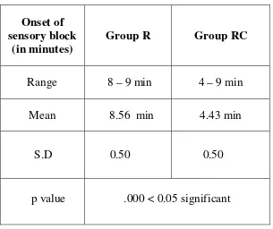
1.Onset of sensory blockade:
It was defined as the time interval between the end of local
anaesthetic administration and complete loss of sensation (Score 0). It
was assessed by pinprick test along the distribution of each nerve with a
needle using a 3 point scale for pain
0 – Loss of sensation to touch (Anaesthesia)
1 – Loss of sensation to pin prick (Analgesia)
2 – Normal sensation
61
2. Onset of motor blockade:
It was defined as the time interval between total local
anaesthetic administration and complete motor block (Grade 2). It was
assessed by thumb abduction (radial nerve), adduction (ulnar nerve),
and opposition (median nerve) according to Modified Bromage Scale.
Grade 0 - Normal motor function with full flexion and extension of
elbow ,wrist and fingers.
Grade 1 - Decreased motor power with the ability to move the fingers
only.
Grade 2 – Complete motor block with inability to move the fingers.
3.Duration of surgery:
It was defined as the time taken from skin incision to skin
closure.
4.Duration of motor blockade:
It was defined as the time interval from complete motor
block to complete recovery of motor function of hand and forearm
62
5. Duration of sensory blockade:
It was defined as the time interval between the complete
sensory block and complete resolution of anaesthesia on all the nerves
(Score 2).
6. Duration of analgesia:
It was defined as the time between the complete sensory
block and the first analgesic request. It was assessed using 10 point
visual analogue scale having 10 cm length numbered from 0 to 10.
Patient was explained about the visual analogue scale as
0 – No pain and 10 the worst possible pain and was asked the score in
visual analogue scale.
Pain was assessed at 2, 4, 6, 8, 12, and 16 h and Injection Tramadol
(100mg) was used as rescue analgesic on patient’s demand.
7.Vital parameters
Heart rate, Blood pressure, Oxygen saturation were