0022-538X/06/$08.00⫹0 doi:10.1128/JVI.00274-06
Copyright © 2006, American Society for Microbiology. All Rights Reserved.
Gamma Interferon Plays a Crucial Early Antiviral Role in Protection
against West Nile Virus Infection
Bimmi Shrestha,
1Tian Wang,
2† Melanie A. Samuel,
3Kevin Whitby,
1Joe Craft,
2,4Erol Fikrig,
2and Michael S. Diamond
1,3,5*
Departments of Medicine,1Molecular Microbiology,3and Pathology and Immunology,5660 S. Euclid Ave., Box 8051, Washington University School of Medicine, St. Louis, Missouri 63110, and Sections of Rheumatology2and
Immunobiology,4Yale University School of Medicine, 300 Cedar Street, New Haven, Connecticut 06520
Received 7 February 2006/Accepted 8 March 2006
West Nile virus (WNV) causes a severe central nervous system (CNS) infection in humans, primarily in the elderly and immunocompromised. Prior studies have established an essential protective role of several innate
immune response elements, including alpha/beta interferon (IFN-␣/), immunoglobulin M,␥␦T cells, and
complement against WNV infection. In this study, we demonstrate that a lack of IFN-␥production or signaling
results in increased vulnerability to lethal WNV infection by a subcutaneous route in mice, with a rise in
mortality from 30% (wild-type mice) to 90% (IFN-␥ⴚ/ⴚor IFN-␥Rⴚ/ⴚmice) and a decrease in the average
survival time. This survival pattern in IFN-␥ⴚ/ⴚand IFN-␥Rⴚ/ⴚmice correlated with higher viremia and
greater viral replication in lymphoid tissues. The increase in peripheral infection led to early CNS seeding
since infectious WNV was detected several days earlier in the brains and spinal cords of IFN-␥ⴚ/ⴚ or
IFN-␥Rⴚ/ⴚmice. Bone marrow reconstitution experiments showed that ␥␦ T cells require IFN-␥ to limit
dissemination by WNV. Moreover, treatment of primary dendritic cells with IFN-␥reduced WNV production
by 130-fold. Collectively, our experiments suggest that the dominant protective role of IFN-␥against WNV is
antiviral in nature, occurs in peripheral lymphoid tissues, and prevents viral dissemination to the CNS.
West Nile virus (WNV) is a mosquito-borne, single-stranded RNA flavivirus that is related to other viruses, including den-gue virus (DENV), yellow fever virus (YFV), and Japanese virus (JEV), St. Louis virus, and tick-borne encephalitis virus, that cause human disease. WNV is endemic in mosquitoes and birds in parts of Africa, Europe, the Middle East, and Asia and has established its presence in North America. Humans de-velop a febrile illness with a subset of cases progressing to meningitis, encephalitis, or an acute flaccid paralysis syndrome (1, 16, 34, 42, 43).
After mosquito inoculation, initial WNV replication is be-lieved to occur in the skin in Langerhans dendritic cells (DCs), which migrate to draining lymph nodes (11, 37), where viral amplification occurs (32, 38, 45, 49, 71). Subsequently, WNV enters the bloodstream and spreads to other peripheral organs, including the spleen. Within days of initial infection, WNV disseminates to the brain and spinal cord (19, 72) and in many animals infects a variety of neurons in the hippocampus, cer-ebellum, brain stem, cerebral cortex, and anterior horn of the spinal cord (19, 22, 63, 72).
In animals and humans the integrity of the immune system response correlates with resistance to WNV infection (1, 20, 22, 23, 28, 29, 31, 34, 42, 43, 50, 53, 64, 70, 73). Recent exper-iments with mice have eluicidated how the innate and adaptive
immune responses protect against WNV dissemination (20, 66). Type I interferon (interferon alpha/beta [IFN-␣/]) con-trols initial WNV infection and restricts tropism in extraneural sites, thus limiting viremia and dissemination to the central nervous system (CNS) (55). The induction of WNV-specific immunoglobulin M (IgM) coincides with the clearance of WNV from the bloodstream (21), and WNV-specific IgG lim-its dissemination and promotes clearance of virus from in-fected cells (4, 19, 24, 51). Changes in blood-brain barrier permeability associated with cytokine production and inflam-mation at peripheral sites facilitate WNV entry into the CNS (17, 68). Finally, CD8⫹T cells contribute to the eradication of WNV from infected cells since CD8-deficient or depleted mice fail to efficiently clear WNV from the CNS, resulting in in-creased morbidity and viral persistence (60, 69).
At present, less is known about the function of type II IFN (IFN-␥) in the control of WNV infection. IFN-␥is produced by natural killer (NK) cells, ␥␦ T cells, and CD8⫹ T cells and generally inhibits virus propagation by several mechanisms, including direct antiviral inhibitory activity, polarization and activation of T-helper-cell responses, enhanced class I major histocompatibility antigen expression and presentation, and activation of phagocytic myeloid cells (14, 30, 58). A recent study showed that␥␦T cells, which produce IFN-␥in response to WNV infection, function to limit early dissemination (67). That study, however, did not directly address whether ␥␦ T cells required IFN-␥to mediate their protective effects. Studies with other flaviviruses have provided some insight as to the potential protective function of IFN-␥. The pretreatment of cells with IFN-␥in vitro reduces the production of DENV (18) and JEV (46). In mice that were genetically deficient in IFN-␥, however, small or no changes in mortality were observed after
* Corresponding author. Mailing address: Departments of Medi-cine, Molecular Microbiology, Pathology and Immunology, Washing-ton University School of Medicine, 660 South Euclid Ave., Box 8051, St. Louis, MO 63110. Phone: (314) 362-2842. Fax: (314) 362-9230. E-mail: diamond@borcim.wustl.edu.
† Present address: Arthropod-Borne and Infectious Diseases Labo-ratory, Department of Microbiology, Immunology, and Pathology, Colorado State University, Fort Collins, CO 80523.
5338
on November 8, 2019 by guest
http://jvi.asm.org/
infection with DENV (35, 59) or YFV (47), and only a mod-erate increase in mortality was seen with Murray Valley en-cephalitis virus (MVEV) (48).
Studies with neurotropic viruses from other families have suggested that IFN-␥plays a more critical role in controlling infection in the CNS (8, 12, 25, 33, 44, 54). Based on experi-ments with Sindbis and measles viruses, IFN-␥is believed to contribute directly to the noncytolytic clearance of virus from neuronal cells and thus minimize host-mediated tissue damage (7, 8, 52). However, CD8⫹ T cells do not require IFN-␥to clear WNV infection since the adoptive transfer of IFN-␥ -deficient CD8⫹T cells into CD8-deficient mice fully controlled peripheral and CNS infection (61). In the present study, using mice that are deficient in either IFN-␥ or IFN-␥ receptor (IFN-␥R), we evaluated the protective effect of IFN-␥against a virulent North American WNV strain. An absence of IFN-␥ function resulted in an increased viral burden in peripheral tissues early during the course of infection, leading to rapid spread to the CNS and early death.
MATERIALS AND METHODS
Virus propagation and titration.The lineage I WNV strain (3000.0259) iso-lated in New York in 2000 was used as described previously (60). The stock virus
(2⫻108PFU/ml) was propagated once in C6/36 cells and used for all in vivo
and in vitro studies. The lineage II Madagascar strain was isolated in 1978 (DakAnMg798) as described previously (3) and was amplified once in Vero
cells (4⫻107
PFU/ml). BHK21 baby hamster kidney fibroblasts and Vero cells were cultivated in Dulbecco modified Eagle medium (DMEM) as de-scribed previously and were used to determine viral burden in the infected mouse cells or tissues by plaque assay (18).
DC infections and IFN treatment.Bone marrow-derived dendritic cells (DCs) were generated according to a published protocol (65). Bone marrow cells were harvested from the femurs of wild-type C57BL/6 mice and cultured in six-well
plates at a density of 105cells per well in RPMI supplemented with 10% defined
fetal calf serum (HyClone, Logan, Utah), granulocyte-macrophage colony-stim-ulating factor, sodium pyruvate, kanamycin sulfate, glutamine, and nonessential amino acids for 7 days. The nonadherent DC fraction was transferred to 12-well
plates at a density of 1.5⫻105cells per well. The purity of the DC population
was confirmed by staining with phycoerythrin-conjugated rat CD11c anti-body by flow cytometry (BD Biosciences Pharmingen). DCs were treated with
100 IU of mouse IFN-␣, IFN-, or IFN-␥(PBL Laboratories, Madison, WI)/ml
24 h prior to infection with WNV (multiplicity of infection [MOI] of 0.2). At 1 h after infection, free virus was removed by washing, and DCs were incubated for 24 h at 37°C in 1 ml of medium. Supernatants were then harvested, and infectious virus was measured by plaque assay on Vero cell monolayers.
Mouse experiments and tissue preparation.C57BL/6J strain (H-2b) inbred
wild-type mice were obtained from a commercial vendor. The congenic
IFN-␥⫺/⫺and IFN-␥R⫺/⫺mice, a gift from H. Virgin and R. Schreiber (Washington
University School of Medicine, St. Louis, MO), were backcrossed onto the C57BL/6 background for 10 generations. All mice were genotyped and bred under pathogen-free conditions in the animal facilities of Washington University School of Medicine. Experiments were performed with approval and under the guidelines of the Washington University Animal Studies Committee. Eight- to
twelve-week-old mice were inoculated with 102PFU of WNV (New York) via a
footpad route or 101
PFU of WNV (Madagascar) via an intracranial (i.c.) route.
Quantitation of viral burden in mouse tissues.To analyze the kinetics of virus
replication in different tissues, groups of wild-type, IFN-␥⫺/⫺, or IFN-␥R⫺/⫺
mice were infected with 102PFU of WNV and euthanized on day 1, 2, 3, 4, 5, 6,
or 8 after infection. Before organs were harvested, blood was collected from the axillary vein of each animal by phlebotomy and centrifuged and the serum was
isolated and stored at⫺80°C. Organs were removed and homogenized using a
Bead-Beater apparatus, and titers were determined on BHK21 cells as previously described (19). Viral RNA was extracted from thawed aliquots of serum samples
(50l) by using a Qia-Amp viral RNA recovery kit (QIAGEN, Palo Alto, CA)
and quantitated by real-time fluorogenic reverse transcription-PCR (RT-PCR) with an ABI 7000 Sequence Detection System (Applied Biosystems, Foster City, CA) (55). Viral RNA levels from the tissues of infected mice were determined as previously described (19) using an RNeasy kit (QIAGEN, Valencia, CA) and
real-time fluorogenic RT-PCR. To normalize the levels of WNV RNA in the samples, eukaryotic 18S rRNA was analyzed in parallel by real-time RT-PCR.
WNV-specific antibody production.An enzyme linked-immunosorbent assay (ELISA) was used to measure the development of WNV-specific IgG and IgM with some modifications of a prior protocol (19, 60). Briefly, purified WNV envelope protein that was generated in baculovirus-infected Hi-5 insect cells (51) was coated on Maxi-Sorp microtiter plates (Nalge Nunc International, Roches-ter, NY) overnight at 4°C. In parallel, plates were also coated with bovine serum albumin as a control antigen. After the saturation of nonspecific sites with blocking buffer (phosphate-buffered saline, 0.05% Tween 20, 3% bovine serum albumin, and 3% horse serum), plates were incubated with serial dilutions of heat-inactivated serum from infected mice for 2 h at room temperature. After extensive washing, plates were incubated with biotin-conjugated goat anti-mouse IgG or IgM (Sigma Chemical Co., St. Louis, MO), followed by incubation with horseradish peroxidase-conjugated streptavidin (Sigma Chemical). A positive signal was detected with tetramethylbenzidine substrate (Dako, Carpinteria,
CA), and the reaction was stopped with H2SO4. Plates were read in a GENios
ELISA plate reader (Tecan US, Inc., Durham, NC) at 450 nm. The background
optical density at 450 nm (OD450) value for binding to bovine serum albumin
alone was subtracted from the E protein wells to obtain an adjusted OD450for
each sample. Titers represent the serum dilution, yielding an OD450value
equiv-alent to three standard deviations above the background of the assay.
PRNT assay.The titer of neutralizing antibodies was determined by using a previously published protocol (19). Briefly, serum samples were diluted and
incubated with 8⫻101
PFU of WNV at 37°C for 1 h. WNV-serum complexes were added to monolayers of BHK21 cells in six-well plates, rocked for 5 min, and incubated at 37°C for 1 h. Subsequently, alpha minimal essential medium supplemented with 4% fetal calf serum and 1% low-melting-point agarose was added, and the plates were incubated at 37°C for 3 days. After fixation with 10% formaldehyde and staining with a 1% crystal violet solution, the plaques were counted and plotted and the plaque reduction neutralization titer for 50%
inhibition (PRNT50) was calculated.
Measurement of IFN activity.The levels of biologically active type I IFN in
serum of wild-type and IFN-␥⫺/⫺mice on days 1, 2, 3, 4, and 5 after WNV
infection were determined by using a previously developed L929 cell bioassay (2). Briefly, serum samples were harvested from infected animals, diluted serially in DMEM containing 10% fetal bovine serum, and incubated in duplicate with
adherent L929 cells (1.8⫻104
/well) for 14 h at 37°C in a 96-well plate. The supernatants were removed and replaced with DMEM containing encephalo-myocarditis virus (MOI of 7). Seven hours later, cells were inspected visually for cytopathic effect, and IFN-mediated protection was measured by colorimetric assay using a CellTiter 96 Aqueous Cell Proliferation Assay according to the manufacturer’s instructions (Promega Corp., Madison, WI).
Cytokine detection by flow cytometric bead array assay.Inflammatory
cyto-kine levels were measured in serum from wild-type and IFN-␥⫺/⫺mice by using
the mouse inflammation cytometric bead array (CBA) kit (BD Biosciences, San Diego, CA). Six different inflammatory cytokines were measured: interleukin-6
(IL-6), IL-10, monocyte chemo-attractant protein (MCP-1), IFN-␥, tumor
ne-crosis factor alpha (TNF-␣), and IL-12p70. For this analysis, serum was collected
at days 0, 1, 2, 3, 4, 6, and 8 postinfection from wild-type and IFN-␥⫺/⫺mice,
mixed with 50l of cytokine detection beads for 2 h at room temperature,
washed, and analyzed by flow cytometry according to the manufacturer’s instruc-tions using CBA software (BD Pharmingen). The concentration of a given cytokine in serum was determined after comparison with a standard curve that was generated as part of the kit.
Bone marrow chimeras and quantitation of WNV after infection.Bone mar-row chimeras were prepared according to a previously described protocol (26). In brief, recipients were irradiated twice with 500 rads, with a 30-min interval between doses. Donor bone marrow cells (mixed equally from two donors, see
Table 1 for details) were injected intravenously into recipients (1.4⫻107cells/
mouse). Antibiotics (sulfamethoxazole and trimethoprim oral pediatric suspen-sion; Hi-Tech Pharmacal Co., Amityville, NY) were added to the drinking water for the first 7 weeks after reconstitution. At 11 weeks after reconstitution, mice were infected with 100 PFU of WNV by footpad inoculation. On days 2, 4, and 6 after infection, blood, spleens, and brains were harvested, and WNV RNA was analyzed by quantitative fluorogenic RT-PCR as described previously (67).
Data analysis.For survival analysis, Kaplan-Meier survival curves were plotted by using Prism software (GraphPad, San Diego, CA). Mortality curves were analyzed by the log-rank test. The viral burden and antibody titer experiments were evaluated by the Mann-Whitney test. Differences in cytokine production
were analyzed by using an unpairedttest.
on November 8, 2019 by guest
http://jvi.asm.org/
RESULTS
Mice that lack IFN-␥function are more susceptible to WNV
infection after peripheral infection.Previous studies have
sug-gested that IFN-␥plays a critical role in controlling infection of some (8, 12, 25, 44), but not all, neurotropic viruses (41). The role of IFN-␥in vivo in modulating flavivirus infection is less certain since studies with DENV and YFV (35, 47, 59) showed little or no increased mortality in IFN-␥⫺/⫺mice. To assess the
effect of IFN-␥on WNV infection and disease, we compared survival rates in wild-type and congenic IFN-␥⫺/⫺ or
IFN-␥R⫺/⫺C57BL/6 mice. After subcutaneous inoculation with 102
PFU of a virulent New York strain of WNV, survival was markedly lower in mice that lacked IFN-␥function: IFN-␥⫺/⫺
or IFN-␥R⫺/⫺mice had a 5 to 10% survival rate compared to
70% in wild-type mice (Fig. 1A, P ⱕ 0.0001). Mortality oc-curred earlier in the IFN-␥⫺/⫺and IFN-␥R⫺/⫺mice since the
mean time to death was shorter (IFN-␥⫺/⫺mice, 9.2 ⫾ 0.8
days; IFN-␥R⫺/⫺mice, 9.7⫾0.7 days; wild-type mice, 10.9⫾
1.3 days; P ⱕ 0.002). Thus, an absence of IFN-␥ function caused a more fulminant WNV infection with increased mor-tality after peripheral infection.
WNV burden in IFN-␥ⴚ/ⴚ
mice. To understand how an
absence of IFN-␥function increased the susceptibility of mice to WNV infection, the levels of WNV RNA and infectious virus in peripheral and CNS tissues were determined by flu-orogenic RT-PCR and plaque assay in BHK21 cells, respec-tively. Wild-type and IFN-␥⫺/⫺ mice were infected with 102
PFU and viral burden was measured at days 1, 2, 3, 4, 6, and 8 after infection in serum, lymph nodes, spleens, brains, and spinal cords.
(i) Viremia.A higher and more prolonged viremia was
ob-served in IFN-␥⫺/⫺mice (Fig. 2A). WNV RNA was detected
in the serum by fluorogenic RT-PCR from day 1 after infection in wild-type and IFN-␥⫺/⫺mice. At days 2 and 4 after infection
approximately 10- to 50-fold-higher levels of WNV RNA were detected in IFN-␥⫺/⫺mice (Pⱕ0.05). At day 6 after infection,
viral RNA was cleared from wild-type serum, and yet signifi-cant levels were still detected in IFN-␥⫺/⫺mice (P⬍0.05). By
day 8, WNV RNA was undetectable in almost all serum sam-ples from IFN-␥⫺/⫺ mice. These results were confirmed by
viral plaque assays in which higher levels of infectious virus were detected in IFN-␥⫺/⫺mice through day 6 after infection
(data not shown).
(ii) Draining lymph node. Because viremia was elevated
early after infection, we evaluated the effect of IFN-␥on viral replication in the draining lymph nodes. Throughout the time course, WNV replication in individual ipsilateral inguinal lymph nodes from wild-type and IFN-␥⫺/⫺mice was below the
limit of detection of our plaque assays. However, when viral RNA was measured by fluorogenic RT-PCR assay, significant differences were observed (Fig. 2B). Although viral RNA was detected between days 1 and 4 after infection in both wild-type and IFN-␥⫺/⫺mice, higher levels (ca. 20- to 60-fold,Pⱕ0.005)
[image:3.585.41.546.82.136.2]were observed in the lymph nodes of IFN-␥⫺/⫺mice.
TABLE 1. Generation of bone marrow chimerasa
Group Donor 1 Donor 2 Recipient Phenotype
1 TCR␦⫺/⫺ IFN-␥⫺/⫺ Wild type ␥␦T cells do not produce IFN-␥; 50% of other cells (␣T cells, NK cells) produce IFN-␥
2 TCR␦⫹/⫹ IFN-␥⫺/⫺ Wild type 50% of␥␦T cells produce IFN-␥; 50% of other cells (␣T cells and NK cells) produce IFN-␥
aBone marrow chimeras were prepared as described in Materials and Methods. Bone marrow cells from IFN-␥⫺/⫺mice were mixed with bone marrow cells from
TCR␦⫺/⫺mice (normal␣T cells without␥␦T cells) or from TCR␦⫹/⫹mice (with␥␦T cells) in a 1:1 ratio and injected intravenously into irradiated wild-type mice.
At 11 weeks after reconstitution, mice were phenotyped and used for infection experiments.
FIG. 1. Survival analysis of wild-type, IFN-␥⫺/⫺, and IFN-␥R⫺/⫺ mice after WNV infection. (A) Wild-type, IFN-␥⫺/⫺, and IFN-␥R⫺/⫺ mice were infected subcutaneously with 102PFU of WNV (New York) and monitored for 28 days for morbidity and mortality. The survival curves were constructed using data from three to five independent experiments. The survival differences between wild-type, IFN-␥⫺/⫺, and IFN-␥R⫺/⫺mice were statistically significant (P⬍0.0001). The numbers of animals weren⫽50 for wild-type,n⫽37 for IFN-␥⫺/⫺, andn⫽29 for IFN-␥R⫺/⫺mice. (B) Wild-type and IFN-␥⫺/⫺mice were infected i.c. with 101PFU of WNV (Madagascar) and monitored for 28 days for morbidity and mortality. The survival curves were constructed with data from two independent experiments. The survival differences between wild-type and IFN-␥⫺/⫺mice were not statistically significant (P⫽0.7). The numbers of animals weren⫽20 for wild-type mice andn⫽20 for IFN-␥⫺/⫺mice.
on November 8, 2019 by guest
http://jvi.asm.org/
[image:3.585.318.519.291.588.2](iii) Spleen.WNV infection was detected at higher levels in the spleens of IFN-␥⫺/⫺mice. Although infectious WNV was
not detected 1 day after infection,⬃30-fold higher levels of viral RNA were measured in spleens from IFN-␥⫺/⫺ mice
compared to wild-type mice at this time (Fig. 2C,P⫽0.001). By day 2 after infection, infectious WNV (⬃102PFU/g, 3 of 10
mice) was detected in the spleen of IFN-␥⫺/⫺mice, whereas
virus was not detected in any of the wild-type mice (Fig. 2D). On days 4 and 6 after infection,⬃100-fold-higher levels of WNV were observed in IFN-␥⫺/⫺mice (Pⱕ0.005).
Nonethe-less, by day 8 all IFN-␥⫺/⫺and wild-type mice had completely
cleared infectious virus from the spleen. Overall, a lack of IFN-␥activity resulted in a failure to control infection in the spleen during the initial stages of infection.
(iv) CNS tissues. (a) Brain. WNV spread earlier into the
brain of IFN-␥⫺/⫺mice (Fig. 2E). At day 4 after infection, 90%
(9 of 10 mice) of IFN-␥⫺/⫺mice had detectable WNV in the
brain (103 to 104 PFU/g), whereas no infectious virus was
present in any of the wild-type mice at this time. By day 6,
⬃100-fold-higher levels of infectious WNV were observed in the brains of IFN-␥⫺/⫺mice (P⫽0.0004). Interestingly, by day
8, the levels of WNV declined in IFN-␥⫺/⫺mice such that no
significant difference was observed compared to wild-type mice (P⫽0.15).
(iv) CNS tissues. (b) Spinal cord. In the spinal cord, a
pattern was observed that is similar to that seen in the brain (Fig. 2F). Earlier entry into the spinal cord was observed in IFN-␥⫺/⫺mice. At day 6 after infection, 100% of IFN-␥⫺/⫺
mice had significant viral infection (⬃104 to 107 PFU/g),
whereas virus was not measurable in the spinal cords of wild-type mice. By day 8, viral burden in the IFN-␥⫺/⫺mice
de-clined slightly such that no statistical difference was observed compared to the wild-type mice (P ⫽0.14). Since there was higher and earlier WNV replication in the CNS of IFN-␥⫺/⫺
mice, we evaluated whether there was a difference in viral tropism. Similar to findings seen with several other immuno-deficient mice (19, 60, 68, 69), immunohistochemical analysis revealed an increase in the number of neurons that expressed WNV antigen in the CNS of IFN-␥⫺/⫺mice (data not shown).
Clearance of WNV from the CNS of surviving IFN-␥ⴚ/ⴚ
mice.A lack of CD8⫹T cells or perforin molecules results in
WNV persistence in the CNS up to 5 weeks after infection (60, 61). Because IFN-␥participates in the clearance of other neu-rotropic viruses from the CNS (7, 8), we analyzed viral
persis-FIG. 2. WNV burden in peripheral and CNS tissues of wild-type and IFN-␥⫺/⫺mice. (A) Levels of viral RNA in serum detected by fluorogenic real-time RT-PCR. The kinetics and level of viral RNA were determined from the sera of wild-type and IFN-␥⫺/⫺mice after WNV (New York) infection at the indicated time point by fluorogenic real-time RT-PCR. The data are expressed as number of copies of WNV RNA per milliliter of serum (n⫽10 mice per time point). (B) Levels of viral RNA in inguinal lymph nodes. Viral RNA was extracted from the draining inguinal lymph nodes of wild-type and IFN-␥⫺/⫺mice at the indicated time points after WNV infection and quantitated by fluorogenic real-time RT-PCR. The data are expressed as the number of copies of WNV RNA per microgram of rRNA after normalization for tissue content (n⫽6 mice per time point). (C) Levels of viral RNA in the spleen on day 1 postinfection. Viral RNA was extracted from wild-type or IFN-␥⫺/⫺spleens at day 1 postinfection and quantitated by fluorogenic real-time RT-PCR. The data are expressed as the number of copies of WNV RNA per microgram of rRNA after normalization for tissue content (n⫽6 mice per group). (D to F) Levels of infectious virus in tissues. Infectious WNV levels were measured in the spleen (D), brain (E), and spinal cord (F) by using a viral plaque assay in BHK21 cells after tissues were harvested and homogenized at the indicated time points after WNV infection. The data are shown as the average PFU per gram of tissue (n⫽10 mice per time point). The dotted line represents the limit of sensitivity of the assay (❋,Pⱕ0.05;❋❋,Pⱕ0.005 [compared to wild-type mice]).
on November 8, 2019 by guest
http://jvi.asm.org/
[image:4.585.57.532.68.330.2]tence in IFN-␥⫺/⫺mice at later time points. The infection of
IFN-␥⫺/⫺ mice resulted in a small (⬃5 to 10%) number of
surviving animals. In surviving wild-type or IFN-␥⫺/⫺ mice,
infectious virus was not detected in the brain after 21 days (data not shown). Thus, a deficiency of IFN-␥did not appear to affect the kinetics of viral clearance from the CNS, results that agree with adoptive transfer experiments that showed that IFN-␥-deficient CD8⫹T cells efficiently cleared WNV from the CNS (61).
WNV burden in IFN-␥Rⴚ/ⴚ
mice. To confirm the role of
IFN-␥ in controlling WNV infection, a subset of analogous virologic experiments were performed with IFN-␥R⫺/⫺mice at
key time points (Fig. 3). Similar to IFN-␥⫺/⫺mice, infectious
virus (⬃103PFU/g) was detected by viral plaque assay in the
serum at days 2 and 4 in 80% (four of five mice) of IFN-␥R⫺/⫺
mice (data not shown). Compared to wild-type mice,
IFN-␥R⫺/⫺mice had significantly enhanced levels of infection in
the spleen at days 4 and 6 (Fig. 3A,Pⱕ0.05); however, these levels were similar to those observed with IFN-␥⫺/⫺mice (Pⱖ
0.1). In addition, early entry and replication of WNV in the brain and spinal cord were observed with IFN-␥R⫺/⫺ mice
(Fig. 3B and C).
Effect of IFN-␥after i.c. infection.Since WNV is naturally
acquired through peripheral inoculation and subsequently spreads to the CNS, an effective immune response to WNV controls viral replication both in the periphery and in the CNS compartments. Our experiments with a subcutaneous infection model show that IFN-␥is required to control peripheral viral replication and spread (see Fig. 2). To determine whether IFN-␥plays an independent role in the CNS after WNV in-fection, we inoculated wild-type and IFN-␥⫺/⫺mice i.c. with
101PFU of an attenuated WNV strain (Madagascar [3]); the
attenuated strain was used because i.c. inoculation of 101PFU
of the virulent New York WNV strain causes complete lethal-ity in wild-type mice (55). As no difference in lethallethal-ity after i.c. infection with the Madagascar strain was observed between wild-type and IFN-␥⫺/⫺mice (Fig. 1B,P⫽0.7), IFN-␥does
not appear to have an essential antiviral effect in the CNS. Importantly, at day 30, surviving wild-type and IFN-␥⫺/⫺mice
all had high-titer WNV-specific antibodies, thus confirming productive infection with the Madagascar strain (data not shown).
WNV-specific IgM and IgG production in the absence of IFN-␥.IFN-␥is an immunomodulatory cytokine with specific roles in the priming of adaptive immune responses (58). Be-cause a higher viremia was observed in mice that lacked IFN-␥ function, we reasoned that this could be due to depressed WNV-specific antibody responses. Blunted WNV-specific an-tibody responses early during the course of infection correlate with higher viremia, earlier spread to the CNS, and increased mortality (19, 21). Interestingly, higher rather than lower titers of WNV-specific IgM and IgG were detected in IFN-␥⫺/⫺mice
(Fig. 4A and B). Increased levels of WNV-specific IgM were detected after day 4 (Pⱕ0.05) and throughout the time course compared to the wild-type mice (Fig. 4A). Although an earlier WNV-specific IgG response was detected in IFN-␥⫺/⫺mice at
day 6 (Fig. 4B,P⬍0.05), by day 8 no statistical difference was observed (P ⫽ 0.12). Despite the somewhat higher level of antibody production in IFN-␥⫺/⫺mice, similar neutralization
titers were detected in both wild-type and IFN-␥⫺/⫺mice from
FIG. 3. Level of infectious WNV burden in the peripheral and CNS tissues of wild-type, IFN-␥⫺/⫺, and IFN-␥R⫺/⫺ mice. The level of infectious virus was measured from the spleens (A), brains (B), and spinal cords (C) of wild-type, IFN-␥⫺/⫺, and IFN-␥R⫺/⫺mice by using a viral plaque assay in BHK21 cells at the indicated time points after WNV (New York) infection. The data are shown as the average PFU per gram of tissue (n⫽5 to 10 mice per time point). The dotted line represents the limit of sensitivity of the assay (❋,Pⱕ0.05;❋❋,Pⱕ
0.005 [compared to wild-type mice]).
on November 8, 2019 by guest
http://jvi.asm.org/
day 4 after infection (Fig. 4C). Because WNV-specific antibody responses are not blunted in IFN-␥⫺/⫺ mice, the increased
WNV infection in the periphery is not due to a compromised ability to prime B-cell responses.
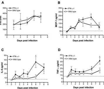
Serum levels of IFN in the absence of IFN-␥.A lack of type
I IFN receptor resulted in a higher WNV burden in peripheral and CNS tissues and uniform mortality in mice by day 4 after
infection (55). Since we observed a similar increase in the peripheral replication of WNV in IFN-␥⫺/⫺mice, we
hypoth-esized that IFN-␥could protect against WNV by amplifying the type I IFN response. To test this, we measured the levels of biologically active type I IFN in serum from wild-type and IFN-␥⫺/⫺mice after WNV infection using an
encephalomyo-carditis bioassay (see Materials and Methods). In both wild-type and IFN-␥⫺/⫺ mice, type I IFN was detected on day 1
after infection and increased through day 5 (Fig. 5A). Impor-tantly, a lack of IFN-␥resulted in no significant difference (Pⱖ
0.1) in the level of the type I IFN antiviral effect throughout the time course compared to wild-type mice. Thus, the early in-creased viral burden in peripheral lymphoid tissues in
IFN-␥⫺/⫺mice was not due to a decreased type I IFN response.
Inflammatory cytokine profile after WNV infection in the
absence of IFN-␥. A prior report suggested that Toll-like
re-ceptor-3-induced production of TNF-␣ in peripheral tissues could promote blood-brain barrier permeability changes and enhanced WNV entry into the CNS (68). To test whether the earlier WNV entry in IFN-␥⫺/⫺mice was due to a difference in
inflammatory cytokine and chemokines, we measured the lev-els of TNF-␣, IL-6, MCP-1, IL-12p70, IL-4, IFN-␥, and IL-10 in sera after WNV infection by using a flow cytometric bead array assay. Among the sera tested, significant levels of IFN-␥, MCP-1, IL-6, and TNF-␣were detected. As expected, IFN-␥ was measurable in sera only from wild-type mice and was present soon after infection (day 1,⬃4 pg/ml), a finding con-sistent with a previous study (28). Significant increases in MCP-1 and IL-6 levels in the serum in IFN-␥⫺/⫺mice were
transiently detected and only at day 6 after infection (Fig. 5B and C). Although increased TNF-␣levels were present in the sera of IFN-␥⫺/⫺mice, statistically significant differences were
not observed until 6 days after infection (Fig. 5D, ⬃8-fold increase,P ⬍ 0.05). Because of the disparity in kinetics be-tween viral CNS entry and TNF-␣levels, we hypothesized that the early dissemination in IFN-␥⫺/⫺mice was not due to
dif-ferential TNF-␣-dependent changes in CNS blood-brain bar-rier permeability. Indeed, no difference in blood-brain barbar-rier permeability was observed by Evans blue staining in IFN-␥⫺/⫺,
IFN-␥R⫺/⫺, or wild-type mice at days 2 to 4 after infection, and
the administration of a neutralizing anti-TNF-␣antibody did not affect the kinetics of viral entry into the CNS or survival in IFN-␥⫺/⫺mice (data not shown).
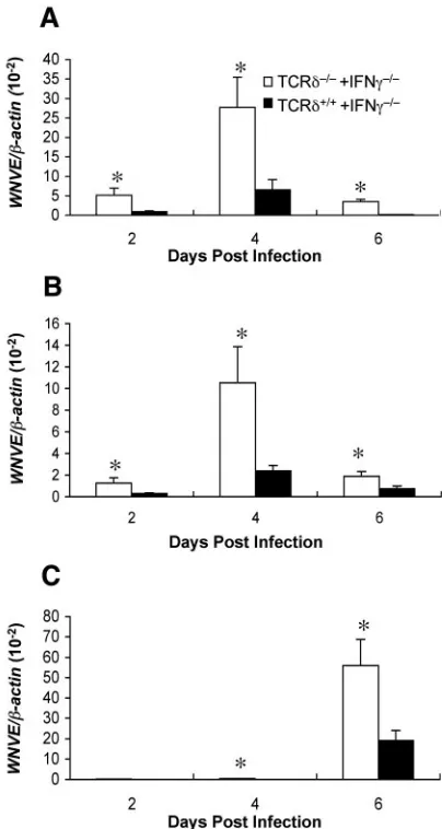
␥␦T cells require IFN-␥to control early WNV infection.␥␦
T cells control the early replication and spread of WNV infec-tion to the CNS (67). To directly assess the role of IFN-␥in
␥␦-T-cell-mediated control of WNV, bone marrow chimeras were generated. To create mice in which only the␥␦ T cells were deficient in IFN-␥, bone marrow cells from TCR␦⫺/⫺and
IFN-␥⫺/⫺mice were mixed at a 1:1 ratio and transferred to
lethally irradiated wild-type mice (see Table 1). Mice were generated in parallel with IFN-␥-sufficient␥␦T cells by using a 1:1 mixture of TCR␦⫹/⫹and IFN-␥⫺/⫺donor cells. Six weeks
later, after reconstitution phenotypes were confirmed, mice were infected with WNV, and viral burdens were analyzed in the sera, spleens, and brains by using fluorogenic quantitative RT-PCR. Mice reconstituted with IFN-␥-deficient␥␦T cells had significantly higher levels of WNV RNA in the sera (Fig. 6A), spleens (Fig. 6B), and brains (Fig. 6C) throughout the time course compared to mice reconstituted with IFN-␥-sufficient␥␦T cells
FIG. 4. WNV-specific antibody responses in wild-type and IFN-␥⫺/⫺ mice. Wild-type and IFN-␥⫺/⫺mice were infected with WNV (New York), and sera were collected at the indicated time points. The development of specific IgM (A) or IgG (B) antibodies to WNV was determined by ELISA using purified WNV E protein. The data are the averages of at least eight mice per time point. (C) Neutralizing anti-body titers. Neutralizing titers were determined by using a PRNT assay. All serum samples were performed in duplicate, and the data are expressed as reciprocal PRNT50s (i.e., the antibody titers that reduced the number of plaques by 50%). The data are the average of eight mice per time point.
on November 8, 2019 by guest
http://jvi.asm.org/
(Pⱕ0.05). These experiments suggest that the early replication and dissemination of WNV observed in IFN-␥⫺/⫺mice is due in
part to the inability of␥␦T cells to produce IFN-␥in the early phases of infection.
Antiviral effect of IFN-␥in bone marrow-derived DCs.Our
studies in vivo suggested that IFN-␥limits WNV replication in peripheral lymphoid tissues within days of infection prior to the development of an adaptive immune response. Based on this, we postulated that IFN-␥, produced by ␥␦ T cells and perhaps by other immune cells, acts as a direct antiviral agent to control WNV infection in lymphoid compartments. To eval-uate this, we assessed the antiviral activity of IFN-␥in vitro on cultured DCs derived from wild-type C57BL/6 mice. DCs, by virtue of their expression of DC-SIGN-like attachment mole-cules (15), are believed to be initial targets for WNV replica-tion after skin inoculareplica-tion (11, 37). CD11c⫹DCs were pre-treated with physiologically relevant doses (100 U/ml) of IFN-␣, IFN-, or IFN-␥and then infected with WNV at an MOI of 0.2. One day later, supernatants were analyzed for the production of infectious virus by plaque assay. Pretreatment of DCs with any of the IFNs significantly reduced (Pⱕ 0.0004) virus titers in the supernatant (Fig. 7). Notably, compared to untreated DCs, there was a 130-fold reduction in WNV pro-duction in cells pretreated with IFN-␥.
DISCUSSION
Previously, we have shown that type I IFN (IFN-␣/) plays a key role in the innate defense against WNV infection by re-stricting the tropism of WNV infection in peripheral tissues (55). Nonetheless, the role of IFN-␥in WNV infection and other flavivirus infections has remained more uncertain since it has both antiviral and immunomodulatory functions against other viruses. Using multiple experimental approaches we in-vestigated how IFN-␥controls WNV infection. C57BL/6 mice that lacked IFN-␥or the IFN-␥R showed increased suscepti-bility to lethal WNV infection with a rise in mortality and a decrease in the average survival time compared to congenic wild-type mice. The increase in mortality in IFN-␥⫺/⫺or
IFN-␥R⫺/⫺ mice was associated with greater viral replication in
lymphoid tissues and higher viremia, which led to a more rapid dissemination of WNV into the brain and spinal cord. These in vivo results, which suggested an early antiviral function of IFN-␥, were supported by in vitro infection experiments with primary DCs.
[image:7.585.115.469.67.371.2]A previous study showed that WNV infection of mice defi-cient in␥␦ T cells resulted in elevated viral loads in the pe-riphery, enhanced dissemination to the CNS, and increased lethality (67). Although the␥␦T cells expanded rapidly in vivo
FIG. 5. Cytokine profiles in the sera of wild-type and IFN-␥⫺/⫺mice. Wild-type and IFN-␥⫺/⫺mice were infected with WNV (New York) via the footpad, and sera were collected at the indicated time points after infection. (A) Type I IFN activity was measured by bioassay using encephalomyocarditis virus and L929 cells. The data are an average of three to five mice per time point. (B to D) Other inflammatory cytokines and chemokines. The levels of MCP-1 (B), IL-6 (C) and TNF-␣(D) in serum were determined by using a flow cytometric bead array. The data are expressed as an average of six mice per time point from two independent experiments. The dotted line represents the limit of sensitivity of the assay (❋,Pⱕ0.05 compared to wild-type mice).
on November 8, 2019 by guest
http://jvi.asm.org/
and produced IFN-␥in ex vivo restimulation assays, experi-ments did not directly identify the mechanism of protection. Here, using bone marrow chimeras, we demonstrate that␥␦T cells require IFN-␥to limit dissemination by WNV. Mice re-constituted with IFN-␥-deficient ␥␦ T cells had significantly higher levels of WNV RNA in the serum, spleen, and brain throughout the time course compared to mice reconstituted with IFN-␥-sufficient ␥␦ T cells. These experiments suggest that the early replication of WNV observed in IFN-␥⫺/⫺mice
is due in part to the inability of␥␦T cells to produce IFN-␥. Interestingly, although NK cells also produce IFN-␥early dur-ing an immune response, antibody depletion studies suggest they do not have a significant role in the control of WNV infection in mice (61).
Our results are most consistent with a model in which IFN-␥ has an early antiviral role, limiting the spread of WNV infec-tion prior to activainfec-tion and control by adaptive B and T-cell responses. The following data from our studies with IFN-␥⫺/⫺
or IFN-␥R⫺/⫺mice support this model: (i) enhanced WNV
infection was observed in lymphoid tissues (spleen and lymph node) by day 1 after infection, (ii) increased viremia was seen within 2 days of infection, and (iii) a more rapid spread of WNV to the brain and spinal cord was consistently observed in all mice lacking the IFN-␥signaling function. Moreover, ex vivo infection of DCs, the cell type that is believed to sustain early rounds of WNV replication (11, 15, 37, 38) was markedly decreased after exposure to physiologically relevant concentra-tions of IFN-␥. In contrast, we did not observe major deficits in the adaptive immune response or function in IFN-␥-deficient mice. IgM and IgG responses against WNV developed with normal kinetics in IFN-␥⫺/⫺ mice. The clearance of WNV
from the spleen was normal, a process that depends on priming and activation of CD8⫹T cells (60, 61). In addition, although most IFN-␥⫺/⫺ mice succumbed rapidly to WNV infection,
those that survived showed a relatively typical pattern of clear-ance of infectious WNV from the CNS. This was reflected by a decrease in viral burden between days 6 and 8 after infection and an absence of CNS viral persistence in any of the few surviving animals.
An absence of IFN-␥in C57BL/6 mice resulted in a marked increase in WNV infection and mortality, results that are somewhat distinct from that previously observed with other flaviviruses. No increase in mortality was observed in IFN-␥⫺/⫺
C57BL/6 mice after primary YFV infection, although the levels of infectious virus in the brain were higher at day 4 (47). For DENV, moderately increased death was observed in IFN-␥⫺/⫺
129 Sv mice (59), with peak mortality reaching 30 and 43% for DENV-1 and DENV-2 infections, respectively. However, com-pared to wild-type mice, no increase in viral burden was ob-served by indirect plaque assay at day 3 or 7 after infection in lymphoid or CNS tissues. Notably, no mortality or illness was observed after DENV-2 infection of IFN-␥⫺/⫺ mice on a
BALB/c background (35). C57BL/6 mice deficient in IFN-␥ showed a moderate (60% compared to 30% for wild-type mice) increase in mortality after infection with MVEV, a
[image:8.585.62.264.69.448.2]re-FIG. 6. WNV burden in peripheral and CNS tissues of bone mar-row chimeras. Bone marmar-row chimeras were prepared as described in Materials and Methods using donors and recipients (see Table 1). Reconstituted mice were infected subcutaneously with 102PFU of WNV (New York). Viral loads were measured at the indicated days in the blood (A), spleens (B), and brains (C) of mice by using quantitative RT-PCR. Theyaxis depicts the ratio of the amplified WNV-E cDNA to-actin cDNA of each sample. At each time point, five mice per group were analyzed (❋,Pⱕ0.05 indicates a significant difference in viral RNA levels between reconstituted groups).
FIG. 7. Inhibition of WNV production from the bone marrow-derived DCs treated with IFN. DCs were generated from the bone marrow of wild-type mice. DCs were pretreated with 100 IU of IFN-␣, IFN-, or IFN-␥24 h before infection. Cells were infected at an MOI of 0.2, and the production of infectious WNV (New York) was deter-mined 1 day later by plaque assay in Vero cells. The data are expressed as an average of two independent experiments performed in triplicate (❋❋,Pⱕ0.005 compared to untreated cells).
on November 8, 2019 by guest
http://jvi.asm.org/
[image:8.585.342.498.70.173.2]lated encephalitic flavivirus (48). Increased MVEV burden was detected in the serum and spleen at day 4 after infection in IFN-␥⫺/⫺mice, and brain titers at day 8 were indistinguishable
from wild-type mice, results that are consistent with our find-ings. Interestingly, higher mortality after MVEV infection was also observed in mice deficient in inducible nitric oxide syn-thase-2, an effector molecule downstream of IFN-␥that has been reported to affect outcome after infection with JEV (56, 57) and other RNA viruses (14, 40).
Surprisingly, our virologic data suggest that IFN-␥has a less significant antiviral role in the CNS. No difference in survival was observed after i.c. infection with the attenuated Madagas-car strain of WNV. Although IFN-␥is induced in CNS tissues by day 8 after WNV infection (28, 39) and is capable of inhib-iting infection⬃10-fold in some primary neuron cultures (55), these effects did not modulate survival. In contrast to other neurotropic RNA viruses such as Sindbis virus (7–9), measles virus (52), mouse hepatitis virus (5, 6) or Theiler’s encephalo-myelitis virus (54), which clear infection through a noncyto-lytic, IFN-␥-dependent mechanism (13, 14), our data suggest that IFN-␥does not have a dominant inhibitory role against WNV in the CNS. Consistent with this, our recent adoptive-transfer studies showed efficient clearance of infection in the spleen and CNS by IFN-␥-deficient WNV-primed CD8⫹ T cells (61).
Our experiments with IFN-␥⫺/⫺and IFN-␥R⫺/⫺mice show
earlier dissemination of WNV to the CNS, with at least 3- to 5-log10-higher levels of viral replication in the brain and spinal
cord at days 4 and 6 after infection, respectively. Earlier entry into the CNS by encephalitic flaviviruses was previously ob-served in mice that lack functional type I IFN-␣ and - re-sponses (48, 55). Notably, normal levels of type I IFN were observed in the sera of IFN-␥⫺/⫺mice; thus, IFN-␥does not
appear to be essential for amplifying the type I IFN response after WNV infection. Early entry into the CNS by WNV cor-relates directly with the increased infection in peripheral lym-phoid tissues and viremia. The direct mechanism for enhanced WNV neuroinvasiveness in IFN-␥⫺/⫺mice remains uncertain.
Although the clearance of WNV from the bloodstream is linked to the development of neutralizing IgM antibodies (21), no significant differences in WNV-specific antibody responses were observed throughout the infection time course. A previ-ous study showed that Toll-like receptor-3-dependent produc-tion of TNF-␣at days 2 and 3 after infection enhanced blood-brain barrier permeability and invasion of the CNS by WNV (68). However, the early entry into the CNS by WNV in
IFN-␥⫺/⫺mice appeared to be independent of TNF-␣: (i) no
dif-ference in blood-brain barrier permeability, as judged by Evans’ blue staining, was seen among wild-type and IFN-␥⫺/⫺
mice, (ii) the levels of TNF-␣in serum were not statistically different until day 8 after infection, and (iii) the administration of a neutralizing TNF-␣ antibody had no effect of the en-hanced mortality observed in IFN-␥⫺/⫺mice. Although future
studies are required to define precisely how IFN-␥regulates WNV neuroinvasion, myeloid cells have been hypothesized to carry viruses in the brain by a “Trojan horse” mechanism (10, 28, 36, 62). Indeed, a recent study shows that equine mono-cytes support productive WNV replication in blood (27). Based on our infection data with DCs, IFN-␥could limit myeloid cell
infection in the intravascular compartment and the transport of infected cells across the blood-brain barrier.
In summary, our experiments demonstrate an essential pro-tective role for IFN-␥in WNV infection. Innate immune re-sponders, including ␥␦ T cells, generate IFN-␥ after WNV infection in peripheral lymphoid tissues. We show here that IFN-␥appears to function as an antiviral agent, possibly lim-iting infection in DCs and other myeloid cells. This decreased viral burden prevents early dissemination to the CNS and in-fection and allows the adaptive immune response to develop before significant irreversible damage to neurons has occurred.
ACKNOWLEDGMENTS
We thank H. Virgin and R. Schreiber for the IFN-␥and IFN-␥R⫺/⫺ mice and R. Schreiber for critical reading of the manuscript. We also acknowledge help provided by Z. Yin, Y. Gao, J. Tao, and D. Zhang in the preparation of the bone marrow chimeras; R. Silverman for advice on the IFN bioassay; M. Gale for the Madagascar WNV isolate; and W. Barchet and M. Colonna for DC isolation and cultivation techniques.
This study was supported by grants and contracts from the National Institutes of Health (M.S.D, E.F., and J.C.), a predoctoral fellowship from the Howard Hughes Medical Institute (M.A.S.) and a New Scholar Award in Global Infectious Disease from the Ellison Medical Foundation (M.S.D).
REFERENCES
1.Asnis, D. S., R. Conetta, A. A. Teixeira, G. Waldman, and B. A. Sampson.
2000. The West Nile virus outbreak of 1999 in New York: the Flushing
Hospital experience. Clin. Infect. Dis.30:413–418.
2.Austin, B. A., C. James, R. H. Silverman, and D. J. Carr.2005. Critical role for the oligoadenylate synthetase/RNase L pathway in response to IFN-beta
during acute ocular herpes simplex virus type 1 infection. J. Immunol.175:
1100–1106.
3.Beasley, D. W., L. Li, M. T. Suderman, and A. D. Barrett.2002. Mouse neuroinvasive phenotype of West Nile virus strains varies depending upon
virus genotype. Virology296:17–23.
4.Ben-Nathan, D., S. Lustig, G. Tam, S. Robinzon, S. Segal, and B. Rager-Zisman.2003. Prophylactic and therapeutic efficacy of human intravenous immunoglobulin in treating West Nile virus infection in mice. J. Infect. Dis.
188:5–12.
5.Bergmann, C. C., B. Parra, D. R. Hinton, R. Chandran, M. Morrison, and S. A. Stohlman.2003. Perforin-mediated effector function within the central
nervous system requires IFN-␥-mediated MHC up-regulation. J. Immunol.
170:3204–3213.
6.Bergmann, C. C., B. Parra, D. R. Hinton, C. Ramakrishna, K. C. Dowdell, and S. A. Stohlman.2004. Perforin and gamma interferon-mediated control of coronavirus central nervous system infection by CD8 T cells in the absence
of CD4 T cells. J. Virol.78:1739–1750.
7.Binder, G. K., and D. E. Griffin.2003. Immune-mediated clearance of virus
from the central nervous system. Microbes Infect.5:439–448.
8.Binder, G. K., and D. E. Griffin.2001. Interferon-gamma-mediated
site-specific clearance of alphavirus from CNS neurons. Science293:303–306.
9.Burdeinick-Kerr, R., and D. E. Griffin.2005. Gamma interferon-dependent, noncytolytic clearance of Sindbis virus infection from neurons in vitro. J.
Vi-rol.79:5374–5385.
10.Burke, D. S., and T. P. Monath.2001. Flaviviruses, p. 1043–1125.InD. M. Knipe and P. M. Howley (ed.), Fields virology, 4th ed., vol. 1. Lippincott Williams & Wilkins, Philadelphia, Pa.
11.Byrne, S. N., G. M. Halliday, L. J. Johnston, and N. J. King.2001. Inter-leukin-1beta but not tumor necrosis factor is involved in West Nile virus-induced Langerhans cell migration from the skin in C57BL/6 mice. J. Investig.
Dermatol.117:702–709.
12.Cantin, E. M., D. R. Hinton, J. Chen, and H. Openshaw.1995. Gamma interferon expression during acute and latent nervous system infection by
herpes simplex virus type 1. J. Virol.69:4898–4905.
13.Chesler, D. A., C. Dodard, G. Y. Lee, D. E. Levy, and C. S. Reiss.2004. Interferon-gamma-induced inhibition of neuronal vesicular stomatitis virus
infection is STAT1 dependent. J. Neurovirol.10:57–63.
14.Chesler, D. A., and C. S. Reiss.2002. The role of IFN-gamma in immune responses to viral infections of the central nervous system. Cytokine Growth
Factor Rev.13:441–454.
15.Davis, C. W., H. Y. Nguyen, S. L. Hanna, M. D. Sanchez, R. W. Doms, and T. C. Pierson.2006. West Nile virus discriminates between DC-SIGN and
DC-SIGNR for cellular attachment and infection. J. Virol.80:1290–1301.
on November 8, 2019 by guest
http://jvi.asm.org/
16.Diamond, M. S.2005. Development of effective therapies against West Nile
virus infection. Expert Rev. Anti-Infect. Ther.3:931–944.
17.Diamond, M. S., and R. S. Klein.2004. West Nile virus: crossing the
blood-brain barrier. Nat. Med.10:1294–1295.
18.Diamond, M. S., T. Roberts, D. Edgil, B. Lu, J. Ernst, and E. Harris.2000. Modulation of Dengue virus infection in human cells by alpha, beta, and
gamma interferons. J. Virol.74:4957–4966.
19.Diamond, M. S., B. Shrestha, A. Marri, D. Mahan, and M. Engle.2003. B cells and antibody play critical roles in the immediate defense of
dissemi-nated infection by West Nile encephalitis virus. J. Virol.77:2578–2586.
20.Diamond, M. S., B. Shrestha, E. Mehlhop, E. Sitati, and M. Engle.2003. Innate and adaptive immune responses determine protection against
dissem-inated infection by West Nile encephalitis virus. Viral Immunol.16:259–278.
21.Diamond, M. S., E. Sitati, L. Friend, B. Shrestha, S. Higgs, and M. Engle.
2003. Induced IgM protects against lethal West Nile virus infection. J. Exp.
Med.198:1–11.
22.Eldadah, A. H., and N. Nathanson.1967. Pathogenesis of West Nile virus encephalitis in mice and rats. II. Virus multiplication, evolution of immuno-fluorescence, and development of histological lesions in the brain. Am. J.
Epidemiol.86:776–790.
23.Eldadah, A. H., N. Nathanson, and R. Sarsitis.1967. Pathogenesis of West Nile virus encephalitis in mice and rats. 1. Influence of age and species on
mortality and infection. Am. J. Epidemiol.86:765–775.
24.Engle, M., and M. S. Diamond.2003. Antibody prophylaxis and therapy against West Nile virus infection in wild type and immunodeficient mice.
J. Virol.77:12941–12949.
25.Finke, D., U. G. Brinckmann, V. ter Meulen, and U. G. Liebert.1995. Gamma interferon is a major mediator of antiviral defense in experimental
measles virus-induced encephalitis. J. Virol.69:5469–5474.
26.Gao, Y., W. Yang, M. Pan, E. Scully, M. Girardi, L. H. Augenlicht, J. Craft, and Z. Yin.2003.␥␦T cells provide an early source of IFN-␥in tumor
immunity. J. Exp. Med.198:433–442.
27.Garcia-Tapia, D., C. M. Loiacono, and S. B. Kleiboeker.2005. Replication of West Nile virus in equine peripheral blood mononuclear cells. Vet.
Immu-nol. Immunopathol.110:229–244.
28.Glass, W. G., J. K. Lim, R. Cholera, A. G. Pletnev, J. L. Gao, and P. M. Murphy.2005. Chemokine receptor CCR5 promotes leukocyte trafficking to
the brain and survival in West Nile virus infection. J. Exp. Med.202:1087–
1098.
29.Glass, W. G., D. H. McDermott, J. K. Lim, S. Lekhong, S. F. Yu, W. A. Frank, J. Pape, R. C. Cheshier, and P. M. Murphy.2006. CCR5 deficiency increases
risk of symptomatic West Nile virus infection. J. Exp. Med.203:35–40.
30.Guidotti, L. G., and F. V. Chisari.2001. Noncytolytic control of viral infec-tions by the innate and adaptive immune response. Annu. Rev. Immunol.
19:65–91.
31.Halevy, M., Y. Akov, D. Ben-Nathan, D. Kobiler, B. Lachmi, and S. Lustig.
1994. Loss of active neuroinvasiveness in attenuated strains of West Nile
virus: pathogenicity in immunocompetent and SCID mice. Arch. Virol.137:
355–370.
32.Ho, L. J., J. J. Wang, M. F. Shaio, C. L. Kao, D. M. Chang, S. W. Han, and J. H. Lai.2001. Infection of human dendritic cells by dengue virus causes cell
maturation and cytokine production. J. Immunol.166:1499–1506.
33.Hooks, J. J., Y. Wang, and B. Detrick.2003. The critical role of IFN-gamma
in experimental coronavirus retinopathy. Investig. Ophthalmol. Vis. Sci.44:
3402–3408.
34.Hubalek, Z., and J. Halouzka.1999. West Nile fever: a reemerging
mosquito-borne viral disease in Europe. Emerg. Infect. Dis.5:643–650.
35.Johnson, A. J., and J. T. Roehrig.1999. New mouse model for dengue virus
vaccine testing. J. Virol.73:783–786.
36.Johnson, R. T.1982. Viral infections of the nervous system. Raven Press, New York, N.Y.
37.Johnston, L. J., G. M. Halliday, and N. J. King.2000. Langerhans cells migrate to local lymph nodes following cutaneous infection with an
arbovi-rus. J. Investig. Dermatol.114:560–568.
38.Johnston, L. J., G. M. Halliday, and N. J. King.1996. Phenotypic changes in Langerhans’ cells after infection with arboviruses: a role in the immune
response to epidermally acquired viral infection? J. Virol.70:4761–4766.
39.Klein, R. S., E. Lin, B. Zhang, A. D. Luster, J. Tollett, M. A. Samuel, M. Engle, and M. S. Diamond.2005. Neuronal CXCL10 directs CD8⫹T-cell
recruitment and control of West Nile virus encephalitis. J. Virol.79:11457–
11466.
40.Komatsu, T., Z. Bi, and C. S. Reiss.1996. Interferon-gamma induced type I nitric oxide synthase activity inhibits viral replication in neurons. J.
Neuro-immunol.68:101–108.
41.Lane, T. E., A. D. Paoletti, and M. J. Buchmeier. 1997. Disassociation between the in vitro and in vivo effects of nitric oxide on a neurotropic
murine coronavirus. J. Virol.71:2202–2210.
42.Leis, A. A., J. Fratkin, D. S. Stokic, T. Harrington, R. M. Webb, and S. A. Slavinski.2003. West Nile poliomyelitis. Lancet Infect. Dis.3:9–10. 43.Leis, A. A., D. S. Stokic, J. L. Polk, V. Dostrow, and M. Winkelmann.2002.
A poliomyelitis-like syndrome from West Nile virus infection. N. Engl.
J. Med.347:1279–1280.
44.Leist, T. P., M. Eppler, and R. M. Zinkernagel.1989. Enhanced virus rep-lication and inhibition of lymphocytic choriomeningitis virus disease in
anti-gamma interferon-treated mice. J. Virol.63:2813–2819.
45.Libraty, D. H., S. Pichyangkul, C. Ajariyakhajorn, T. P. Endy, and F. A. Ennis.2001. Human dendritic cells are activated by Dengue virus infection: enhancement by gamma interferon and implications for disease
pathogene-sis. J. Virol.75:3501–3508.
46.Lin, Y. L., Y. L. Huang, S. H. Ma, C. T. Yeh, S. Y. Chiou, L. K. Chen, and C. L. Liao.1997. Inhibition of Japanese encephalitis virus infection by nitric oxide: antiviral effect of nitric oxide on RNA virus replication. J. Virol.
71:5227–5235.
47.Liu, T., and T. J. Chambers.2001. Yellow fever virus encephalitis: properties of the brain-associated T-cell response during virus clearance in normal and
gamma interferon-deficient mice and requirement for CD4⫹lymphocytes.
J. Virol.75:2107–2118.
48.Lobigs, M., A. Mullbacher, Y. Wang, M. Pavy, and E. Lee.2003. Role of type I and type II interferon responses in recovery from infection with an
en-cephalitic flavivirus. J. Gen. Virol.84:567–572.
49.Marovich, M., G. Grouard-Vogel, M. Louder, M. Eller, W. Sun, S. J. Wu, R. Putvatana, G. Murphy, B. Tassaneetrithep, T. Burgess, D. Birx, C. Hayes, S. Schlesinger-Frankel, and J. Mascola.2001. Human dendritic cells as targets
of dengue virus infection. J. Investig. Dermatol. Symp. Proc.6:219–224.
50.Mashimo, T., M. Lucas, D. Simon-Chazottes, M. P. Frenkiel, X. Montagutelli, P. E. Ceccaldi, V. Deubel, J. L. Guenet, and P. Despres.2002. A nonsense
mutation in the gene encoding 2⬘-5⬘-oligoadenylate synthetase/L1 isoform is
associated with West Nile virus susceptibility in laboratory mice. Proc. Natl.
Acad. Sci. USA99:11311–11316.
51.Oliphant, T., M. Engle, G. Nybakken, C. Doane, S. Johnson, L. Huang, S. Gorlatov, E. Mehlhop, A. Marri, K. M. Chung, G. D. Ebel, L. D. Kramer, D. H. Fremont, and M. S. Diamond.2005. Development of a humanized monoclonal antibody with therapeutic potential against West Nile virus. Nat.
Med.11:522–530.
52.Patterson, C. E., D. M. Lawrence, L. A. Echols, and G. F. Rall.2002. Immune-mediated protection from measles virus-induced central nervous system disease is noncytolytic and gamma interferon dependent. J. Virol.
76:4497–4506.
53.Perelygin, A. A., S. V. Scherbik, I. B. Zhulin, B. M. Stockman, Y. Li, and M. A. Brinton.2002. Positional cloning of the murine flavivirus resistance
gene. Proc. Natl. Acad. Sci. USA99:9322–9327.
54.Rodriguez, M., L. J. Zoecklein, C. L. Howe, K. D. Pavelko, J. D. Gamez, S. Nakane, and L. M. Papke.2003. Gamma interferon is critical for neuronal viral clearance and protection in a susceptible mouse strain following early
intracranial Theiler’s murine encephalomyelitis virus infection. J. Virol.77:
12252–12265.
55.Samuel, M. A., and M. S. Diamond.2005. Type I IFN protects against lethal West Nile virus infection by restricting cellular tropism and enhancing
neu-ronal survival. J. Virol.79:13350–13361.
56.Saxena, S. K., A. Mathur, and R. C. Srivastava.2001. Induction of nitric oxide synthase during Japanese encephalitis virus infection: evidence of
protective role. Arch. Biochem. Biophys.391:1–7.
57.Saxena, S. K., A. Singh, and A. Mathur.2000. Antiviral effect of nitric oxide
during Japanese encephalitis virus infection. Int. J. Exp. Pathol.81:165–172.
58.Schroder, K., P. J. Hertzog, T. Ravasi, and D. A. Hume.2004. Interferon-gamma: an overview of signals, mechanisms and functions. J. Leukoc. Biol.
75:163–189.
59.Shresta, S., J. L. Kyle, H. M. Snider, M. Basavapatna, P. R. Beatty, and E. Harris.2004. Interferon-dependent immunity is essential for resistance to primary dengue virus infection in mice, whereas T- and B-cell-dependent
immunity are less critical. J. Virol.78:2701–2710.
60.Shrestha, B., and M. S. Diamond.2004. The role of CD8⫹T cells in the
control of West Nile virus infection. J. Virol.78:8312–8321.
61.Shrestha, B., M. A. Samuel, and M. S. Diamond.2006. CD8⫹T cells require
perforin to clear West Nile virus from infected neurons. J. Virol.80:119–129.
62.Solomon, T., and D. W. Vaughn.2002. Pathogenesis and clinical features of Japanese encephalitis and West Nile virus infections. Curr. Top. Microbiol.
Immunol.267:171–194.
63.Steele, K. E., M. J. Linn, R. J. Schoepp, N. Komar, T. W. Geisbert, R. M. Manduca, P. P. Calle, B. L. Raphael, T. L. Clippinger, T. Larsen, J. Smith, R. S. Lanciotti, N. A. Panella, and T. S. McNamara.2000. Pathology of fatal West Nile virus infections in native and exotic birds during the 1999 outbreak
in New York City, New York. Vet. Pathol.37:208–224.
64.Tsai, T. F., F. Popovici, C. Cernescu, G. L. Campbell, and N. I. Nedelcu.
1998. West Nile encephalitis epidemic in southeastern Romania. Lancet
352:767–771.
65.Veeraswamy, R. K., M. Cella, M. Colonna, and E. R. Unanue.2003. Den-dritic cells process and present antigens across a range of maturation states.
J. Immunol.170:5367–5372.
66.Wang, T., and E. Fikrig.2004. Immunity to West Nile virus. Curr. Opin.
Immunol.16:519–523.
67.Wang, T., E. Scully, Z. Yin, J. H. Kim, S. Wang, J. Yan, M. Mamula, J. F. Anderson, J. Craft, and E. Fikrig.2003. IFN-␥-producing␥␦T cells help
control murine West Nile virus infection. J. Immunol.171:2524–2531.
on November 8, 2019 by guest
http://jvi.asm.org/
68.Wang, T., T. Town, L. Alexopoulou, J. F. Anderson, E. Fikrig, and R. A. Flavell.2004. Toll-like receptor 3 mediates West Nile virus entry into the
brain causing lethal encephalitis. Nat. Med.10:1366–1373.
69.Wang, Y., M. Lobigs, E. Lee, and A. Mullbacher.2003. CD8⫹T cells mediate recovery and immunopathology in West Nile virus encephalitis. J. Virol.
77:13323–13334.
70.Weiner, L. P., G. A. Cole, and N. Nathanson.1970. Experimental encepha-litis following peripheral inoculation of West Nile virus in mice of different
ages. J. Hyg.68:435–446.
71.Wu, S. J., G. Grouard-Vogel, W. Sun, J. R. Mascola, E. Brachtel, R. Putvatana, M. K. Louder, L. Filgueira, M. A. Marovich, H. K. Wong, A. Blauvelt, G. S.
Murphy, M. L. Robb, B. L. Innes, D. L. Birx, C. G. Hayes, and S. S. Frankel.
2000. Human skin Langerhans cells are targets of dengue virus infection.
Nat. Med.6:816–820.
72.Xiao, S. Y., H. Guzman, H. Zhang, A. P. Travassos da Rosa, and R. B. Tesh.2001. West Nile virus infection in the golden hamster (Mesocricetus auratus): a model for West Nile encephalitis. Emerg. Infect. Dis.7:714– 721.
73.Yakub, I., K. M. Lillibridge, A. Moran, O. Y. Gonzalez, J. Belmont, R. A. Gibbs, and D. J. Tweardy.2005. Single nucleotide polymorphisms in genes
for 2⬘-5⬘-oligoadenylate synthetase and RNase L inpatients hospitalized with
West Nile virus infection. J. Infect. Dis.192:1741–1748.