To Rhona, Hannah, Douglas, Alice, Kathleen and Euan for being a great family, and especially to Fiona for her support during this and many other projects (LK)
Problem Solving in
Endocrinology and
Metabolism
Lee Kennedy
James Cook University, Queensland, Australia
Ansu Basu
City Hospital, Birmingham, UK
O X F O R D
C L I N I C A L P U B L I S H I N G
An imprint of Atlas Medical Publishing Ltd
Oxford Centre for Innovation Mill Street, Oxford OX2 0JX, UK
T: +44 1865 811116
F: +44 1865 251550
W: www.clinicalpublishing.co.uk
Distributed in the USA and Canada by: Clinical Publishing
30 Amberwood Parkway Ashland OH 44805 USA
T: 800 247 6553 (toll free within U.S. and Canada) F: 419 281 6883
Distributed in UK and Rest of World by: Marston Book Services Ltd
PO Box 269, Abingdon Oxon OX14 4YN, UK T: +44 1235 465500 F: +44 1235 465555
©
Atlas Medical Publishing Ltd 2007 First published 2007All rights reserved. No part of this publication may be reproduced, stored in a retrieval system, or transmitted, in any form or by any means, without the prior permission in writing of Clinical Publishing or Atlas Medical Publishing Ltd
Although every effort has been made to ensure that all owners of copyright material have been acknowledged in this publication, we would be glad to acknowledge in subsequent reprints or editions any omissions brought to our attention
A catalogue record for this book is available from the British Library
Electronic ISBN 978 1 84692 566 5 ISBN 978 1 904392 79 8
The publisher makes no representation, express or implied, that the dosages in this book are correct. Readers must therefore always check the product information and clinical procedures with the most up-to-date published product information and data sheets provided by the manufacturers and the most recent codes of conduct and safety regulations. The authors and the publisher do not accept any liability for any errors in the text or for the misuse or misapplication of material in this work
Project manager: Gavin Smith, GPS Publishing Solutions, Herts, UK Series design by Pete Russell, Faringdon, Oxon, UK
Contents
Abbreviations
viiS E C T I O N 01
Thyroid
11 Graves’ disease 1
2 Hyperthyroidism — multinodular goitre 6
3 Thyroid nodule 11
4 Sick euthyroid syndrome 16
5 Amiodarone and the thyroid 21
6 Subclinical hypothyroidism 27
7 Thyroid function in early pregnancy 31
8 Post-partum thyroid disturbance 35
9 Thyrotoxic crisis 39
10 Thyroid eye disease 43
S E C T I O N 0 2
Adrenal
4911 Addison’s disease 49
12 Autoimmune polyglandular syndromes 54
13 The incidental adrenal nodule 59
14 Cushing’s syndrome 63
15 Congenital adrenal hyperplasia 68
S E C T I O N 0 3
Pituitary
7516 Acromegaly 75
17 Prolactinoma 80
18 Non-functioning pituitary adenoma 85
19 Hypopituitarism: investigation and treatment 90
S E C T I O N 0 4
Reproductive
9520 Primary amenorrhoea 95
21 Secondary amenorrhoea 99
22 Polycystic ovarian syndrome — subfertility 104
23 Premature ovarian failure 108
24 Hirsutism 113
25 Erectile dysfunction 119
26 Male hypogonadism 125
Contents
vi
S E C T I O N 0 5
Growth
13127 Delayed puberty 131
28 Gynaecomastia 136
29 Turner’s syndrome 142
30 Klinefelter’s syndrome 147
S E C T I O N 0 6
Calcium
15331 Primary hyperparathyroidism 153
32 Hypocalcaemia 158
S E C T I O N 07
Hypertension
16333 Hypertension — is it endocrine? 163
34 Phaeochromocytoma 169
35 Conn’s syndrome 174
S E C T I O N 0 8
Electrolytes
17936 Hyponatraemia 179
37 Hypokalaemia 185
38 Hypomagnesaemia 190
39 Diabetes insipidus 194
40 Spontaneous hypoglycaemia 200
S E C T I O N 0 9
Therapeutic
20541 Corticosteroid and mineralocorticoid replacement 205
42 Neutropaenia on carbimazole 210
43 Lithium 214
44 Calcium and vitamin D 219
45 Oestrogen and progesterone 223
46 Thyroid hormone replacement 228
17-OHP 17-hydroxyprogesterone ACTH adrenocorticotrophic hormone ADH antidiuretic hormone
AECA anti-endothelial cell antibodies AIDS acquired immune deficiency syndrome AIT amiodarone-induced thyrotoxicosis AITD autoimmune thyroid disease ALD adrenoleukodystrophy AMI acute myocardial infarction AMP adenosine monophosphate
ANCA antineutrophil cytoplasmic antibody anti-TPO antithyroid peroxidase
APA aldosterone-producing adenoma APS autoimmune polyendocrine deficiency
syndromes
autoimmune polyglandular syndromes adrenergic postprandial syndrome AQP2 aquaporin-2
ARR ratio of plasma aldosterone to plasma renin ATP adenosine triphosphate
AVP arginine vasopressin BAH bilateral adrenal hyperplasia BMD bone mineral density BMI body mass index BMR basal metabolic rate
CAH congenital adrenal hyperplasia CBZ carbimazole
CC clomiphene citrate
CEE conjugated equine oestrogen CI confidence interval
CRH corticotrophin-releasing hormone CT computed tomography
CTLA-4 cytotoxic T lymphocyte antigen DA dopamine agonist
DDAVP 1-desamino-8-d-arginine vasopressin DHEA dehydro-3-epiandrosterone
DHEAS DHEA sulphate
DI deiodinase DIT diiodothyronine
DITPA 3, 5-diiodothyropropionic acid DOC deoxycorticosterone
DST dexamethasone suppression test ECG electrocardiogram
ED erectile dysfunction
EDTA ethylenediamintetraacetic acid
EPHESUS Eplerenone Neurohormonal Efficacy and Survival Study
FAI free androgen index
FNAC fine needle aspiration cytology FSH follicle-stimulating hormone GFR glomerular filtration rate GH growth hormone GLP glucagon-like peptide GMP guanosine monophosphate GnRH gonadotrophin-releasing hormone GTP guanosine triphosphate
hCG human chorionic gonadotrophin HIV human immunodeficiency virus HLA human leucocyte antigen
HPA hypothalamic–pituitary–adrenal axis HRT hormone replacement therapy HU Hounsfield Unit
ICSI intracytoplasmic sperm injection IGF insulin-like growth factor IPSS inferior petrosal sinus sampling ITU intensive therapy unit
JNC7 Joint National Committee 7 LH luteinizing hormone
LOD laparoscopic ovarian drilling MDT multidisciplinary team MEN multiple endocrine neoplasia MIBG 123I-metaiodobenzylguandine
MIVAT minimally invasive video-assisted thyroidectomy
Abbreviations
viii
MMAS Massachusetts Male Aging Study MMI methimazole
MNG multinodular goitre
MORE Multiple Outcomes of Raloxifene Evaluation MRI magnetic resonance imaging
NAION non-arteritic ischaemic optic neuropathy NANC non-adrenergic non cholinergic [neurones] NEFA non-esterified fatty acid
NHANES National Health and Nutrition Examination Study
NS non-significant
oGTT oral glucose tolerance test OR odds ratio
PADAM partial androgen deficiency in ageing men PCOS polycystic ovarian syndrome
PDE-5 phosphodiesterase-5 inhibitor PKA protein kinase A
POF premature ovarian failure
PPAR-␥ peroxisome proliferator-activated receptor-␥
PPTD post-partum thyroid disturbance PSV peak systolic velocity
PTH parathyroid hormone PTHrP parathyroid-related protein PTU propylthiouracil
RALES Randomised Aldactone Evaluation Study RR relative risk
SAGH subclinical autonomous glucocorticoid hypersecretion
SAME Syndrome of apparent mineralocorticoid excess
SCA silent corticotroph adenomas SCC side chain cleavage
SERM selective oestrogen receptor modulator SERPINA serine protease inhibitor superfamily
member A7 SES sick euthyroid syndrome SHBG sex hormone-binding globulin SIADH syndrome of inappropriate ADH
secretion
SMR standard mortality ratio
SPECT single photon emission computed tomography
SST Short synacthen test T3 triiodothryronine T4 thyroxine
TBG thyroxine-binding globulin TBI traumatic brain injury TBII TSH receptor antibodies
(TSH binding inhibitory immunoglobulins) TED thyroid eye disease
TNF tumour necrosis factor TPO thyroid peroxidase TRAB TSH receptor antibody TRH thyrotrophin-releasing hormone TSH thyroid-stimulating hormone TTR transthyretin
UFC urine free cortisol
01 Graves’ disease
02 Hyperthyroidism — multinodular goitre
03 Thyroid nodule
04 Sick euthyroid syndrome
05 Amiodarone and the thyroid
06 Subclinical hypothyroidism
07 Thyroid function in early pregnancy
08 Post-partum thyroid disturbance
09 Thyrotoxic crisis
10 Thyroid eye disease
P R O B L E M
01
Graves’ Disease
Case History
A previously fit 32-year-old woman notices tremor and heat intolerance. She has lost one and a half stones (9.5 kg) in weight over the past 6 months. You note signs of hyperthyroidism and a diffuse goitre. Her mother is treated for hypothyroidism. The patient smokes 20 cigarettes per day. She and her husband want to start a family in the foreseeable future.
How should she be investigated?
Does she require a thyroid scan?
What is the preferred first line of treatment?
If she has a child, how likely is the child to be affected by Graves’ disease?
©Atlas Medical Publishing Ltd 2007
Thyroid
S E C T I O N O N E
01
§01 Thyroid
2
Background
Thyrotoxicosis occurs in 2% of women and 0.2% of men. In younger people, Graves’ dis-ease is by far the commonest diagnosis, with peak onset at 20–40 years. Treatment is with drugs, radioactive iodine or surgery. Thionamide drugs are generally the first line of therapy in young women.1,2They have been used for over 50 years. They are safe and well tolerated. Up to 10% of patients experience mild side effects including urticaria, skin rash, joint pain, altered taste and nausea. These do not usually necessitate stopping the drug. The most seri-ous side effect is agranulocytosis which occurs in less than 0.4%. Patients should always be warned to report skin rash, sore throat or any other untoward side effect, and this warning should be recorded in their notes. If side effects are reported, full blood count and differen-tial should be requested urgently and consideration should be given to stopping the drug. There are three thionamide drugs—carbimazole (CBZ), methimazole (MMI), and propylthiouracil (PTU). They are similar in their clinical effect. There have been no sub-stantial head-to-head studies comparing them. CBZ is the most commonly used drug in the UK, whereas MMI is used in the USA and in many European countries. PTU is usual-ly used as second line treatment. It has a shorter duration of action and therefore is best given in divided doses. PTU may have free radical scavenging activity, and it is not the drug of first choice before or after radioactive iodine because it may diminish the effect-iveness of the latter. Skin rashes may be commoner with MMI—reported rate in trials was 7% for CBZ compared with 12% for MMI.2PTU is the drug of choice in acute severe thyrotoxicosis as it decreases conversion of T4to T3.
In practice, duration of antithyroid treatment does not appear to be critical. Endocrinologists have all encountered patients who stop taking their drugs after a few months and do not relapse and others who relapse even after prolonged treatment. There is consensus that patients should be treated for at least 6 months, and certainly until serum thyrotropin (TSH) is no longer suppressed and levels of TSH receptor antibodies (TBII) have decreased. Longer treatment may lead to decrease in goitre size, and thus lower risk of relapse. Evidence slightly favours longer than 6 months’ treatment; common practice is between 12 and 18 months, and there is no evidence to favour longer treatment.
Most endocrinologists commence patients on high dose and gradually decrease to main-tenance dose according to response. Block and replace regimens were based on the hypoth-esis that antithyroid drugs had immune-modulating and antioxidant properties, and thus may modify the natural history of the disease. Exposure to higher doses of the drug for longer necessitates concurrent thyroid hormone treatment. The two regimens have been compared in 12 studies involving a total of over 1700 patients. The compliance with follow-up varied in these studies. On an intention-to-treat basis, and with follow-follow-up greater than 2 years, relapse rate is just over 50% with either regimen. Higher dose of drug increases risk of side effects. There was no difference in the incidence of agranulocytosis. However, skin rashes were more common in block and replace studies—10% for block and replace vs. 5% for titration (odds ratio [OR] 2.62; 95% confidence interval [CI] 1.20 to 5.75). More people withdrew because of side effects in the block and replace groups.
01 Graves’ disease 3
a period of thyroxine treatment. In these studies relapse rate was 31% in the thyroxine-treated patients and 29% in those thyroxine-treated with placebo (not significant).
Thyrotoxicosis may temporarily worsen after 131I because of a combination of
radi-ation-induced thyroiditis and increased TBII. Severe exacerbation occurs in less than 1%. Antithyroid drugs are frequently used prior to 131I to achieve more rapid symptom
con-trol. There is no real proof that pre-treatment with antithyroid drugs prevents exacerba-tion of thyrotoxicosis after treatment, but the increase in TBII is less marked, and exacerbations may thus be less severe.3Resumption of antithyroid drugs after radioactive iodine achieves symptom control but does not alter the outcome.4Antithyroid drugs are generally stopped 4–10 days before therapy and resumed 7 days after.
Genetics of Graves’ disease
Graves’ disease results from interaction between genetic and environmental factors. Up to 60% of patients have family history of autoimmune thyroid disease (AITD). About a third of first-degree relatives will develop, or have developed, AITD, and around half will be positive for autoantibodies. Concordance rates are higher for monozygotic twins than for dizygotic twins. Genetic influences are thought to account for up 80% of the suscepti-bility to Graves’ disease.5
The human leucocyte antigen (HLA) complex located at chromosome 6p21 has three classes of antigen:
쎲class I—HLA-A, B and C
쎲class II—HLA DP, DQ and DR
쎲class III—complement, tumour necrosis factor (TNF)-␣, heat shock protein-70 and other immune regulatory genes.
This is a highly polymorphic region of the genome, conferring susceptibility to a range of diseases. HLA-DR3 is the most useful marker. Among patients with Graves’ disease 40–50% are HLA-DR3 positive, compared with 15–30% of the general population. Recent studies have identified associations with other HLA alleles, most notably DQA1*0501. HLA is probably important in all ethnic groups, but the precise associations in non-Caucasians differ from the above. Cytotoxic T lymphocyte antigen-4 (CTLA-4), located at chromosome 2q33, is a costimulatory molecule involved in interaction between T lymphocytes and antigen-presenting cells. At least four polymorphisms have been identified and confer susceptibility to autoimmune endocrine disease.6Together, HLA antigens and CTLA-4 confer around half the susceptibility to Graves’. Other candi-date genes include immune regulatory genes, such as the vitamin D receptor, TSH recep-tor and thyroglobulin.
Recent Developments
1 Wang et al.7 have shown that the A/G polymorphism at position 40 in exon 1 of CTLA-4 may be a marker for relapse after antithyroid drug therapy. Early identifica-tion of patients liable to relapse may allow us to target definitive treatment early.
§01 Thyroid
4
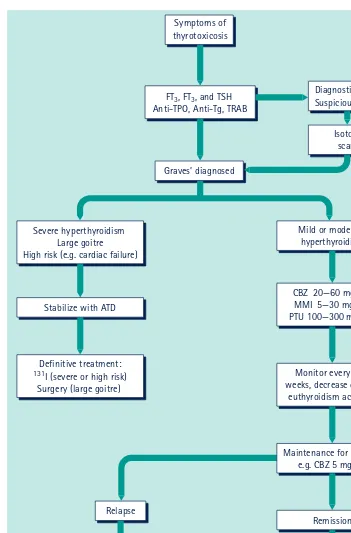
Symptoms of thyrotoxicosis
Isotope scan* Diagnostic doubt Suspicious goitre
Graves’ diagnosed
Stabilize with ATD Severe hyperthyroidism
Large goitre High risk (e.g. cardiac failure)
Mild or moderate hyperthyroidism
Definitive treatment:
131I (severe or high risk)
Surgery (large goitre)
CBZ 20—60 mg/day MMI 5—30 mg/day PTU 100—300 mg/day
Monitor every 4-6 weeks, decrease dose as
euthyroidism achieved
Monitor 3/12 for 1st year then annually
Remission Relapse
Definitive treatment (Usually 131I)
2nd course ATD
Maintenance for (12/12), e.g. CBZ 5 mg OD FT3, FT3, and TSH
[image:11.595.71.422.58.591.2]Anti-TPO, Anti-Tg, TRAB
01 Graves’ disease 5
1.93). Obesity was associated with lower risk of Graves’—hazard ratio for individuals with body mass index (BMI) greater than 30 kg/m2was 0.68 (95% CI 0.49 to 0.92).
3 Colour Doppler sonography may be useful in diagnosis of thyroid disorders. This is a safe, non-invasive technique to assess blood flow in the thyroid arteries. Results correlate highly with thyroid volume and function. In a preliminary study,9thyroid blood flow at baseline was highly correlated with outcome after 14 months of antithyroid drug therapy. Relapse could be predicted with a sensitivity of 71% and specificity of 100%.
Conclusions
Initial investigations should include thyroid hormone, TSH and thyroid antibodies, including TBII. Full blood count and liver tests should be requested at baseline and at intervals in patients taking antithyroid drugs (Figure1.1). Thyroid scanning is not rou-tinely warranted unless there is doubt about the diagnosis. Antithyroid drug treatment is usually the first line treatment. Radioactive iodine has been increasingly used in recent years. There is no evidence of teratogenicity. Obviously, it is absolutely contraindicated during pregnancy and most endocrinologists would avoid its use within 6–12 months of conception. The above patient should not be overly concerned about the implications of the disease for her children although, if female, they will inherit a roughly one in three lifetime chance of developing AITD.
Further Reading
1 Cooper DS. Antithyroid drugs.N Engl J Med2005;352: 905–17.
2 Abraham P, Avenell A, Watson WA, Park CM, Bevan JS. Antithyroid drug regimen for treating Graves’ hyperthyroidism (Review).Cochrane Library2005;3: 1–48.
3 Andrade VA, Gross JL, Maia AL. Serum thyrotropin-receptor autoantibody levels after 131I
therapy in Graves’ patients: effect of pretreatment with methimazole evaluated in a prospective, randomized study.Eur J Endocrinol2004;151: 467–74.
4 Bonnema SJ, Bennedbaek FN, Gram J, Veje A, Marving J, Hegedus L. Resumption of
methimazole after 131I therapy of hyperthyroid diseases: effect on thyroid function and volume
evaluated by a randomised clinical trial.Eur J Endocrinol2003;149: 485–92.
5 Tomer Y, Davies TF. Searching for the autoimmune thyroid disease susceptibility genes: from gene mapping to gene function.Endocr Rev2003;24: 694–717.
6 Vaidya B, Pearce S. The emerging role of the CTLA-4 gene in autoimmune endocrinopathies.
Eur J Endocrinol2004;150: 619–26.
7 Wang PW, Liu RT, Juo SHH,et al. Cytotoxic T lymphocyte-associated molecule-4 polymorphism and relapse of Graves’ hyperthyroidism after antithyroid withdrawal.J Clin Endocrinol Metab
2004;89: 169–73.
8 Holm I, Manson JE, Michels KB, Alexander EK, Willett WC, Utiger RD. Smoking and other lifestyle factors and the risk of Graves’ hyperthyroidism.Arch Intern Med2005;165: 1606–11.
9 Saleh A, Cohnen M, Fürst G, Mödder U, Feldkamp J. Prediction of relapse after antithyroid drug therapy of Graves’ disease: value of color Doppler sonography.Exp Clin Endocrinol Diabetes
2004;112: 510–13.