Chicago, Illinois
Received 13 August 2004/Accepted 26 October 2004
Nectin-1 is an immunoglobulin (Ig)-like entry receptor for herpes simplex virus (HSV). Like other nectins, nectin-1 forms dimers and mediates cell adhesion through interactions with other nectins. We constructed a second-domain deletion mutant of nectin-1 (nectin-1-⌬2) to examine the role of the second Ig-like domain in HSV entry. Nectin-1-⌬2 exhibited a severely reduced ability to mediate HSV entry and accumulated in the endoplasmic reticulum but retained the ability to interact with its HSV ligand, gD. The failure of nectin-1-⌬2 to mediate HSV entry probably resulted from its failure to be transported to a membrane targeted by HSV for viral entry.
Herpes simplex virus (HSV) entry into susceptible cells in-volves binding of the viral glycoproteins gC and/or gB to hepa-ran sulfate chains on the cell surface, followed by fusion of the viral envelope with a cell membrane. The fusion step requires four glycoproteins (gD, gB, gH, and gL) and a cellular receptor that binds gD. At least four different membrane-bound mole-cules can serve as gD receptors (20): HVEM, a member of the tumor necrosis factor receptor superfamily; nectin-1 and nec-tin-2, which are immunoglobulin (Ig) superfamily members; and a modified form of heparan sulfate. Members of the nectin family of molecules, such as nectin-1 and nectin-2, contain three extracellular Ig-like domains, a transmembrane domain, and a cytoplasmic tail. They normally serve functions in cell adhesion, including formation of adherens junctions and tight junctions, certain neuronal synapses, and cell junctions re-quired for spermatogenesis (22). These activities require inter-actions of nectin homodimers on one cell with the same or different member of the nectin family on an adjacent cell, as well as interactions with other junctional proteins.
Numerous studies have shown that the first or N-terminal Ig-like domains of nectin-1 and nectin-2 contain the regions that are critical for homotypic and heterotypic trans- interac-tions (4, 13, 16), binding to gD (2, 12, 14), and HSV entry (1, 3, 5, 15), but little is known about the functions of the second and third domains. In a recent study (17), Momose et al. investigated the role of the second Ig-like domain of nectins in
cis-andtrans-interactions and cell adhesion by using a mouse nectin-2 second-domain deletion mutant. They concluded that the second domain is necessary forcis-interactions (formation
of dimers in the plane of the membrane) and thatcis- interac-tions are required for homotypictrans-interactions and cell-cell adhesion. Based on this study, we aimed to investigate the role of the second Ig-like domain for the function of nectin-1 as a receptor for HSV type 1 (HSV-1).
(Parts of this work were presented at the American Society for Virology 23rd Annual Meeting, Montreal, Canada, 10 to 14 July 2004.)
We constructed a mutant form of human nectin-1 (GenBank accession number NP_002846), named nectin-1-⌬2, from which amino acids 144 to 244 were deleted (Fig. 1A). Based on a CLUSTALW alignment of mouse nectin-2 and human nec-tin-1, this deletion corresponds to the one described by Mo-mose et al. (17). The overlap extension PCR method (9, 10) was used to generate this deletion from pBG38 (7), which contains the coding region of nectin-1␣(referred to here as nectin-1) in pcDNA3.1 (Invitrogen). After insertion of the PCR product into the pcDNA3.1 vector, the resulting nectin-1-⌬2-expressing plasmid was named pAP12. The coding region of pAP12 was then PCR amplified withPfuUltra DNA poly-merase (Stratagene) using primers that add a 5⬘NotI site and a 3⬘ClaI site, and the PCR product was inserted in frame into the 3⫻ FLAG cytomegalovirus 14 vector (Sigma) to yield a C-terminal FLAG-tagged version of the second-domain-de-leted form of nectin-1 (pFS37). A hemagglutinin (HA)-tagged version of the full-length nectin-1 in pcDNA3.1 (pCJ4) was described previously (6).
To test whether nectin-1-⌬2 could mediate HSV-1 entry, we used the previously described reporter virus HSV-1(KOS)tk12 (below named HSV-1), in which the viral thymidine kinase gene was replaced with the Escherichia coli lacZgene (23). Chinese hamster ovary (CHO) cells, which are normally resis-tant to HSV-1 infection, were used for this entry assay. The cells were grown to 70% confluency in six-well tissue culture
* Corresponding author. Mailing address: Northwestern University, The Feinberg School of Medicine, Department of Microbiology and Immunology, Searle 6-447, 320 E. Superior St., Chicago, IL 60611. Phone: (312) 503-1343. Fax: (312) 503-1339. E-mail: p-spear @northwestern.edu.
3841
on November 8, 2019 by guest
dishes and transfected with plasmids expressing nectin-1 or nectin-1-⌬2 by using Lipofectamine (Invitrogen) according to the manufacturer’s instructions. The cells were transferred to 96-well tissue culture plates 24 h after transfection. Thirty-six hours after transfection, the cells were washed once with phos-phate-buffered saline (PBS) plus 0.1% glucose and 1% heat-inactivated calf serum. After addition of serial dilutions of virus prepared in the same buffer, the cells were incubated for 6 h at 37°C. The cells were washed with PBS, incubated with PBS containing 3 mg ofo-nitrophenyl--D-galactopyranoside
(ONPG; Sigma) substrate/ml and 0.5% NP-40 (Calbiochem). Generation of ONPG reaction product, as a measure of viral entry and-galactosidase expression, was monitored at 405 nm in a Victor microplate reader (Perkin-Elmer) at various time intervals. As is shown in Fig. 1B, entry of HSV-1 was strongly reduced in nectin-1-⌬2-expressing CHO cells compared to cells expressing the full-length form of nectin-1. Similar results were obtained when the CHO cells expressed the tagged forms of nectin-1 and nectin-1-⌬2.
We tested for cell surface expression of the wild-type and mutant forms of nectin-1 by using the monoclonal antibodies CK8 and CK41 (12). Both antibodies recognize epitopes in the V-like domain of nectin-1, a linear epitope in the case of CK8 and a conformational epitope in the case of CK41. The cell-based enzyme-linked immunosorbent assay, used for detection of cell surface proteins on live cells, was described in detail elsewhere (6). As is shown in Fig. 1C, binding of both CK8 and CK41 was strongly reduced for nectin-1-⌬2 compared to wild-type nectin-1, indicating reduced cell surface expression for the mutant. However, nectin-1-⌬2 was expressed in the CHO cells, as shown by the results presented in Fig. 2 to 4, below.
We hypothesized that the nectin-1-⌬2 protein was unable to reach the cell surface because of retention in the endoplasmic reticulum (ER) or Golgi apparatus. To investigate potential
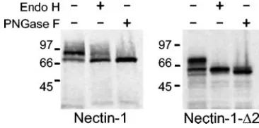
[image:2.585.112.470.69.227.2]incomplete processing of the nectin-1-⌬2 protein through the exocytic pathway, we studied its electrophoretic mobility after endoglycosidase treatment. The N-linked glycans of glycopro-teins retained in the ER are sensitive to cleavage by both Endo H and PNGase F, whereas the N-linked glycans of glycopro-teins that transit through the Golgi apparatus are usually mod-ified to become resistant to cleavage by Endo H, but not by PNGase F. Nectin-1 and nectin-1-⌬2, tagged at the C terminus with an HA or a 3⫻ FLAG epitope, respectively, were ex-pressed in CHO-K1 cells, and the cell lysates were treated with Endo H or PNGase F as described elsewhere (19) and ana-lyzed by immunoblotting with anti-HA or anti-FLAG antibod-ies. Figure 2 shows that the high-molecular-weight band of nectin-1-⌬2 was completely sensitive to Endo H treatment. Wild-type nectin-1, on the other hand, showed a high-molec-ular-weight band that was only partially sensitive to Endo H treatment and fully sensitive to PNGase F treatment. The
FIG. 1. Structure, HSV entry activity, and cell surface expression of nectin-1-⌬2. (A) Schematic representation of the nectin-1-⌬2 mutant, with amino acid numbering (based on GenBank reference sequence NP_002846) shown below. (B) Nectin-1-⌬2 does not mediate entry of HSV-1. CHO-K1 cells transfected with receptor or vector control were grown to confluency in 96-well tissue culture plates and exposed to increasing doses of reporter virus HSV-1(KOS)tk12 that expresses-galactosidase upon viral entry (23). After 6 h of incubation at 37°C, cells were permeabilized and ONPG substrate was added. Viral entry was quantified by measuring optical densities at 405 nm. Error bars represent 1 standard deviation of triplicate determinations within the same experiment, and the results shown here are representative of three independent experiments. (C) Cell surface expression of nectin-1 and nectin-1-⌬2. CHO-K1 cells were transfected with plasmids expressing nectin-1, nectin-1-⌬2, or vector control, replated in 96-well plates, and then incubated with anti-nectin-1 monoclonal antibodies CK8 or CK41. Following washes to remove unbound antibodies, the cells were fixed and incubated with secondary antibodies and the horseradish peroxidase detection system described elsewhere (6). Results of the cell-based enzyme-linked immunosorbent assays are expressed as optical density (OD) at 380 nm and are means of triplicate measurements, with error bars indicating 1 standard deviation. The results shown are representative of three independent experiments.
FIG. 2. Western blot analysis of nectin-1 and nectin-1-⌬2 after endoglycosidase treatment. CHO-K1 cells were transfected with tagged versions of nectin-1 and nectin-1-⌬2. The cells were lysed 48 h after transfection, cell lysates were treated with Endo H or PNGase F and run on a denaturing SDS-PAGE gel, and the expressed proteins were visualized by Western blotting using antibodies specific for the tags.
on November 8, 2019 by guest
http://jvi.asm.org/
[image:2.585.328.511.571.662.2]acquisition of Endo H resistance by wild-type nectin-1 but not the mutant nectin-1 indicates that nectin-1-⌬2 failed to transit to the Golgi, compatible with its retention in a pre-Golgi com-partment of the cell.
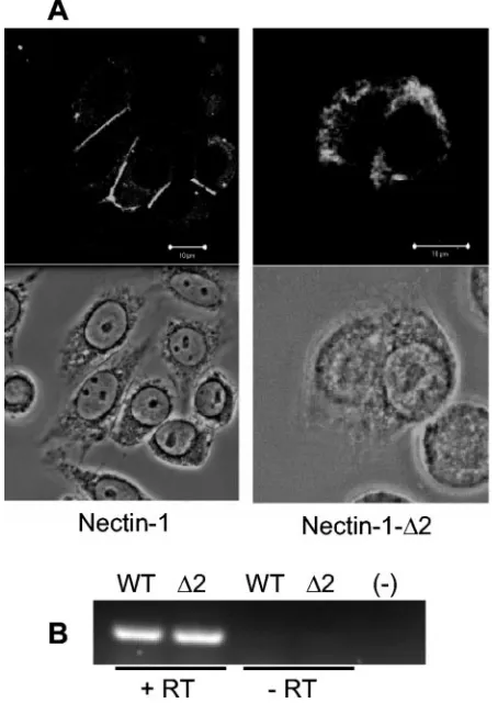
Confocal microscopy (Fig. 3A) showed that the wild-type form of nectin-1 localized largely to cell-cell contact sites at the cell surface, whereas nectin-1-⌬2 was retained in cytoplasmic compartments of the cell. In this experiment, CHO-K1 cells transfected with full-length nectin-1 or nectin-1-⌬2 were incu-bated with chicken polyclonal anti-HveC (nectin-1) serum (6) at a 1:500 dilution, stained, and examined on an LSM510 confocal microscope (Zeiss) as described previously (21). Sim-ilar amounts of nectin-1 and nectin-1-⌬2 mRNA were detected by reverse transcriptase PCR (Fig. 3B) with primers HVECiF1 (5⬘-TGA CCT GGA TCC TTC GAG C-3⬘) and HVECiR1 (5⬘-TGA AGC CAT ACA TGG AGT CG-3⬘), which amplify a 247-bp region upstream of the deletion in nectin-1-⌬2. These
within the ER until they are folded properly (8), the removal of this site may contribute to ER retention of the nectin-1-⌬2 protein. An alternative explanation is that the second domain mediates dimerization (17), which may be required for transit of nectin-1 from the ER.
To investigate whether the deletion of the second Ig-like domain would alter the gD binding ability of the V-like do-main, we cotransfected CHO-K1 cells with HSV-1 gD and wild-type or mutant nectin-1 and examined the cells by confo-cal microscopy. As shown in Fig. 4C and previously (11, 21), expression of full-length nectin-1 caused a redistribution of gD towards cell-cell contact sites where both proteins colocalized. Nectin-1-⌬2 also colocalized with gD, which indicated that the V-like domain probably retained gD binding activity. Both proteins colocalized with a distribution similar to that of nec-tin-1-⌬2 alone (Fig. 3).
Geraghty and coworkers (5) created chimeras between hu-man nectin-1 and CD4, an Ig-like molecule without HSV entry activity, and showed that HSV-1 gD binding, cell surface ex-pression, and entry activity were retained when the first Ig-like domain (amino acids 1 to 144) was fused to the C-terminal part of CD4, including its second and third Ig-like domains, the transmembrane domain, and the cytoplasmic tail. Presumably, the domains of the fusion protein from CD4 enabled the hy-brid protein to transit from the ER to the cell surface and to present the N-terminal nectin-1 domain in a conformation that was functional for HSV entry. This suggested that only the N-terminal V-like domain of nectin-1 was important for HSV entry, provided it was folded and presented properly. Our results suggest that deletion of the second Ig-like domain limits accessibility of the virus to the receptor by altering the intra-cellular localization of the receptor. This may also provide an explanation for the findings of Cocchi et al. (2), who reported that a nectin-1 mutant from which the second and third Ig-like domains were deleted was expressed at levels similar to those of wild-type nectin-1, but nevertheless mediated entry of HSV-1 with lower efficiency than the full-length form. While we cannot exclude that the second and third domains may serve other functions, such as mediating dimerization (14) or acting as a spacer to present the V-like domain to the virus, ER retention of the mutant receptor could explain a diminished entry activity.
Reduced cell surface expression of a receptor in itself may not necessarily imply that it is unable to mediate HSV entry, as was suggested by a recent study (18). In this study, Nicola and Straus showed that HSV can enter HeLa cells and
1-FIG. 3. Cellular localization and expression of 1 and nectin-1-⌬2. (A) CHO-K1 cells were transfected with plasmids expressing nectin-1 or nectin-1-⌬2 and stained with anti-nectin-1 antibody (top panels) or visualized by phase-contrast microscopy (bottom panels). Nectin-1 localized to cell-cell junctions, whereas nectin-1-⌬2 expres-sion was mainly confined to the cytoplasm. Bars, 10m. (B) Similar amounts of nectin-1 (WT) and nectin-1-⌬2 (⌬2) mRNA were detected by reverse transcriptase PCR using primers that amplify a 247-bp region upstream of the deletion in nectin-1-⌬2. Total RNA extracted from transfected CHO cells using the RNeasy kit (QIAGEN) was subjected to DNase I treatment (Invitrogen). RNA samples before (-RT) or after (⫹RT) reverse transcription (Promega) using an oli-go(dT) primer were used as templates in the PCR. (-), negative control (water).
on November 8, 2019 by guest
[image:3.585.50.277.71.391.2]FIG. 4. Costaining of wild-type and mutant nectin-1 with calreticulin or viral gD. (A) Nectin-1 does not colocalize with calreticulin in CHO cells transfected with nectin-1. (B) Nectin-1-⌬2 colocalizes with calreticulin in CHO cells transfected with nectin-1-⌬2. Primary antibodies used in panels A and B were chicken anti-nectin-1 (green) and rabbit anti-calreticulin (red; Abcam). Bottom right of each panel shows the overlay, and the bottom left shows a phase-contrast image. (C) Colocalization of HSV gD and wild-type or mutant nectin-1. CHO-K1 cells were transfected with plasmid pCJ3, expressing HSV-1 gD (6), or cotransfected with plasmids expressing HSV-1 gD and either nectin-1 or nectin-1-⌬2, stained with chicken anti-nectin-1 antibody (green) and rabbit anti-gD antibody R7 (red), and visualized by confocal microscopy. As in cells transfected with receptor alone, nectin-1 localized to cell-cell junctions, whereas nectin-1-⌬2 localized to the cytoplasm. HSV-1 gD was mostly colocalized with nectin-1 or nectin-1-⌬2 but, when expressed alone, had a different intracellular localization than when expressed with either form of nectin-1. Bars, 10m.
on November 8, 2019 by guest
http://jvi.asm.org/
We thank Gary Cohen and Roselyn Eisenberg (University of Penn-sylvania) for antibodies and Nanette Susmarski (Northwestern Uni-versity) for excellent technical assistance.
This work was supported by grants AI-36293 and AI-049394 from the National Institutes of Health. F.S. was supported by Public Health Service training grant T32 AR07593.
REFERENCES
1.Cocchi, F., M. Lopez, P. Dubreuil, G. Campadelli-Fiume, and L. Menotti.
2001. Chimeric nectin 1-poliovirus receptor molecules identify a nectin 1 region functional in herpes simplex virus entry. J. Virol.75:7987–7994. 2.Cocchi, F., M. Lopez, L. Menotti, M. Aoubala, P. Dubreuil, and G.
Cam-padelli-Fiume.1998. The V domain of herpesvirus Ig-like receptor (HIgR) contains a major functional region in herpes simplex virus-1 entry into cells and interacts physically with the viral glycoprotein D. Proc. Natl. Acad. Sci. USA95:15700–15705.
3.Cocchi, F., L. Menotti, P. Mirandola, M. Lopez, and G. Campadelli-Fiume.
1998. The ectodomain of a novel member of the immunoglobulin subfamily related to the poliovirus receptor has the attributes of a bona fide receptor for herpes simplex virus types 1 and 2 in human cells. J. Virol.72:9992– 10002.
4.Fabre, S., N. Reymond, F. Cocchi, L. Menotti, P. Dubreuil, G. Campadelli-Fiume, and M. Lopez.2002. Prominent role of the Ig-like V domain in trans-interactions of nectins. Nectin3 and nectin 4 bind to the predicted C-C⬘-C⬙-D beta-strands of the nectin1 V domain. J. Biol. Chem.277:27006– 27013.
5.Geraghty, R. J., A. Fridberg, C. Krummenacher, G. H. Cohen, R. J. Eisen-berg, and P. G. Spear.2001. Use of chimeric nectin-1(HveC)-related recep-tors to demonstrate that ability to bind alphaherpesvirus gD is not necessar-ily sufficient for viral entry. Virology285:366–375.
6.Geraghty, R. J., C. R. Jogger, and P. G. Spear.2000. Cellular expression of alphaherpesvirus gD interferes with entry of homologous and heterologous alphaherpesviruses by blocking access to a shared gD receptor. Virology
268:147–158.
13.Krummenacher, C., I. Baribaud, J. F. Sanzo, G. H. Cohen, and R. J. Eisen-berg.2002. Effects of herpes simplex virus on structure and function of nectin-1/HveC. J. Virol.76:2424–2433.
14.Krummenacher, C., A. H. Rux, J. C. Whitbeck, M. Ponce-de-Leon, H. Lou, I. Baribaud, W. Hou, C. Zou, R. J. Geraghty, P. G. Spear, R. J. Eisenberg, and G. H. Cohen.1999. The first immunoglobulin-like domain of HveC is sufficient to bind herpes simplex virus gD with full affinity, while the third domain is involved in oligomerization of HveC. J. Virol.73:8127–8137. 15.Martinez, W. M., and P. G. Spear.2001. Structural features of nectin-2
(HveB) required for herpes simplex virus entry. J. Virol.75:11185–11195. 16.Miyahara, M., H. Nakanishi, K. Takahashi, K. Satoh-Horikawa, K.
Tachi-bana, and Y. Takai.2000. Interaction of nectin with afadin is necessary for its clustering at cell-cell contact sites but not for its cisdimerization or
transinteraction. J. Biol. Chem.275:613–618.
17.Momose, Y., T. Honda, M. Inagaki, K. Shimizu, K. Irie, H. Nakanishi, and Y. Takai.2002. Role of the second immunoglobulin-like loop of nectin in cell-cell adhesion. Biochem. Biophys. Res. Commun.293:45–49. 18.Nicola, A. V., and S. E. Straus.2004. Cellular and viral requirements for
rapid endocytic entry of herpes simplex virus. J. Virol.78:7508–7517. 19.Potel, C., K. Kaelin, L. Danglot, A. Triller, C. Vannier, and F. Rozenberg.
2003. Herpes simplex virus type 1 glycoprotein B sorting in hippocampal neurons. J. Gen. Virol.84:2613–2624.
20.Spear, P. G., and R. Longnecker.2003. Herpesvirus entry: an update. J. Vi-rol.77:10179–10185.
21.Struyf, F., W. M. Martinez, and P. G. Spear.2002. Mutations in the N-terminal domains of nectin-1 and nectin-2 reveal differences in requirements for entry of various alphaherpesviruses and for nectin-nectin interactions. J. Virol.76:12940–12950.
22.Takai, Y., and H. Nakanishi.2003. Nectin and afadin: novel organizers of intercellular junctions. J. Cell Sci.116:17–27.
23.Warner, M. S., R. J. Geraghty, W. M. Martinez, R. I. Montgomery, J. C. Whitbeck, R. Xu, R. J. Eisenberg, G. H. Cohen, and P. G. Spear.1998. A cell surface protein with herpesvirus entry activity (HveB) confers susceptibility to infection by mutants of herpes simplex virus type 1, herpes simplex virus type 2, and pseudorabies virus. Virology246:179–189.