ANALYSIS OF IMMUNOHISTOCHEMICAL
EXPRESSION OF CD10 IN THE LESIONS OF PROSTATE
Dissertation submitted in
Partial fulfillment of the requirements for the award of
M.D. DEGREE in
PATHOLOGY – BRANCH III
THE TAMILNADU
DR. M.G.R. MEDICAL UNIVERSITY
CHENNAI - 32
ii
DECLARATION
I hereby declare that the dissertation entitled “Analysis of Immunohistochemical Expression of CD10 in the Lesions of Prostate” was done by me in the Department of Pathology, Chengalpattu Medical College from June 2012 to May 2016 under the guidance and
supervision of Dr. S. Sasikala M.D., Associate Professor, Department of Pathology, Chengalpattu Medical College.
This dissertation is submitted to the Tamil Nadu Dr.MGR Medical
University, Chennai towards the partial fulfillment of the requirements for the
award of M.D. Degree in Pathology.
I have not submitted this dissertation on any previous occasion to any University for the award of any degree.
Place:
iii
CERTIFICATE
This is to certify that the dissertation entitled “Analysis of Immunohistochemical Expression of CD10 in the Lesions of Prostate” is a record of bonafide work done by Dr. D. Saranya in the Department of Pathology, Chengalpattu Medical College, Chengalpattu under
the supervision of Dr. S. Ravi M.D., Professor and Head, Department of
Pathology and submitted in partial fulfillment of the requirements for the
award of M.D. Degree in Pathology by The Tamilnadu Dr. MGR Medical University, Chennai. This work has not previously formed the basis for the
award of a degree or diploma.
Dr. N. Gunasekaran M.D.,D.T.C.D., Dr. S. Ravi M.D.
Dean, Professor and Head,
Chengalpattu Medical College, Department of pathology,
Chengalpattu Chengalpattu medical college
iv
CERTIFICATE FROM THE GUIDE
This is to certify that the dissertation entitled, “Analysis of Immunohistochemical Expression of CD10 in the Lesions of Prostate” submitted by the candidate Dr. D. Saranya in partial fulfillment of the requirements for the award of M.D. Degree in Pathology by The Tamil
Nadu Dr.M.G.R. Medical University, Chennai is a bonafide research work done by her under my direct guidance and supervision, in the Department of
Pathology, Chengalpattu Medical College, Chengalpattu. This work has not
previously formed the basis for the award of any degree or diploma.
Dr. S.Sasikala.,MD.
Associate Professor Department of Pathology,
Chengalpattu Medical College,
ix
ACKNOWLEDGEMENT
To think with, I thank the almighty GOD in making this project a successful one.
I express my deep gratitude to Dr. N. Gunasekaran M.D.,D.T.C.D
Dean, Chengalpattu Medical College, for granting me permission to undertake this study.
I profusely thank and express my sincere gratitude to Dr. S. Ravi M.D., Professor and Head, Department of Pathology, Chengalpattu Medical College, for having suggested this topic for dissertation and for having rendered his valuable support and encouragement without which this project work would not have been feasible.
I wish to record my sincere thanks to Dr. I. Vijay Sathish Kumar M.D., Dr. S. Sasikala M.D., Dr. S. Premalatha M.D., Associate Professors, Department of Pathology, Chengalpattu Medical College, for their constant support and encouragement throughout my work.
I also wish to record my sincere thanks to Dr. K.R. Mohan M.D., Dr. G. Selvambigai M.D., Associate professors for their timely support throughout my work.
I also wish to record my sincere thanks to Dr. M. Kuzhalmozhi, M.D., Dr.S.Suryalakshmi, M.D., Dr.V.Palaniappan, M.D., Dr.M.Malathi M.D., Dr.G.Devi Priya, M.D, Dr.S.Rohini Priya, M.D, Dr.V.Dhamodharan,M.D, Dr.D.Pushpalatha,M.D, Dr.C.Arunprabakaran,M.D, Dr.P.S.Vanitha,M.D
Assistant Professors, Dr.Sivashankari,D.C.P, Dr.Shanmugapriya,D.C.P
tutor in Department of Pathology, Chengalpattu Medical College, for their constant support and encouragement throughout my work.
x
I thank all the technical staff in the Department of Pathology, Chengalpattu Medical College, for their sincere and timely technical assistance.
Also, I am indebted to my husband Mr. K. Siddharthan for his constant support, encouraging words and source of strength all the way through this endeavour.
To my lovable family members, I express my gratitude for their extreme patience and tireless support while pursuing this study.
Last, but not the least I am indebted to all the patients who made it possible for me to carry out this study.
xi
CONTENTS
SL. NO PARTICULARS PAGE NO.
1 INTRODUCTION 1
2 AIMS AND OBJECTIVES 3
3 REVIEW OF LITERATURE 4
4 MATERIALS AND METHODS 44
5 OBSERVATION AND RESULTS 49
6 DISCUSSION 71
7 SUMMARY 81
8 CONCLUSION 83
9 BIBLIOGRAPHY 84
10
ANNEXURES
I: Proforma 95
II: Master chart 97
xii
LIST OF FIGURES
Sl.no TITLE
1 Zones of prostate
2 Hypothesised model of prostate cancer in relation to vitamin D deficiency
3 List of risk factors in prostate cancer
4 Pathogenesis of Benign prostatic hyperplasia 5 Dihydrotestosterone and nuclear transcription. 6 Pathogenesis of prostatic carcinoma
7 Evolution of Gleason grading system
xiii
LIST OF TABLES
Table No TITLE
1. Age vs Histopathological diagnosis
2. P value for Age vs Histopathological diagnosis 3. Gleasons score and Prostatic carcinoma
4. Serum PSA vs Prostatic carcinoma 5. Serum PSA level vs Gleason score 6. Age vs Gleasons score
7. Immunohistochemical diagnosis vs benign and premalignant lesions
8. Different proportion Gleason pattern 2,3,4,5 9. Gleason grade vs CD10 expression
10 Serum PSA level vs CD 10 expression
11 Age wise distribution of prostatic carcinoma - Comparison 12 Gleason score wise distribution in prostatic carcinoma -
Comparison
xiv
LIST OF CHARTS
Sl. No TITLE
1. Case distribution of prostatic lesions – Pie chart
2. Age wise distribution of histopathological diagnosis- Bar chart
3. Prostatic adenocarcinoma case distribution according to Gleason score –Pie chart
4. Prostatic adenocarcinoma case distribution according to serum PSA level – Pie chart
5. Serum PSA and Gleason score case distribution – Bar chart 6. Age wise distribution of Gleason score – Bar chart
7. CD 10 expression in benign and premalignant lesion – Bar chart 8. Case distribution according to Gleason grade – Bar chart
xv
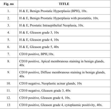
[image:15.595.110.532.202.626.2]LIST OF COLOUR PLATES
Fig. no TITLE
1. H & E, Benign Prostatic Hyperplasia (BPH), 10x.
2. H & E, Benign Prostatic Hyperplasia with prostatitis, 10x. 3. H & E, Prostatic Intraepithelial Neoplasia, 10x.
4. H & E, Gleason grade 3, 10x 5. H & E, Gleason grade 4, 10x 6. H & E, Gleason grade 5, 40x 7. CD10 positive, BPH,10x.
8. CD10 positive, Apical membranous staining in benign glands, 40x
9. CD10 positive, Diffuse membranous staining in benign glands, 40x
10. CD10 negative, Neoplastic acinar glands, 10x 11. CD10 negative, Gleason grade 3, 40x
12. CD10 positive, Gleason grade 4, 10x.
1
INTRODUCTION
The lesions of prostate are responsible for significant morbidity and mortality among the males worldwide (1).The age range of males presenting
with symptoms due to prostatic lesions is 40- 90 years, with majority of the
cases were in the age group of 60 – 70 years (1).
Prostatic lesions are broadly categorized as inflammatory and
neoplastic lesions. The neoplastic lesions are inturn subclassified as benign, in situ and malignant lesions .
Prostate cancer is the most aggressive malignant neoplasm with varied
clinical presentations. This tumor does not show any warning signs in its early
course of development.
The most widely used screening test for detecting prostatic cancer is the measurement of serum Prostate specific antigen (PSA) level , in conjunction
with digital rectal examination for all the suspected cases.
Prostate Specific antigen is secreted by normal and malignant prostatic
epithelial cells. Therefore their level in the serum increases significantly in
men with prostate cancer. Though it gives the suspicion for the underlying
tumor, it isnot specific.There are many benign conditions like benign prostatic hyperplasia and prostatitis which increases the serum PSA levels.Therefore it
is of at most significance to use a newer marker to identify the prostatic cancer
2
Cluster of Differentiation (CD) 10, also known as Common Acute
Lymphoblastic Leukemia Antigen (CALLA) was first described on human
leucocytes (20). Several studies on CD10 revealed that it is not only seen in lymphocytes, but also found to be expressed in other human cells both in
normal and in pathological states.
Regarding the prostate gland, CD10 is expressed constantly in the
apical luminal surface of the normal prostatic epithelial cells. In various
lesions of prostate the pattern of expression varies ranging from altered
expression to loss of expression.
In prostatic cells CD 10 acts as a transmembrane peptidase .It plays an
important role in the pathogenesis of prostatic cancer. Generally it cleaves the
excessive growth factor from the stroma thereby it prevents the continuous
and unwanted growth in the luminal epithelial cells.
Literature review shows that loss of CD10 expression is seen in lower
Gleason score prostatic tumors whereas increased and altered expression in
high Gleason score tumors, lymph node metastasis and in bone metastatic
prostatic carcinoma. This concept signifies the use of CD10 as a diagnostic
and prognostic marker in prostatic carcinoma.
Based on this we are analysing the expression of CD 10 in a various
3
AIMS AND OBJECTIVES
1. To identify the expression of CD 10 in the lesions of prostate.
2. To analyse the pattern of expression (membranous, cytoplasm, both).
3. To correlate the expression of CD10 with the age of the prostatic
carcinoma patients.
4. To correlate the expression of CD10 with histopathological grading and
4
REVIEW OF LITERATURE
ANATOMY:
The prostate is an exocrine gland constituting an important organ of
male reproductive system.
It is located in the pelvis just below the urinary bladder encompasses
the urethra and in front of the rectum.
It is a walnut shaped organ and its average weight is around 11-16 grams.
Anatomically it is divided into 5 lobes namely anterior, posterior, 2
lateral lobes and one median lobe but widely used terms are three zones
[image:19.595.212.449.501.689.2]namely peripheral zone, central zone, transitional zone.
5
The transition zone surrounds the prostatic urethra; central zone that
lies posterior to the transition zone encircles the ejaculatory duct. Peripheral
zone forms the main bulk of the gland.
Each zone has got its own significance. Prostatic cancer usually arises
from the peripheral zone and prostatic hyperplasia from the transition zone.
This anatomical knowledge is important because any lesion in a
particular zone can give a clue to the underlying pathology.
VASCULAR AND NERVE SUPPLY:
The arterial supply of prostate gland is through the Internal pudendal
artery, inferior vesical artery and branches of middle rectal artery.
The blood from the prostatic gland drains via the vesico prostatic
plexsus to the internal iliac veins. These plexus are particularly strong under
the puboprostatic ligaments
The autonomic innervation reaches the prostate gland together with the
arterial branches and perforates the capsule of the prostate. Parasympathetic
signals stimulate glandular activity and the sympathetic innervation of α 1-receptors mediates smooth muscle contraction.
6 HISTOLOGY:
Prostatic gland is mainly composed of branching duct, acinar glands
embedded in dense fibromuscular stroma.
Prostatic glands show mild convolutions. It is lined by 2 layers of
epithelial cells. Inner tall columnar cells with basally located nucleus that
performs the secretory function and the outer layer of flattened basal cells.
Functions:
The gland secretes milky white colour fluid that constitutes around 30% of volume of the semen.It is alkaline in nature. Its function is to preserve the
sperm and maintain its motility, and also neutralizes the acidity of the vagina.
EPIDEMIOLOGY OF PROSTATIC LESIONS:
Benign prostatic hyperplasia is the most common benign neoplastic lesion of prostate of aging men. It poses major public health problem causing
significant morbidity thereby affecting quality of life of aging men.
The prevalence of BPH increases with increasing age.It is about 8% in
the age group of 30 to40 years and 50% and 80% in the 8th and 9th decade
respectively. The risk of developing BPH in men aged 70 -79 years are
7
4.5 years around the age group of 35- 50 and 10 years for the age range of
51 – 70(4)
Prostate cancer is the second most common cause of cancer (1) and the sixth leading cause of cancer death among men worldwide (2). The worldwide
prostate cancer burden is expected to grow to 1.7 million new cases and
499 000 new deaths by 2030 simply due to the growth and aging of the global
population.
The incidence rates of prostate cancer are considered low in Asian and
North African countries, ranging from 1 to 9/100,000 persons.The prevalence of prostate cancer in India is far lower as compared to the western countries
but with the increased migration of rural population to the urban areas,
changing life styles, increased awareness, and easy access to medical facility,
more cases of prostate cancer are being picked up. The data regarding the exact incidence of prostate cancer in India is limited mainly because of the fact
that it is not a notifiable disease and only limited population based cancer
registries are available in India.
In India, when comparing the incidence rates of different cancers, there
is a highest incidence of oral cancers and lowest incidence and prostate cancers (3). The estimated incidence of prostate cancer in India is around
3.75/100,000 persons. The incidence rate varies among the major cities in
8
Chennai it is 4.2% and lowest in northeast India like in Manipur the rate being
0.8%.(4)
ETIOPATHOGENESIS FOR PROSTATIC LESIONS:
The major proven risk factors for prostatic lesions are age, hormonal
factors and family history. Considering the importance of prostatic malignancy
detailed analysis of risk factors of prostatic carcinoma are as follows.
AGE:
As the age advances the risk for prostate cancer also increases. The risk
for prostate cancer begins to rise after 55 years and it peaks at around 70 and
declines thereafter (6). According to statistics of United States of America it
was estimated that every one in 10,000 men in their 40s and one in 15 men in
their 60s will be affected by prostate cancer.
FAMILY HISTORY:
An individual with a positive family history has a significant risk in
developing prostate cancer. In a family if there is a first degree relative
(brother or father) with prostatic cancer then there is 2 to 3 fold risk for the
individual to develop the same. It is further increased by the early age at onsetin relative or multiple relatives with the disease (5).Whole genome or
partial genome analysis by linkage mapping studies among the high risk
pedigrees revealed many prostatic cancer specific foci.Several studies
9
genes. It states that those high risk genes responsible for cancer follows
Mendelian Autosomal dominant expression thereby results in early age of
onset of the disease (12)
RACE:
Incidence rate for African American is much higher, around 60 fold
when compared to men in Asian countries. This variations are mainly due to
the screening programmes, diagnostic advancement, increase accessibility to
total health care services etc. Migrants from Asian countries also shows
similar incidence rate of prostatic cancer to that of Americans. It is the environmental factors prevailing in that region contributing to the
development of cancer (13)
DIET:
Diet constituting large amounts of fat and increased intake of total
calories is associated with the increased risk. Reduced intake of antioxidant
like selenium, vitamin C, plays a significant role in the development of cancer.
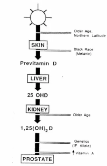
Vitamin D deficiency has been identified as one of the possible risk
factors for prostatic carcinoma. Increased age, black race and northern
latitudes which was proved as an independent risk factors, are all associated with Vitamin D deficiency.
Possible explanations are increased age is associated with decreased
10
In black race there is more melanin pigments which can directly inhibits the
synthesis of vitamin D. When compared to Americans, Asian men have
decreased incidence rate of prostatic carcinoma again can be associated with their dietary habits. Asian diet constitute rich in fish that has higher amount of
[image:25.595.244.429.243.529.2]Vitamin D (66).
Figure 2: Hypothesised model of prostate cancer in relation to vitamin D deficiency
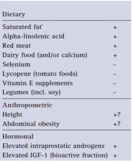
Other less important factors are anthropometric factors, hormonal
profiles, and other co-morbid health factors. They play a minor role in the
11 ANTHROPOMETRIC FACTORS:
Anthropometric factors like height and obesity and their association
with prostate cancer risk has been extensively studied. It was hypothesized that adult height is due to the hormone Insulin like growth factor. This
hormone carries significant risk for the development of prostate cancer.
Regarding obesity, it was hypothesized that increased obesity reduces sex
hormone binding globulins, therefore more availability of free sex hormones
in the circulation which can stimulate cancer progression (14). But both the
[image:26.595.206.435.373.653.2]hypothesis has not been proved so far.
Figure 3: List of risk factors in prostate cancer (14)
12
An increased level of IGF-1 that mediates the action of growth
hormone was identified as a independent risk factor for prostate cancer. It was
proven that administration of IGF-1 promotes growth of prostate and tumor development in animal models (15)
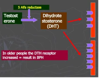
PATHOGENESIS:
It is the hormone dihydrotestosterone plays a major role in the
development of prostatic lesions including benign and malignant lesions.
BPH is characterized by increase in the epithelial and stromal cells
commonly in the periurethral zone of prostate.This increased cell number
could be due to either increased proliferation of cells or decreased
[image:27.595.157.484.455.713.2]apoptosis (67).
13
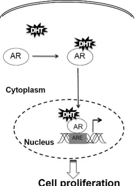
Dihydrotestosterone (DHT) which is the active metabolite of hormone
testosterone is the main androgen responsible for development of BPH.
The stromal cells of prostate gland convert testosterone into DHT through the enzymatic action of 5 Alpha reductase. This DHT is more potent
and has more affinity towards Androgen receptor when compared to hormone
testosterone. It binds to the androgen receptor (AR) of epithelial and stromal
cells thereby stimulating transcription of genes resulting in proliferation of
[image:28.595.203.429.347.658.2]epithelial cells and stromal cells.
14
Though the pathogenesis for the BPH has been well understood, the
exact triggering or inciting event is still not clear. One possible hypothesis is
that prostatic inflammation could be a triggering factor for the cell proliferation (16). Inflammatory cytokines such as Interleukin 2, Interleukin 6,
interleukin 8, interleukin 15 and Interferon alpha causes tissue damage and
oxidative stress to the stromal cells. This leads to compensatory cellular
proliferation thereby promoting the growth of the gland. (19).
In case of prostatic carcinoma these circulating androgens are essential
for the onset of prostate cancer through their interactions with Androgen receptor. Therefore surgical treatment like bilateral removal of testes which is
the source for androgens and antiandrogen drugs causes disease regression.
But some of the tumors become androgen resistant by following mechanisms:
(I) Androgen receptor gene (AR) amplification results in hypersensitivity to even low levels of androgens.
(II) Mutation in androgen receptor gene causing ligand independent
AR activation.
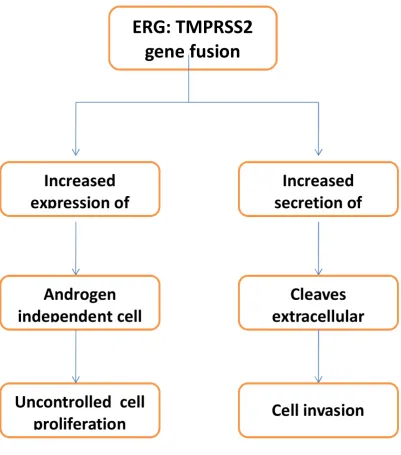
In addition to the androgens, prostatic cancer usually acquires large
number of genetic alterations including point mutations, deletions, amplifications and translocations. There are prostate cancer specific
chromosomere arrangements. It commonly involves E26 Transformation
15
which belongs to the ETS family, fuses with Transmembrane protease serine 2
(TMPRSS2) resulting in Androgen independent tumor progression. This
results in overexpression of transcription factors that causes upregulation of matrix metalloproteinase. Increased matrix metalloproteinase makes the
malignant prostatic epithelial cells more invasive.
Benign prostatic hyperplasia (BPH) and normal epithelium are negative
for ERG rearrangements and fusion transcripts. TMPRSS2: ERG fusions are
reported in 10 – 21% of high grade prostatic intraepithelial neoplastic
lesions (17) and 29 – 59 % in hormone refractory and metastatic prostatic carcinoma. (18)
Other common genetic alterations in prostate cancer includes mutation
in BRCA 2 and PTEN tumor suppressor gene, MYC oncogene and at later
16
Figure 6: Pathogenesis of prostatic carcinoma
ERG: TMPRSS2
gene fusion
Increased
expression of
secretion of
Increased
Androgen
independent cell
extracellular
Cleaves
Uncontrolled cell
17 COMMON DISEASES OF PROSTATE:
1. INFLAMMATION:
Inflammation of the prostate is known as prostatitis. Presenceof
inflammatory cells in the prostate is commonly seen in the biopsy specimen.
Prostatitis is further divided into asymptomatic inflammatory prostatitis, acute
prostatitis and granulomatous prostatitis.
Acute prostatits:
It most commonly results from ascending infection from urethra,
bladder and epididymis. Patient often presents with acute symptoms like fever,
dysuria, urinary urgency intense pelvic pain. Microscopically prostatic biopsy
composed of neutrophils in the glandular lumen and macrophages in the stroma. It cannot be diagnosed by histology alone. Often combined clinical
picture, urine culture aids in the diagnosis.
Asymptomatic inflammatory prostatitis:
It includes the presence of inflammatory cells like neutrophils,
lymphocytesand histiocytes in the prostate but most of the patients are without any clinical symptoms. Since inflammation of the prostate also raises the
serum PSA level and since the patient is also asymptomatic it can be clinically
misdiagnosed for prostatic carcinoma.Therefore it is important to mention in
the biopsy report. Inflammation is commonly associated with benign prostatic
18 Granulomatous inflammation:
Granulomatous prostatitis is a rare entity. This inflammation is due to
the release of prostatic secretions into the stroma. It elicits the granulomatous reaction in the prostate. This condition is also seen in postbiopsy, after
instillation of BCG injection into the bladder for bladder carcinoma patients.
Histologically it is composed of granulomas destroying the glands often
associated with multinucleated giant cells, histiocytes, lymphocytes and
plasma cells. Differential diagnosis could be tuberculosis which often consists
of caseating necrosis or could be of fungal etiology, metastatic deposits which are very rare causes.
2. METAPLASIA:
Metaplasia of prostatic epithelium is a secondary phenomenon.It occurs in response to inflammation or injury.Various metaplasias in prostate are
squamous, eosinophilic, mucinous, urothelial etc. Reversibility of these
metaplasias is very unlikely. These changes are not the preneoplastic
conditions.
Squamous metaplasia: It is most commonly an incidental finding. This is
mostly a secondary phenomenon due to infarction of the usual nodular hyperplasia, or after post hormonal and radiotherapy for prostatic carcinoma.
Histologically it is composed of nests of squamous epithelium with adjacent
19
carcinoma and Urothelial carcinoma which shows more cytological atypia and
stromal invasion.
Mucinous metaplasia: It is very rare entity composed of acini lined by tall columnar cells filled with mucin secretion with basally located nucleus.
Clinical significance is that it can mimic adenocarcinoma prostate which
should be differentiated by the lack of infiltrative nature, cytological atypia,
and presence of basal cells
Urothelial metaplasia: It is defined as urothelial lining the prostatic acini and larger ducts. It is a great mimicker of Urothelial carcinoma spreading via
prostatic ducts. Cytologically these two conditions can be differentiated by the
presence of nuclear atypia in malignancy and also with the help of
immunohistochemistry. Other differential diagnosis is high grade prostatic
intraepithelial neoplasia that shows prominent nucleoli.
3. HYPERPLASIA:
Hyperplasia of the prostate includes both epithelial and stromal cell
proliferation. It is a benign condition. It includes
a. Usual nodular hyperplasia:
It is the most common microscopic finding in the patient clinically
diagnosed of BPH. It commonly arises from the transition zone and
20
symptoms. It is composed of glandular and stromalelements with varied
percentage. The glands are more complex with prominent luminal infoldings.
Cystic dilatation of glands can also occur.
b. Basal cell hyperplasia:
This subtype includes the proliferation of basal cells. It defines the
presence of two or more layers of basal cells with scant cytoplasm around the
prostatic ducts and acini.
PROSTATIC INTRAEPITHELIAL NEOPLASIA:
It is the proliferation of epithelial cells with features of atypia in the
ducts and acini.
Originally PIN was classified as grade I, II, III, according to increasing
degrees of abnormality. Nowadays it is been termed as low grade PIN (grade
I) and high grade PIN (grade II, III)
Low grade PIN was formerly known as mild dysplasia. LGPIN mostly
do not progress and carries no significance for the subsequent development of
prostatic carcinoma. Microscopically the glands show complex architecture
with intact basal cell layer and cellular stratification. Cytologically the cells have eosinophilic cytoplasm with enlarged nuclei with increased variability in
21
High grade PIN has epithelial cell proliferation in four different
architectural patterns: Tufting, micropapillary, cribriform and flat. Regarding
the basal cells, high grade PIN shows reduced number of basal cells.
When nuclear features are taken into consideration High grade PIN has
increased nuclear atypia and prominent nucleoli.
The significance of reporting high grade PIN is that the patients should
be considered for rebiopsy (time interval for rebiopsy not standardised) and
they are at increased risk of development of carcinoma.
PROSTATIC CARCINOMA:
Prostatic carcinoma causes significant morbidity among elderly
individuals. It has been classified by WHO 2016 as follows.
WHO HISTOLOGICAL CLASSIFICATION OF TUMOURS OF
PROSTATE (2016)
EPITHELIAL TUMOURS
Adenocarcinoma(acinar)
o Atrophic
o Pseudohyperplastic o foamy
o colloid
22 o Microcystic variant
o Pleomorphic giant cell adenocarcinoma
o Sarcomatoid carcinoma
Prostatic intraepithelial neoplasia (PIN), High grade
Intraductal carcinoma NOS
Ductal adenocarcinoma
Cribriform
Papillary
Solid
Urothelial tumors
Urothelial carcinoma
Squamous tumors
Adenosquamous carcinoma
Squamous cell carcinoma
BASAL CELL TUMOURS
Basal cell adenoma
23 NEUROENDOCRINE TUMOURS:
Adenocarcinoma with Neuroendocrine differentiation
Well differentiated neuroendocrine tumor
Small cell neuroendocrine carcinoma
Large cell neuroendocrine carcinoma
MESENCHYMAL TUMOURS
Stromal tumor of uncertain malignant potential
Stromal sarcoma
Leiomyosarcoma
Rhabdomyosarcoma
Chondrosarcoma
Angiosarcoma
Synovial sarcoma
Inflammatory myofibroblastic tumour
Osteosarcoma
Undifferentiated pleomorphic sarcoma
24 Chondroma
Leiomyoma
Granular cell tumor
Solitary fibrous tumor
Solitary fibrous tumor , Malignant
HEMATOLYMPHOID TUMORS
Diffuse Large B cell lymphoma
Chronic lymphocytic leukemia/ Small lymphocytic lymphoma
Follicular lymphoma
Mantle cell lymphoma
Acute myeloid leukemia
B lymphoblastic leukemia/Lymphoma
MISCELLANEOUS TUMOUR
Cystadenoma
Nephroblastoma
Rhabdoidtumor
25 Clear cell adenocarcinoma
Melanoma
Paraganglioma
Neuroblastoma
SECONDARY/METASTATIC TUMOUR
NEWER ENTITY ADDED IN WHO 2016:
Intraductal carcinoma
New variants of acinar adenocarcinoma
Microcystic variant
Pleomorphic large cell variant
Large Cell carcinoma of prostate
Though we have numerous histological classification of prostatic
tumor, the term prostatic carcinoma commonly refers to prostatic adenocarcinoma.
Prostatic adenocarcinoma usually arises in the peripheral zone of
prostate gland. Grossly the tumor is grey white, irregular or nodular in
26
Histologically prostatic adenocarcinoma is characterised by abnormal
glandular architectural pattern with single layer of epithelial cells and with
absence of basal cells.
Numerous variants of acinar adenocarcinoma have been described.
Microcystic variant and Pleomorphic giant cell variant are the newly added
entity in WHO 2016 blue book.
VARIANTS OF PROSTATE ACINAR ADENOCARCINOMA:
ATROPHIC VARIANT:
Atrophic variant is a rare entity. The criteria to report this entity is
presence of malignant atrophic glands occupying at least 50 % of the tumor.
The glands should be of infiltrative nature. The glands are lined by flattened
cells with scant cytoplasm and hyperchromatic nuclei with prominent nucleoli.
This variant has been graded as Gleason pattern 3 or 4 and grouped under moderately differentiated carcinoma.
PSEUDOHYPERPLASTIC VARIANT:
The glands are benign looking ,dilated with papillary infoldings but
show malignant cytological features like enlarged hyperchromatic nuclei with prominent nucleoli. Mostly graded as Gleason’s grade 3 and generally has
27 FOAMY GLAND VARIANT:
The glands will have foamy appearance with round pinpoint nuclei
rather than prominent hyperchromatic features. Infiltrative nature of glands proves a diagnostic feature. It has got a good prognosis and mostly comes
under Gleason’s grade 3 (54)
MUCINOUS VARIANT:
Atleast 25% of the tumor should have aggregates of tumor cells floating in mucin lakes. No significant intracellular mucin is seen.This variant
has to be differentiated from metastasis before diagnosing the same. These
tumors are given Gleason grade 4 and considered as aggressive variant. Since
only few cases have been reported the exact behavior is not known (51)
SIGNET RING VARIANT:
25% of the tumor should have single cells with intracellular mucin vacuole that pushes the nuclei to the periphery.This need to be differentiated
from metastasis. It is assigned Gleason grade 5 and has aggressive behavior. It
is regarded as high grade adenocarcinoma with poor patient survival. (52)
MICROCYTSIC VARIANT:
28
in atrophied cells due to cystic dilatation of the glands. It has been graded as
Gleason 2 or 3, with favorable prognosis (55)
PLEOMORPHIC GIANT CELL VARIANT:
It is the highly aggressive variant of adenocarcinoma composed of giant
cells with marked pleomorphism, lack of cohesiveness and areas of extensive
necrosis (56). It has a poor outcome and generally assigned high Gleason
score 9 or 10.
The different architectural patterns forms the basis for Gleason’s
grading system which forms the important prognostic factor in the
management of prostatic carcinoma. It is the most commonly used grading
system which consists of five basic grades. The original Gleason’s grading
system was developed by Dr. Donald Gleason, who is a pathologist worked at Minneapolis Veterans Affairs Hospital. He developed it in the year 1965 (45)
Then it underwent several modifications and popularly called as
modified Gleason’s grading system. It was first modified by International
Society of Urological Pathology (ISUP) consensus meeting in the year 2005
(46). Again in the year 2014 ISUP Prostate Cancer Grading Panel consensus
meeting made further alterations.
Grade 1 and 2 encloses well differentiated tumor. It is composed of
well circumscribed nodules of closely packed small glands of uniform sizes.
29
Grade 3: It is the most common grade and considered as well
differentiated grade like grades 1 and 2. It consists of small and irregularly
dilated infiltrating individual glands with wide stromal separation. The cells are darker when compared to the normal epithelial cells. WHO 2016 includes
microcystic and pesudohyperplastic glands in this grade and removed
cribriform glands from this grade.
Grade 4 consists of attempted glandular formation glands fusion, glands
with cribriform pattern and glands with glomeruloid pattern.
Grade 5 composed of sheets, cords and in single cells. WHO 2016 also
includes linear arrays, solid nests which was not recognized by WHO 2004.
Post hormonal and post radiotherapy tumors are excluded from Gleason’s
grading.
EVOLUTION OF GLEASON GRADING:
Donald Gleason included cribriform pattern of glands in both grade 3
and grade 4. He doesn’t forms a strict criteria to differentiate cribriform
pattern of Gleason grade3 from Gleason grade 4.
In 2005 ISUP modified and developed strict criteria to overcome the above difficulties.
Cribriform Grade 3 : It includes small round glands with round contours
30
Cribriform Grade 4: It includes irregular gland with irregular contours with
irreguilar lumens or slit like lumensunder pattern 4.
According to 2014 ISUP modification all the cribriform pattern are considered as grade 4.
Newer modifications in Gleasons grading system in WHO 2016 are as
follows:
1. All cribriform patterns are grouped under pattern 4. 2. Glomeruloid pattern gouped under pattern 4.
3. Mucinous adenocarcinoma may be pattern 3 or pattern 4.
31
Figure 7: Evolution of Gleason grading system
(a)Original Gleasom grading system
(b)2005 ISUP Modification of Gleason grading system
(c)2014 Isup Modificationof Gleason grading system
32
Figure 8 : 2016 WHO modified Gleason grading system.
Reporting of Gleasons grading
Based on the above patterns in H and E sections the tumors are scored.
The most predominant pattern is called as primary pattern and the
second most common pattern is known as secondary pattern and the least
pattern is known as tertiary pattern.
Generally the primary and secondary pattern is added together for
Gleason’s scoring in cases of radical prostatectomy and TURP specimens. In case of needle biopsy the most common grade and worst grades are added
33
The lowest possible Gleason score is 2 (1 + 1), where both the primary
and secondary patterns have a Gleason grade of 1.
The highest possible Gleason score is 10 (5 + 5), when the primary and secondary patterns both have the most disordered Gleason grades of 5.
Based on Gleason’s score prostatic adenocarcinoma is divided into well
differentiated, moderately differentiated and poorly differentiated tumors.
Gleason’s score 2- 6 : Well differentiated tumors, with excellent prognosis.
Gleason’s score 7 (3+4): Moderately differentiated tumors.
Gleason’s score 7(4+3): Moderately to poorly differentiated tumors.
Gleason’s score 8 – 10: Poorly to undifferentiated tumors, aggressive in
nature.
Recently based on Gleason’sscore prostatic carcinoma is divided into 5
prognostic groups by ISUP (International Society of Urologic Pathology). The prognostic groups are as follows:
Grade group 1 (Gleason score 3 + 3 = 6): Only individual discrete
well-formed glands.
34
Grade group 3 (Gleason score 4 + 3 = 7): Predominantly poorly formed/
fused/cribriform glands with lesser component of well-formed glands.
Grade group 4 (Gleason score 8) - Only poorly formed/fused/cribriform glands or predominantly lacking glands and lesser component of well-formed
glands
Grade group 5 (Gleason scores 9–10): Lack of gland formation (or with
necrosis) with or without poorly formed/fused/cribriform glands.
They analysed that the 5 year risk free survival for grade groups 1 to 5
[image:49.595.129.509.383.658.2]are 96%, 88%, 63%, 48% & 26% respectively (fig 9)
35 RISK STRATIFICATION:
Based on diagnostic serum PSA level , Gleason score and clinical stage
of localized prostate cancer, D’Amico et al in the year 1998 (50) proposed risk stratification that was included in the WHO 2016 Blue book. They provide
better options for treatment recommendations than just using stage of cancer
alone. It forms the basis for initial treatment for men with prostate cancer and
it avoids overtreatment for early stage of cancer (49).
Low risk -Diagnostic PSA <10.0 ng/ml and highest biopsy Gleason score <6 and clinical stage T1c or T2a.
Intermediate risk - Diagnostic PSA >10.0 ng/ml but <20ng/ml or highest
biopsy Gleason score =7 or clinical stage T2b.
High risk – Diagnostic biopsy >20 ng/ml or highest biopsy Gleason sc ore >8
or clinical stage T2c /T3.
Thus according to ISUP recommendations It was decided that any
biopsy report for prostate carcinoma it is mandatory that it should contain
modified Gleason scoring system and Prognostic groups. Treatment options
are categorized according to risk stratification.
PROSTATIC INTRADUCATL CARCINOMA (IDC – P)
It is added as a new entity in the recent WHO 2016 classification of
36
grade (Gleason 4 and 5) adenocarcinoma. The basal cells are focally
preserved.
Rather than infiltrating borders the edges are smooth. It may have any one of the following three architectural and cytological features (57)
1. Solid cribriform architecture – less than 50 % cribriform spaces
2. Loose cribriform architecture> 50 % of lumen formation.
3. Marked nuclear pleomorphism with 6 times larger than the normal and associated with comedo necrosis.
The main differential diagnosis is high grade PIN, which can be
distinguished from Intraductal carcinoma Prostate by the absence of nuclear
[image:51.595.138.503.416.696.2]pleomorphism, comedo necrosis.
37
IMMUNOHISTOCHEMISTRY IN PROSTATIC CARCINOMA:
Though predominantly architectural pattern of glands and their
cytological features helps in differentiating benign and malignant, immunohistochemistry proves to be a valuable tool in a subset of cases.
There are separate immunostains for basal cells and malignant
epithelial cells.
The loss of basal cells is an early and the most important diagnostic feature in prostate carcinomas.. The lack of basal cell layer staining must be
validated against the simultaneous demonstration of a positive basal cell layer
in adjacentfociof benign glands. Basal cell markers are cytokeratins (CK
HMW, CK 5/6, CK 14) and p63.
AMACR (Alpha-methylacyl-CoA racemace) stains the malignant
epithelial cells.AMACR is a mitochondrial and peroxisomal enzyme that is involved in beta-oxidation of branched-chain fatty acids. It is expressed in the
cytoplasm of malignant epithelial cells.
It is highly sensitive marker showing positivity in both PIN and
prostatic carcinoma.
When it is combinedly used with basal cell markers we can significantly
increase the diagnostic accuracy and thereby avoiding unnecessary re-biopsies. Prostate-specific antigen (PSA) is widely used marker to confirm the
38
In case of distinguishing from poorly differentiated prostatic carcinoma
and poorly differentiated urothelial carcinoma prostatic carcinoma shows
positivity for PSA, PSAP (prostate specific acid phosphatase) and negative for thrombomodulin and Uroplakin. Others newer marker used in diagnosing
prostatic origin are Prostein and NKX3.1
CD 10 MARKER:
CD 10 commonly called as CALLA antigen is a neutral endopeptidase present on many cell surface. Their major function is to inactivate biopeptides
(20). It is widely used as cell surface marker for categorizing acute leukemia
and malignant lymphomas. It is expressed by germinal center B cells, and
lymphoid precursor cells. The role and expression of CD10 in non
hematolymphoid tissue in both normal and pathological state has been well
documented in many literatures.
CD 10 IN NORMAL TISSUE:
Normal Tissue in Which CD10 Antigen Was Detected are
myoepithelial cells of breast and in apocrine metaplasia of breast tissue, apical
surface of normal epithelial cells of small and large intestines (25), glomerular
cells and proximal convoluted tubules of kidney, Apical surface of large
prostate ducts (23), Apical surface of epididymalducts, Endometrial stromal cells (26), Bone marrow stromal cells, Liver canaliculi (24) and alveolar
39 CD10 IN HEMATOPOIETIC TUMORS:
CD10 is used as a diagnostic marker in B-lymphoblastic leukemia/
lymphoma and in mature B-cell lymphomas like plasma cell myeloma, follicular lymphoma, diffuse large B-cell lymphoma and Burkitt lymphoma
and very rarely in T-cell lymphoma.
CD 10 positivity in B cell lymphoma indicates a good prognosis. Such
patients receive less intensified chemotherapy, proving it to be prognostic marker as well.(27)
CD10 IN SOLID TUMORS
From the extensivestudies on CD10, it was revealed that CD10 is
notspecific to hematopoietic malignancies but is also expressed by several non
hematopoietic tumors such as solid tumors of childhood like nephroblastoma
and neuroblastoma (28), and in several carcinomas originating from kidney (30), lung (31), pancreas (33), prostate (34), liver (35), breast (36), stomach
(37), cervix (36), and bladder (38), Malignant melanoma (29) and various
other skin tumors (32) also show CD10 positivity.
ROLE OF CD 10 IN EPITHELIAL CELLS:
CD 10 is a transmembrane enzyme present over the surface of epithelial
cells has got dual functions.
The extracellular portion of CD10 has peptidase enzymatic activity
which cleaves the several peptides like endothelin, bombesin, enkephalin etc.
40
resulting in either proliferation and differentiation towards the epithelial cell
[image:55.595.142.452.146.410.2]lineage or it can be inhibited from proliferation.
Figure 11: CD10 structure and function
The intracellular portionof CD10 governs major signaling pathway that
are required for cell proliferation. Thus it can integrate signals from
extracellular and intracellular compartment and can bring important changes in the cells according to the microenvironment present around the cells.
ROLE OF CD 10 IN NORMAL PROSTATE:
In normal prostate gland CD 10 is expressed by luminal epithelial cells.
Some studies state that it also expressed by basal cell layer of prostate (39)
41
ROLE OF CD10 IN PROSTATE CANCER:
CD10 is strongly expressed by normal prostatic luminal epithelial cells.
Primary tumors of the Prostate especially adenocarcinoma shows different pattern of expression of CD10 .Low Gleason grade tumors shows loss of
expression whereas high Gleason grade tumors shows altered and strong
expression. The expression of CD10 is cytoplasmic when compared to apical
membranous of normal epithelial cells. Cytoplasmic accumulation of CD10 may activate signaling pathway constantly leading to uncontrolled
proliferation and invasion. But the hypothesis has not been proved yet (60)
MOLECULAR ANALYSIS OF CD 10 EXPRESSION IN PROSTATE:
The neutral endopeptidase or CD10 gene was analysed by several
molecular studies including tissue microarray, human cell lines study in
vitro andin vivo etc. It was identified that transcription of this gene is
androgen hormone mediated in prostate cancer cells. They have also identified
in the NEP gene an androgen responsive element (NEP-ARE) and an
androgen responsive region (NEP-ARR) suggesting the role of androgen ion
their expression. Thus both the regions are involved in the transcriptional
activation of CD10. (41).
After transcription, translation of CD10 is enhanced by methylation of
gene promoter (42). There is evidence of hypermethylation of gene promoter
42
mutation. So far no studies have demonstrated the mutations in CD10 gene per
se.
It has been well analysed that CD 10 is expressed in the membranes of prostatic cells. High grade tumors of prostate showed cytoplasmic positivity
and low grade tumors shows absence of expression. This variation in
cytoplasmic localization of CD10 possibly due to following mechanisms:
1. Internalization of membrane bound CD10 (43)
2. Strong association of CD10 with intracytoplasmic heat shock
proteins.(44)
The reason for absence of its expression in low grade tumors is not well
understood.
Most lymph node metastases of prostatic carcinomastrongly express
CD10 positivity in the malignant epithelial cells thus contributing to the fact that it could be involved in the pathogenesis of lymph node metastasis.(39).
High CD10 expression directly correlates with advanced Gleason score,
and thus tumor expressing CD10 may be considered aggressive tumors with
poor pathological outcome.
Because of the strong association of CD10 with high grade tumors and
in lymph node metastasis one can consider the possibility that CD10 can be used to categorise the lesion as aggressive and such patients can be closely
43
Anti CD10 drug therapy can also be used to treat the patients, if its
exact role in the pathogenesis is identified.
An immunohistochemistry based test can be used in the clinical setting to identify CD10-positive tumors on prostate needle biopsies, which may
warrant more aggressive initial therapy or closer surveillance post-operatively.
A number of drugs against CD10 are available and potential targeted
44
MATERIALS AND METHODS
Study Place: Department of Pathology, Chengalpattu Medical College and Hospital, Chengalpattu.
Study Design: The present study is an observational study conducted in the
Department of Pathology during the period of June 2012 to May2016 .
Ethical clearance for the study was obtained from the Institutional
Ethics Committee of Chengalpattu Medical College, Chengalpattu.
A total sample of 40 cases of prostatic lesions was analyzed during the
period of June 2012 to May 2016.
Study Population
INCLUSION CRITERIA
Tissue blocks of patients who are diagnosed as having benign and malignant prostatic lesions.
EXCLUSION CRITERIA:
Tissue blocks of patients who are diagnosed as prostatic carcinoma and
underwent preoperative Radiotherapy or Chemotherapy.
During the period of June 2012 to May 2016, as per the inclusion and exclusion criteria, biopsies received in the Department of Pathology were
45
History written in the histopathology request form was recorded on
predesigned and pretested proforma (Annexure I).
MATERIALS USED
Tissue sections prepared from paraffin embedded formalin fixed tissues
Haematoxylin and eosin staining kit
CD 10 monoclonal antibody kit
Secondary antibody kit Positive control
Negative control
METHOD:
Formalin fixed paraffin embedded blocksand haematoxylin eosin
stained sections of 40 prostatic biopsies are taken up for the study. On
histopathological examination, they were categorized as follows: 1. Benign prostatic hyperplasia,
2. Benign prostatic hyperplasia with prostatitis
3. Prostatic intraepithelial neoplasia high grade and low grade,
4. Prostatic adenocarcinoma.
Prostatic adenocarcinoma was assigned Gleason grade ranging from
grade 1 to grade 5 according to modified Gleason grading system.
Immunohistochemistry was performed on the tissue sections taken
46 Immunohistochemistry
Procedure
1. 4μ thick sections were cut from formalin fixed paraffin embedded tissue samples and transferred to gelatin-chrome alum coated slides.
2. The slides were incubated at 58ºC for overnight.
3. The sections were deparaffinized in xylene for 15 minutes x 2 changes.
4. Rehydrated through descending grades of alcohol as follows : (i)Absolute alcohol x 2 changes 5 minutes each
(ii) 90% alcohol x 5 minutes
(iii) Washed in distilled water 2 changes, 2 minutes each
5. Heat induced antigen retrieval was done with microwave oven at 150
degree Celsius with citrate buffer (pH 6.0) for 15 to 20 minutes.
6. Then cooled for 10 minutes.
7. Washed in distilled water 2 changes, 2 minutes each.
8. Washed in Tris Buffer Saline (TBS) for 2 minutes.
9. Endoperoxidase blocking was done by adding hydrogen peroxide on
the section and kept for 5 minutes.
10. Washed in the wash buffer for 2 minutes twice.
11. Primary antibody CD 10(Mouse monoclonal;prediluted) was added and kept for 30 minutes in a moist chamber.
12. Then washed in wash buffer 2 minutes 2 times each.
47
14. Washed in two changes of buffer 2 minutes each.
15. Poly excel HRP (Horse Radish Peroxidase) was added and incubated
for 15 minutes.
16. Washed with buffer – 2 minutes, 2 changes.
17. Working DAB Dchromogen (1ml DAB buffer + 1 drop chromogen,
mix well) was added and kept for 2-5 minutes.
18. Then washed in distilled water.
19. Counter stained with hematoxylin for 30 seconds.
20. The slides were washed in running tap water for 3 minutes.
21. The slides were air dried, cleared with xylene and mounted with DPX.
Positive control included blocks containing normal prostatic gland-
internal control Negative control included Primary antibody replaced
with PBS.
Immunostained sections were reviewed for CD 10 expression
CD10Expression:
1. CD 10 immunoreactivity was observed and assigned as positive or negative.
2. CD 10 immunreactivity was analysed for pattern of expression.
Pattern of expression as follows:
1. Apical membranous positivity 2. Diffuse membranous positivity
3. Membranous and cytoplasmic positivity 4. Cytoplasmic positivity
48
STATISTICAL ANALYSIS
Datas obtained were coded and entered into the Microsoft excel spread sheet (Annexure II). Datas were compared between groups using Pearson
Chi-square or Fisher‘s exact tests (p<0.05).
All statistical analysis was performed using SPSS statistical software
49
OBSERVATION AND RESULTS
We took a sample size of 40. Among the 40 cases .We have the
following distribution
15 cases - Benign prostatic hyperplasia 15 cases -Prostatic adenocarcinoma
5 cases - Benign prostatic hyperplasia with prostatitis,and
5 cases - Prostatic intraepithelial neoplasia.
The case distribution is represented as follows
AGE WISE DISTRIBUTION OF PROSTATIC LESION:
The age wise distribution of prostatic lesions in the present study
showed the following observations.
For benign prostatic hyperplasia majority of the patients are in the age
group of 60 – 80 years.
50
Prostatic intraepithelial neoplasia - 60 – 80 years
Prostatic adenocarcinoma - 60 – 70 years.
The mean age group for various lesions is as follows Benign prostatic hyperplasia–67years
Benign prostatic hyperplasia with prostatitis-70years
Protatic intraepithelial neoplasia –66yearsand
Prostatic carcinoma -68years.
Age Vs HPE
Benign Prostatic Hyperplasia
% BPH with Prostatitis % Intraepithelial Prostatic Neoplasia
% AdenocarcinProstatic oma
%
≤ 50 years 1 6.67 0 0.00 1 20.00 0 0.00
51-60 years 3 20.00 1 20.00 1 20.00 4 26.67
61-70 years 5 33.33 1 20.00 1 20.00 7 46.67
71-80 years 5 33.33 3 60.00 1 20.00 2 13.33
81-90 years 1 6.67 0 0.00 1 20.00 2 13.33
Total 15 100 5 100 5 100 15 100
1
0 1 0
3 1 1 4 5 1 1 7 5 3 1 2 1 0 1 2 0 1 2 3 4 5 6 7 8 Benign Prostatic
Hyperplasia Benign ProstaticHyperplasia with Prostatitis Prostatic Intraepithelial Neoplasia Prostatic Adenocarcinoma
Chart 2: Age Vs Histopathological diagnosis
51
Table 1: Age and histopathological diagnosis
HPE Distribution Benign Prostatic Hyperplasia BPH with Prostatitis Prostatic Intraepithelial Neoplasia Prostatic Adenocarcinoma
Mean 67.93 70.80 66.80 68.47
SD 10.26 7.85 12.83 9.53
P value
Single Factor ANOVA Test 0.9307
GLEASON SCORE AND PROSTATIC ADENOCARCINOMA :
The prostatic carcinoma cases were scored according to Gleason score
[image:66.595.106.533.385.515.2]as<3+3, 3+4, 4+3, >4+4. Their distribution as follows:
Table 4: Gleasons score and prostatic carcinoma.
Gleason Score Prostatic Adenocarcinoma %
<3+3 6 40.00
3+4 2 13.33
4+3 3 20.00
>4+4 4 26.67
Total 15 100
6 cases were having the score of <3+3
2 cases were having the score of 3+4
3 cases were having the score of 4+3
4 cases were having the score of >4+4 .
Their percentage is represented as follows:
Among the study cases 40% showed Gleason score <3+3, 26% showed Gleason score >4=4, 20 % showed with Gleason score 4+3, and 13.33%
52
SERUM PSA LEVEL AND PROSTATIC CARCINOMA:
We collected Serum PSA level from the patients with prostatic
[image:67.595.125.517.102.370.2]carcinoma. The cases were categorised as follows
Table 5: Serum PSA level vs prostatic carcinoma
Serum PSA Levels Prostatic Adenocarcinoma %
< 10 ng/ml 0 0.00
10 – 20 ng/ ml 7 46.67
>20 ng/m 8 53.33
Total 15 100
40%
13% 20%
27%
Gleason Score Vs Prostatic Carcinoma
53 In our present study,
There were no cases under <10 ng/ml,
There were 7 cases under the range of 10 – 20 and
There were 8 cases coming under range of >20 ng /ml.
The percentage of cases are 46.67% in the range of 10 – 20ng/ml, and
53.33% for >20 ng/ml.
SERUM PSA LEVEL AND GLEASON SCORE:
Analysing serum PSA levels with Gleason score we had the following
results
Serum PSA level 10 – 20ng/ml,
Gleason score <3+3 - 83.33%
Gleason score 3+4- Nil cases
Gleason score 4+3 - 33.33% and
Gleason score>4+4. - 25%
0, 0%
7, 47%
8, 53%
Serum PSA Levels Vs Prostatic Carcinoma
54
For serum PSA level more than 20 ng/ml, we have the following percentage
Gleason score<3+3 - 16.67%
Gleason score 3+4- 100%
Gleason score 4+3 - 66.67% and
Gleason score>4+4. - 75% .
Using fischers exact test increases serum PSA level shows strong
[image:69.595.109.522.319.501.2]association with high Gleason score with the P value being <0.001
Table 6: Serum PSA level vs Gleason score
Serum PSA Levels Vs Gleason Score
<3+3 % 3+4 % 4+3 % >4+4 %
< 10 ng/ml 0 0.00 0 0.00 0 0.00 0 0.00
10 – 20 ng/ ml 5 83.33 0 0.00 1 33.33 1 25.00
>20 ng/m 1 16.67 2 100.00 2 66.67 3 75.00
Total 6 100 2 100 3 100 4 100
P value
Fishers Exact Test <0.0001
0 0 0 0
5 0 1 1 1 2 2 3 0 2 4 6
<3+3 3+4 4+3 >4+4
Serum PSA Levels Vs Gleason Score
55 AGE AND GLEASON SCORE:
In our present study we analysed the age wise distribution of Gleason
score. The distribution and mean age group for the different gleason score are as follows.
For Gleason score<3+3 the mean age group is 68 years
For Gleason score 3+4 the mean age group is 62 years
For Gleason score 4+3 the mean age group is 66 years For Gleason score >4+4 the mean age group is 73 years.
Age Vs Gleason Score <3+3 % 3+4 % 4+3 % >4+4 %
≤ 50 years 0 0.00 0 0.00 0 0.00 0 0.00
51-60 years 1 16.67 0 0.00 1 33.33 1 25.00
61-70 years 4 66.67 2 100.00 1 33.33 1 25.00
71-80 years 0 0.00 0 0.00 1 33.33 1 25.00
81-90 years 1 16.67 0 0.00 0 0.00 1 25.00
[image:70.595.107.531.324.462.2]Total 6 100 2 100 3 100 4 100
Table 7: Age vs Gleasons score
Age Vs Gleason Score Distribution <3+3 3+4 4+3 >4+4
Mean 68.33 62.50 66.67 73.00
SD 10.29 3.54 7.64 12.25
P value
Single Factor ANOVA Test 0.8036
Gleasons score <3+3 and 3+4 Gleason score was observed predominantly in the age group of 61 – 70 years constituting 66.67% and
100% respectively.Gleason score 4+3 and 4+4 shows equal distribution of age
56
DIFFERENT PROPORTION OF GLEASON GRADE AMONG
PROSTATIC CARCINOMA:
We also analysed the percentage of Gleason grade 2, 3, 4 and 5 among
the total sample of 15 malignant lesions. Their percentage are as follows
Grade 2 – 7% Grade 3 – 53% Grade 4 – 34% Grade 5 – 7%
In our study Gleason pattern 3 form major proportion of cases.
0 0 0 0
1 0 1 1 4 2 1 1 0 0 1 1 1 0 0 1 0 1 2 3 4 5
<3+3 3+4 4+3 >4+4
Age Vs Gleason Score
57
[image:72.595.112.524.86.290.2]We also analysed the different combinations among various Gleason pattern. The results are as follows:
Table 8: Different combination of Gleason pattern 2,3,4,5
Gleason Score Expression
Grade
2 %
Grade
3 %
Grade
4 %
Grade
5 %
Grade 2 0 0% 1 7% 0 0% 0 0%
Grade 3 2 13
% 3
20
% 2
13
% 1 7%
Grade 4 0 0% 3 20% 1 7% 1 7%
Grade 5 0 0% 1 7% 0 0% 0 0%
IMMUNOOHISTOCHEMICAL EXPRESSION IN BENIGN AND
PREMALIGNANT LESIONS:
Regarding the immunohistochemical expression of CD10 in benign and
premalignant lesions the results are as follows. In case of benign prostatic hyperplasia 73% showed apical membranous staining whereas only 13%
showed diffuse membranous staining and 7% cytoplasmic staining. The 13% 7% 20% 20% 7% 13% 7% 7% 7% 0% 20% 40% 60% 80% 100%
Grade 2 Grade 3 Grade 4 Grade 5
Gleason Score Expression
58
staining pattern is only apical membranous in all the 5 cases of BPH with
prostatitis. In case of PIN 20% showed diffuse membranouspositivity,
predominantly around 60 % showed both membranous and cytoplasmic positivity and 20 % showed only cytoplasmic positivity.. This showed a
[image:73.595.106.537.289.696.2]significant correlation with p value <0.0021
Table 3: Immunohistochemical diagnosis vs benign and premalignant lesions
IHC Diagnosis Vs Benign and Premalignant Lesions Benign Prostatic Hyperplasia % Benign Prostatic Hyperplasia with Prostatitis % Prostatic Intraepithelial Neoplasia %
Negative 0 0.00 0 0.00 0 0.00
Apical Membranous
Positivity
11 73% 5 100.00 0 0.00
Diffuse Membranous
Positivity
2 13% 0 0.00 1 20.00
Membranous and Cytoplasmic
Positivity
1 7% 0 0.00 3 60.00
Cytoplasmic
Positivity 1 7% 0 0.00 1 20.00
Total 15 100 5 100 5 100
P value
59
GLEASON GRADE AND CD10 EXPRESSION:
Having been categorized the malignant lesions of prostate according to
the age, Gleason score, and serum PSA levels, we also analysed the
immunohistochemical pattern of expression of CD 10 and its significance with
different Gleason score and serum PSA levels.
All the prostatic carcinoma cases were graded according to the Gleason grade and the percentage of each grade was estimated. We had the following
results:
Out of 15 cases grade 3 component was seen in 13 cases, grade 4
component in 7 cases, grade 2 and grade 5 component in 3 cases each. The percentage of staining in each pattern was estimated. We observed the
60
Table 9: Gleason grade vs. CD10 expression
Gleason Grade Vs CD 10 Expression Negative Apical Membranous Positivity Diffuse Membranous Positivity Membranous and Cytoplasmic Positivity Cytoplasmic Positivity Total
Grade 2 3 0 0 0 0 3
grade 3 10 3 0 0 0 13
grade 4 2 0 0 0 5 7
Grade 5 0 0 0 0 3 3
P value, Fishers Exact Test 0.0021
Analysing the expression of CD10 all the grade 2 components showed
absence of expression (100%), 76.92% of grade 3 componentsshowed absence
of expression and 23.07% showed apical membranous positivity.None of the
grade 3 lesions showed combined and cytoplasmic positivity. Among grade 4
lesions 71.43% showed intense cytoplasmic positivity and 28.57% showed absence of expression. All cases of grade 5 lesions (100%) showed diffuse
61
SERUM PSA LEVEL AND CD10 EXPRESSION:
In the present study we classified the serum PSA level of the prostatic
carcinoma cases as <10ng/ml, 10-20 ng/ml, >20ng/ml.
CD 10 expression also varied depending upon the PSA level.
In the PSA range of 10 – 20 ng/ml
4 cases showed absence of expression
2 cases showed apical membranous positivity and
1 case with cytoplasmic positivity.
In cases with serum PSA level >20ng/ml, we had
2 cases withneagtive expression
1 case with apical membranous positivity
5 cases with cytoplasmic positivity. 3 10 2 3 5 3 0 2 4 6 8 10 12 14
Grade 2 grade 3 grade 4 Grade 5
Gleason Grade Vs CD 10 Expression
Negative Apical Membranous Positivity