SERUM ALBUMIN LEVEL AS A PREDICTOR OF ISCHEMIC
STROKE OUTCOME
Dissertation submitted to
The Tamil Nadu Dr. M.G.R Medical University, Chennai
In fulfilment of the requirements for the award of the degree of
Doctor of Medicine in General Medicine
Under the guidance of
Dr. SUJAYA MENON M.D.,MRCP.,
DEPARTMENT OF GENERAL MEDICINE
PSG INSTITUTE OF MEDICAL SCIENCES & RESEARCH,
COIMBATORE
THE TAMILNADU DR. M.G.R MEDICAL UNIVERSITY,
CHENNAI, TAMILNADU
CERTIFICATE BY THE GUIDE
This is to certify that the dissertation entitled, “SERUM ALBUMIN LEVEL AS A PREDICTOR OF ISCHEMIC STROKE OUTCOME” is the bonafide original work of Dr.JOEL FRANKLIN.F, done under my direct guidance and supervisionin the Department of General Medicine, PSG Institute of Medical Sciences and Research, Coimbatore in fulfilment of the regulations by The Tamil Nadu Dr.MGR Medical University, Chennai for the degree of Doctor of Medicine in General Medicine.
Signature of the guide
Dr.SUJAYA MENON M.D, MRCP., Professor of Medicine,
CERTIFICATE BY THE HOD AND DEAN OF THE INSTITUTION
This is to certify that the dissertation entitled, “SERUM ALBUMIN LEVEL AS A PREDICTOR OFISCHEMIC STROKE OUTCOME” is the bonafide original research work of Dr .JOEL FRANKLIN.F under the guidance ofDr.SUJAYA MENON, M.D., MRCP., Professor of Medicine, PSG IMS&R, Coimbatore in partial fulfilment of the requirements for the degree of Doctor of Medicine in General Medicine.
Seal and Signature of the HOD Seal and Signature the Dean
Dr.JAYACHANDRAN.K, M.D., Dr.RAMALINGAM.S,M.D.,
Professor of HOD, Dean
DECLARATION BY THE CANDIDATE
I hereby declare that this dissertation entitled “SERUM ALBUMIN LEVEL AS A PREDICTOR OF ISCHEMIC STROKE OUTCOME” is a bonafide and genuine research work carried out by me under the guidance of Dr .SUJAYA MENON, M.D., MRCP,Professor of Medicine, PSG IMS&R, Coimbatore. This dissertation is submitted to The Tamil Nadu Dr.M.G.R Medical University in fulfilment of the university regulations for the award of MD degree in General Medicine. This dissertation has not been submitted for award of any other degree or diploma.
CERTIFICATE – II
This is to certify that this dissertation work titled SERUM ALBUMIN LEVEL AS A PREDICTOR OF ISCHEMIC STROKE OUTCOME of the candidate JOEL FRANKLIN.F with registration Number 201511502 for the award of DOCTOR OF MEDICINE in the branch of GENERAL MEDICINE. I personally verified the urkund.com website for the purpose of plagiarism Check. I found that the uploaded thesis file contains from introduction to conclusion pages and result shows 3% percentage of plagiarism in the dissertation.
CONTENTS
1. INTRODUCTION 2
2. AIM 6
3. MATERIALS AND METHODS 7
4. REVIEW OF LITERATURE 14
5. RESULTS 60
6. DISCUSSION 80
7. CONCLUSION 85
8. BIBLIOGRAPHY 9. ANNEXURES
i. PROFORMA
ii. ABBREVIATIONS iii. CONSENT FORM iv. LIST OF FIGURES
ACKNOWLEDGEMENT
I would like to express my deep sense of gratitude to my respected guide and teacher Dr. SUJAYA MENON, Professor, Department of General Medicine for her valuable advice and guidance. I am very much thankful for her constant inspiration and timely suggestions without which this study would have not been completed.
I would also extend my gratitude to Dr.K.Jayachandran, Professor and Head of Department, Department of General Medicine, for his constant encouragement and structural support in carrying out this study.
It will be immense pleasure to take this opportunity forthanking Dr.Balakrishnan.R, Professor, Department of Neurology, who had lended a very big hand with the expertise and valuable suggestions and never ending guidance that played an essential role in initiation and completion of this study.
I am very much thankful to Dr.Ramdoss,and Dr.Gnanashanmugham, from the Department of Neurology, for their keen enthusiasm and constant supervision during the study.
My heartful thanks to Dr.Anithkumar M.D, MRCP, Dr.DeneshNarasimham M.D, Dr.Jagadeeshwaran, Associate Professors, Department of General Medicine for their support and guidance.
My heartful thanks to Dr.Santni, Dr.Zeya Ansari, Dr.Velammal, Dr.Yoganathan Assistant professors, Department of general medicine for their support.
I also extend my sense of gratitude to all my colleague post graduates and my friends for their constant help and cooperation during the study.
I also extend my thanks to all the staff of Department of general medicine, department of neurology and emergency staff for their help in carrying out the study.
TITLE
SERUM ALBUMIN LEVEL AS A PREDICTOR OF ISCHEMIC
INTRODUCTION
Stroke is a global health problem. It is the second most common cause of death and fourth leading cause of disability worldwide[1]. Around 20 million people each year will suffer from stroke and about 5 million people among them will not survive [2,3]. In developed countries, stroke is the first leading cause for disability, second leading cause of dementia and third leading cause of death. Stroke is also a predisposing factor for epilepsy, falls and depression in developed countries [4] and is a leading cause of functional impairments, with 20% of survivors requiring institutional care after 3 months and 15% - 30% being permanently disabled[5].
The World Health Organization (WHO) definition of stroke is: “rapidly developing clinical signs of focal (or global) disturbance of cerebral function, with symptoms lasting 24 hours or longer or leading to death, with no apparent cause other than of vascular origin.”
Morbidity and Mortality associated with Stroke Global Stroke estimates
• 400-800 strokes per 100,000 [6] • 5.7 million Deaths [7]
• 16 million new acute strokes every year[1]
Morbidity and Mortality in India
• Prevalence 90-222 per 100,000 [2] •
102, 620 million deaths [10]
• 1.44-1.64 million cases of new acute strokes per year[11,12] • 6,398,000 DALYs[13]
• 12% of strokes occur in the population aged <40 years [14] • 28-30 day case fatality ranges from 18-41% [2,3]
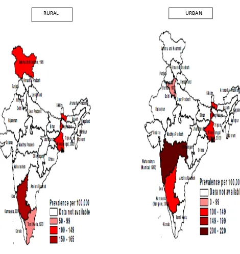
FIGURE 1Crude prevalence rates for stroke Rural India 1970-2004 Urban India 1973-2004
FIGURE 2: Stroke mortality rates across world
AIM
MATERIALS AND METHODS
STUDY DESIGNHospital based prospective study
SAMPLE SIZE: 50
INCLUSION CRITERIA:
All patients of age group more than 18 years with clinical and radiological evidence of acute ischemic stroke
EXCLUSION CRITERIA:
1) Cerebral hemorrhage of any etiology
2) Liver disease (Alcohol intake >20 units/week for males,>14 units/week for females)
3) Cardiac failure
4) Nephrotic syndrome/diabetic nephropathy(urine albumin >2+) 5) Protein losing enteropathies
6) Malignancy
7) Conditions mimicking stroke
a) Metabolic -severe hyponatremia
-hypoglycemia
b) Psychiatric disorders -conversion disorder -malingering
-factitious disorder
c) Infectious conditions -viral encephalitis -Bacterial meningitis -Brain abscess
d) Cardiovascular
- syncope
- hypertensive encephalopathy e) Neurological conditions
-seizure with Todd’s paralysis
-brain tumor
-demyelinating disorders
-myasthenia gravis
METHODOLOGY:
The study is based on prospective collection of data in Ischemic stroke patients who fulfill the inclusion criteria stated above and who were admitted in medicine and neurology wards in a tertiary care centre(PSGIMSR) where systematic computer coding for registry is used.Diagnosis of ischemic stroke is based on clinical observation and radiological imaging.
Blood samples for assessment of albumin was collected at admission within 36 hours after stroke onset. Strokeseverity at presentation was determined by NIHSS score. Functional outcome was measured 1 week post admission and after 3 months during follow up using modified Rankin scale.
Favourable score mRS:0-3
Unfavourable score mRS:4-6
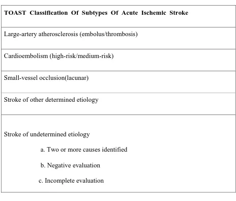
TABLE 1
TOAST Classification Of Subtypes Of Acute Ischemic Stroke
Large-artery atherosclerosis (embolus/thrombosis)
Cardioembolism (high-risk/medium-risk)
Small-vessel occlusion(lacunar)
Stroke of other determined etiology
Stroke of undetermined etiology
a. Two or more causes identified b. Negative evaluation
c. Incomplete evaluation
TABLE 2: National Institute of Health stroke scale (NIHSS):
STROKE STROKE SEVERITY
0 No stroke symptoms
1-4 Minor Stroke
5-15 Moderate Stroke
16-20 Moderate to Severe Stroke
21-42 Severe Stroke
TABLE 3: Modified Rankin Scale
SCORE SYMPTOMS
0 - No symptoms
1 - No significant disability. Able to carry out all usual activities, despite some symptoms
2 - Slight disability. Able to look after own affairs without assistance, but unable to carry out all previous activities
3 - Moderate disability. Requires some help, but able to walk unassisted
4 - Moderately severe disability. Unable to attend to own bodily needs without assistance, and unable to walk unassisted
5 - Severe disability. Requires constant nursing care and attention, bedridden, incontinent
Statistical tools:
REVIEW OF LITERATURE
History and Background:Stroke was first recognized by Hippocrates (460 to 370 BC), father of medicine some 2400 years ago. Initially it was termed apoplexy in Greek (meaning “struck down by violence”).
Johann Jacob Wepfer (1620–1695) was the next well renowned person in the field of stroke. He studied the corpses deceased due to apoplexy. He discovered that blood supply might be disrupted to the brain due to blocked arteries in a few or due to massive bleeding into the brain tissue [28].
Rudolf Virchow, known as father of modern pathology, was the first to describe the mechanism of thromboembolism as a major factor causing stroke. In the mid nineteenth century , he described the term thrombosis which can detached to form an embolus causing cardio-embolic stroke[29].
EPIDEMIOLOGY
In India, the prevalence of stroke is increasing. The incidence of stroke increases exponentially from 30 years of age, and etiology varies by age. About two-thirds of strokes occur in those over the age of 65[23,24]. It has been observed now that the prevalence is becoming high among younger age group and those of low socioeconomic status[25]
The AIRs of stroke in India, as observed in the Kolkata, Mumbai, and Trivandrum studies are higher than that in United States (107 per100,000 per year), European countries (61-111 per 100,000 per year)[17,18,19],and Australia (99 per 100,000 per year)[21] but similar to that reportedfrom one Chinese city - Changasha. [35]
of Indian descent.[22] The fatality rates were higher among women, as women outnumber men and also have higher prevalence of uncontrolled hypertension than men.[24] In a study done in Trivandrum, the 28-day CFRs were 24.5% for urban and 37.1% for rural areas.[23]
TABLE 5:Data from Trivandrum stroke registry showing urban and rural distribution
DEFINITION AND CLASSIFICATION:
Although more common in older adults, stroke also occurs in neonates, infants, children and young adults, resulting in significant morbidity and mortality[36].Stroke is a clinical syndrome which is classified broadly into:
Ischaemic strokes – These are caused by sudden occlusion of arteries supplying the brain, either due to thrombus at the site of occlusion or formed in another part of the circulation causing restriction of blood supply to the part of brain causing cerebral infarction. It accounts for 50-85% of all strokes worldwide [37]
[image:26.612.106.510.144.377.2]Transient ischemic attacks (TIAs) are defined as temporary neurological deficit with symptoms lasting less than 24hrs which is thought to be due to inadequate cerebral or ocular blood supply as a result of thrombosis or embolism associated with arterial, cardiac or haematological disease. It serves as a warning signal for impending stroke. It leaves no clinical residue or abnormality on imaging [39].
Risk factors :
Risk factors are defined as any attribute, characteristic or exposure of an individual that increases the likelihood of developing a disease or injury. Risk factors may be further classified as modifiable or non-modifiable.
Non modifiable risk factors:
Risk factors like age, gender, race/ethinicity and family genetics come under non modifiable risk factors.
1. Gender: Globally, males are affected predominantly regardless of age and stroke subtype. In the Indian scenario, this difference observed is due to high prevalence of smoking and alcohol among males. The male :female sex ratio among affected individuals is 7:1 in India.[40,41]
2. Race and ethnicity: Blacks are more commonly affected with stroke compared to whites. Lack of proper awareness, affordability and no insurance are among the contributory factors to this observed difference.[42,43]
3. Family genetics: Family members may have a genetic tendency or share lifestyles and behavioural patterns that significantly contribute to the occurrence of stroke. Genetic factors for hypertension, Von Willebrand disease, Sickle cell disease may also contribute to increased risk of stroke. Risk of stroke further increases with a past history of stroke. Although heredity plays only a minor role in the pathogenesis of stroke, an increased risk is seen among first-degree relatives with a family history of stroke. Heredity factors when combined with an unhealthy lifestyle leads to increased risk among individuals. [44,45]
Modifiable
Cigarette smoking – It is one of the most important risk factors of stroke among
Alcohol consumption – This is another modifiable and preventable risk factor for
stroke in young adults. It differs among men and women. In men, binge drinking of alcohol carries a higher risk of developing ischemic stroke than in non -alcoholics or alcoholic abstainers. Binge drinking has the effect of raising blood pressure which further increases risk of stroke[47].In men, binge drinking is defined as six or more alcoholic drinks whereas it is four or more in women. In women the risk increases with increase in intake of alcohol. Consumption of wine has a protective effect.
Dyslipidaemia – Atherogenic dyslipidaemia is an independent risk factor of stroke
Abdominal obesity – defined as BMI more than 30kg/sq.m. Predisposing mechanism
of stroke in obese individuals is likely due to its effect on arterial hypertension associated with elevated cholesterol levels. It mainly plays an important contributory factor for stroke. Upper body obesity is more important than lower body obesity as a risk factor[49]. Increased waist hip ratio is associated with early death. People should be encouraged to increase daily physical activity and reduction of weight in order to lessen the risk of stroke.Increased physical activity also decreases platelet aggregation and increase insulin sensitivity[50].
Sedentary life style – It has been a contributory factor for occurrence of stroke in
young individuals. Blood pressure, impaired glucose tolerance, insulin resistance are associated with altered lipid profile with increasing risk for stroke. Lifestsyle modifications like regular exercise, weight reduction measures and dietary modifications may help decrease incidence.
Hypertension – It is defined as systolic blood pressure (SBP) more than 140 mm Hg
factor for ischaemic stroke[51,52]. Isolated systolic blood pressure and wide pulse pressures are at greater risk.
In chronic hypertension, there is vessel wall thickening and luminal narrowing which limit the capacity of the resistance vessels for dilation. In acute stroke, auto-regulation may be impaired in regions surrounding an acute lesion and even in the hemisphere contralateral to the lesion because of dilation of cerebral resistance vessels in an attempt to increase blood flow in response to tissue ischemia and acidosis. About 10-20 mm of Hg decrease in SBP causes reduction of incidence of stroke of 28% in young adults[53].
Diabetes mellitus: Increasing prevalence of diabetes results in an increase of
deficits. It is therefore said that prompt treatment of hypoglycaemia in emergency conditions in suspicion of stroke.
Oral contraceptives – risk of stroke in young women taking oral contraceptives is
four times higher. High dose oestrogen doubles the risk of stroke [56]in women with other associated factors like smoking, pro-thrombotic genetic variants and migraine. High prevalence of thromboembolic stroke is seen in these conditions[57]. Newer oral contraceptives with low levels of estrogen are being used to decrease the incidence of stroke in women.
Pregnancy: Pregnancy and puerperium - Estrogen related stroke – Risk of stroke is
high in third trimester and 6 weeks of post-partum period[58]. Preeclampsia and eclampsia play a synergistic role leading to nine fold increase in stroke occurrence[59]. Amniotic fluid embolism, postpartum angiopathy and postpartum cardiomyopathy can result in cardio-embolism or infarction due to hypotension. It may also be influenced by underlying haemotological or thrombotic conditions
Illicit drug abuse: Recreational drug abuse in young adults increases the
and arterioles causing stroke. Drugs like heroin, opiates, cannabinoids also play a role in etiology of ischemic stroke.
Migraine headache – There is increased risk of ischemic stroke among young women (35-45years) with migraine headache with aura (MA). The risk is accelerated with concomitant use of oral contraceptives, smoking and blood pressure. Cerebral ischemia may induce migraine headache[62,63]. Most commonly occipital headaches are present. It is seen in about one third of total cases.
ETIOLOGY:
Atherosclerotic – thrombotic and cerebral embolic stroke are the predominant cause of AIS.
Atherosclerosis:
It mainly involves large vessels both intra-cranial and extra-cranial arteries and small vessels (lacunar arteries)[64]. It begins with damage to the endothelial lining of vessel wall. The damaged area attracts platelets, calcium, fatty substances, fibrin and cellular debri which accumulate to form an atherosclerotic plaque. These plaque eventually increase in size and block the blood flow through the vessel thereby decreasing the blood supply leading to hypoxia.
blood vessel[65]. The second type of mechanism of stroke called cardio-embolic stroke. Cardioembolism is responsible for 20% of all ischemic strokes. Stroke caused by heart disease is primarily due toembolism of thrombotic material forming on the
atrial or ventricular wall or the left heart valves. These thrombi then detach and
embolizeinto the arterial circulation. The thrombus may fragment or lyse quickly,
producing only TIA. Alternatively, the arterial occlusion may last longer, producing
stroke. Embolic strokes tend to occur suddenly with maximum neurologic deficit
present at onset. Nonrheumatic atrial fibrillation is the most common cause of cerebral
embolism overall. The presumed stroke mechanism is thrombus formation in the
fibrillating atrium or atrial appendage, with subsequentembolization. Atrial fibrillation
causes stasis of blood due to hypocontractile nature of atrium. There occurs atrial
remodeling, endothelial dysfunction, release of coagulation factors like Xa and
thrombin. All these factors promote thrombus formation and ultimately lead to
cerebral embolism.Patients with atrial fibrillation have an average annual risk of
stroke of -5%[66].Recent MI may be a source ofEmboli, especially when transmural and involving the anteroapical ventricular wall, and prophylactic anticoagulation
following MI has been shown to reduce stroke risk. Mitral valve prolapse is not
usually a source of emboli unless the prolapse is severe. Paradoxical embolization
occurs when venous thrombi migrate to the arterial circulation, usually via a patent
Atheromatous plaques mostly form preferentially at branching points and curves of cerebral arteries. The most frequent sites involved are
Internal carotid artery at its origin from the common carotid artery
In the cervical part of vertebral arteries and at their junction to form basilar arteries In the stem or at the main bifurcation of middle cerebral arteries
In the proximal posterior cerebral arteries as they wind around the midbrain
In the proximal anterior cerebral arteries as they pass anteriorly and curve around corpus callosum.
Large artery disease:
Carotid and vertebral dissections - Defined as tear in intimal layer of vessel leading to blood flow in between the layers of vessel wall. The stasis of blood in between the vessel layers leads to formation of a clot which can embolise leading to formation of stroke[67]. It is one of the factors in causing stroke in the young though it becomes a rarer cause in older age group. It is commonly caused by trauma to blood vessel. It also causes haemorrhagic stroke in young adults. Spontaneous dissection is also seen in few individuals, especially in those with connective tissue disorders, increases predisposition to stroke.
Small artery disease: systemic hypertension and migraine effect the small
microvasculature causing lacunar infarcts in individuals.
Cardioembolism: It is the most common cause of arterial ischemic stroke (AIS) of
atherosclerosis in older age group. It accounts for about one fifth of ischemic stroke [68]. Most of the cases the emboli arises from a thrombus within heart. The arterial ischemic stroke caused by embolism from the heart can only be diagnosed, if there is an identifiable cardioembolic source, by using transoesophageal echocardiography. It can provide the anatomic location regarding the source of emboli.
Cardiac diseases – they may classified as congenital and acquired.
o Congenital heart disease is a major risk factor for cardio-embolic stroke especially in the perioperative period or following catheterization or extracorporeal membrane oxygenation (ECMO).
o Rheumatic Heart Disease(RHD) – it plays a significant role in causing stroke in
young adults in developing countries. About 3-8% strokes are attributed to RHD. It also plays a role in recurrent stroke. It remains as an occult cause where early identification and preventive measures must be taken to reduce the incidence and recurrence[69].
o Acquired conditions –These commonly include Dilated cardiomyopathy (DCM),
Elevated Homocysteinelevels: Inborn errors of metabolism characterized by
defect in methionine metabolism due to deficiency of enzyme cystathione b-synthase(CBS). Common pattern of inheritance is autosomal recessive. Deficiency of vitamin B12 and serum folate levels is another factor having a contributory role. These enzyme and vitamin deficiencies cause increased accumulation of homocysteine levels in plasma and urine. Elevated homocysteine levels causes endothelial dysfunction and thromboembolic events affecting both small and large vessels causing cerebrovascular ischemia[71]. It is mainly diagnosed by measuring the levels of homocysteine in urine and plasma. Most of them respond to vitamin supplements.
Fibromuscular dysplasia – This condition mainly affects the medium sized
vasculature in young women of childbearing age group. It causes medial fibrosis of vessel wall due to an unknown mechanism, predominantly involving the carotid vasculature[72]. Stroke may be thromboembolic or vascular stenosis may be the underlying cause. On angiography, a typical beading pattern of involved vessel is formed.
Radiotherapy – Patients who have received radiation for head and neck
Haematological disorders:
o Thrombophilia: Defined as a group of condition related to the impairment of
haemostatic mechanism which manifest as an increased tendency to form thrombus. They may be classified as inheritable or acquired conditions. Deficiency in natural coagulants like protein C, protein S and antithrombin III deficiency, polymorphisms in activated protein C and disturbance of clotting mechanisms by mutation in prothrombin gene 20210G/A are classified under the inheritable causes. Among the inherited conditions, Factor V leiden and prothrombin 20210 mutation are most commonly associated with arterial ischemia stroke(AIS)[74]. Among acquired conditions, Anti-phospholipid antibody syndrome (APLA) is most commonly associated with AIS. Acquired conditions are relatively at higher risk of causing stroke than inherited conditions. Workup for these conditions with a background of prior episode suggestive of thromboembolic event or having a positive family history or recurrent pregnancy loss or absence of other identifiable risk factors[75]. Family members and siblings should be screened.
o Myeloproliferative syndromes – They mainly include polycthemiavera (PV),
o Sickle cell anaemia – Stroke in this condition is a near fatal complication. Considered
mechanism in the etiology of stroke in this condition is that, the deformed sickle cells cause vaso-occlussion of vessels leading to stroke. Another mechanism is by hemolysis within the vasculature altering the endothelial structure of vessel wall[77]. This condition mainly affects the large arteries.
o Vasculitides: Defined as occurrence of inflammation and formation of necrosis of
vessel wall. It is classified as primary or secondary in nature. Among the primary conditions Takayasu’sarteritis,Polyarteritisnodosa (PAN), Wegener’s granulomatosis (WG), Behcet’s are common conditions associated with stroke in young individuals. Another rare disorder called primary angitis of central nervous system (CNS) a multifocal disorder considered one of the cause of stroke in young[78]. In these conditions, there is inflammation of blood vessel wall which increases the thrombogenecity and alter the vessel tone predisposing to stroke.
TABLE 6
TYPES: GENE MUTATION VESSEL AFFECTED
CADASIL Notch 3 receptor Small vessel disease
CARASIL Notch 3 receptor Small vessel disease
Fabry disease (X‐linked recessive)
α‐galactosidase A Large and small vessel disease
MELAS (maternal) Transfer RNA Complex
Marfan syndrome (autosomal dominant)
Fibrillin 1 Cardioembolism and arterial dissection
Ehlers–Danlos
syndrome (autosomal dominant) – type IV
Collagen type III
o CARASIL(cerebral autosomal recessive arteriopathy with subcortical infarcts and leukoencephalopathy ) – autosomally recessively inherited CADASIL associated with acute features of lumbago, spondylosisdeformans, diffuse baldness and progressive mental and motor deterioration. Most common age of onset is 25-30years of age (younger age comparatively to CADASIL). On imaging, it is typically identified as diffuse and homogenous lesions.
o MELAS – mitochondrial myopathy, encephalopathy, lacto-acidosis and stroke. It is a multisystemicmaternally inherited disorder which are characterised by stroke like episodes[80]. In this condition mostly posterior regions like temporo-parietal and occipital regions are involved. Due to mitochondrial dysfunction, nitric oxide metabolism is impaired and free radicals are released which cause impaired auto-regulation. Cardiomyopathy due to mitochondrial disease may cause cardio-embolic stroke. They are transient in nature and reflected in abnormalities on neuroimaging[81]. Muscle biopsy helps in diagnosis, by identifying the abnormal proliferation of mitochondria.
o Heritable disorders of connective tissue – this mainly involves the mutation in collagen and elastin content which constitute the vessel wall. Of the disorders affecting collagen fibres, Ehlers Danlos syndromes (EDS), osteogenesisimperfecta (OI), autosomal dominant polycystic kidney disease (ADPKD) and collagen type IV related small vessel disease are most commonly associated with stroke[84]. These are associated with formation of aneurysms in the vessel wall and are complicated with carotid and vertebral artery dissections. Among those affecting the elastin content of vessel wall, Marfan’s syndrome, Loeyz Dietz syndrome (LDS type I and II) and Pseudoxanthomaelasticum are commonly associated with occurrence of stroke in young.
o Amyloid angiopathy: it is associated with amyloid deposition in leptomeningeal
walls and cerebral arteries and arterioles. Most commonly involves the occipital region and considered severe if it involves the same region. Mostly it causes haemorrhagic stroke.
o Moyamoyadisease: It is a genetic disorder with AD pattern of inheritance
ROLE OF ALBUMIN IN ACUTE ISCHEMIC STROKE ALBUMIN:
A) SYNTHESIS:
Albumin is quantitatively the most important plasma protein. Adult liver synthesizes around 15 grams per day. Approximately 300 to 500 grams of albumin is distributed in the body fluids. The synthesis rate can double in situation in which there is rapid albumin loss or a fall in serum albumin concentration. Rate of albumin transcription can be affected due to trauma, sepsis, hepatic disease, diabetes , fasting.
B) STRUCTURE:
C) FUNCTION:
Major functions of albumin include regulation of colloid osmotic pressure of plasma,transportation of hormones,fattyacids,drugs and metabolites across plasma,regulation of microvascularpermeability,anti-oxidant activity,anti thrombotic and anti inflammatory activity.
D) DISTRIBUTION:
It is a major component of ECF including CSF,interstitial fluid and lymph.Around 40-60% albumin is degraded in muscle and skin,15% in liver,10% in kidney and 10% in GI tract.
E) USES:
Because of its unique properties,albumin has been used as therapeutics in the field of hepatology. The volume expanding property of albumin is widely used for benefit of patients with cirrhosis. Among neurological diseases albumin has a role in ischemic stroke,Alzhemier’s disease and epilepsy. Human albumin in high doses has been used in clinical trials for acute ischemic stroke based on its neuroprotective effects.
VOLUME OF INFARCT AND CLINICAL OUTCOME
The standard measure of therapeutic success in animal stroke models is reduction in infarct volume. As a replacement for disability and global clinical scales, reduction in infarct volume can be considered an auxiliary outcome measure for human stroke clinical trials. Few studies have compared infarct volume and clinical outcome.
Several small series showed that infarct volume and clinical outcomein terms of BI, NIHSS, Rankin Disability Scale had no statistical significance.[91,92]Some larger series , revealed correlations between infarct volume and clinical scales (NIHSS, Rankin Disability Scale, Oxford Disability Scale, aphasia severity scale).[93–95]
Volume of infarct is calculated using the formula ABC/2. The infarct was measured in three perpendicular axes A,B and C
A = Longest dimension in axis x
B = Longest perpendicular dimension to axis x (y) C = Total length in z dimension
ANATOMY AND CLINICAL MANIFESTATIONS
o ANATOMY OF CEREBRAL CIRCULATION:
Brain is the highest perfused organ in the body. It receives about 20% of total circulation and also has the maximum consumption of oxygen in the blood. It is supplied by two pairs of large arteries – internal carotid arteries and vertebral arteries[78].
Internal carotid artery supplies about 3/5th of cerebrum. The two vertebral arteries join together to form basilar artery which supplies cerebellum and brain stem. These two arterial circulation join together with the help of communicating branches to form circle of willis (COW)[79]
o The internal carotid group produce three main vessel branches which include – 1. Ophthalmic artery – supplies the meninges, contents of orbit.
o Clinical relevance –
• Occlusion of ACA may cause following symptoms:
• Contralateral lower limb upper motor neuron type (UMN) of weakness • Contralateral sensory loss in lower limb
• Due to frontal lobe involvement – Behavioural abnormalities, cortical release reflexes – grasp reflex, sucking reflex, gegenhalten phenomenon. • Transcortical aphasia.
3. Middle cerebral artery (MCA) – It is also a paired artery which supplies anterior temporal and insular cortices. They are connected to ACA with the help of anterior communicating branches and connected with PCA with the help of posterior communicating branches. They are further divided into 4 parts or segments in their course of supply. They supply the bulk of lateral surface of the hemispheres along with speech areas (Broca’s and Wernicke’s areas)
o Clinical relevance –
• Contralateral upper and lower limb UMN type of paralysis • Contralateral sensory loss over face and arm
• If lenticulostriate branches of MCA are involved –
If involvement of dominant hemisphere aphasia
4. Posterior cerebral artery (PCA) – it is one of the paired arteries which supply the posterior part of the brain which includes occipital lobe. It is divided into 2 branches – cortical and ganglionic vessels.
o Clinical relevance –
• Contralateral loss of pain and temperature
• Contralateral homonymous hemianopia with macular sparing • Alexia and agraphia
MANAGEMENT:
NON INVASIVE METHODS:
In patients with AIS, diagnosing stroke early plays a key role in many ways: To assess the clinical outcome and prognostication of the condition. To institute thrombolysis whenever possible.
It helps further management – mainly in the ED, where it plays a crucial role in order to decide about further management.
Therefore, a standard approach for diagnosis and treatment for stroke must be established in primary health care centres and educational institutions for management of stroke in emergency.It also helps in assessing the arterial territory involved and area of brain affected by examining clinically.Identification and control of modifiable risk factors and especially hypertension, is the best strategy to reduce the burden of stroke, and the total
Stroke scoring systems:
Routine baseline investigations to be done are (TABLE 8): Complete haemogram
Blood sugars Hypo or hyperglycemia Urine routine Diabetes, infection
Serum electrolytes Hyponatremia or hypokalaemia or hyperkalaemia Renal function tests Renal failure
Fasting lipids Dyslipidemia Homocysteine levels Homocystinemia
Serology Vasculitis , Infections, HIV, VDRL
ECG Left ventricular hypertrophy (LVH), Atrial fibrillation, arrhythmias, AMI
Echocardiography Infective endocarditis, atrial myxoma,mural thrombus ESR, CRP Autoimmune causes-vasculitis, SLE
To do in young stroke
ANA profile, APLA APLA syndrome, SLE, vasculitis
Coagulation profile Protein C and S deficiency, anti thrombin III deficiency, hyperfibrinogenemia.
Genetic studies (optional)
TREATMENT:
Treatment is mainly aimed at reversing the hypoxic brain injury and lessen further damage of brain from hypoxia caused by decreased blood supply due to occlusion of the blood vessels supplying the brain.
At the presentation to ED with acute stroke, the primary management is to assess the airway, breathing and circulation initially[81].Treat the hypoglycaemia (<60mg/dl) identified on presentation as it may act as a stroke mimic. Hyperglycemia leads to poor outcomes in stroke. Hence, early treatment of blood sugars is needed.
TABLE 8
Early fibrinolysis :
r-tPA(recombinant tissue plasminogen activator). Streptokinase which has a major role in AMI, has high incidence of complications in acute AIS. Hence this drug is of not in regular use for AIS. It must be decided whether a patient is a feasible candidate for fibrinolysis[81].They must be thrombolysed within a window period of maximum of 4.5hrs from the onset of symptoms.
The FDA approved dose of IV r-tPA is 0.9 mg per kilogram of body weight, withamaximum dose of 90 mg. A bolus of 10% of the dose to be given over 1 minute, with the remaining 90% to be infused over 60 minutes. Weight should be determined reliably. Treatment with a lower dose of r-tPA (0.6 mg per kilogram) in Japan suggest that it had similar efficacy but the lower dose has not yet been assessed in large, randomized trials.
Third-generation plasminogen activators, such as Tenecteplase and Desmoteplase, are more fibrin specific than second generations r-tPA and cause less activation of systemic lytic activity.
Symptomatic intracranial hemorrhage occurs in 1.7 to 8.0% of treated patients.[87,88]Recommendations for the treatment of intracranial or major systemic bleeding after thrombolytic therapy often includes administration of cryoprecipitate and platelets,although evidence-based guidelines for such an approach are lacking[89,90]
In patient receiving UFH consider giving protamine 1mg for every 100 U UFH
INCLUSION AND EXCLUSION CRITERIA FOR THROMBOLYSIS IN AIS: INCLUSION CRITERIA:
Onset of symptoms <3 hours before beginning treatment (Onset time is defined as either the witnessed onset of symptoms or the time last known normal)
Age ≥18 years
Potential risks and benefits of IV t-PA treatment discussed with patient and/or family members and they have verbalized understanding (to be documented in patient’s record). If patient unable to give verbal consent and no family available, IV t-PA can be given under Emergency Doctrine. Written informed consent not required for IV tPA when given within 3 hours of symptom onset.
EXCLUSION CRITERIA:
Significant head trauma or prior stroke in previous 3 months Any history of haemorrhage stroke
Intracranial neoplasm, arteriovenous malformation, or aneurysm Recent intracranial or intra-spinal surgery
Elevated blood pressure (systolic >185 mm Hg or diastolic >110 mm Hg) Active internal bleeding
Blood glucose concentration <50mg/dl (2.7mmol/L)
IV rtPA can be initiated before availability of platelet count but should be discontinued if platelet count is <100 000/mm³.)
Heparin received within 48 hours, resulting in abnormally elevated aPTT greater than the upper limit of normal
Current use of anticoagulant with INR >1.7 or PT >15 seconds and current use of direct thrombin inhibitors or direct factor Xa inhibitors with elevated sensitive laboratory tests (such as aPTT, INR, platelet count, and ECT; TT; or appropriate factor Xa activity assays)
CT demonstrates multilobar infarction (hypodensity>1/3 cerebral hemisphere)
RELATIVE EXCLUSION CRITERIA:
Only minor or rapidly improving stroke symptoms (clearing spontaneously) Seizure at onset with postictal residual neurological impairments
Major surgery or serious trauma within previous 14 days
Recent gastrointestinal or urinary tract haemorrhage (within previous 21 days) Pregnancy
To extend IV tPA to 4.5 hours from symptom onset/last known normal, the following additional criteria MUST be met:
Patient is < 80 years of age
Patient is not taking Warfarin (Coumadin) or any other anticoagulant regardless of INR/coagulation results
NIHSS is < 25
Written informed consent obtained from patient and/or family – required when IV tPA given within the 3-4.5 hour window.
Anti coagulants:
Anticoagulation is not recommended in all patients with AIS in emergency condition. Drugs most commonly used are low molecular weight heparin (LMWH) or unfractionated heparin (UFH)[84]. They can be used to prevent venous thromboembolism. Only in the below mentioned specified clinical situations, anticoagulation therapy is indicated :
Conditions with potential high risk of early cardiogenic reembolization Symptomatic dissection of arteries supplying the brain
Symptomatic extra-cranial or intra-cranial atherosclerotic stenosis
Basilar artery occlusion before or after intra-arterial pharmacological or mechanical thrombolysis
Anti-thrombotic agents :
They are mainly used for secondary prevention of stroke. Drugs most commonly used are aspirin,clopidogrel and extended release dipyridamole. Newer antiplateletsdabigatran, apixaban and rivaroxaban are being studied for anticoagulation comparing with warfarin [85].
According to guidelines, aspirin should be given within 24 to 48hrs of onset of stroke to prevent mortality and primary end point.
Endovascular techniques – intra-arterial fibrinolysis, thrombo-embolectomy, suction thrombectomy, angioplasty and revascularisation are among the common endovascular procedures. The main aim of these is to recanalise the thrombosed vessel to improve the blood flow. For this a team of skilled neurologists, interventional radiologists, anaesthesiology, nursing and technical support is required for optimal success.
Neuro protection:
Several novel neuro-protective agents like citicholine, traxoprodil, ONO-2506, magnesium, DP-b99 and NXY-059 have been identified. They limit the infarct size and improve functional outcome (primary end point)[85]. They act as free radicals scavengers and inhibit further occurrence of neuronal cell death.
Rehabilitation centres :
RESULTS
In this study, about 50 consecutive cases admitted with AIS, which met inclusion and exclusion criteria were evaluated. At presentation, along with complete history taking, relevant clinical examination, scoring was done based on NIHSS score.mRSscoring 1 week post admission,3 months later and Volume of infarct in CT scan and clinical outcome were analysed in this study.
GENDER
Among the study population of 50 patients, 44(88%) were males and 6(12%) were females. Out of 50 patients,3 patients were lost to followup.
FIGURE 12: Sex Distribution
SEX DISTRIDUTION
TABLE 9: Presenting symptoms in study population
SYMPTOMS NO OF PATIENTS %
HEADACHE 5 10
VOMITING 4 8
MOTOR WEAKNESS 18 36
UNCONSIOUSNESS 6 12
DYSARTHRIA 11 22
VERTIGO 3 6
TINGLING 2 4
DIPLOPIA 0 0
TABLE 10: Distribution according to systolic blood pressure ( SBP ) on admission
SBP MALE FEMALE TOTAL %
< 138 5 1 6 12
140 – 158 21 2 23 46
160 – 178 9 2 11 22
180 – 198 7 1 8 16
>200 2 0 2 4
TOTAL 44 6 50 100
TABLE 11:Distribution according to diastolic blood pressure (DBP) on admission
DBP MALE FEMALE TOTAL %
<78 4 0 4 8
80 – 88 13 2 15 30
90 – 98 18 3 21 42
100 – 108 7 1 8 16
>110 2 0 2 4
TABLE 12:Past medical history on study population
RISK FACTORS TOTAL MALE FEMALE
TOBACCO 1 0 1
OLD CVA 5 4 1
ALCOHOLISM 11 11 0
IHD 12 12 0
SMOKING 12 12 0
DLP 14 13 1
DM 20 19 1
SHT 34 30 4
FIGURE 13: 0 5 10 15 20 25 30 35 0 4
11 12 12 13
19
30
1
1
0 0 0
1
1
4
FEMALE
TABLE 13: Frequency of lesion in MRI / CT brain
FREQUENCY OF LESION IN MRI/CT NO OF PATIENTS
MCA INFARCT 30
MULTI INFARCT 9
POSTERIOR CIRCULATION INFARCT 5
LACUNAR INFARCT 6
TOTAL 50
FIGURE 14: 0 5 10 15 20 25 30 35 40 45 50
MCA INFARCT MULTI INFARCT POSTERIOR CIRCULATION
INFARCT
LACUNAR INFARCT
TOTAL
TABLE 14: Gender and mRS @ 1 week
Gender
mRS (1 week)
Total Less than or equal
to 3 More than 3
Male 29 14 43
Female 3 1 4
Total 32 15 47
FIGURE 15:
Out of the 43 male patients , 14 patients had significant disability. Among the 14, 3 had death as outcome. 29 patients had mild disability. Out of the 4 female patients, 1 had significant disability and died during the first week. Remaining 3 patients had mild disability.
0 10 20 30 40 50
Male Female G en d er 29 3 14 1
mRS (1 week) Less than or equal to 3
TABLE 15: Gender and mRS @ 3 months
Gender
mRS (3 months)
Total Less than or equal
to 3 More than 3
Male 39 4 43
Female 3 1 4
[image:75.612.72.513.105.560.2]Total 42 5 47
FIGURE 16:
Among the male patients, no of patients with significant disability dropped from 14 to 4.In the female group,the 3 patients with mild disability had improvement in the mRS score and didn’t worsen.
0 10 20 30 40 50
Male Female G en d er 39 3 4 1
mRS (3 months) Less than or equal to 3
TABLE 16 :Gender and serum albumin level
Gender
Serum Albumin Level
Total 3-3.4 3.5-3.9 4-4.4 >4.4
Male 1 11 24 8 44
Female 0 2 3 1 6
Total 1 13 27 9 50
FIGURE 17:
Among 50 patients , only one patient had low serum albumin levels. Majority of patients had albumin levels between 4 to 4.4.Both male and female groups were similar.
0 10 20 30 40 50 Male Female Gender 1 0 11 2 24 3 8 1
Serum Albumin Level >4.4
Serum Albumin Level 4-4.4
Serum Albumin Level 3.5-3.9
TABLE 17: Gender and NIHSS score
Gender
NIHSS Score
Total Less than or equal
to 10 More than 10
Male 37 7 44
Female 4 2 6
Total 41 9 50
FIGURE 18:
On admission, no. of males who had NIHSS more than 10 were 7. No of females who had NIHSS more than 10 were 2.
0 20 40 60
Male Female G en d er 37 4 7 2
NIHSS Score Less than or equal to 10
TABLE 18: Age and mRS @ 1 week
Age
mRS (1 week)
Total Less than or equal to 3 More than 3
26-35 1 1 2
36-45 2 2 4
46-55 9 5 14
56-65 9 6 15
>65 11 1 12
Total 32 15 47
FIGURE 19:
Among the 47 patients , no of people who had significant disability were 15.Among them 6 were in the age group of 56 to 65. No of people who had mild disability were
0 2 4 6 8 10 12 14 16
26-35 36-45 46-55 56-65 >65
Age
1 2
9 9 11
1
2
5 6 1
mRS (1 week) More than 3
[image:78.612.73.520.237.616.2]TABLE 19: Age and mRS @ 3 months
Age
mRS (3 months)
Total Less than or equal to 3 More than 3
26-35 2 0 2
36-45 3 1 4
46-55 12 2 14
56-65 13 2 15
>65 12 0 12
Total 42 5 47
FIGURE 20:
After 3 months , number of people with significant disability were 5 and number of people with mild disability were 42.Significant improvementwas seen in the age group of 56 to 65 ( 9 to 13 ).
0 2 4 6 8 10 12 14 16
26-35 36-45 46-55 56-65 >65
Age
2 3
12 13 12
0
1
2 2
0
mRS (3 months) More than 3
[image:79.612.74.500.223.578.2]TABLE 20: Age and Serum Albumin Level
Age
Serum Albumin Level
Total
3-3.4 3.5-3.9 4-4.4 >4.4
26-35 0 0 1 1 2
36-45 0 0 3 2 5
46-55 1 4 6 3 14
56-65 0 5 9 2 16
>65 0 4 8 1 13
Total 1 13 27 9 50
FIGURE 21:
Mean albumin is 3.8.Only one patient in the age group of 46 to 55 had low serum albumin .Each group had high no of patients in the albumin range 4 to 4.4.
0 2 4 6 8 10 12 14 16
26-35 36-45 46-55 56-65 >65
Age
0 0 1 0 0
0 0 4 5 4 1 3 6 9 8 1 2 3 2 1
Serum Albumin Level >4.4
Serum Albumin Level 4-4.4
Serum Albumin Level 3.5-3.9
[image:80.612.71.511.101.618.2]TABLE 21: Age and NIHSS score
Age
NIHSS Score
Total Less than or equal to 10 More than 10
26-35 2 0 2
36-45 5 0 5
46-55 9 5 14
56-65 13 3 16
>65 12 1 13
Total 41 9 50
FIGURE 22:
No. of people with NIHSS score less than or equal to 10 was 41 and more than 10 was 9. NIHSS score of 10 or less than 10 was seen in high numbers in 56-65 and >65 years age group. 0 2 4 6 8 10 12 14 16
26-35 36-45 46-55 56-65 >65
Age 2 5 9 13 12 0 0 5 3 1
NIHSS Score More than 10
[image:81.612.73.520.180.612.2]TABLE 22:Serum Albumin with mRS after 1 week
Serum Albumin Level
mRS (1 week)
Total P value Less than or
equalto 3 More than 3
<3.5 g/dl 0 1 1
0.319
> or = 3.5 g/dl 32 14 46
Total 32 15 47
FIGURE 23:
Serum albumin level was compared with mRSscore. No of people with serum albumin >3.5 and mRS less than or equal to 3 are 32 and with mRS> 3 are 14. No of people with serum albumin<3.5 and mRS>3 are 1. Pvalue was 0.319. Hence there was no significant
0 10 20 30 40 50
<3.5 g/dl > or = 3.5 g/dl
Se ru m Alb u m in L ev el 0 32 1 14
mRS (1 week) Less than or equal to 3
TABLE 23: Serum Albumin with mRS after 3 months
Serum Albumin Level mRS (3 months)
Total P value Less than or equal
to 3 More than 3
<3.5 g/dl 1 0 1
0.894
> or = 3.5 g/dl 41 5 46
Total 42 5 47
FIGURE 24:
Serum albumin level and mRS score after 3 months were compared. Number of patients with serum albumin >3.5 and mRS< 3 increased from 32 to 41. The patient with serum albumin <3.5 had an improvement in mRS score. Eventhough there was improvement in patients clinically, statistically there was no significant correlation as the p value was 0.894.
0 20 40 60
<3.5 g/dl > or = 3.5 g/dl
Se ru m Alb u m in L ev el 1 41 0 5
MRS (3 months) Less than or equal to 3
TABLE 24: Volume of Infarct in CT Scan withmRS after 1 week
FIGURE 25:
N Mean
Std.
Deviation Std. Error
95% Confidence Interval for
Mean
Minimum Maximum P value Lower Bound Upper Bound
Less than or equal to 3 32 5.5948 12.00600 2.12238 1.2662 9.9234 .11 52.54
.001 More than 3 14 26.5182 26.47135 7.07477 11.2341 41.8023 .30 81.60
FIGURE 26:Scatter Plot of infarct volume VsmRS @ 1 week
Out of 47 patients , volume of infarct could not be calculated for one patient . For remaining patients volume of infarct and mRS scale was compared using ANOVA method. P value was 0.001.This shows that there was significant correlation between the volume of infarct and outcome during first week.
0 1 2 3 4 5 6 7
0 20 40 60 80 100
mRS (1 week)
TABLE 25: Volume of Infarct in CT Scan with MRS after 3 months
N Mean
Std.
Deviation Std. Error
95% Confidence Interval for
Mean
Minimum Maximum P value Lower
Bound Upper Bound
Less than or equal to 3 42 10.6385 17.63608 2.72131 5.1428 16.1343 .11 62.70
0.146 More than 3 4 25.8675 37.60486 18.80243 -33.9702 85.7052 1.57 81.60
Total 46 11.9628 19.91184 2.93584 6.0497 17.8759 .11 81.60
FIGURE 28:Scatter Plot of infarct volume VsmRS @ 3 months
The volume of infarct and mRS scale was compared during 3rd month using ANOVA method. P value was 0.146 . This showed there was no correlation between volume of infarct and outcome during third month.
0 1 2 3 4 5 6 7
0 20 40 60 80 100
mRS (3 months)
TABLE 26 :Serum Albumin with NIHSS Score
Serum Albumin Level
NIHSS Score
Total P value Less than or
equal to 10 More than 10
3-3.4 0 1 1
0.124
3.5-3.9 10 3 13
4-4.4 24 3 27
>4.4 7 2 9
[image:88.612.70.524.90.527.2]Total 41 9 50
FIGURE 29:
Most of the previous studies showed that patients with good outcome had lower NIHSS score and high serum albumin level on admission and those with worst outcome showed high NIHSS score and low serum albumin level. In this study also patient with good outcome had low NIHSS and high albumin. Only one patient had low albumin and high NIHSS score. P value was 0.124.Hence there is no significant correlation between serum
0 5 10 15 20 25 30
3-3.4 3.5-3.9 4-4.4 >4.4
Serum Albumin Level 0 10 24 7 1 3 3 2
NIHSS Score More than 10
DISCUSSION
The mean age group of the study population was 53 years, predominantly middle aged group which is a decade lower than the peak group of stroke in western countries.[96]
STUDY NO OF PATIENTS MEAN ALBUMIN P VALUE
Reeta et al[100] 100 3.73 Significant
Idicula et al[98] 444 3.76 <0.05
Dziedzic et al[97] 759 3.55 <0.01
Gaurav et al[99] 50 3.81 <0.001
Present study 50 3.85 0.894
term functional outcome. These two studies didn’t compare serum albumin levels with functional outcome at third month. Most western studies and Indian studies didn’t take volume of infarct into account which also plays a significant role in the outcome. A study by Mallemoggala et al has found that patients with low albumin levels had high incidence of recurrence.
Albumin has a wide range of intravascular effects. It reduces hematocrit level and also plays a vital role in aggregation of erythrocytes by increasing low shear viscosity and decreasingerythrocyte sedimentation under no-flow conditions. Albumin has a good antioxidant property. Other neuroprotective effect of albumin in stroke includes
A) Prevention of thrombosis and
B) Prevention of leukocyte adhesion within postcapillary microcirculation in the early reperfusion phase.
Albumin captures the oxygen free radicals and slows the production of reactive hydroxyl radical species. Albumin has a peculiar property of binding to copper ions by doing so it inhibits the process of copper ion dependent lipid peroxidation at cell membrane. It has also been postulated that albumin exerts neuroprotection by binding to lysophosphatidylcholine. Free lysophosphatidylcholine increases leukocyte adhesion molecules which leads to inflammatory mediated damage on vascular endothelium. It also causes apoptosis when it is present in high concentration. Based on the above properties it was postulated that albumin infusion post ischemic stroke may be beneficial in long term outcome.
CONCLUSION
BIBLIOGRAPHY
1. WHO. Stroke, Cerebrovascular accident [Internet]. Health topics. 2013. Available from: http://www.who.int/topics/cerebrovascular_accident/en/
2. Warlow C, Sudlow C, Dennis M, Wardlaw J, Sandercock P. Stroke. Lancet. 2003. p. 1211–24.
3. Strong K, Mathers C, Bonita R. Preventing stroke: saving lives around the world.
The Lancet Neurology. 2007 Feb 28;6(2):182-7.
4. WHO. Global burden of stroke. atlas Hear Dis stroke. 2004;15:50–1.
5. Lipska K, Sylaja PN, Sarma PS, Thankappan KR, Kutty VR, Vasan RS,
Radhakrishnan K. Risk factors for acute ischaemic stroke in young adults in
South India. Journal of Neurology, Neurosurgery & Psychiatry. 2007 Sep
1;78(9):959-63.
6. Maaijwee NA, Rutten-Jacobs LC, Schaapsmeerders P, Van Dijk EJ, de Leeuw
FE. Ischaemic stroke in young adults: risk factors and long-term consequences.
Nature Reviews Neurology. 2014 Jun 1;10(6):315-25.
7. Mehndiratta MM, Agarwal P, Sen K, Sharma B. Stroke in young adults: a study from a university hospital in north India. Medical Science Monitor. 2004 Sep 1;10(9):CR535-41.
9. Putaala J, Metso AJ, Metso TM, Konkola N, Kraemer Y, Haapaniemi E, Kaste M, Tatlisumak T. Analysis of 1008 consecutive patients aged 15 to 49 with
first-ever ischemic stroke. Stroke. 2009 Apr 1;40(4):1195-203.
10. Lipska K, Sylaja PN, Sarma PS, Thankappan KR, Kutty VR, Vasan RS,
Radhakrishnan K. Risk factors for acute ischaemic stroke in young adults in
South India. Journal of Neurology, Neurosurgery & Psychiatry. 2007 Sep
1;78(9):959-63.
11. Leys D, Bandu L, Henon H, Lucas C, Mounier-Vehier F, Rondepierre P,
Godefroy O. Clinical outcome in 287 consecutive young adults (15 to 45 years)
with ischemic stroke. Neurology. 2002 Jul 9;59(1):26-33.
12. Johnston SC, Mendis S, Mathers CD. Global variation in stroke burden and mortality: estimates from monitoring, surveillance, and modelling. The Lancet Neurology. 2009 Apr 30;8(4):345-54.
13. Lyden P, Lu M, Jackson C, Marler J, Kothari R, Brott T, Zivin J. Underlying
structure of the National Institutes of Health stroke scale. Stroke. 1999 Nov
1;30(11):2347-54.
14. Rouhl RP, Van Oostenbrugge RJ, Knottnerus IL, Staals JE, Lodder J.
Virchow-Robin spaces relate to cerebral small vessel disease severity. Journal of
neurology. 2008 May 1;255(5):692-6.
15. Finger S, Stone JL. Landmarks of surgical neurology and the interplay of
16. Zeman A. Neurology is psychiatry—and vice versa. Practical neurology. 2014 Feb 1:practneurol-2013.
17. Kokotailo RA, Hill MD. Coding of stroke and stroke risk factors using
international classification of diseases, revisions 9 and 10.Stroke. 2005 Aug
1;36(8):1776-81.
18. Jamieson DG. Diagnosis of ischemic stroke.The American journal of medicine.
2009 Apr 30;122(4):S14-20.
19. Smith SD, Eskey CJ. Hemorrhagicstroke.Radiologic Clinics of North America.
2011 Jan 31;49(1):27-45.
20. Chimowitz MI, Lynn MJ, Howlett-Smith H, Stern BJ, Hertzberg VS, Frankel
MR, Levine SR, Chaturvedi S, Kasner SE, Benesch CG, Sila CA. Comparison of
warfarin and aspirin for symptomatic intracranial arterial stenosis. New England
Journal of Medicine. 2005 Mar 31;352(13):1305-16.
21. Towfighi A, Saver JL. Stroke declines from third to fourth leading cause of death
in the United States. Stroke. 2011 Aug 1;42(8):2351-5.
22. Feigin VL, Forouzanfar MH, Krishnamurthi R, Mensah GA, Connor M, Bennett
DA, Moran AE, Sacco RL, Anderson L, Truelsen T, O'Donnell M. Global and
regional burden of stroke during 1990–2010: findings from the Global Burden of
Disease Study 2010. The Lancet. 2014 Jan 24;383(9913):245-55.
23. Grinnon ST, Miller K, Marler JR, Lu Y, Stout A, Odenkirchen J, Kunitz S.
National institute of neurological disorders and stroke common data element
24. Capistrant BD, Wang Q, Liu SY, Glymour MM. Stroke‐Associated Differences in Rates of Activity of Daily Living Loss Emerge Years Before Stroke Onset.
Journal of the American Geriatrics Society. 2013 Jun 1;61(6):931-8.
25. Leistner S, Michelson G, Laumeier I, Ahmadi M, Smyth M, Nieweler G, Doehner
W, Sobesky J, Fiebach JB, Marx P, Busse O. Intensified secondary prevention
intending a reduction of recurrent events in TIA and minor stroke patients
(INSPiRE-TMS): a protocol for a randomised controlled trial. BMC neurology.
2013 Jan 24;13(1):11.
26. Singhal AB, Biller J, Elkind MS, Fullerton HJ, Jauch EC, Kittner SJ, Levine DA,
Levine SR. Recognition and management of stroke in young adults and
adolescents. Neurology. 2013 Sep 17;81(12):1089-97.
27. Hankey GJ, Warlow CP. Lacunar transient ischaemic attacks: a clinically useful
concept?.The Lancet. 1991 Feb 9;337(8737):335-8.
28. Jain S, Maheshwari MC. Cerebrovascular diseases: a review of the Indian
experience in the last 35 years. Neuroepidemiology. 1986;5(1):1-6.
29. Anjana RM, Pradeepa R, Deepa M, Datta M, Sudha V, Unnikrishnan R, Nath
LM, Das AK, Madhu V, Rao PV, Shukla DK. The Indian Council of Medical
Research—India Diabetes (ICMR-INDIAB) Study: Methodological
Details.Journal of diabetes science and technology. 2011 Jul;5(4):906-14.
30. Ferri CP, Schoenborn C, Kalra L, Acosta D, Guerra M, Huang Y, Jacob KS,