0022-538X/11/$12.00 doi:10.1128/JVI.05035-11
Copyright © 2011, American Society for Microbiology. All Rights Reserved.
Epstein-Barr Virus Latent Membrane Protein 1 (LMP1)
C-Terminal-Activating Region 3 Contributes to
LMP1-Mediated Cellular Migration via
Its Interaction with Ubc9
䌤
Gretchen L. Bentz,
2Christopher B. Whitehurst,
2and Joseph S. Pagano
1,2*
Departments of Medicine and Microbiology and Immunology1and Lineberger Comprehensive Cancer Center,2The University of
North Carolina, Chapel Hill, North Carolina
Received 5 May 2011/Accepted 19 July 2011
Epstein-Barr virus (EBV) latent membrane protein 1 (LMP1), the principal viral oncoprotein and a member of the tumor necrosis factor receptor superfamily, is a constitutively active membrane signaling protein that regulates multiple signal transduction pathways via its C-terminal-activating region 1 (CTAR1) and CTAR2, and also the less-studied CTAR3. Because protein sumoylation among other posttranslational modifications may regulate many signaling pathways induced by LMP1, we investigated whether during EBV latency LMP1 regulates sumoylation processes that control cellular activation and cellular responses. By immunoprecipita-tion experiments, we show that LMP1 interacts with Ubc9, the single reported SUMO-conjugating enzyme. Requirements for LMP1-Ubc9 interactions include enzymatically active Ubc9: expression of inactive Ubc9 (Ubc9 C93S) inhibited the LMP1-Ubc9 interaction. LMP1 CTAR3, but not CTAR1 and CTAR2, participated in the LMP1-Ubc9 interaction, and amino acid sequences found in CTAR3, including the JAK-interacting motif, contributed to this interaction. Furthermore, LMP1 expression coincided with increased sumoylation of cellular proteins, and disruption of the Ubc9-LMP1 CTAR3 interaction almost completely abrogated LMP1-induced protein sumoylation, suggesting that this interaction promotes the sumoylation of downstream targets. Additional consequences of the disruption of the LMP1 CTAR3-Ubc9 interaction revealed effects on cellular migration, a hallmark of oncogenesis. Together, these data demonstrate that LMP1 CTAR3 does in fact function in intracellular signaling and leads to biological effects. We propose that LMP1, by interaction with Ubc9, modulates sumoylation processes, which regulate signal transduction pathways that affect phenotypic changes associated with oncogenesis.
Epstein-Barr virus (EBV) is a ubiquitous gammaherpesvirus that establishes life-long latent infection within its hosts. Types I, II, and III latent EBV infections are characteristically asso-ciated with distinct lymphoid and epithelial malignancies (36). EBV nuclear antigen 1 (EBNA-1) is the only viral gene con-sistently expressed in type I latency, and Burkitt’s lymphoma exemplifies this latent state. EBNA-1 and latent membrane protein 1 (LMP1), LMP2A, and LMP2B are expressed in type II EBV latency, which characterizes nasopharyngeal carci-noma and EBV-positive Hodgkins lymphoma. EBNA-1, -2, -3A, -3B, and -3C, along with LMP1, LMP2A, and LMP2B, are expressed in type III EBV latency, the viral phenotype in immunoblastic lymphomas, such as posttransplant lymphopro-liferative disorder (PTLD) and AIDS-associated central ner-vous system lymphomas. Type III EBV latency is captured in lymphoblastoid cell lines (LCLs), which can be established following EBV infection of primary B cells and exhibit sus-tained cellular proliferation and increased cellular survival due to the constitutive activation of cellular signaling pathways.
The main viral protein important in regulating these signal transduction events is LMP1.
LMP1 is the principal viral oncoprotein that is essential for immortalization of B cells into proliferating LCLs. It is an integral membrane signaling protein that mimics the tumor necrosis factor (TNF) receptor family members (such as CD40), with the exception that its activation is ligand indepen-dent and it is constitutively active (26). LMP1 consists of a short 24-amino-acid cytoplasmic N-terminal domain, six trans-membrane domains (required for oligomerization of LMP1 and its constitutive activity), and a 200-amino-acid cytoplasmic C-terminal domain, which contains three C-terminal activating regions (CTARs) (3, 21, 26). Most LMP1-mediated signal transduction events are mediated via the extensively charac-terized CTAR1 and CTAR2. Function for CTAR3 is less well defined. CTAR1 contains a PXQXT motif through which it interacts with TNF receptor-associated factors (TRAF) 1, 2, 3, and 5. TRAF2 acts as a linker between CTAR1 and TRAF6. CTAR2 contains a YYD motif that binds to TNF receptor-interacting protein (RIP) and the TNF-associated death do-mains (TRADD), which enable indirect interaction between LMP1, TRAF2, and TRAF6 (15, 26, 35, 39). As a result of the protein-protein interactions at CTAR1 and CTAR2, multiple signal transduction events are initiated (15, 26, 28, 35).
Not much is known about the role of CTAR3, which falls between CTAR1 and CTAR2, in LMP1-induced signaling.
* Corresponding author. Mailing address: Departments of Medicine and Microbiology and Immunology, Lineberger Comprehensive Can-cer Center, The University of North Carolina, Chapel Hill, NC 27599-7295. Phone: (919) 966-5907. Fax: (919) 966-9673. E-mail: joseph [email protected].
䌤Published ahead of print on 27 July 2011.
10144
on November 7, 2019 by guest
http://jvi.asm.org/
CTAR3 has been shown to bind JAK3 via PXXPXP motifs located at amino acids 275 to 280 and 302 to 307 that activate the DNA binding of STAT1 (11). However, Higuchi et al. reported that JAK3 only weakly associated with both LMP1 and LMP1 lacking CTAR3 (13), suggesting that CTAR3 does not bind JAK3, and Brennan et al. showed that CTAR3 was not capable of inducing STAT1 transcriptional activity (5). Others have reported that deletion of the 120 amino acids between LMP1 CTAR1 and CTAR2 (amino acids 231 to 351) did not alter the ability of LMP1 to interact with TRAFs, TRADDs, or RIP (17) or its ability to activate JAK3, STAT2, STAT5 (13), NF-B, JNK1/2, and Bcl-2 (17), suggesting that deletion of CTAR3 does not affect the protein interactions of CTAR1 and CTAR2. Deletion of amino acids 231 to 351 was also reported not to affect cellular growth, viral growth, or B-cell transformation (7, 17), but it did abolish the ability of LMP1 to induce colony formation (49). Together, these re-ports raise the question of whether CTAR3 does in fact have a function, which is addressed here.
The C-terminal region of LMP1 induces different types of posttranslational modifications of proteins that regulate their function. The two most common are phosphorylation and ubiquitination. Induction of a third major posttranslational modification, sumoylation, by LMP1 has not been reported, although most of the signaling pathways activated by LMP1, such as phosphatidylinositol 3-kinase (PI3K), NF-B, and in-terferon regulatory factor 7 (IRF7), can also be regulated by sumoylation or regulate sumoylation of additional proteins (12, 25, 29, 38).
Protein sumoylation is a posttranslational modification in which the small ubiquitin-like modifier (SUMO), a 12-kDa protein that shares 20% homology with ubiquitin (20), is conjugated to the protein typically at lysine residues found within the conserved ⌿KxE motif, where ⌿ represents a hydrophobic residue (20). Only 5 to 10% of a given protein is found in a sumoylated form at any given time. Sumoyla-tion results in the regulaSumoyla-tion of protein funcSumoyla-tion through alteration of intracellular location, affecting the ability of the protein to interact with other proteins and modifying the ability of the protein to interact with DNA (1, 19, 20, 22, 34, 43, 50). In a manner similar to ubiquitination, proteins can be mono- or polysumoylated, but little is known of how these forms differ in regulating protein function (20). Pro-tein sumoylation is a dynamic and reversible process requir-ing the activatrequir-ing enzyme SAE1/SAE2, the SUMO-conjugating enzyme Ubc9, and one of the few identified SUMO-E3 ligases.
Reports on the effects of EBV latency proteins on protein sumoylation are limited and have mostly focused on the EBNAs. SUMO-1 and SUMO-3 interact with EBNA3C (27), allowing it to coactivate the LMP1 promoter with EBNA2 (37). In addition, EBNA3C and EBNA3B are sumoylated (37). Fi-nally, even though it is not sumoylated, interaction of EBNA2 with sumoylated proteins modulates its transactivational func-tion (14). A role for LMP1 in the sumoylafunc-tion process has not been reported.
Because LMP1 can induce several different posttranslational modifications, most LMP1-regulated pathways can be affected by sumoylation, and preliminary data suggest that LMP1 ex-pression induces sumoylation of IRF7 and STAT1 (data not
shown), we hypothesized that EBV LMP1 may also be involved in sumoylation processes during EBV latent infection and con-trol cellular activation and cellular responses. We show here that EBV LMP1 interacts with Ubc9 in a process that requires its enzymatic activity through its CTAR3 region. Neither CTAR1 nor CTAR2 appears to contribute to the LMP1-Ubc9 interaction, which is critical for the viral oncogene to induce sumoylation of cellular proteins. In addition, the LMP1-Ubc9 interaction can bring about phenotypic changes that affect cel-lular migration, a prominent feature in oncogenesis. Together, these findings demonstrate that LMP1 CTAR3 exerts regula-tory functions by enabling sumoylation of cellular proteins, which results in biological effects.
MATERIALS AND METHODS
Cells.Human embryonic kidney (HEK) 293T cells (293T cells), 293 cells, and U20S cells were maintained in Dulbecco’s modified Eagle’s medium (DMEM; CellGro) plus 10% fetal bovine serum (FBS; Gemini). Raji cells, EBV-positive cells derived from Burkitt’s lymphoma, were maintained in RPMI (Cellgro) plus 10% FBS. EBV-infected MDA-MB-231 cells (16, 41, 47) were maintained in
RPMI with 10% FBS and 700g/ml G418 (Cellgro).
Plasmids. FLAG-LMP1, FLAG-LMP1 1–231, FLAG-LMP1 1–187, and
FLAG-LMP1⌬187–351 were gifts from Nancy Raab-Traub (31). FLAG-LMP1
⌬33bpr (base pair repeats), FLAG-LMP1 1xbox1, and FLAG-LMP1 1x33bpr
were gifts from Wolfgang Hammerschmidt (11). Hemagglutinin (HA)-Ubc9 and HA-Ubc9 C93S were obtained from Addgene (48).
Construction of FLAG-LMP1⌬275–307.A FLAG-tagged LMP1 CTAR3 de-letion mutant was constructed with wild-type FLAG-LMP1 and use of a
site-directed mutagenesis kit (Stratagene). Using forward primer LMP C3 F (5⬘
-GGA CCC TGA CAA CAC TGA TGA CAA TGG CCA TAG CCC TAG CGA
CTC TGC TGG AAA TG-3⬘) and reverse primer LMP C3 R (5⬘-CAT TTC
CAG CAG AGT CGC TAG GGC TAT GGC CAT TGT CAT CAG TGT TGG
CAG GGT CC-3⬘), PCR was performed. DNA was denatured at 95°C for 30 s
followed by 20 cycles of 95°C for 30 s, 60°C for 60 s, and 68°C for 8 min. Final elongation was performed at 68°C for 10 min. The PCR product was subjected to DpnI (Stratagene) digestion, transformed into XL-1 supercompetent cells (Stratagene), and grown on LB-ampicillin plates. Colonies were selected and sequenced to verify the deletion of amino acids 275 to 307 and the correct sequence of the remaining amino acids. Protein expression was verified by West-ern blot analyses.
Immunoprecipitation.A total of 1⫻106
293T cells were grown in 60-mm
dishes and transfected with a total amount of 2g of DNA with use of Effectene
transfection reagent (Qiagen). At 48 h posttransfection, cells were lysed in NP-40 lysis buffer (50 mM Tris [pH 7.5], 150 mM NaCl, 1% NP-40, 5 mM dithiothreitol,
50M Na3VO4, 100 mM NaF, 1 mM phenylmethylsulfonyl fluoride, and
Com-plete protease inhibitors). Supernatant fluids were collected after freezing and thawing the cells to lyse cells. Ten percent of the supernatants fluids were collected to examine protein expression and labeled whole-cell lysates (WCL). The remaining 90% of the fluids were used for immunoprecipitations for which
lysates were incubated with 1g of antibody (FLAG or HA) for 1 h at 4°C.
Washed protein A/G-agarose beads (Santa Cruz Biotechnology) were added to the samples, which were then incubated overnight at 4°C. Beads were washed four times with NP-40 lysis buffer, and sodium dodecyl sulfate (SDS; Sigma) loading buffer was added to the tubes.
Immunoprecipitations for endogenous interactions were performed in a sim-ilar manner with LMP1, Ubc9, and isotype control antibodies.
Western blot analysis.Whole-cell lysates and immunoprecipitates were dena-tured in SDS loading buffer and boiled for 5 min. Samples were separated by SDS-polyacrylamide gel electrophoresis (SDS-PAGE) and transferred to poly-vinylidene difluoride membranes (PVDF). Membranes were blocked with 5% milk in Tris-buffered saline–Tween 20 (TBST) and incubated overnight at 4°C with primary antibodies. The membranes were washed and incubated with ap-propriate horseradish peroxidase-conjugated secondary antibodies for 1 h at room temperature. Membranes were washed again and bands visualized with enhanced chemiluminescence reagent.
Antibodies.Mouse anti-FLAG (M2) antibodies were purchased from Sigma. Mouse and rabbit anti-HA (F-7 and Y-11), mouse anti-Ubc9 (67AT1237.95.90), mouse anti-SUMO-1 (D-11), and rabbit anti-glyceraldehyde 3-phosphate dehy-drogenase (GAPDH; FL-335) antibodies were purchased from Santa Cruz
Bio-VOL. 85, 2011 EBV LMP1 CTAR3 INTERACTS WITH Ubc9 10145
on November 7, 2019 by guest
http://jvi.asm.org/
technology. Anti-LMP1 (S12) antibodies were a gift from Nancy Raub-Traab (University of North Carolina at Chapel Hill).
Cellular migration assays.Modified scratch assays were performed (2, 4). U20S or 293 cells were grown in 12-well plates and transfected with Effectene. At 42 h posttransfection, once cells were confluent, monolayers were scratched with a pipette tip and washed, and fresh DMEM supplemented with 1% FBS was added back to the wells. At 17 h later, medium was removed, and cells were fixed
and stained in H2O with 0.1% crystal violet and 20% ethanol. Fifteen images
were taken along the scratches (at 0 and 6 h), and scratch widths, percent scratch recovery, and the fold change in migration were calculated (2, 4). Experiments were performed in triplicate.
RESULTS
EBV LMP1 interacts with Ubc9.We hypothesized that EBV LMP1 regulates sumoylation processes during EBV latent in-fections, so we first examined if LMP1 interacted with Ubc9. 293T cells transfected with FLAG-LMP1 or control expression constructs showed that when immunoprecipitations were per-formed with FLAG antibodies (IP:FLAG) for FLAG-LMP1, endogenous Ubc9 could only be detected by immunoblotting when FLAG-LMP1 was expressed (Fig. 1A, lane 2). Control immunoprecipitations were performed with either FLAG an-tibodies on samples not expressing FLAG-LMP1 (Fig. 1A, lane 1) or with control antibodies, which did not interact with FLAG-LMP1, on samples expressing FLAG-LMP1 (Fig. 1A, lane 3). Endogenous Ubc9 was not pulled down in either sam-ple. To confirm these data, experiments were repeated with overexpression of FLAG-LMP1 and HA-Ubc9. In immuno-precipitations performed with FLAG antibodies (IP:FLAG),
HA-Ubc9 was detected only when FLAG-LMP1 was coex-pressed (Fig. 1B). Immunoprecipitations with HA antibodies (IP:HA) for HA-Ubc9 further confirmed that Ubc9 interacted with LMP1 (Fig. 1B). When only FLAG-LMP1 or HA-Ubc9 was expressed (Fig. 1B, lanes 1 and 2), neither interactions between the two proteins nor nonspecific pulldown of the proteins was detected. In addition, we verified that nonspecific immunoprecipitation of Ubc9 did not occur when we used control antibodies (Fig. 1C).
Next we investigated whether LMP1 interacted with Ubc9 endogenously in three different cell lines. Raji cells, EBV-positive B lymphoblastoid cells, express high levels of LMP1 (Fig. 1C, lane 1). MDA-MB 231 EBV⫹cells are EBV-infected human breast cancer cell lines (16, 41). Clone C3B4 expresses intermediate levels of LMP1 (Fig. 1D, lane 2). Clone C4A3 does not express detectable levels of LMP1 (Fig. 1D, lane 3). Western blot analyses showed that endogenous Ubc9 expres-sion was similar in all three cell lines (Fig. 1D), suggesting that LMP1 does not regulate Ubc9 expression.
[image:3.585.93.492.69.295.2]Immunoprecipitations with LMP1 antibodies or isotype con-trol antibodies were performed with equal amounts of proteins from lysates of all three cell lines. Western blot analyses re-vealed that when immunoprecipitations were performed with LMP1 antibodies, both Ubc9 as well as LMP1 were detected in Raji cells and clone C3B4 (Fig. 1E), but neither protein was detected in clone C4A3 or with isotype control antibodies (IgG) (Fig. 1E). Immunoprecipitations performed with Raji
FIG. 1. EBV LMP1 interacts with Ubc9. (A) 293T cells were transfected with FLAG-LMP1 or vector control and cultured for 48 h. Cell lysates were collected and immunoprecipitations (IP) were performed with FLAG or isotype control antibodies. Western blot analyses of the immuno-precipitates, and whole-cell lysates (WCL; 10g, or 2% of total lysates) were obtained for detection of FLAG-LMP1 and endogenous Ubc9. (B) 293T cells were transfected with FLAG-LMP1 and/or HA-Ubc9 and cultured for 48 h. Cell lysates were collected, and immunoprecipitations were performed with FLAG and HA antibodies. Western blot analyses were performed to detect FLAG-LMP1 and HA-Ubc9 expression. (C) 293T cells were transfected with FLAG-LMP1 and/or HA-Ubc9 and cultured for 48 h. Cell lysates were collected, and immunoprecipitations were performed with FLAG antibodies or isotype control antibodies. Western blot analyses were performed to detect FLAG-LMP1 and HA-Ubc9 expression. (D) WCL of Raji cells and MDA-MB 231 EBV⫹cell clones C3B4 and C4A3 were subjected to Western blot analyses for detection of endogenous LMP1 and Ubc9. (E) Cell lysates were collected from Raji cells and MDA-MB 231 EBV⫹cell clones C3B4 and C4A3. One milligram of total protein from each cell lysate was subjected to immunoprecipitations with LMP1 and IgG isotype control antibodies. Western blot analyses were performed to detect LMP1 and Ubc9 expression.
on November 7, 2019 by guest
http://jvi.asm.org/
cell extracts, which expressed higher levels of LMP1 than clone C3B4, pulled down larger amounts of Ubc9 than clone C3B4, suggesting a dose-dependent interaction between LMP1 and Ubc9. Taken together, these data demonstrated that LMP1 interacts with Ubc9 endogenously as well as when overex-pressed.
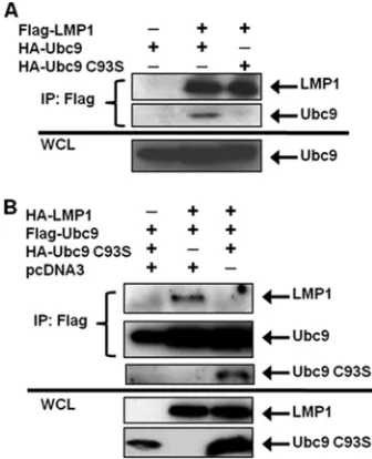
EBV LMP1 interacts only with enzymatically active Ubc9.
Cysteine residue 93 of Ubc9 is essential for its SUMO-conju-gating activity; mutation of this amino acid (Ubc9 C93S) ren-ders Ubc9 enzymatically inactive (32). However, Ubc9 C93S still interacts with certain proteins and can regulate protein function without mediating sumoylation (18, 23, 24). To test whether LMP1 could interact with Ubc9 C93S, 293T cells were transfected with expression vectors for FLAG-LMP1 and HA-Ubc9 or HA-HA-Ubc9 C93S. Results showed that FLAG-LMP1 only interacted with enzymatically active HA-Ubc9 (Fig. 2A); when HA-Ubc9 C93S was expressed, interaction with FLAG-LMP1 was not detected (Fig. 2A). Thus, Ubc9 activity is nec-essary for the LMP1-Ubc9 interaction.
Next, because overexpression of Ubc9 C93S suppresses the function of endogenous Ubc9 (10, 33), and in some cases mutation of cysteine residue 93 of Ubc9 results in loss of protein-protein interaction (9), we tested whether its expres-sion could disrupt the LMP1-Ubc9 interaction by transfecting 293T cells with FLAG-Ubc9, HA-LMP1, and HA-Ubc9 C93S. The results showed that FLAG-Ubc9 only interacted with HA-LMP1 when HA-Ubc9 C93S was not coexpressed (Fig. 2B). With coexpression of HA-Ubc9 C93S, interaction between wild-type Ubc9 and LMP1 was no longer detected. Instead, wild-type FLAG-Ubc9 pulled down HA-Ubc9 C93S, suggest-ing that the inactive Ubc9 mutant interacted with wild-type FLAG-Ubc9, possibly inhibiting the interaction between Ubc9
and LMP1. These data demonstrate that the LMP1-Ubc9 in-teraction can be disrupted by the overexpression of the enzy-matically inactive Ubc9 mutant.
Ubc9 does not interact with EBV LMP1 CTAR1 or CTAR2.
We next determined the region of LMP1 required for its in-teraction with Ubc9. The C-terminal region of EBV LMP1 contains two major C-terminal activating regions (CTAR1 at amino acids 204 to 208 and CTAR2 at amino acids 384 to 386), and we selected deletion mutants lacking CTAR1 (FLAG-LMP1⌬187–351), CTAR2 (FLAG-LMP1 1–231), and CTAR1 and CTAR2 (FLAG-LMP1 1–187) (Fig. 3A) to test in this study (31). 293T cells were transfected with expression con-structs for HA-Ubc9 and FLAG-LMP1 or its deletion mutants. The results showed that HA-Ubc9 interacted with wild-type FLAG-LMP1 but not with any of the deletion mutants (Fig. 3B). Thus, neither LMP1 CTAR1 nor LMP1 CTAR2 was sufficient for LMP1 to interact with Ubc9.
To confirm these results, which suggested that CTAR1 and CTAR2 were not critical for the LMP1-Ubc9 interaction, LMP1 constructs containing point mutations (Fig. 3A) that inactivate CTAR1 (PQAA), CTAR2 (YIID), and both CTAR1 and CTAR2 (DM) were examined for their ability to interact with Ubc9. 293T cells were transfected with expression constructs for HA-Ubc9 and FLAG-LMP1 or its point mu-tants. The results showed that HA-Ubc9 interacted not only with wild-type FLAG-LMP1 but also the four mutated con-structs (Fig. 3), demonstrating that functional CTAR1 and CTAR2 are not required for LMP1 to interact with Ubc9. Taken together, these data suggest that the region of LMP1 between CTAR1 and CTAR2 (amino acids 231 to 351) is critical for the LMP1-Ubc9 interaction.
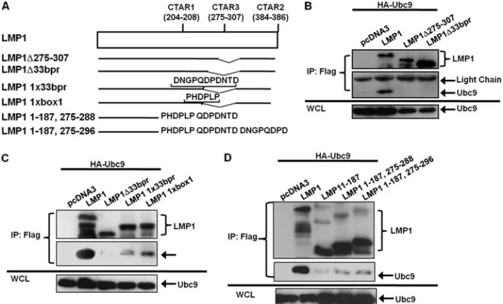
LMP1 CTAR3 is required for the LMP1-Ubc9 interaction.
Because LMP1 amino acids 231 to 351, which encompass CTAR3, were deleted in all LMP1 mutants that did not inter-act with Ubc9 and were present in all LMP1 point mutants that did interact with Ubc9, we tested if CTAR3 was necessary by using two CTAR3 deletion mutants. We constructed FLAG-LMP1⌬275–307 and obtained another construct from Wolf-gang Hammerschmidt (FLAG-LMP1⌬33bpr, missing amino acids 245 to 307) (Fig. 4A). 293T cells were transfected with HA-Ubc9 and FLAG-LMP1 or either of the CTAR3 deletion mutant expression constructs. Results showed that HA-Ubc9 interacted only with wild-type FLAG-LMP1 but with neither of the deletion mutants (Fig. 4B), suggesting that CTAR3, spe-cifically amino acids 275 to 307, was necessary for the observed LMP1-Ubc9 interaction.
Amino acid sequences found in LMP1 CTAR3 aid the in-teraction of LMP1 and Ubc9.Next we examined if reconstitu-tion of one copy of the 11-amino-acid repeats or one copy of the JAK-binding motif found in CTAR3 restored the LMP1-Ubc9 interaction. LMP1 CTAR3 deletion mutants with one copy of the 11-amino-acid repeats (LMP1 1x33bpr) (Fig. 4A) or one copy of the JAK-binding motif (LMP1 1xbox1) (Fig. 4A) were obtained from Wolfgang Hammerschmidt (11). 293T cells were transfected with expression constructs for HA-Ubc9 and FLAG-LMP1 or the LMP1 mutants. The results con-firmed that when CTAR3 was missing, the interaction between LMP1 and Ubc9 was not observed (Fig. 4C, lane 3). Recon-stitution with one copy of the JAK-binding motif (Fig. 4C, lane 5) restored approximately 10 to 20% of this interaction.
Re-FIG. 2. EBV LMP1 interacts only with enzymatically active Ubc9. (A) 293T cells were transfected with FLAG-LMP1 and HA-Ubc9 or HA-Ubc9 C93S. (B) 293T cells were transfected with FLAG-Ubc9, HA-LMP1, and increasing amounts of HA-Ubc9 C93S. At 48 h after transfection, cell lysates were harvested and immunoprecipitations (IP) were performed with FLAG antibodies. Western blot analyses of the immunoprecipitates and WCL were used to detect FLAG and HA expression.
VOL. 85, 2011 EBV LMP1 CTAR3 INTERACTS WITH Ubc9 10147
on November 7, 2019 by guest
http://jvi.asm.org/
[image:4.585.77.245.70.277.2]constitution with one copy of the 11-amino-acid repeats re-stored 5 to 10% of the Ubc9-LMP1 interaction (Fig. 4C, lane 4). Thus, while both the JAK-interacting motifs and the 11-amino-acid repeats aid the interaction of LMP1 and Ubc9, the
JAK-interacting motif may be somewhat more important for the interaction.
To determine if CTAR3 alone was sufficient to interact with Ubc9, we reconstituted FLAG-LMP1 1–187 with LMP1 amino
[image:5.585.110.472.455.674.2]FIG. 3. LMP1 CTAR1 and CTAR2 are not critical for the LMP1-Ubc9 interaction. (A) Diagrammatic alignment of wild-type LMP1 and selected LMP1 mutants. LMP 1–187 is missing both CTAR1 and CTAR2. LMP1 1–231 only contains CTAR1. LMP1⌬187–351 only contains CTAR2. LMP1 PQAA, YIID, and DM are point mutants with inactive CTAR1 and/or CTAR2. (B and C) 293T cells were transfected with FLAG-LMP1 or the indicated FLAG-LMP1 mutants and HA-Ubc9. Cell lysates were harvested 48 h after transfection, and immunoprecipitations were performed with FLAG antibodies. Western blot analyses of the immunoprecipitates and WCL were used to detect FLAG-LMP1 and HA-Ubc9 expression.
FIG. 4. LMP1 CTAR3 is required for its interaction with Ubc9. (A) Diagrammatic alignment of LMP1 mutants. LMP1⌬275–307 was constructed to delete both JAK3-binding motifs. LMP1⌬33bpr is missing the 33-bp repeats, which includes the JAK3-binding motifs. LMP1 1xbox 1 and LMP1 1x33bor are reconstituted LMP1⌬33bpr mutants. (B to D) 293T cells were transfected with HA-Ubc9 and FLAG-LMP1 or the indicated FLAG-LMP1 mutant. Cell lysates were harvested 48 h after transfection, and immunoprecipitations were performed with FLAG antibodies. Western blot analyses of the immunoprecipitates and WCL were performed to determine FLAG and HA expression.
on November 7, 2019 by guest
http://jvi.asm.org/
acids 275 to 288 (LMP1 1–187, 275–288) or 275 to 296 (LMP1 1–187, 275–296). 293T cells were transfected with expression constructs for HA-Ubc9 and FLAG-LMP1 or the LMP1 mu-tants. The results confirmed that LMP1 1–187 did not interact with Ubc9 (Fig. 4D). Reconstitution of LMP1 amino acids 275 to 288 or 275 to 296 partially restored the interaction between LMP1 and Ubc9 (Fig. 4D, lanes 4 and 5), suggesting CTAR3 is sufficient for the LMP1-Ubc9 interaction.
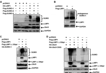
LMP1 induces the sumoylation of cellular proteins.Because our findings showed that LMP1 interacted with Ubc9, we next examined if LMP1 regulated sumoylation processes within the cells. 293T cells were transfected with FLAG-SUMO-1, FLAG-SUMO-2, or FLAG-SUMO-3 expression constructs along with HA-LMP1. Results showed that small amounts of cellular proteins were detected as modified by SUMO-1 and SUMO-3 when coexpressed with the vector control (pcDNA3) (Fig. 5A). However, when SUMO-1, SUMO-2, and SUMO-3 were coexpressed with HA-LMP1, increased amounts of cel-lular proteins appeared to be modified. These data were con-firmed by examining effects of endogenous SUMO-1 on mod-ification of cellular proteins. Western blot analyses showed that LMP1 expression coincided with detection of increases in sumoylated cellular proteins (Fig. 5B). Together, these data demonstrate that LMP1 expression increases levels of endog-enously sumoylated cellular proteins and suggest that the viral oncoprotein induces sumoylation processes within the cell.
To determine if CTAR3 was required for LMP1-induced sumoylation, 293T cells were transfected with HA-SUMO-1 and FLAG-LMP1 or FLAG-LMP1⌬33bpr expression con-structs. Results showed that LMP1 expression coincided with
increased amounts of cellular proteins modified by SUMO-1 (Fig. 5C), confirming our earlier data. In contrast, when FLAG-LMP1⌬33bpr was expressed, amounts of sumoylated proteins detected decreased. Repeat experiments revealed that FLAG-LMP1⌬33bpr expression coincided with 80% less su-moylation than when FLAG-LMP1 was expressed. The results suggest that LMP1 CTAR3 accounts for the majority of LMP1-induced protein sumoylation.
The importance of the Ubc9 interaction in LMP1-induced protein sumoylation was verified by utilizing overex-pression of Ubc9 and its inactive mutant. 293T cells were transfected with HA-Ubc9 or HA-Ubc9 C93S along with HA-SUMO-1 and vector control, LMP1, or FLAG-LMP1⌬33bpr expression constructs. Western blot analyses showed that when wild-type Ubc9 was overexpressed, LMP1 expression coincided with increased amounts of cellular pro-teins modified by SUMO-1, whereas expression of FLAG-LMP1⌬33bpr decreased protein sumoylation. In addition, ex-pression of HA-Ubc9 C93S resulted in decreased modification by SUMO-1 with either the wild-type or mutant LMP1 con-structs (Fig. 5D). Together, these data demonstrate the impor-tance of the Ubc9 interaction, via CTAR3, in LMP1-induced sumoylation of cellular proteins.
[image:6.585.111.476.68.322.2]Inhibition of the Ubc9-LMP1 CTAR3 interaction reduces cellular migration. Increased protein sumoylation has been implicated in tumorigenesis (8, 44). Because our findings sug-gested that LMP1 CTAR3 could function to induce sumoyla-tion of cellular proteins, we examined whether the LMP1-Ubc9 interaction affected phenotypic changes associated with LMP1-mediated oncogenesis. In a previous report, deletion of
FIG. 5. LMP1 induces the sumoylation of cellular proteins. (A) 293T cells were transfected with vector control or HA-LMP1 and FLAG-SUMO-1, FLAG-SUMO-2, or FLAG-SUMO-2. (B) 293T cells were transfected with vector control or FLAG-LMP1. (C) 293T cells were transfected with vector control, FLAG-LMP1, or FLAG-LMP1⌬33bpr and HA-SUMO-1. (D) 293T cells were transfected with HA-Ubc9 or HA-Ubc9 C93S along with HA-SUMO-1 and vector control, FLAG-LMP1, or FLAG-LMP1⌬33bpr. At 48 h after transfection, whole-cell lysates were collected and Western blot analyses were performed for SUMO-1, FLAG, HA, Ubc9, and GAPDH expression.
VOL. 85, 2011 EBV LMP1 CTAR3 INTERACTS WITH Ubc9 10149
on November 7, 2019 by guest
http://jvi.asm.org/
CTAR3 abrogated LMP1-induced colony formation (49), suggesting a function for LMP1 CTAR3 in oncogenesis in epithelial cells. We next investigated whether the Ubc9-LMP1 CTAR3 interaction could affect cellular migration, as commonly observed during oncogenesis. HA-LMP1-, HA-LMP1⌬33bpr-, and vector-expressing stable cell lines were generated, starting with MDA-MB 231 EBV⫹cells, clone C4A3, which are in-fected with EBV but do not express detectable levels of LMP1 (16, 41). Scratch assays were performed on confluent cell monolayers, and cellular migration was quantitated (2, 4). Re-sults showed that vector control-expressing cells migrated and filled in approximately 50% of the lesion in the monolayer, whereas HA-LMP1-expressing cells produced a significant (P⬍0.05) increase in cellular migration, filling almost 100% of the lesion (Fig. 6A), equivalent to a 2-fold increase in migra-tion. HA-LMP1⌬33bpr-expressing cells exhibited an interme-diate phenotype, producing a 1.6-fold increase in migration compared with the vector control, as expected, because CTAR1 and CTAR2 also affect cellular migration (6, 40). These data demonstrated a role for LMP1 CTAR3 in onco-protein-mediated cellular migration.
To confirm the contribution of LMP1 CTAR3 in cellular migration, 293 cells were transfected with FLAG-LMP1, FLAG-LMP1⌬33bpr, or control expression plasmids along with HA-Ubc9 or HA-Ubc9 C93S constructs, and migration was examined by scratch assays (Fig. 6B). The results again showed that FLAG-LMP1-expressing cells exhibited a signifi-cant (P ⬍ 0.05) increase in cellular migration compared
with vector control cells (Fig. 6B). In contrast, FLAG-LMP1⌬33bpr-expressing cells migrated at significantly (P ⬍ 0.05) lower levels, with only a 1.6-fold increase, than LMP1-expressing cells, but still at significantly (P⬍0.05) higher levels than vector control-expressing cells (Fig. 6B). Coexpression of HA-Ubc9 C93S and FLAG-LMP1 or FLAG-LMP1⌬33bpr sig-nificantly (P⬍0.05) inhibited LMP1-mediated cellular migra-tion (Fig. 6B). However, levels of migramigra-tion were still higher than with vector control-expressing cells. Results were similar with U20S cells (data not shown), and cellular proliferation assays yielded similar growth curves for all experimental arms (Fig. 7A and B).
We next used LMP1 constructs with mutations in CTAR1 and CTAR2 as well as CTAR3. In scratch assays, FLAG-LMP1-expressing cells again migrated at significantly (P ⬍ 0.05) higher levels than vector control-expressing cells and at lower levels in FLAG-LMP1⌬33bpr-expressing cells than LMP1-expressing cells, but still higher than vector control cells. Results were similar in cells expressing FLAG-LMP1 1–231 (Fig. 6C), which expressed CTAR1 but not CTAR2 or CTAR3. FLAG-LMP1 ⌬187–351 and FLAG-LMP1 1–187, both of which lacked CTAR1 and CTAR3, were not able to induce significant levels of cellular migration (Fig. 6C). These data suggest that the three terminal activating regions are required for LMP1-induced cellular migration, and CTAR3 has an independent effect on this event.
[image:7.585.114.471.71.318.2]Scratch assays were also performed on cells expressing the CTAR1 and CTAR2 point mutations tested earlier. Again,
FIG. 6. Inhibition of the Ubc9-LMP1 CTAR3 interaction decreases cellular migration. (A) MDA-MB 231 EBV⫹ clone C4A3 cells were transfected to stably express vector control, HA-LMP1, or HA-LMP1⌬33bpr. Following selection, cells were plated and grown to confluence. Confluent cell layers were scratched with a pipette tip and washed, and fresh RPMI was added to wells. After 24 h, medium was removed and cells were stained and fixed in a crystal violet and ethanol solution. Multiple images were captured along the scratch, and the wound width was calculated (2, 4). Representative images are shown, and the fold change in migration was determined. (B to D) 293 cells were plated and transfected as indicated. Confluent cell layers were scratched with a pipette tip and washed, and fresh 1% DMEM was added to wells. After 18 h, medium was removed and cells were stained and fixed in crystal violet-ethanol. The wound width was calculated (2, 4), and the fold change in migration was determined. Data are the means⫾standard deviations for experiments performed in triplicate.
on November 7, 2019 by guest
http://jvi.asm.org/
FLAG-LMP1-expressing cells showed a 2-fold increase in cell migration, whereas FLAG-LMP1⌬33bpr-expressing cells ex-hibited a 1.6-fold increase compared to vector control cells (Fig. 6D). FLAG-LMP1 YIID-expressing cells yielded similar results as FLAG-LMP1⌬33bpr-expressing cells (Fig. 6D). Both FLAG-LMP1 PQAA and FLAG-LMP1 DM, which bind Ubc9 but lack a functional CTAR1 domain, induced significantly (P⬍ 0.05) lower levels of cellular migration. However, they were both still able to induce significantly (P⬍ 0.05) higher levels of migration than the vector control-expressing cells. These data confirm that regardless of the presence of func-tional CTAR1 and CTAR2 domains, the ability of LMP1 to interact with Ubc9 contributes to its induction of cellular mi-gration. Furthermore, CTAR3 contributes to the ability of LMP1 to induce cell migration. Together, these data suggest that the LMP1 CTAR3-Ubc9 interaction contributes to phe-notypic changes associated with LMP1’s oncogenic potential.
DISCUSSION
In this paper, we have shown that EBV LMP1 interacts with the single-known SUMO-conjugating enzyme, Ubc9, suggest-ing that it may influence sumoylation processes. The interac-tion between Ubc9 and LMP1 is localized to LMP1’s intracel-lular signaling domain, the CTAR. This interaction does not involve the much-studied CTAR1 or CTAR2, but it is depen-dent on the much-less-characterized CTAR3. Expression of LMP1 induced sumoylation of cellular proteins, and disruption
of its interaction with Ubc9, either by deletion of LMP1 amino acids 275 to 307 or by overexpression of enzymatically inactive Ubc9, significantly reduced sumoylation. These observations confirmed our hypothesis that LMP1 regulates sumoylation processes within the cell. Furthermore, disruption of LMP1’s interaction with Ubc9 suppressed cellular migration, a pheno-typic change associated with basic oncogenic properties, in-duced by the viral oncoprotein.
This is one of the few reports on a role for CTAR3 in LMP1-induced cellular signaling and its functional conse-quences. CTAR3 was first defined by findings that suggested that the domain interacts with JAK3 and activates binding of STAT1 to DNA (11). However, a later report claimed that LMP1 CTAR3 was insufficient for activation of STAT1 tran-scriptional activity (5). An explanation for these seemingly conflicting results may lie in the sumoylation status of STAT1. STAT1 can be sumoylated (45, 51), and STAT1 sumoylation at K703 depends on its phosphorylation status. Specifically, phos-phorylation of STAT1 Y701 inhibits its sumoylation (51), whereas phosphorylation of STAT1 Y727 induces its sumoy-lation (46). Therefore, it is possible that LMP1 CTAR3 may mediate Y727 phosphorylation of STAT1, induce its sumoyla-tion, and thereby inhibit its transcriptional activity. We have begun studies to test this possibility. Preliminary results have demonstrated that STAT1 can be sumoylated in a CTAR3-dependent manner (data not shown). Our findings not only support the existence of LMP1 CTAR3 as a functional domain, but also they may help to resolve how it activates STAT1 DNA binding yet is not required for STAT1 activation (5, 11).
Others have reported that the 120-amino-acid region be-tween LMP1 CTAR1 and CTAR2 (amino acids 231 to 351) did not influence protein interactions of CTAR1 and CTAR2. Specifically, deletion of this region did not alter the ability of LMP1 to interact with TRAFs, TRADDs, or RIP (17) or its ability to activate JAK3, STAT2, STAT5 (13), NF-B, JNK1/2, and Bcl-2 (17). Nor did this region of LMP1 influence cellular growth or B-cell transformation (7, 17). However, deletion of these 120 amino acids did abolish the ability of LMP1 to induce colony formation (49). While the LMP1 CTAR3 mutants used in our study (FLAG-LMP1⌬33bpr lacks 65 amino acids and LMP1⌬275–307 lacks 32 amino acids [Fig. 4]) differed slightly from published mutants, we also found that whereas LMP1 CTAR3 did not affect cellular growth (Fig. 7), it did affect another phenotypic response typical of oncogenic processes, specifically, cellular migration (Fig. 6).
Previous reports in which both deletion and point mutants were used documented the importance of CTAR1 and CTAR2 in LMP1-mediated cellular migration (6, 40). Shair et al. tested epithelial cell migration by using LMP1 deletion mutants, while Dawson et al. tested epithelial cell migration using point mutants to inactivate CTAR1 and CTAR2 (6). Both reported that LMP1-induced epithelial cell migration required both CTAR1 and CTAR2 (40). When we used similar mutants, our data confirmed these findings. Additionally, the CTAR3 dele-tion mutant was still capable of inducing migradele-tion, but not to the same extent as full-length LMP1. Therefore, we now pro-pose that all three regulatory regions contribute to LMP1-induced migration. The role of CTAR3 in B-lymphocyte mi-gration needs to be examined.
While the importance of LMP1 CTAR3 in the LMP1-JAK3
FIG. 7. The LMP1-Ubc9 interaction does not affect cellular growth. (A) A total of 1⫻104293 cells were plated and transfected
with vector control, FLAG-LMP1, or FLAG-LMP1⌬33bpr. (B) A total of 1⫻104293 cells were plated and transfected with HA-Ubc9 or
HA-Ubc9 C93S and either the vector control, FLAG-LMP1, or FLAG-LMP1⌬33bpr. Cells and supernatant fluids were collected at 24, 48, 72, and 96 h after transfection, and the total numbers of cells were determined.
VOL. 85, 2011 EBV LMP1 CTAR3 INTERACTS WITH Ubc9 10151
on November 7, 2019 by guest
http://jvi.asm.org/
interaction has been contentious (11, 13), the data shown here clearly demonstrate the criticalness of CTAR3 in the interac-tion of LMP1 with Ubc9. The selected deleinterac-tion mutants (LMP1 1–187, LMP1 1–231, LMP1⌬187–351, LMP1⌬275–307, and LMP1⌬33bpr) all failed to interact with Ubc9. A similarity between these 5 mutants is that they all lack amino acids 275 to 308, suggesting these amino acids are necessary for the LMP1-Ubc9 interaction. Because the two CTAR3 deletion mutants (LMP1⌬275–307 and LMP1⌬33bpr) retain functional CTAR1 and CTAR2 domains, the possibility existed that CTAR1 and CTAR2 could cooperate to mediate Ubc9 association and that deletion of CTAR3 inhibited certain protein-protein interac-tions needed for CTAR1 or CTAR2. We have now discounted these possibilities by testing point mutants that inactivated either CTAR1 or CTAR2 or both domains. Our data showed that when either region is inactivated, LMP1 still interacted with Ubc9 (Fig. 3C). Thus, neither CTAR1 nor CTAR2 is required for the LMP1-Ubc9 interaction. These findings also rule out the possibility that CTAR1 and CTAR2 are together required for the LMP1-Ubc9 interaction. Instead, the data strongly suggest that CTAR3 is critical for this protein-protein interaction.
Further support for the role of LMP1 CTAR3 in the LMP1-Ubc9 interaction are obtained from the data demonstrating that a JAK-interacting motif and a 33bpr could both partially restore the LMP1-Ubc9 interaction in mutants lacking CTAR3 (LMP1-⌬33bpr). Reconstitution of part of CTAR3 to LMP1 1–187, which lacks the cytoplasmic tail of LMP1, also partially restored the LMP1-Ubc9 interaction. Taken together, these data converge to indicate that CTAR3 is necessary and suffi-cient, while CTAR1 and CTAR2 are neither required nor sufficient, for LMP1 to interact with Ubc9.
In light of the interaction between LMP1 and enzymatically active Ubc9, we examined if LMP1 was itself sumoylated. However, LMP1 does not contain the conserved sumoylation motif (20), and in the systems tested, sumoylation of the viral oncoprotein was not detected (data not shown).
In any case, sumoylation of cellular proteins was strongly induced by the LMP1-Ubc9 interaction and may regulate LMP1-mediated signaling. Our findings show that expression of LMP1 coincides with increased amounts of cellular proteins modified by SUMO-1, SUMO-2, and SUMO-3. These results were confirmed by examining endogenous sumoylation (by SUMO-1). Together, these findings demonstrate that the effect was LMP1 specific and not due to LMP1-induced plasmid expression. Because LMP1 can induce protein expression, the possibility existed that LMP1-induced sumoylation of cellular proteins was caused by increased expression of Ubc9. How-ever, levels of endogenous Ubc9 were not affected by LMP1 or LMP1⌬33bpr (Fig. 1A and D and 5A and C), thus demon-strating a link between LMP1 expression and the induction of sumoylation of cellular proteins. Additional experiments dem-onstrated that LMP1 CTAR3 and its ability to interact with Ubc9 was responsible for mediating the majority of LMP1-induced sumoylation. When the LMP1-Ubc9 interaction was inhibited, either by deletion of CTAR3 or by overexpression of Ubc9 C93S, approximately 80% less sumoylation of cellular proteins was observed.
Because LMP1 expression coincided with increased sumoy-lation of cellular proteins, it is possible that LMP1 acts as a
SUMO E3 ligase. However, SUMO E3 ligases are not required for protein sumoylation (19, 20). Therefore, the ability of LMP1 to serve as a ligase will need to be investigated. Addi-tionally, LMP1 may not directly bind to Ubc9 but instead requires another protein for this interaction. It is also probable that the binding of Ubc9 to the CTAR3 region influences signaling via CTAR1 and CTAR2. For instance, our prelimi-nary data suggest that CTAR3 is critical for LMP1-induced sumoylation of IRF7. We recently reported that CTAR2 is necessary for regulating IRF7 activity (35). These findings sug-gest that LMP1-induced sumoylation is mediated by CTAR3 but may modulate signal transduction pathways induced by CTAR1 and CTAR2. Therefore, examination of how sumoy-lation processes are involved in cross talk among the different regulatory regions is necessary to clarify the function of CTAR3.
Finally, our data show that overexpression of inactive Ubc9 (Ubc9 C93S) inhibited the LMP1-Ubc9 interaction, resulting in decreased cell motility. Ubc9 is a candidate target for anti-cancer therapies (7, 30). Means proposed to target Ubc9 in-clude use of small interfering RNA to inhibit Ubc9 expression (21, 26, 42), expression of the avian adenovirus protein Gam1 to induce Ubc9 degradation (4), and expression of a dominant-negative Ubc9 mutant (Ubc9 C93S) to suppress the function of endogenous Ubc9 (8, 31). This last method could be an ap-proach for treatment of LMP1-associated malignancies, be-cause we have shown that expression of Ubc9 C93S inhibits the LMP1-Ubc9 interaction and controls a cellular behavior that is characteristic of the oncogenic effects of the viral protein.
Together, these data disclose a protein-protein interaction and function unique for this region of LMP1. Our findings support the existence of LMP1 CTAR3 as a significant func-tional domain and suggest that further study of mechanisms by which LMP1 CTAR3 affects cellular behavior will be revealing.
ACKNOWLEDGMENTS
This work was supported by National Institutes of Health grants HL-64851, CA09156, and CA142132.
REFERENCES
1.Adamson, A. L., and S. Kenney.2001. Epstein-Barr virus immediate-early protein BZLF1 is SUMO-1 modified and disrupts promyelocytic leukemia
bodies. J. Virol.75:2388–2399.
2.Albuquerque, M. L., C. M. Waters, U. Savla, H. W. Schnaper, and A. S. Flozak.2000. Shear stress enhances human endothelial cell wound closure in
vitro. Am. J. Physiol. Heart Circ. Physiol.279:H293–H302.
3.Aviel, S., G. Winberg, M. Massucci, and A. Ciechanover.2000. Degradation of the Epstein-Barr virus latent membrane protein 1 (LMP1) by the ubiq-uitin-proteasome pathway. Targeting via ubiquitination of the N-terminal
residue. J. Biol. Chem.275:23491–23499.
4.Bentz, G. L., and A. D. Yurochko.2008. Human CMV infection of endothe-lial cells induces an angiogenic response through viral binding to EGF
receptor and1 and3 integrins. Proc. Natl. Acad. Sci. U. S. A.105:5531–
5536.
5.Brennan, P., J. E. Floettmann, A. Mehl, M. Jones, and M. Rowe.2001. Mechanism of action of a novel latent membrane protein-1 dominant
neg-ative. J. Biol. Chem.276:1195–1203.
6.Dawson, C. W., L. Laverick, M. A. Morris, G. Tramoutanis, and L. S. Young.
2008. Epstein-Barr virus-encoded LMP1 regulates epithelial cell motility and
invasion via the ERK-MAPK pathway. J. Virol.82:3654–3664.
7.Dirmeier, U., et al.2003. Latent membrane protein 1 is critical for efficient growth transformation of human B cells by Epstein-Barr virus. Cancer Res.
63:2982–2989.
8.Duan, X., J. O. Trent, and H. Ye.2009. Targeting the SUMO E2 conjugating enzyme Ubc9 interaction for anti-cancer drug design. Anticancer Agents
Med. Chem.9:51–54.
9.Fan, Z., et al.2006. SARS-CoV nucleocapsid protein binds to hUbc9, a
on November 7, 2019 by guest
http://jvi.asm.org/
ubiquitin conjugating enzyme of the sumoylation system. J. Med. Virol.
78:1365–1373.
10.Giorgino, F., et al.2000. The sentrin-conjugating enzyme mUbc9 interacts with GLUT4 and GLUT1 glucose transporters and regulates transporter
levels in skeletal muscle cells. Proc. Natl. Acad. Sci. U. S. A.97:1125–1130.
11.Gires, O., et al.1999. Latent membrane protein 1 of Epstein-Barr virus
interacts with JAK3 and activates STAT proteins. EMBO J.18:3064–3073.
12.Guo, B., S. H. Yang, J. Witty, and A. D. Sharrocks.2007. Signalling pathways
and the regulation of SUMO modification. Biochem. Soc. Trans.35:1414–
1418.
13.Higuchi, M., E. Kieff, and K. M. Izumi.2002. The Epstein-Barr virus latent membrane protein 1 putative Janus kinase 3 (JAK3) binding domain does not mediate JAK3 association or activation in B-lymphoma or
lymphoblas-toid cell lines. J. Virol.76:455–459.
14.Hille, A., A. Badu-Antwi, D. Holzer, and F. A. Grasser.2002. Lysine residues of Epstein-Barr virus-encoded nuclear antigen 2 do not confer secondary modifications via ubiquitin or SUMO-like proteins but modulate
transcrip-tional activation. J. Gen. Virol.83:1037–1042.
15.Huye, L. E., S. Ning, M. Kelliher, and J. S. Pagano.2007. Interferon regu-latory factor 7 is activated by a viral oncoprotein through RIP-dependent
ubiquitination. Mol. Cell. Biol.27:2910–2918.
16.Imai, S., J. Nishikawa, and K. Takada.1998. Cell-to-cell contact as an efficient mode of Epstein-Barr virus infection of diverse human epithelial
cells. J. Virol.72:4371–4378.
17.Izumi, K. M., et al.1999. The residues between the two transformation effector sites of Epstein-Barr virus latent membrane protein 1 are not critical
for B-lymphocyte growth transformation. J. Virol.73:9908–9916.
18.Kaul, S., J. A. Blackford, Jr., S. Cho, and S. S. Simons, Jr.2002. Ubc9 is a novel modulator of the induction properties of glucocorticoid receptors.
J. Biol. Chem.277:12541–12549.
19.Kerscher, O.2007. SUMO junction—what’s your function? New insights
through SUMO-interacting motifs. EMBO Rep.8:550–555.
20.Kerscher, O., R. Felberbaum, and M. Hochstrasser.2006. Modification of proteins by ubiquitin and ubiquitin-like proteins. Annu. Rev. Cell Dev. Biol.
22:159–180.
21.Kieser, A.2008. 13th Bienn. Conf. Int. Assoc. Res. Epstein-Barr Virus Assoc. Dis., Guangzhou, China, abstr. 27. International Association for Research on Epstein-Barr Virus and Associated Diseases, Houston, TX.
22.Kroetz, M. B.2005. SUMO: a ubiquitin-like protein modifier. Yale J. Biol.
Med.78:197–201.
23.Kurihara, I., et al.2005. Ubc9 and protein inhibitor of activated STAT 1 activate chicken ovalbumin upstream promoter-transcription factor
I-medi-ated human CYP11B2 gene transcription. J. Biol. Chem.280:6721–6730.
24.Kurtzman, A. L., and N. Schechter.2001. Ubc9 interacts with a nuclear localization signal and mediates nuclear localization of the paired-like homeobox protein Vsx-1 independent of SUMO-1 modification. Proc. Natl.
Acad. Sci. U. S. A.98:5602–5607.
25.Lang, V., and M. S. Rodriguez.2008. Innate link between NF-B activity and
ubiquitin-like modifiers. Biochem. Soc. Trans.36:853–857.
26.Li, H. P., and Y. S. Chang.2003. Epstein-Barr virus latent membrane protein
1: structure and functions. J. Biomed. Sci.10:490–504.
27.Lin, J., E. Johannsen, E. Robertson, and E. Kieff.2002. Epstein-Barr virus nuclear antigen 3C putative repression domain mediates coactivation of the
LMP1 promoter with EBNA-2. J. Virol.76:232–242.
28.Luftig, M., et al.2003. Epstein-Barr virus latent membrane protein 1
acti-vation of NF-B through IRAK1 and TRAF6. Proc. Natl. Acad. Sci. U. S. A.
100:15595–15600.
29.Mabb, A. M., and S. Miyamoto.2007. SUMO and NF-B ties. Cell. Mol. Life
Sci.64:1979–1996.
30.Maul, G. G., D. Negorev, P. Bell, and A. M. Ishov.2000. Review: properties and assembly mechanisms of ND10, PML bodies, or PODs. J. Struct. Biol.
129:278–287.
31.Miller, W. E., G. Mosialos, E. Kieff, and N. Raab-Traub.1997. Epstein-Barr
virus LMP1 induction of the epidermal growth factor receptor is mediated
through a TRAF signaling pathway distinct from NF-B activation. J. Virol.
71:586–594.
32.Mo, Y. Y., and S. J. Moschos.2005. Targeting Ubc9 for cancer therapy.
Expert Opin. Ther. Targets9:1203–1216.
33.Mo, Y. Y., Y. Yu, Z. Shen, and W. T. Beck.2002. Nucleolar delocalization of human topoisomerase I in response to topotecan correlates with sumoylation
of the protein. J. Biol. Chem.277:2958–2964.
34.Muller, S., M. J. Matunis, and A. Dejean.1998. Conjugation with the ubiq-uitin-related modifier SUMO-1 regulates the partitioning of PML within the
nucleus. EMBO J.17:61–70.
35.Ning, S., A. D. Campos, B. G. Darnay, G. L. Bentz, and J. S. Pagano.2008. TRAF6 and the three C-terminal lysine sites on IRF7 are required for its ubiquitination-mediated activation by the tumor necrosis factor receptor
family member latent membrane protein 1. Mol. Cell. Biol.28:6536–6546.
36.Pagano, J. S.2009. EBV diseases, p. 217–240.InB. Damania and J. M. Pipas (ed.), DNA tumor viruses. Springer Science and Business Media, New York, NY.
37.Rosendorff, A., et al.2004. EBNA3C coactivation with EBNA2 requires a
SUMO homology domain. J. Virol.78:367–377.
38.Rui, H. L., et al.2002. SUMO-1 modification of the C-terminal KVEKVD of Axin is required for JNK activation but has no effect on Wnt signaling.
J. Biol. Chem.277:42981–42986.
39.Schultheiss, U., et al.2001. TRAF6 is a critical mediator of signal
transduc-tion by the viral oncogene latent membrane protein 1. EMBO J.20:5678–
5691.
40.Shair, K. H., C. I. Schnegg, and N. Raab-Traub.2008. EBV latent membrane protein 1 effects on plakoglobin, cell growth, and migration. Cancer Res.
68:6997–7005.
41.Shimizu, N., H. Yoshiyama, and K. Takada.1996. Clonal propagation of Epstein-Barr virus (EBV) recombinants in EBV-negative Akata cells. J.
Vi-rol.70:7260–7263.
42.Sides, M. D., et al.2011. Arsenic mediated disruption of promyelocytic leukemia protein nuclear bodies induces ganciclovir susceptibility in
Epstein-Barr positive epithelial cells. Virology416:86–97.
43.Song, J., L. K. Durrin, T. A. Wilkinson, T. G. Krontiris, and Y. Chen.2004. Identification of a SUMO-binding motif that recognizes SUMO-modified
proteins. Proc. Natl. Acad. Sci. U. S. A.101:14373–14348.
44.Stewart, M. J., K. Smoak, M. A. Blum, and B. Sherry.2005. Basal and reovirus-induced beta interferon (IFN-beta) and IFN-beta-stimulated gene expression are cell type specific in the cardiac protective response. J. Virol.
79:2979–2987.
45.Ungureanu, D., S. Vanhatupa, J. Gronholm, J. J. Palvimo, and O. Silven-noinen.2005. SUMO-1 conjugation selectively modulates STAT1-mediated
gene responses. Blood106:224–226.
46.Vanhatupa, S., D. Ungureanu, M. Paakkunainen, and O. Silvennoinen.
2008. MAPK-induced Ser727 phosphorylation promotes SUMOylation of
STAT1. Biochem. J.409:179–185.
47.Wakisaka, N., et al.2004. Epstein-Barr virus latent membrane protein 1
induces synthesis of hypoxia-inducible factor 1 alpha. Mol. Cell. Biol.24:
5223–5234.
48.Yasugi, T., and P. M. Howley.1996. Identification of the structural and functional human homolog of the yeast ubiquitin conjugating enzyme UBC9.
Nucleic Acids Res.24:2005–2010.
49.Zhang, Z., Q. Zhang, Y. Yu, Y. Ouyang, and Z. He.2008. Construction and function analysis of the Epstein-Barr virus-encoded latent membrane
pro-tein-1 of CTAR3 region. Wei Sheng Wu Xue Bao48:1308–1313. (In
Chi-nese.)
50.Zhong, S., et al.2000. Role of SUMO-1-modified PML in nuclear body
formation. Blood95:2748–2752.
51.Zimnik, S., M. Gaestel, and R. Niedenthal. 2009. Mutually exclusive STAT1 modifications identified by Ubc9/substrate dimerization-dependent
SUMOylation. Nucleic Acids Res.37:e30.
VOL. 85, 2011 EBV LMP1 CTAR3 INTERACTS WITH Ubc9 10153